Plant Physiology and Biochemistry 167 (2021) 851–861
Available online 13 September 2021
0981-9428/© 2021 The Authors. Published by Elsevier Masson SAS. This is an open access article under the CC BY-NC-ND license
(http://creativecommons.org/licenses/by-nc-nd/4.0/).
Computational prediction of NO-dependent posttranslational modifications in plants: Current status and perspectives
Zsuzsanna Kolbert
a,*, Christian Lindermayr
b,**aDepartment of Plant Biology, University of Szeged, K¨oz´ep fasor 52, 6726, Szeged, Hungary
bInstitute of Biochemical Plant Pathology, Helmholtz Zentrum München, German Research Center for Environmental Health, Ingolstaedter Landstr. 1, D-85764, Oberschleißheim, München, Germany
A R T I C L E I N F O Keywords:
Computational prediction Nitric oxide
Posttranslational modification S-Nitrosation
Tyrosine nitration
A B S T R A C T
The perception and transduction of nitric oxide (NO) signal is achieved by NO-dependent posttranslational modifications (PTMs) among which S-nitrosation and tyrosine nitration has biological significance. In plants, 100-1000 S-nitrosated and tyrosine nitrated proteins have been identified so far by mass spectrometry. The determination of NO-modified protein targets/amino acid residues is often methodologically challenging. In the past decade, the growing demand for the knowledge of S-nitrosated or tyrosine nitrated sites has motivated the introduction of bioinformatics tools. For predicting S-nitrosation seven computational tools have been developed (GPS-SNO, SNOSite, iSNO-PseACC, iSNO-AAPAir, PSNO, PreSNO, RecSNO). Four predictors have been devel- oped for indicating tyrosine nitration sites (GPS-YNO2, iNitro-Tyr, PredNTS, iNitroY-Deep), and one tool (DeepNitro) predicts both NO-dependent PTMs. The advantage of these computational tools is the fast provision of large amount of information. In this review, the available software tools have been tested on plant proteins in which S-nitrosated or tyrosine nitrated sites have been experimentally identified. The predictors showed distinct performance and there were differences from the experimental results partly due to the fact that the three- dimensional protein structure is not taken into account by the computational tools. Nevertheless, the pre- dictors excellently establish experiments, and it is suggested to apply all available tools on target proteins and compare their results. In the future, computational prediction must be developed further to improve the precision with which S-nitrosation/tyrosine nitration-sites are identified.
1. Introduction
Nitric oxide (NO), previously known as an air pollutant gas, has been shown to be an endogenously produced jack-off-all-trades plant signal molecule. In higher plants, nitrite is the major substrate for NO forma- tion (Astier et al., 2018), while in primitive algae, similar to animals, NO is primarily derived from the amino acid L-arginine (Astier et al., 2021), indicating that reductive pathways of endogenous NO formation have become dominant during the evolution of terrestrial plants (Fr¨ohlich and Durner, 2011). NO is an integral regulator in a wide range of physiological processes such as vegetative-reproductive development (S´anchez-Vicente et al., 2019), photosynthesis (Lopes-Oliveira et al., 2021), stomatal movements (Van Meeteren et al., 2020), abiotic stress responses (Fancy et al., 2017), symbiotic interactions (Berger et al., 2019) and defence mechanisms against phytopathogens (Lubega et al.,
2021; Jedelsk´a et al., 2021). In biological systems, NO reacts among other things, with molecular oxygen, reactive oxygen species, gluta- thione, and amino acids to form the diverse group of reactive nitrogen species (RNS) including peroxynitrite (ONOO−) and S-nitro- soglutathione (GSNO) as the most relevant ones. While the blood pres- sure regulating effect of NO in animals and humans is mediated by cGMP-dependent signalling and soluble guanylate cyclase (sGC) func- tions as a NO receptor (Horst et al., 2019), in plants NO-induced cGMP signalling seems to be unlikely (Astier et al., 2019). In recent years, the view has become prevalent that the transfer of NO’s bioactivity is conveyed mainly through posttranslational modifications (PTMs) of specific protein targets. PTMs occurring following or during translation aim to increase the size and complexity of the proteome. Protein mod- ifications result from enzymatic or non-enzymatic bounding of specific chemical groups to amino acid side chains (Santos and Lindner, 2017).
* Corresponding author.
** Corresponding author.
E-mail addresses: ordogne.kolbert.zsuzsanna@szte.hu (Z. Kolbert), lindermayr@helmholtz-muenchen.de (C. Lindermayr).
Contents lists available at ScienceDirect
Plant Physiology and Biochemistry
journal homepage: www.elsevier.com/locate/plaphy
https://doi.org/10.1016/j.plaphy.2021.09.011
Received 9 June 2021; Received in revised form 4 September 2021; Accepted 8 September 2021
Due to the alterations in the protein structure, protein activity, stability, localization, and molecular interactions may be modified (Vu et al., 2018). The biological function of more than 200 different enzymatic and non-enzymatic PTMs has been revealed so far (Vir´ag et al., 2020).
Among them, NO and its reaction products are responsible for the in- duction of PTMs called nitration, S-nitrosation and metal nitrosylation.
Nitration may covalently modify tyrosine, tryptophan, cysteine and methionine (Corpas et al., 2009), S-nitrosation affects cysteine-containing proteins (Hess et al., 2005), and during metal nitrosylation NO reacts with metallo-enzymes (Ignarro et al., 1999).
S-Nitrosation is also known as S-nitrosylation. However, nitrosylation involves direct addition of NO to a reactant and is derived from chem- istry terminology that describes the coordination of NO to a metal centre (Ford et al., 2005). Since the transfer of a nitrosonium ion (NO+) is the primarily mechanism for the oxidation of protein cysteine thiols, the term S-nitrosation is the more applicable expression for this chemical process (Gupta et al., 2019). In biological systems, the most actively studied NO-dependent PTMs are S-nitrosation and tyrosine nitration affecting a large number of proteins thus having wide-ranging impact in the cells. Protein S-nitrosation has been established as a significant route by which NO transmits its ubiquitous cellular function (Hess et al., 2005;
Spadaro et al., 2010; Astier and Lindermayr, 2012), while tyrosine nitration seems to have a major role as an irreversible modification leading to protein inactivation (Kolbert et al., 2017).
2. S-nitrosation: mechanism, specificity, selectivity, identification in plants
The mechanism of S-nitrosothiol formation is an important issue for understanding the biological actions of NO. Often thiol-containing molecules like cysteine and glutathione have been used for S-nitro- sation to yield low-molecular-weight S-nitrosothiols such as S-nitro- socysteine (CysNO) and GSNO and to study the S-nitrosation mechanism. However, the reactivity of NO with thiol groups is very low.
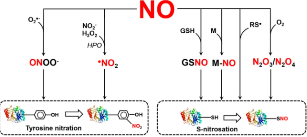
Therefore, the formation of SNOs depends on the generation of reactive intermediates (Hill et al., 2010; Broniowska and Hogg, 2012). As a free radical (●NO), it can lose or gain electrons to become oxidized nitro- sonium cation (NO+) or reduced nitroxyl anion (NO−) species, each with different oxidation state for the nitrogen atom (+2, +3, and +1 respectively) (Arnelle and Stamler, 1995). Moreover, in aerobic, bio- logical milieu, NO can be oxidized to its +5 oxidation state to form non-reactive nitrate anion (NO3−). The existence of NO in different redox status multiplies the possibilities to form S-nitrosothiols via various pathways (Fig. 1). For instance, NO can be oxidized to the highly reactive dinitrogen trioxide (N2O3), which is an effective S-nitrosating agent. Moreover, the NO radical can react with highly electrophilic thiyl (RS●) radicals. Furthermore, redox-active metals, e. g. such as those present in heme groups, can catalyze SNO formation. Finally, S-nitro- sothiols can transfer their NO moiety to cysteine thiol in a
trans-nitrosylation reaction. This is of special importance in context of the physiological NO donors CysNO and GSNO (Hess et al., 2005; Smith and Marletta, 2012; Kovacs and Lindermayr, 2013). But also S-nitro- sated protein cysteine residues can function as NO donors. Several nitrosated proteins are described to transferring their NO group to target proteins or low molecular weight thiols, e. g. hemoglobin (Pawloski et al., 2001), thioredoxin (Mitchell and Marletta, 2005; Mitchell et al., 2007; Wu et al., 2010), caspase-3 (Nakamura and Lipton, 2013), cyclin-dependent kinase 5 (Qu et al., 2011), glyceraldehyde 3-phosphate dehydrogenase (Kornberg et al., 2010; Zaffagnini et al., 2013), and non-canonical catalase ROG1 (Chen et al., 2020).
The microenvironment around a cysteine residue defines its NO accessibility and reactivity. Cysteine residues exhibiting a low-pKa sulfhydryl group are particularly susceptible to certain types of redox modification (Spadaro et al., 2010). In the past, different consensus motifs for S-nitrosation have been defined by comparing the amino acid sequences around identified target cysteine residues. In general, such NO sensitive cysteine residues are often located within an acid-base or hydrophobic motif (Stamler et al., 2001), while Greco et al. (2006) supported the idea of extending the motif beyond the primary sequence including hydrophobic motifs nearby the target cysteine residues (Greco et al., 2006). Based on amino acid sequence comparison of S-nitrosated proteins, several different consensus sequences for S-nitrosation have been described. Stamler et al. (1997) proposed an acid-base motif for protein S-nitrosation and denitrosation. The acid-base motif comprises flanking acidic (Asp (D), Glu (E)) and basic (Arg (R), His (H), Lys (K)) residues to the reactive thiol cysteine sites ([KRHDE]-C-[DE]). More- over, a GSNO binding motif is described ([HKR]-C-[hydrophobic]X [DE]) (Hess et al., 2005). Analysis of 1195 sequences of S-nitrosated peptides identified in GSNOR-KO plants (Hu et al., 2015) revealed 10 motifs, including EXC, EC, CD, CE, CXXE, CXD, CXE, DXXC, DC, and EXXXC, harboring conserved negatively charged amino acids glutamate (E) or aspartate (D) in close proximity of the S-nitrosated cysteine res- idue. Although such charged motifs have been shown to be predictive in a number of cases, the common features of acid-base motifs are still object of intense discussions and there are still no general rules, which can explain which cysteine residue is a target for NO.
In contrast, other studies have demonstrated on the peptide level that the sequence of the surrounding amino acids has no significant effect on the reactivity of cysteines towards S-nitrosation (Taldone et al., 2005).
Moreover, analysis of 70 S-nitrosation sites revealed that proximal acid–base motif, Cys pKa, sulfur atom exposure, and hydrophobicity in the vicinity of the modified cysteine do not predict S-nitrosation speci- ficity. Instead, a revised acid-base motif that is located farther from the target cysteine and in which the charged groups are exposed has been identified (Marino and Gladyshev, 2010). This emphasizes also the importance of the three-dimensional folding, which needs to be considered whenever defining the NO sensitivity of a cysteine residue (Kovacs and Lindermayr, 2013).
Fig. 1.Reactions leading to the formation of reactive nitrogen species which are responsible for post- translational modifications such as S-nitrosation and tyrosine nitration. See explanations in the text. Ab- breviations: NO, nitric oxide; GSH, glutathione;
GSNO, S-nitrosoglutathione; M, metal; RS●, thiyl radical; O2, oxygen; N2O3, dinitrogen trioxide; N2O4, dinitrogen tetroxide, O2●-, superoxide anion radical;
ONOO−, peroxynitrite; NO2−, nitrite; H2O2, hydrogen peroxide; HPO, hemoperoxidase; ●NO2, nitrogen di- oxide radical.
In recent two decades, much effort has been made to identify S- nitrosated proteins in plants. A number of indirect mass spectrometry (MS)-based proteomics approaches have been developed to identify S- nitrosated proteins and their modification sites from complex biological samples (Jaffrey and Snyder, 2001; Hao et al., 2006; Camerini et al., 2007; Chouchani et al., 2010; Hu et al., 2015). The biotin switch tech- nique (BST) is the most widely used method and is based on the con- version of S-nitrosated Cys to biotinylated Cys (Jaffrey and Snyder, 2001). Such a labelling allows the detection of S-nitrosated proteins using specific anti-biotin antibodies and their enrichment by affinity chromatography using neutravidin matrices. Finally, the enriched pro- teins are identified by MS. Variants of the BST assay, including quanti- tative approaches and the use of protein microarrays have been reported and successfully used (Torta et al., 2008; Astier et al., 2011; Seth and Stamler, 2011; Wang and Xian, 2011; Lee et al., 2014). Including a digest step before purification allows the enrichment of peptides con- taining NO-targeted cysteine residues (SNOSID) (Hu et al., 2015).
Modification of the BST method enabled quantification of S-nitrosated proteins via fluorescent labelling (Santhanam et al., 2008) or via the use of isobaric iodoacetyl tandem mass tags (iodoTMT) (Qu et al., 2014).
Furthermore, proteins can also react with a thiol-reactive resin allowing on-resin enzymatic digestion before MS analysis. This resin-assisted capture (SNO-RAC) requires fewer steps, detects high-mass S-nitro- sated proteins more efficiently, and facilitates identification and quan- tification of S-nitrosated sites by mass spectrometry (Forrester et al., 2009; Kolbert et al., 2019).
Until now, several hundreds of endogenously S-nitrosated proteins have been identified in proteome wide-scale studies in plants, whereas NO donor treatments are often used to increase the amount of S-nitro- sated proteins. S-nitrosated proteins function in major cellular activities of the primary and secondary metabolism and regulate processes related to biotic and abiotic stress response (Astier et al., 2012). However, these candidates need confirmation by candidate-specific approaches for the physiological relevance. This includes also the identification of the NO-sensitive cysteine residue(s) of these proteins.
3. Tyrosine nitration: mechanism, specificity, selectivity, identification in plants
Tyrosine is a moderately hydrophilic aromatic amino acid, which is
therefore often on the surface of the protein and thus subject to modi- fications. Nitration reaction may be catalysed by ONOO− or by nitrogen dioxide radical formed in the reaction between hydrogen peroxide and nitrite in the presence of hemoperoxidase enzyme. Peroxynitrite is a strong oxidizing and nitrating agent resulting from the reaction between superoxide anion radical and NO, mainly at the sites of superoxide formation (Radi et al., 2001; Szabo et al., 2007, Fig. 1). During nitration ´ of the tyrosine amino acid, a nitro group is attached to the hydroxyl group of the ortho carbon atom in the aromatic ring leading to the for- mation of 3-nitrotyrosine (YNO2). The process takes place in two steps, since the attachment of the nitro group is preceded by a one electron oxidation of the tyrosine aromatic ring to tyrosyl radical. The major oxidants are hydroxyl radical and carbonate radical which originate from ONOO− due to diverse reactions (Kolbert et al., 2017). As a consequence of YNO2 formation, the key physical and chemical prop- erties including pKa, redox potential, hydrophobicity/hydrophilicity, molecular size of amino acids may be modified (Sabadashka et al., 2021). Due to these physico-chemical alterations, the structure and function of the target protein may be changed. In animal systems, accumulating evidence suggest the reversibility and consequently the signalling function of tyrosine nitration (Sabadashka et al., 2021). In contrast, most of the nitrated plant enzyme proteins examined in detail so far show activity loss indicating that tyrosine nitration may be a signal for degradation (Kolbert et al., 2017).
Protein tyrosine nitration is a relatively widespread PTM because it affects numerous proteins in different organs of plants grown under diverse conditions (both unstressed and stressed). At the same time tyrosine nitration can be considered as highly selective, since only 1–2%
of the total tyrosine proteome (3% of the whole proteome) may be exposed to in vivo nitration (Bartesaghi et al., 2007). Consequently, the total yield (expressed as mole of 3-nitrotyrosine/mole tyrosine) is low, as was determined in hypocotyls of sunflower grown under physiolog- ical conditions (Chaki et al., 2009). Nitration of protein tyrosine is a selective process despite the fact that no consensus sequence ensuring selectivity has been convincingly confirmed (Bartesaghi and Radi, 2018). Rather, some common features appear to affect YNO2 formation such as the presence of acidic residues next to the YNO2 site, cysteine or methionine neighbouring the target tyrosine residue and the presence of loop-forming amino acids such as proline or glycine (Souza et al., 2008).
Beyond the amino acid sequence, additional factors influence the Table 1
List of software tools developed so far for predicting NO-dependent PTMs (S-nitrosation and tyrosine nitration). Modified from Bignon et al. (2018).
Name Year Availability Link Number of citations (1st of June
2021) Citation Note
SNO prediction
GPS-SNO 2010 web server,
standalone http://sno.biocuckoo.org/ 157 Xue et al. (2010)
SNOSite 2011 web server http://csb.cse.yzu.edu.tw/SNOSite/ 69 Lee et al. (2011) link doesn’t
work iSNO-
PseACC 2013 web server http://app.aporc.org/iSNO-PseAAC/index.
html 345 Xu et al. (2013a)
iSNO-
AAPAir 2013 web server http://app.aporc.org/iSNO-AAPair/ 249 Xu et al. (2013b)
PSNO 2014 web server http://59.73.198.144:8088/PSNO/ 82 Zhang et al. (2014) link doesn’t
work PreSNO 2019 web server http://kurata14.bio.kyutech.ac.
jp/PreSNO/ 21 Hasan et al. (2019)
RecSNO 2021 web server http://nsclbio.jbnu.ac.kr/tools/RecSNO/ 1 Siraj et al. (2021)
YNO2 prediction
GPS-YNO2 2011 web server,
standalone http://yno2.biocuckoo.org/ 66 Liu et al. (2011)
iNitro-Tyr 2014 web server http://app.aporc.org/iNitro-Tyr/ 209 Xu et al. (2014)
PredNTS 2021 web server http://kurata14.bio.kyutech.ac.jp
/PredNTS/ 1 Nilamyani et al.
(2021)
iNitroY-Deep 2021 webserver http://3.15.230.173/ 0 Naseer et al. (2021) link doesn’t
work Both SNO and YNO2 prediction
DeepNitro 2018 web server http://deepnitro.renlab.org 33 Xie et al. (2018)
nitration process including the centrifugal-centripetal position of the tyrosine residue within the three-dimensional (3D) structure of the protein and the cellular and redox environment of the target protein (Yeo et al., 2015; Bartesaghi and Radi, 2018).
In plant studies, the one- and two-dimensional gel electrophoresis followed by immunochemical detection of nitrated proteins are frequently used approaches. Protein identification by regular MS/MS in combination with immuno-enrichment of tyrosine-nitrated peptides is possible. For detecting the nitrated peptides matrix-assisted laser desorption/ionization-time of flight (MALDI-TOF) MS and LC-MS/MS can be used (Yeo et al., 2015; Batthy´any et al., 2017). In most plant studies, immune-affinity based approaches was optimized for identi- fying tyrosine nitrated-proteins (e.g. Corpas et al., 2008; Lozano-Juste et al., 2011; Cecconi et al., 2009; Tanou et al., 2012; Begara-Morales et al., 2013a, 2019; Takahashi and Morikawa, 2019). However, false positive detection may happen due to non-specific antibody binding and the identified protein occasionally mismatch the protein database (Corpas et al., 2013a). Thus MS assays are being continuously improved in order to provide more accurate detection of tyrosine nitrated proteins and peptides (Ng et al., 2013; Tsikas and Duncan, 2013; Yeo et al., 2015;
Batthy´any et al., 2017; Chaki et al., 2018). To date, large-scale studies identified more than one hundred plant proteins as in vivo targets of tyrosine nitration in the organs of healthy and stressed plants. For most of these proteins, the YNO2 site and the change in activity/function have not been studied.
4. Computational tools for predicting NO-dependent PTMs Although many different experimental methods have been devel- oped for accurate identification of NO target cysteine residues, these are often still associated with technical difficulties based on the instability of SNOs. For instance, direct detection of NO-modified thiols by MS or X- ray crystallography is still very challenging and only possible on re- combinant proteins. Moreover, such approaches are time-consuming and cost-intensive. The situation is similar with the analytical determi- nation of YNO2, as there are methodological challenges during the detection: (i) endogenous levels of YNO2 are very low, (ii) the vast excess of tyrosine in the samples disturbs the detection and quantification of YNO2 (iii) special precautions must be taken since YNO2 may be formed during sample preparation (Tsikas and Duncan, 2013). Therefore, the computational approach of screening proteins for NO sensitive cysteine or tyrosine residues is an attractive alternative since the recent progress of machine learning makes possible the efficient use of computational prediction preceding the laboratory experimentation. With the avail- ability of a huge amount of amino acid sequences, it is possible to develop computational methods for predicting SNO or YNO2 sites in proteins. Such kind of information is very useful for both basic research and application. Table 1 summarizes the developed tools either for predicting SNO sites or YNO2 sites, or both.
4.1. Tools for computational prediction of S-nitrosation sites and testing their performance
The algorithms developed to identify NO-sensitive cysteine residues include GPS-SNO, SNOSite, iSNOPseAAC, iSNO-AAPair, RecSNO, Pre- SNO, and DeepNitro (Lee et al., 2011; Xu et al., 2013a, 2013b; Xue et al., 2010; Hasan et al., 2019; Xie et al., 2018; Siraj et al., 2021; Zhang et al., 2014). A big disadvantage of these computational methods is still the non-consideration of the 3D structure of the proteins. Cysteine residues, which might be predicted as target for S-nitrosation could be buried inside the protein and in this way inaccessible for NO. Moreover, for calculating the NO-sensitivity of a cysteine residue, the algorithms consider only amino acids, which are nearby a cysteine residue in the primary structure. However, in the folded protein amino acids, which are far away in the primary structure, could get in close vicinity of a cysteine residue and affect its microenvironment.
The first released online tool for SNO-site prediction was GSP-SNO 1.0 in 2010 (Xue et al., 2010). The leave-one-out validation and 4-, 6-, 8-, 10-fold cross-validations were calculated to evaluate the prediction performance and system robustness. The GPS 3.0 algorithm performed quite well with an accuracy of 75.70%, a sensitivity of 55.32% and a specificity of 80.11% under the low threshold. The online service and local packages of GPS-SNO 1.0 were implemented in JAVA 1.4.2 and freely available at: http://sno.biocuckoo.org/.
One year later, the software tool SNOSite was presented (Lee et al., 2011). The authors used a total of 586 experimentally identified S-nitrosation sites from S-nitroso-L-penicillamine (SNAP)/L-cysteine-s- timulated mouse endothelial cells for an informatics analysis on S-nitrosation sites including structural factors such as the flanking amino acids composition, the accessible surface area and physicochemical properties, i.e. positive charge and side chain interaction parameter.
Maximal dependence decomposition (MDD) has been applied to obtain statistically significant conserved motifs. Support vector machine (SVM) is applied to generate predictive model for each MDD-clustered motif.
According to five-fold cross-validation, the MDD-clustered SVMs could achieve an accuracy of 0.902, and provides a promising performance in an independent test set. The MDD-clustered model was adopted to construct an effective web-based tool, named SNOSite (http://csb.cse.
yzu.edu.tw/SNOSite/), for identifying S-nitrosation sites on the uncharacterized protein sequences. At the time of writing this review, SNOSite is not available.
In 2013, a new predictor, called iSNO-PseAAC, was developed for identifying the SNO sites in proteins by incorporating the position- specific amino acid propensity (PSAAP) into the general form of pseudo amino acid composition (PseAAC) (Xu et al., 2013a). The pre- dictor was implemented using the conditional random field (CRF) al- gorithm. The overall cross-validation success rate achieved by iSNO-PseAAC in identifying nitrosated proteins on an independent dataset was over 90%, indicating that the new predictor is quite prom- ising. A web server for iSNO-PseAAC is available at http://app.aporc.
org/iSNO-PseAAC/, where users can easily obtain the desired results without the need to follow the mathematical equations involved during the process of developing the prediction method. Then same group published another prediction tool called iSNO-AAPair (Xu et al., 2013b).
This algorithm was developed by considering the coupling effects for all the pairs formed by the nearest residues and the pairs by the next nearest residues along protein chains. A web server for iSNO-AAPair was established at http://app.aporc.org/iSNO-AAPair/.
In 2014, Zhang and co-workers presented a new bioinformatics tool, named PSNO, to identify SNOs from amino acid sequences (Zhang et al., 2014). They explored various promising sequence-derived discrimina- tive features, including the evolutionary profile, the predicted secondary structure and the physicochemical properties and used the relative en- tropy selection and incremental feature selection approach to select the optimal feature subsets. Afterwards, they trained their model by the technique of the k-nearest neighbour algorithm. Using both informative features and an elaborate feature selection scheme, the PSNO method achieved good prediction performance with a mean Mathews correla- tion coefficient (MCC) value of about 0.5119 on the training dataset using 10-fold cross-validation. The PSNO web server was established at http://59.73.198.144:8088/PSNO/, but at the time of writing this re- view it is not accessible.
Four years later, Xie and colleagues developed a computational tool for predicting nitration and nitrosation sites in proteins (Xie et al., 2018). They constructed positional amino acid distributions, sequence contextual dependencies, physicochemical properties, and position-specific scoring features, to represent the modified residues.
Based on these encoding features, they established a predictor called DeepNitro using deep learning methods for predicting S-nitrosation.
Using n-fold cross-validation, the evaluation shows great AUC value for DeepNitro, of 0.70 for cysteine nitrosation, demonstrating the robust- ness and reliability of the predictor. The application of deep learning
Table 2
List of plant proteins in which the S-nitrosated cysteine residues have been experimentally identified. S-nitrosated sites in the listed proteins were computationally predicted using GSP-SNO 1.0, iSNO-PseAAC, iSNA-AAPair, DeepNitro, PreSNO and RecSNO software. Bold indicates matched cysteine residue.
Protein name Accession
number Total
number of Cys
Identified by LC-MS/
MS or mutation
Predicted by GPS-SNO 1.0 (2010) (medium threshold)
Predicted by iSNO- PseAAC (2013)
Predicted by iSNO- AAPair (2013)
Predicted by DeepNitro (2018) (medium threshold)
Predicted by PreSNO (2019)
Predicted by RecSNO (2020) 0.6 threshold
Citation
NPR1 At1g64280 17 C156 C156, C385 C212, C306 C223,
C306, C394, C457
non non C82, C212,
C216, C223, C378, C394, C457
Tada et al. (2008)
SAMS1 At1g02500 8 C114 C114 C161 C31, C90,
C161 non C114 C45, C73,
C90, C161 Lindermayr et al.
(2006) OST1
(SnRK2.6) At4g33950 7 C137 non C107,
C159, C203
C131,
C203 non C137 C159, Wang et al.
(2015)
ASK1 At1g75950 3 C37, C118 C118 non C59,
C118 C118 non C59,
C118 Iglesias et al.
(2018)
SCE1 At3g57870 4 C139 non C94, C139 C139 C139 non non Skelly et al.
(2019)
SRG1 At3g46080 7 C87 C87 C87 C28 non non C18,
C28, Cui et al. (2018)
AHP1 At3g21510 4 C115 non C104 C115 non non non Feng et al. (2013)
cALD2 At2g36460 6 C173 C68, C326 C326 C208 C326 non C197, C208,
C326 van der Linde et al. (2011)
TIR1 At3g62980 23 C140 C516, C551 C34, C53,
C121, C140, C155, C210, C269, C288, C311, C405, C480, C491
C121, C140, C405, C551
C53, C516 non C53, C121,
C551 Terrile et al.
(2012)
MC9 At5g04200 7 C147 C17, C147 C17, C29 C117 C17, C29,
C147 C147 C17, C29,
C117, C147, C251
Belenghi et al.
(2007)
PRXII E At3g52960 2 C121 C121 C121, C146 C121 C121 C121 C121, C146 Romero-Puertas
et al. (2007)
GAPDH At1g13440 2 C156,
C160 C156, C160 non non C156, C160 C156,
C160 C156 Holtgrefe et al.
(2008)
SABP3 At3g01500 7 C280 C34, C173,
C280 C230, C257 C34 non non C34, C173,
C230 Wang et al.
(2009) NADPH
Oxidase (RBOHD)
At5g47910 10 C890 non C208, C387,
C433, C480, C695
C412, C480, C695, C890
C695, C890 non C433, C695,
C890 Yun et al. (2011)
TGA1 At5g65210 4 C172,
C287 C172 non non non C260,
C266 non Lindermayr et al.
(2010)
CDC48 Q1G0Z1
Nicotiana tabacum
14 C110,
C526, C664
C426, C576 C74, C82, C110, C526, C539, C576, C664, C699
C74, C426,
C539, C576 C110, C419, C539, C664 C526,
C539 C74, C82, C110, C272, C539, C695
Astier et al.
(2012)
MYB30 At3g28910 7 C53 C6 C6, C7, C49,
C53, C257, C289
C6, C7 C49 C49, C53 non Tavares et al.
(2014)
PDK1 Q5I6E8,
Solanum lycopersicum
4 C128 C214 C128, C214,
C244, C466 non non non non Liu et al. (2017)
GSNOR At5g43940 15 C10 C10, C283 C10, C59,
C77, C117, C125, C189, C283, C296, C385
C59 non non C10, C382,
C385 Guerra et al.
(2016);
Zhan et al.
(2018)
ROG1 At1g20620 7 C343 non C230, C370,
C402, C420 C402 C230 non C86, C230,
C370, C402 Chen et al.
(2020)
cFBP1 AAD10213,
Pisum sativum
7 C153 C173 C178 C92, C306 C306 non C49, C92,
C306 Serrato et al.
(2018)
APX1 At1g07890 5 C32 C119 C32, C138 C19, C32 C138 C32, C49 C32, C49,
C138 Yang et al.
(2015)
ABI5 At2g36270 4 C153 C153, C440 C56, C440 non C440 non C153, C293 Albertos et al.
(2015)
PRMT5 At4g31120 12 C125 C125 C17, C70,
C125, C141, C189, C238,
C238, C260 non non C125, C160 Hu et al. (2015)
(continued on next page)
method and novel encoding schemes, especially the position-specific scoring feature, seems to improve the accuracy of S-nitrosation site prediction. DeepNitro is implemented in JAVA and PHP and is freely available for academic research at http://deepnitro.renlab.org.
A novel predictor PreSNO has been developed that integrates mul- tiple encoding schemes by the support vector machine and random forest algorithms (Hasan et al., 2019). The PreSNO achieved an accuracy and MCC value of 0.752 and 0.252 respectively in classifying between SNO and non-SNO sites when evaluated on the independent dataset, outperforming the existing methods. The web application of the PreSNO and its associated datasets are freely available at http://kurata14.bio.ky utech.ac.jp/PreSNO/.
The latest SNO-site prediction tool is called RecSNO and was pub- lished in 2021 by Siraj and colleagues (Siraj et al., 2021). They proposed an end-to-end deep learning based S-nitrosation site predictor with an embedded layer and bidirectional long short-term memory. This method uses amino acid sequences as inputs without any need for complex features interventions. This sequence-based protein prediction method is associated with a significant improvement in identification of S-nitrosation sites. The best prediction of the proposed architecture showed an improvement of in MCC 3% on 5-fold cross validation and 5%
on an independent test dataset. The user-friendly publicly available web server is accessible at http://nsclbio.jbnu.ac.kr/tools/RecSNO/.
It has to be emphasized that the prediction tools GPS-NO and DeepNitro have both an option for selecting a threshold (low, medium, high) allowing to altering the stringency of the SNO site prediction.
Similarly, a threshold between 0 and 1.0 can be selected in RecSNO. All other available SNO site prediction tools work with a fixed stringency.
NO-sensitive cysteine residues can be experimentally identified/
verified by MS or by generation and analysis of cysteine mutants.
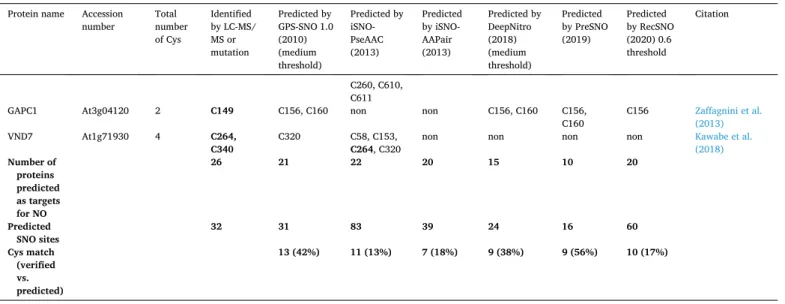
Although MS allows the direct identification of the modified cysteine residues, cysteine mutants are often additionally analysed, especially if the physiological function of the S-nitrosated protein needs to be char- acterized. In this case, knock-out/knock-down plants of the NO-sensitive protein is complemented with corresponding cysteine mutants to get hints to the physiological function of the S-nitrosated proteins and to verify the NO-sensitivity of the cysteine residue(s) in vivo. This approach is the gold standard for characterisation of protein S-nitrosation. How- ever, because of different reasons such as in vivo analyses are not always possible, e. g. if knock-out/knock-down lines are not available. In this case, recombinant proteins of the cysteine mutants can be produced and analysed for their NO-sensitivity, provided, that enzymatic or functional assays are available. Until now, 32 NO-sensitive cysteine residues have been identified/verified in 26 plant proteins by MS or by generation and
analysis of cysteine mutants (Table 2). We have chosen these 26 proteins to compare the prediction efficiency of the available SNO site prediction software. Table 2 shows that the different computational programs have predicted SNO sites in the selected proteins with different efficiency.
GPS-SNO, iSNO-PseAAC, iSNO-AAPair and RecSNO identified between 20 and 22 of the 26 analysed proteins as targets for S-nitrosation, whereas DeepNitro and PreSNO identified 15 and 10, respectively.
Moreover, the first published online tool for SNO site detection, GPS- SNO, as well as the newer tools DeepNitro and PreSNO predict 31, 24 and 16 putative SNO sites, respectively, including 13 (GPS-SNO) and 9 (DeepNitro and PreSNO) verified SNO sites. These three prediction tools have a hit rate (number of matched SNO sites divided by the total number of predicted SNO sites) of 42% (GPS-SNO), 38% (DeepNitro) and 56% (PreSNO). The other computational tools, such as iSNO- PseAAC, iSNO-AAPair or RecSNO predict a much higher number of putative NO-sensitive cysteine residues - 83, 39, and 60, respectively – whereas only 11 (iSNO-PseAAC), 7 (iSNO-AAPair) and 10 (RecSNO) are matching with experimentally identified/verified SNO sites. This quite high rate of mis-prediction is making these three tools less useful. The prediction efficiency of the different online tools is further characterized by calculating their sensitivity (Sn), specificity (Sp) and accuracy (AC) as described by Nilamyani et al. (2021) (Table 3). Sensitivity is the pro- portion of true positives that are correctly identified by the prediction algorithm, specificity is the proportion of the true negatives correctly identified by the software and accuracy is the proportion of true results, either true positive or true negative, in a population (Wihinen, 2012).
Table 2 (continued) Protein name Accession
number Total
number of Cys
Identified by LC-MS/
MS or mutation
Predicted by GPS-SNO 1.0 (2010) (medium threshold)
Predicted by iSNO- PseAAC (2013)
Predicted by iSNO- AAPair (2013)
Predicted by DeepNitro (2018) (medium threshold)
Predicted by PreSNO (2019)
Predicted by RecSNO (2020) 0.6 threshold
Citation
C260, C610, C611
GAPC1 At3g04120 2 C149 C156, C160 non non C156, C160 C156,
C160 C156 Zaffagnini et al.
(2013)
VND7 At1g71930 4 C264,
C340 C320 C58, C153,
C264, C320 non non non non Kawabe et al.
(2018) Number of
proteins predicted as targets for NO
26 21 22 20 15 10 20
Predicted
SNO sites 32 31 83 39 24 16 60
Cys match (verified vs. predicted)
13 (42%) 11 (13%) 7 (18%) 9 (38%) 9 (56%) 10 (17%)
Table 3
Values of sensitivity, specificity and accuracy of SNO predicting software tools.
Metrics were calculated based on the predictions in 26 experimentally identified S-nitrosated plant proteins listed in Table 2.
Software Sensitivity (Sn,
%) Specificity (Sp,
%) Accuracy (Acc,
%) GPS-SNO (medium
threshold) 46.07 67.48 61.59
iSNO-PseAAC 42.66 45.14 46.89
iSNO-AAPair 26.00 63.92 56.64
DeepNitro (medium
threshold) 24.66 47.96 43.38
PreSNO 29.20 29.96 29.77
RecSNO (0.6 threshold) 33.20 42.69 44.00
4.2. Tools for computational prediction of tyrosine nitration sites and testing their performance
The first software for predicting YNO2 sites in proteins using the FASTA format of peptide sequence was GPS-YNO2 1.0 which was pub- lished in 2011 by Liu and co-workers (Liu et al., 2011). The algorithm is based on the biochemical properties of neighbouring amino acids and it showed promising performance (accuracy of 76.51%, sensitivity of 50.09%, specificity of 80.18%) using leave-one-out validation and 4-, 6-, 8-, 10-fold cross-validations. The tool can be used online or as a local
package both implemented in JAVA. It is freely available at: http://
yno2.biocuckoo.org/.
In 2014, a novel predictor algorithm called iNitro-Tyr was developed (Xu et al., 2014). It is based on the incorporation of the position-specific dipeptide propensity into the general pseudo amino acid composition which allows the proper discrimination of the YNO2 sites from the non-nitrated ones. It was demonstrated by the rigorous jackknife tests that iNitroTyr shows higher success rate and stability and is less noisy than GPS-YNO2. This algorithm indicates the total number of tyrosine residues within the protein sequence which is useful information.
Table 4
List of plant proteins in which the nitrated tyrosine residues have been experimentally identified. Nitration sites in the listed proteins were computationally predicted using GSP-YNO2 1.0, iNitro-Tyr, DeepNitro, PredNTS software. Bold indicates matched tyrosine reidue.
Protein name Accesion
number Total
number of Tyr
Identified by LC-MS/MS or mutation
Predicted by GPS-YNO2 (2011) (medium threshold)
Predicted by iNitro-Tyr (2014)
Predicted by DeepNitro (2018) (medium threshold)
Predicted by PredNTS
(2021) Citation
MS1 At5g17920 26 Y287 Y463, Y469,
Y698 Y141, Y623,
Y650 Y141, Y287,
Y463 Y8, Y132, Y141, Y161, Y188, Y226, Y243, Y287, Y453, Y463, Y581, Y740
Lozano-Juste et al. (2011)
OASA1 At4g14880 7 Y302 Y158 non Y302 Y20, Y91,Y143, Y158,
Y192, Y203, Y302
Alvarez et al. ´ (2011)
psbA AtCg00020 12 Y262 Y73, Y107,
Y237, Y246 Y246 Y237, Y246 Y262 Galetskiy et al.
(2011) IDH (NADP) Q6R6M7 Pisum
sativum 14 Y392 Y69, Y210,
Y221, Y274 Y172, Y185, Y221, Y233, Y241, Y259, Y274
Y274 Y43, Y69, Y141, Y172, Y185, Y210, Y221, Y233, Y274, Y392
Begara-Morales et al. (2013a) APX, cytosolic P48534 Pisum
sativum 7 Y5, Y235 Y5 Y5, Y93 non Y5, Y12, Y224, Y235 Begara-Morales
et al. (2013b) HPR, peroxisomal At1g68010 11 Y97, Y108,
Y198 Y10, Y108,
Y150 Y10,Y150,
Y251 Y97, Y180 Y97 Corpas et al.
(2013b)
PYR1 At4g17870 4 Y23, Y58,
Y120 non non Y23 Y23, Y58, Y120, Y143 Castillom et al.,
2015 MnSOD1,
mitochondrial At3g10920 10 Y38, Y40,
Y63, Y67, Y198, Y199, Y202
Y63, Y226 Y63, Y67,
Y226 Y63 Y63, Y67, Y209, Y221,
Y226 Holzmeister
et al., 2015 Leghemoglobin-1 P02232 Vicia
faba 3 Y25, Y30,
Y133 Y134 non non Y25,Y30, Y134 Sainz et al. (2015)
MDHAR Q66PF9 Pisum
sativum 22 Y213, Y292,
Y345 Y154, Y34 Y7, Y192,
Y292 Y292, Y383 Y7, Y44, Y53, Y89, Y114, Y143, Y154, Y172, Y292, Y305, Y383
Begara-Morales et al. (2015)
PSBO1 At5g66570 8 Y9 Y94, Y102,
Y328 Y236 Y94, Y102,
Y131, Y236 Y94, Y102, Y169, Y328 Takahashi et al.
(2015)
NADP-MAE1 At2g19900 25 Y73 Y129, Y204,
Y235, Y248, Y522, Y528, Y550
Y235, Y263, Y286, Y550, Y580
Y235, Y248,
Y550 Y66, Y92, Y99, Y114, Y235, Y148, Y263, Y343, Y522, Y528, Y550, Y573, Y577, Y580
Begara-Morales et al. (2019)
CDKA1 A0A3L6F4W4
Zea mays 11 Y15, Y19 Y11, Y178,
Y222 Y78, Y231 Y15 Y11, Y15, Y73, Y78,
Y178, Y194 M´endez et al.
(2020)
NIA1 At1g77760 30 Y548, Y614,
Y714, Y771, Y10, Y548,
Y908 Y10, Y83,
Y431, Y851, Y908
Y241, Y266,
Y395, Y624 Y10, Y62, Y82, Y83, Y266, Y286, Y330, Y331, Y333, Y390, Y395, Y397, Y548, Y614, Y624, Y714, Y771, Y802, Y851, Y862, Y908
Costa-Broseta et al. (2021)
NIA2 At1g37130 34 Y545, Y714,
Y771 Y10, Y77,
Y545, Y908 Y77, Y392, Y428, Y568, Y802, Y908
Y182, Y271 Y10, Y76, Y77, Y99, Y182, Y271, Y328, Y387, Y382, Y394, Y545, Y611, Y621, Y771, Y802, Y819, Y822, Y843, Y851, Y908
Costa-Broseta et al. (2021)
Number of proteins predicted as targets for NO
15 14 12 13 15
Predicted YNO2
sites 36 41 41 27 123
Tyr match (verified vs.
predicted)
5 (12%) 4 (10%) 7 (26%) 21 (17%)
iNitroTyr is freely available online at: http://app.aporc.org/iNitro-Tyr/.
In 2018, DeepNitro a predictor simultaneously identifies sites of S- nitrosation, tyrosine nitration and tryptophan nitration has been developed (Xie et al., 2018, see in 3.1).
One of the most recent computational predictors for identifying YNO2 sites is PredNTS published by Nilamyani et al. (2021). The algo- rithm was developed by integrating multiple sequence features including K-mer, composition of k-spaced amino acid pairs, AAindex and binary encoding schemes. Using a comprehensive dataset, PredNTS outperformed the previously developed predictors. The software is freely available at: http://kurata14.bio.kyutech.ac.jp/PredNTS/.
The other recently developed predictor is iNitroY-Deep which uses pseudo amino acid compositions and deep neural networks (DNNs) (Naseer et al., 2021). Using widely-accepted model evaluation mea- sures, iNitroY-Deep outperformed the previously published nitro- tyrosine predictor tools. The web server was established at http://3.15 .230.173/, but at the time of writing this review it is not accessible.
In order to evaluate the performance of the available tyrosine nitration predicting tools, we performed in silico analysis of proteins with nitrated tyrosine residues identified by LC-MS/MS. Among those, 11 proteins were tested by GPS-YNO2 and iNitro-Tyr in our previous work (Kolbert et al., 2017) and the list has been supplemented by recently identified proteins (Table 4). Of the 15 nitrated proteins, 14 were identified as candidates by GPS-YNO2 software, 12 by iNitro-Tyr, 13 by DeepNitro and 15 by PredNTS. In the 15 proteins, 36 YNO2 sites have been experimentally identified and the number of YNO2 sites predicted by the software tools was variable. The DeepNitro tool assigned 27 tyrosine amino acids as candidates for being nitrated (which is the 75% of the experimentally identified sites), while the recently developed PredNTS indicated 104 sites in 15 proteins, which is 3-fold more than the experimentally identified sites. Both GPS-YNO2 and iNitro-Tyr predicted 41 YNO2 sites in 15 proteins. The highest number of YNO2 sites were assigned by PredNTS, and accordingly this tool showed the highest match rate, since one or more predicted nitrated sites matched the experimentally identified ones for 12 of the 15 proteins.
When we calculated the hit rate, we found that those are relatively low, and DeepNitro had the highest hit value (26%). It has to be noted that of the 36 MS-identified YNO2 sites only 18 sites matched the predictions of one of the programmes indicating 50% agreement between in silico and experimental results. This number was significantly lower (only 4 out of 26, 15%) when two software tools (GPS-YNO2 and iNitro-Tyr) were tested (Kolbert et al., 2017). It can be concluded that all available tools are advisable to use for a certain protein in order to predict as many YNO2 sites as possible.
Furthermore, based on the previously identified 15 nitrated proteins, sensitivity, specificity and accuracy were calculated in order to evaluate the performance of the software tools (Table 5). The highest Sn value (~62%) was obtained in case of PredNTS, but the Sp and AC values of this tool were relatively low. The highest AC value was shown by the DeepNitro software supporting its better performance compared to the other programmes. In general, the above mentioned values are relatively low which indicates that the agreement of the in silico predictions with experimental data is moderate. This is partly due to the limitations of
MALDI based methods used for identifying YNO2 sites in proteins (Ytterberg and Jensen, 2010) and to the fact that prediction algorithms do not consider the 3D structures of the proteins which greatly affect the sensitivity to tyrosine nitration.
5. Conclusion and future perspectives
Both S-nitrosation and tyrosine nitration are NO-dependent PTMs affecting plant proteins of various kinds from structural proteins to transporters and enzymes. S-nitrosation is directly involved in cell sig- nalling while tyrosine nitration is thought to result in protein instability and degradation and it may indirectly affect signal transduction. Both PTMs are selective and specific, since not every Cys/Tyr is nitrosated/
nitrated in a protein’s amino acid chain and not every Cys/Tyr- containing proteins are targets of these modifications. In the case of S- nitrosation various consensus amino acid sequences have been sug- gested; however, there is still no general rule explaining which cysteine residue is a target for NO. Similarly, there is no amino acid motif or any definite pattern in the protein structure which determines the target tyrosine for nitration. For both NO-dependent PTMs, some common physico-chemical features have been revealed. In the future, intensive effort should be directed on revealing the high-resolution structure of the microenvironment around each cysteine/tyrosine residue to get in- formation about the physicochemical features that determine S-nitro- sation/tyrosine nitration specificity.
In order to assign the target Cys and Tyr residues within a certain protein, specific computational tools have been developed. In the last ten years, 11 computational tools for predicting S-nitrosation, tyrosine nitration or both based on different algorithms have been created. In Table 1, the number of references indicates that these tools are frequently used by the scientific community. This is not surprising, since the predictors rapidly generate extensive information, while the labo- ratory experiments are lengthy and often technically cumbersome. Our tests on plant proteins showed that there are discrepancies between the experimentally confirmed and the predicted PTM sites, which may be due in part to the fact that the algorithms don’t take into account the 3D protein structure.
Therefore, computational prediction of SNO or YNO2 sites can’t substitute laboratory work but can provide a starting point for experi- mental verification and the combination of computer-based prediction and experimental verification represents still a promising approach for a better understanding of the molecular mechanisms and the regulatory functions of protein S-nitrosation and tyrosine nitration. Before planning experiments, it is advisable to use all the available tools on the proteins of interest and compare the results of the predictions. Based on our analyses on plant proteins, S-nitrosation sites can be predicted by the available tools with higher confidence compared to the sites of tyrosine nitration. However, computational prediction still must be developed further to improve the precision with which S-nitrosation/tyrosine nitration-sites are identified. In this context, probably machine learning systems (artificial intelligence) based on experimentally verified S- nitrosated cysteine residues and nitrated tyrosine residues and 3D pro- tein structures could provide a step further to successful prediction of NO-dependent PTM sites. But all these prediction approaches can finally not replace the experimental analysis of the function of S-nitrosated or tyrosine nitrated proteins, including recombinant proteins, site-directed mutagenesis and in vivo experiments.
Funding
NO research in the Kolbert Lab is supported by the National Research, Development and Innovation Office (Grant no. K135303).
CRediT authorship contribution statement
Zsuzsanna Kolbert: Conceptualization, Formal analysis, Writing – Table 5
Values of sensitivity, specificity and accuracy of YNO2 predicting software tools.
Metrics were calculated based on the predictions in 15 experimentally identified nitrated plant proteins listed in Table 4.
Software Sensitivity (Sn,
%) Specificity (Sp,
%) Accuracy (Acc,
%) GPS-YNO2 (medium
threshold) 10.00 65.70 57.36
iNitro-Tyr 7.40 60.66 52.36
DeepNitro (medium
threshold) 24.21 77.11 62.86
PredNTS 61.76 39.15 49.38