"This accepted author manuscript is copyrighted and published by Elsevier. It is posted here by
agreement between Elsevier and MTA. The definitive version of the text was subsequently published in Seminars in Cell & Developmental Biology, 2019 Jan; 85:122‐131. doi: 10.1016/j.semcdb.2017.12.018..
Available under license CC‐BY‐NC‐ND."
Complement factor H family proteins in their non-canonical role as modulators of cellular functions
Mihály Józsi,1,2 Andrea E. Schneider,1 Éva Kárpáti,1 Noémi Sándor1
1Department of Immunology, ELTE Eötvös Loránd University, Budapest, Hungary;
2MTA-ELTE Immunology Research Group, Department of Immunology, ELTE Eötvös Loránd University, Budapest, Hungary
Corresponding author: Mihály Józsi, Department of Immunology, ELTE Eötvös Loránd University, Pázmány Péter sétány 1/c, H-1117 Budapest, Hungary; Phone: +36 1 3812175; Fax:
+36 1 3812176; E-mail: mihaly.jozsi@ttk.elte.hu.
Abbreviations used: AMD, age-related macular degeneration; CCP, complement control protein domain; CR3, complement receptor type 3 (CD11b/CD18); CR4, complement receptor type 4 (CD11c/CD18); CRP, C-reactive protein; ECM, extracellular matrix; FHR, factor H-related;
GAG, glycosaminoglycan; NET, neutrophil extracellular trap; PTX3, pentraxin 3; ROS, reactive oxygen species
Abstract
Complement factor H is a major regulator of the alternative pathway of the complement system.
The factor H-related proteins are less characterized, but recent data indicate that they rather promote complement activation. These proteins have some common ligands with factor H and have both overlapping and distinct functions depending on domain composition and the degree of conservation of amino acid sequence. Factor H and some of the factor H-related proteins also appear in a non-canonical function that is beyond their role in the modulation of complement activation. This review covers our current understanding on this emerging role of factor H family proteins in modulating the activation and function of various cells by binding to receptors or receptor ligands.
Keywords:
cell activation; complement; complement receptor; factor H; factor H-related protein;
inflammation; innate immunity; neutrophil extracellular traps
1. Introduction
The complement system is an essential humoral arm of innate immunity, with versatile functions involved in maintaining host homeostasis [1, 2]. Complement has a major role in the recognition of microbes and defence against pathogens, which is based on its ability to discriminate self from non-self, and promote opsonophagocytosis and lysis of microbes. Complement also participates in the disposal of immune complexes, dying cells and cell debris, in the regulation of inflammation and activation of various cells. The complement system consists of more than 40 soluble and cell membrane-bound proteins. Complement can be activated through three major pathways in a cascade-like manner [1]. The classical pathway is activated when C1q in the C1 complex binds to selected microbes and host ligands, such as antigen-bound immunoglobulins and pentraxins, and to apoptotic/necrotic cells. The lectin pathway is activated upon the interaction of mannan-binding lectin or ficolins with specific carbohydrates. The alternative pathway is activated constitutively at a low rate by the spontaneous hydrolysis of C3, which is the central molecule of complement. In addition, all activation pathways, via the generation of C3b from C3, feed into the alternative pathway, which then serves as an amplification mechanism of complement activation (Fig. 1). The effector mechanisms of complement are mediated by the direct lytic effect of the common terminal pathway, and by cellular receptors that bind the various activation fragments generated from the complement components during propagation of the activation cascades. Fragments of the central C3 molecule bind to complement receptors type 1 (CR1, binds C3b), type 2 (CR2, binds C3d), types 3 and 4 (CR3 and CR4, bind iC3b), and to the anaphylatoxin receptor C3aR (binds the C3a fragment) [3]. CR3 and CR4 serve as major opsonin receptors on myeloid cells, and have several additional ligands (discussed in more detail in section 3.1). CR2 is part of the B-cell receptor co-receptor complex and its engagement with C3d lowers the threshold for B cell activation.
Regulatory molecules in body fluids and on cell surfaces keep complement activation in check, which otherwise could lead to inflammation and tissue damage. Uncontrolled or misdirected complement activation is implicated in a growing number of diseases, both systemic and organ-specific ones [4, 5]. Dysregulation of the alternative pathway in particular is associated with diseases such as atypical hemolytic uremic syndrome, C3 glomerulopathy and age-related macular degeneration (AMD) [5-8]. The major soluble inhibitor of the alternative pathway is complement factor H (Fig.1) [9].
2. The role of the complement factor H family proteins in the modulation of complement activation
2.1. Factor H as a complement regulator
Factor H is a 155-kDa glycoprotein that consists of 20 complement control protein (CCP) domains, from which the four N-terminal domains provide the complement inhibitory functions of factor H. This activity is realized both in fluid phase and on host cellular and non-cellular surfaces, thus factor H supports the protection of host cells which are in contact with complement (e.g., platelets, erythrocytes, leukocytes and endothelial cells) [10, 11]. To perform this latter function, factor H has to properly discriminate self surfaces from non-self surfaces, which ability highly depends on the most C-terminal part (CCP19-20) of the molecule [12-17]. These domains include binding sites for C3b/C3d that is deposited on a surface, as well as for polyanionic host markers, such as glycosaminoglycans (GAGs) and sialic acid [12, 18-20]. The dual recognition of C3b and GAGs/sialic acid allows docking of factor H on the host surface in a conformation that favours its complement regulatory activity (mediated by the N-terminal domains). CCP7 harbors an additional binding site for negatively charged host markers [21]. These and other domains also mediate important interactions with ligands such as pentraxins, extracellular matrix (ECM)
proteins, malondialdehyde adducts and DNA (Fig. 2) (reviewed in [11, 22]). Interaction of factor H with these ligands is thought to direct the complement inhibitory activity of factor H to sites of ongoing complement activation and inflammation (Fig. 3A) [10, 11, 23]. Factor H polymorphisms, mutations and autoantibodies that influence factor H levels or interaction with certain ligands, or affect its function, were described in association with several diseases [24-27].
Factor H-like protein 1 (FHL-1), a splice variant derived from the CFH gene, includes the CCPs 1-7 of factor H plus four amino acids at its C-terminal end. Thus, FHL-1 can also bind C3b and inhibit complement activation [28]. While this protein has not been extensively investigated, it might be more important than previously appreciated, because recent evidence suggests that at specific anatomic sites, such as the Bruch’s membrane in the eye, FHL-1 may pass this barrier whereas factor H, due to its large size, cannot pass [29]. The CCP7 and CCPs 19-20 domains contain recognition sites for different GAGs, which can target FHL-1 and factor H to different sites; FHL-1 also lacks the sialic acid-binding site present in CCP20 [18, 29].
2.2. The factor H-related (FHR) proteins as positive modulators of complement activation Factor H is a member of a protein family that also includes FHL-1, as well as five factor H- related proteins (FHRs) that are derived from five highly related genes [30]. FHRs are also composed exclusively of CCPs of varying numbers, and show immunological cross-reactivity with each other and with factor H. Each FHR has domains homologous to the CCP19-20 domains of factor H, and some domains homologous to factor H domains CCPs 6-9 (also CCPs 10-14 in FHR-5), but lacks the N-terminal complement regulatory domains (Fig. 2).
The FHRs may share binding ability to some of the factor H ligands due to these homologies [30]. However, in spite of the relatedness between the respective domains, the FHRs may do not, or with markedly different avidity, bind to certain factor H ligands. For example,
while both factor H and FHR-4 bind to C-reactive protein (CRP), factor H binds predominantly the modified monomeric CRP form, whereas FHR-4 binds the native pentameric CRP form [31- 33]. FHR-5 binds pentraxin 3 (PTX3) stronger, and FHR-1 binds PTX3 weaker than factor H, and FHR-4 does not show significant PTX3 binding [34-36]. These ligand binding profiles influence the function of the proteins. FHR-4 can activate the classical pathway via recruited CRP [37]. FHR-5 can inhibit factor H binding to pentraxins and ECM and thus indirectly promote complement activation [34]. FHR-1, FHR-4 and FHR-5 can bind C3b via their C- terminal domains and allow for the formation of the C3bBb alternative pathway C3 convertase and thus activate the alternative pathway, an activity similar to that previously described for properdin (Fig. 3A) [34, 38, 39]. In addition, CCPs 1-2 of FHR-1, FHR-2 and FHR-5 mediate dimerization of the proteins, which increases the avidity of these FHRs to e.g. surface-bound C3b, resulting in more efficient competition with factor H [40-42].
Indeed, the FHR proteins recently emerged as positive complement regulators, in contrast with the inhibiting role of factor H [30]. For several of them the ability to competitively inhibit factor H binding to certain host ligands has been demonstrated. This includes surface-bound C3b, ECM and the pentraxins CRP and PTX3 [34, 38, 40-43]. In addition, FHR-3 was shown to compete with factor H for a bacterial ligand, namely, to inhibit factor H binding to the fHbp protein of Neisseria meningitidis [43].
This gene cluster evolved through segmental duplications, which due to the homologies make this cluster prone to gene rearrangements. These give rise to gene deletions and duplications, as well as partial gene deletions and duplications leading to the formation of hybrid genes, and thus abnormal hybrid proteins. By removing or introducing FHRs as competitors of factor H, by altering their oligomeric structure or exchanging their recognition domains, these changes alter the balance between complement activation and inhibition, and are associated with
a number of human disorders, e.g. the kidney diseases atypical hemolytic uremic syndrome, C3 glomerulopathy (including CFHR5 nephropathy), IgA nephropathy; systemic lupus erythematosus; and also AMD, by conferring susceptibility or protection (reviewed in [30, 44]).
For example, the lack of FHR-1 and FHR-3 due to the prevalent CFHR3-CFHR1 gene deletion is strongly associated with lower risk of developing IgA nephropathy [45] and AMD [46], likely due to the less amount of FHRs to interfere with the complement regulatory activity of factor H [30]. It was very recently shown indeed, that FHR-1 serum levels and the FHR-1:factor H ratio are higher in IgA nephropathy patients compared with healthy controls [47, 48]. Factor H pathogenic variants causing decreased factor H activity and certain haplotypes that determine different factor H and FHR-1/FHR-3 serum levels modulate the FH/FHR balance and influence susceptibility to diseases [48-52].
3. Factor H family proteins in their non-canonical role as modulators of cellular functions In addition to their role in modulating complement activation, factor H family proteins were shown to bind to receptors and modulate cell functions / cellular activation (summarized in Table 1 and Fig. 3B). In the next sections, this non-canonical role of factor H and the FHR proteins will be reviewed and discussed.
Factor H can bind to the surface of various cells through three major mechanisms:
(1) First, as described in section 2.1., factor H binds on host surfaces to acceptor molecules, such as GAGs and sialic acid, which allow attachment of this regulator in order to protect the cells from complement attack. Other examples include host ligands exposed under specific conditions, such as malondialdehyde epitopes due to oxidative modification; DNA, annexin II and histones on apoptotic and necrotic cells [53, 54]; and binding to ECM proteins.
(2) Second, factor H may also bind to host surfaces through soluble host ligands, as exemplified by the recruitment of factor H to apoptotic cells by the pentraxins PTX3 and CRP [33, 35].
(3) Third, factor H can bind to various cells through receptors. Receptor-bound factor H could still act as cofactor for factor I and aid in cellular protection against complement attack [55]. In addition, receptor-bound factor H can mediate or regulate cellular adhesion and activation, i.e.
exert a function not related to its complement-regulatory capacity, as discussed below. Of note, the interaction of factor H with cells is often complex, as described for example for its interaction with platelets, involving both direct interaction with sialic acid, binding mediated by thrombospondin-1 and binding to the GPIIb/IIIa (αIIbβ3 integrin) receptor [56-58].
3.1. CR3 and CR4, the major factor H receptors
Binding of factor H to several cell types is mediated by the complement receptors CR3 (CD11b/CD18) and CR4 (CD11c/CD18) [55, 59-64]. In addition to factor H, these receptors bind a wide variety of other ligands, such as iC3b (the inactivated fragment of C3) fibrinogen, ICAM- 1, factor X, LPS etc. [65-68]. CR3 and CR4 belong to the family of beta2 integrins and are expressed mainly by myeloid cell types (monocytes, macrophages, dendritic cells, neutrophil granulocytes) in humans. They mediate several functions including cell-cell and cell-ECM contacts, proliferation, phagocytosis and transendothelial migration of immune cells [69-71].
Most important of them is the iC3b-mediated phagocytosis that is a crucial step in the elimination of complement-opsonized pathogens and apoptotic cells [72-75].
3.2. Factor H binding to monocytes, macrophages and dendritic cells
Monocytes, macrophages and dendritic cells are essential innate immune cells involved in the initiation and resolution of inflammation, phagocytosis and killing of pathogens, antigen presentation, and instruction of the adaptive immune response.
Early studies described functional effects of factor H on monocytes, without identifying the receptor, such as chemotactic effect [76, 77], stimulation of respiratory burst [78] and induction of IL-1β secretion [79]. Factor H was also shown to stimulate release of thromboxane and prostaglandin E from guinea pig peritoneal macrophages [80]. Direct interaction of factor H and myeloid cells has recently been studied by several groups in more detail. In a murine model of AMD, factor H was found to inhibit the resolution of inflammation by restraining the macrophages at the site of inflammation. This effect was mediated by factor H binding to CR3 on the surface of mononuclear phagocytes, which interaction blocked thrombospondin-1–CD47 signaling that otherwise promotes the elimination of the phagocytes [81]. The immune modulating effect of two factor H variants were also analyzed in the context of AMD. The factor H H402 isoform was found to be more potent in inducing phagocyte accumulation compared to the more common (protective) variant Y402 [81]. In addition, in AMD patients elevated plasma levels of factor H bearing nitrated tyrosines was detected. Purified nitrated factor H showed impaired GAG and C3b binding, loss of cofactor activity, and induced inflammatory IL-8 production in monocytes that had been stimulated with peroxidated lipids [82].
In the context of apoptotic cell clearance, complement plays a major role and factor H seems to play important roles as well [83-85]. The presence of factor H on apoptotic cells under physiological and pathological conditions was demonstrated, and shown to be involved in the regulation of complement activation on dead cells [33, 53, 54, 60, 83]. Early apoptotic cells were found to bind and internalize soluble factor H and use it to increase iC3b deposition on their surface and to enhance their uptake by monocytes [86]. In contrast to this, in
the study of Kang et al., enhanced uptake of the factor H-coated apoptotic material was not observed despite its binding to CR3 on monocytes [60]. In the study of Martin et al., factor H- coated apoptotic cells and nucleosomes induced, via Siglec-9, elevated production of the anti- inflammatory cytokine IL-10 in monocytes; however, the possible role of CR3 in binding factor H and inducing tolerance was not investigated [86]. In addition, the modified (monomeric) form of CRP was shown to recruit factor H to apoptotic cells and particles and promote their uptake by macrophages in an anti-inflammatory manner [33, 83]. The induction of anti-inflammatory cytokine production upon binding of factor H-coated apoptotic cells to CR3 is in good agreement with studies showing that iC3b-coated apoptotic cells induce tolerance via CR3 signaling [87-90].
These studies indicate that factor H has an important role in the silent removal of apoptotic material by enhancing iC3b deposition and inducing tolerogenic signals. How factor H and iC3b – both deposited on the surface of dying cells and both ligands of CR3 – interact in this process needs further investigation; moreover, the role of CR4 needs to be addressed, as well.
The anti-inflammatory effect of cell bound factor H was also demonstrated for dendritic cells. Monocyte-derived dendritic cells (MDCs) differentiated in the presence of factor H did not respond to LPS stimulation with the well known activation markers, such as costimulatory receptor upregulation and pro-inflammatory cytokine production; rather they secreted IL-10 [91].
The authors concluded that factor H has to act on monocytes to induce these changes in MDC behavior, however, this is not achieved by factor H binding to CR3 or CR4 on monocytes [91].
3.3. Factor H and neutrophil granulocytes
Neutrophil granulocytes are rapidly recruited to infected tissue and have several killing mechanism to eliminate pathogens, thus they provide a first line of host cellular defense, and are
involved in a variety of pathophysiological processes [92-94]. Factor H binding to neutrophils was demonstrated by Avery and Gordon in 1993, which binding was found to be specific and could be enhanced by neutrophil activation [95]. This receptor-mediated binding was confirmed by other groups and the β2 integrin CR3 was identified as the major factor H receptor [55, 59, 63]. While factor H can bind to CR4, as well, it is difficult to dissect CR3- and CR4-specific factor H effects on neutrophils because these cells express little amount of CR4. Upon binding to CR3, factor H retains its cofactor activity [55]. Because factor H could serve as an adhesion ligand for neutrophils [59] it is expected to affect several functions of neutrophil granulocytes, as migration, phagocytosis, degranulation and oxidant generation are all influenced by adhesion.
Indeed, immobilized factor H could trigger calcium signal and spreading in adhered neutrophils [55]. DiScipio et al. [59] found that generation of hydrogen peroxide by neutrophils stimulated with C5a or TNFα was enhanced by factor H; lactoferrin release was also enhanced by adherence to factor H and stimulation by TNFα. Soluble factor H did not inhibit the adhesion of neutrophils to immobilized factor H [59]. In addition, factor H itself was shown to support neutrophil migration [55]. Immobilized factor H significantly enhanced the release of IL-8, a known migratory chemokine, by neutrophils, while for soluble factor H a similar effect was not observed. In contrast, the generation of reactive oxygen species (ROS) and the release of neutrophil extracellular traps (NETs) by neutrophils stimulated with phorbol 12-myristate 13- acetate (PMA) or with immobilized fibronectin plus β-glucan were inhibited by immobilized factor H [55]. NETs can trap microorganism that are too large to be phagocytosed, and also play a direct role in killing pathogens [92, 94, 96]. Thus, factor H present in the body fluids appears not to stimulate neutrophils, only when deposited on a pathogen surface or in a tissue; it may affect the recruitment and activation of these inflammatory cells. On the other hand, factor H
could reduce host damage caused by an inflammatory environment through the inhibition of NET and ROS production.
3.4. Factor H family proteins in the interaction of phagocytes with pathogens
Complement factor H can also influence the immune response by modulating the interaction of phagocytes with pathogens (reviewed in [97]). Several microbes were shown to be able to bind factor H, e.g. Pseudomonas aeruginosa, Borrelia burgdorferi, Francisella tularensis, Candida albicans, Neisseria meningitidis, etc. [97-102]. On one hand, pathogens can avoid complement attack and opsonophagocytosis by recruiting factor H on their surface [97, 103-107]. On the other hand, factor H can enhance adhesion of the pathogen to phagocytes by binding to specific receptors [62, 63].
Candida albicans-bound factor H was shown to enhance the binding of the yeast to neutrophil granulocytes, and to enhance neutrophil migration, ROS production, phagocytosis and fungal killing [63]. These effects were mediated via factor H binding to CR3 on neutrophils.
FHL-1, FHR-1 and FHR-4A were also shown to bind to neutrophils; FHL-1 and FHR-1 bind via CR3, whereas the FHR-4A receptor is distinct from CR3. Similar to factor H, FHR-1 when bound to C. albicans, supported neutrophil migration, enhanced adhesion, phagocytosis and antimicrobial activities, such as lactoferrin release, ROS production and fungal killing. Although FHL-1 could also enhance neutrophil adhesion to yeast cells, it had no measurable effect on migration and did not affect the antimicrobial activities under the given experimental conditions.
For FHR-4A, no such functional activities were found [63].
Factor H-opsonized C. albicans acted similarly on macrophages, namely, factor H enhanced the production of the pro-inflammatory cytokines IL-1β and IL-6 by macrophages exposed to yeasts [64]. However, this pathogen can release a protease that – among others –
cleaves and inactivates factor H and its receptors on macrophages [64]. The pro-inflammatory cytokine production of macrophages was elevated also by stimulation with factor H-coated Mycobacterium bovis compared to stimulation with the bacteria only, although factor H inhibited the uptake of the bacteria by the cells [108].
Recently, extracellular DNA trap formation in the case of monocytes (MoETs) when exposed to C. albicans was demonstrated and, like NETs, these MoETs were shown to activate complement and bear factor H bound to the extracellular DNA [109]. While deposited factor H in neutrophils induced IL-8 production, MoET-associated factor H had an anti-inflammatory action on monocytes by blocking their IL-1β production [55, 109]. Thus, it is possible that by interacting with surface-bound factor H, neutrophils can alert and recruit other cells to the site of infection or inflammation through secretion of the chemotactic cytokine IL-8, while monocytes take control over the extent of inflammation.
In summary, while some pathogens can exploit host factor H to their benefit in order to down-regulate opsonization, factor H can promote interaction between phagocytes and pathogens in a direct receptor-mediated manner. Since complement activation is a rapid process, and bound factor H can effectively reduce the amount of deposited C3b and C3b-derived opsonins on the pathogen surface, it is likely that microbial complement evasion is particularly relevant in body fluids where complement is abundant and the microbes can more easily disseminate. The role of factor H in modulating phagocyte responses in the context of infection is more likely to be relevant in the tissues. In this way, factor H can protect the host not only by limiting complement- mediated damage, but also by mediating cellular responses, including the regulation of the inflammatory microenvironment and the elimination of the pathogens. Further studies will also be needed to investigate whether and how other pattern-recognition receptors, particularly Toll-
like receptors expressed on the phagocytes take part in these processes and influence the effect of factor H bound on the microbes.
3.5. Factor H, FHR-3 and B cells
Factor H was shown to bind to B lymphocytes, but the factor H receptor(s) could not be identified at the molecular level [110-112]. Factor H was shown to induce complement factor I release from the B cells [113], block immunoglobulin secretion [114], as well as support their proliferation [115]. These early studies, however, were not followed upon, thus a direct B cell activation modulating role of factor H remains uncertain.
An indirect role of modulating B cell activation was recently shown for FHR-3. Like other FHR proteins, FHR-3 can bind to certain fragments of C3b. In their study, Buhlmann et al.
demonstrated that FHR-3 can block the activation of B cells by inhibiting C3d binding to the B- cell co-receptor CR2, whereas factor H and FHR-1 showed no such effect [116].
While data regarding the interaction and modulatory function of factor H family proteins on B cells are scarce, such mechanisms may provide a further link between innate and adaptive immunity, and may prove relevant in disease settings, such as certain autoimmune disorders. For example, the deletion of the CFHR1 gene has been found to predispose to the autoimmune form of atypical hemolytic uremic syndrome, which is associated with the appearance of pathogenic autoantibodies against factor H, possibly due to lack of induction of tolerance against FHR-1 [117]. Similarly, the loss of the FHR-3 and FHR-1 proteins due to deletion of the CFHR3 and CFHR1 genes predisposes to systemic lupus erythematosus [118]. Further studies are needed to clarify the role of these proteins in developing autoantibodies and study the possibility of their direct effects on B cells.
3.6. Other cell types
Factor H and FHL-1 were shown to interact also with some non-immune cells via specific receptors. The binding of factor H to CR3 was shown to promote the adherence of Streptococcus pneumoniae to epithelial cells and facilitate the uptake of pneumococci by these cells (also by neutrophils) [62]. In an in vitro model, factor H also facilitated the adherence of Neisseria gonorrhoeae to CHO cells transfected with CR3; however, FHL-1 and FHR-1 could not bridge gonococci with the CR3-CHO cells [61]. FHL-1 was demonstrated to support spreading and attachment of three anchorage-dependent cells lines, CCL-64 mink lung epithelial-like cells, C32 human melanoma cells, and MRC-5 human fibroblast-like cells, presumably via integrin receptors [119].
Thus, factor H and other factor H family proteins may bind to and modulate the activity of non-immune cells, as well. Additional factor H receptors, such as L-selectin [120], were also proposed, which need to be further investigated in the future.
4. Conclusion
By regulating and modulating complement activation, factor H, FHL-1 and the FHR proteins influence opsonization and opsonophagocytosis of host and nonhost particles and cells [30, 44, 97]. In their additional role, these proteins bind to cellular receptors and regulate cell activation and inflammation. Apparently, depending on the context, e.g. the nature of additional stimuli like pathogens, factor H can exert both pro- and anti-inflammatory activities on immune cells. Recent exciting studies signal a renewed interest in such cellular effects of particularly factor H. Future studies are expected to advance our understanding of the mechanisms of receptor-mediated cellular functions of the factor H family proteins and answer the question, to what extent this non-canonical role of factor H or the FHRs contribute to diseases, such as it was suggested for
AMD. This knowledge may be applied in the future to manipulate these proteins or their receptor/ligand interactions in a therapeutically useful way.
Acknowledgements
The work of the authors was supported by the Lendület program of the Hungarian Academy of Sciences (grant nr. LP2012-43) and the National Research, Development and Innovation Office (grant nr. K 109055).
References
[1] N.S. Merle, S.E. Church, V. Fremeaux-Bacchi, L.T. Roumenina, Complement System Part I - Molecular Mechanisms of Activation and Regulation, Front Immunol 6 (2015) 262.
[2] D. Ricklin, G. Hajishengallis, K. Yang, J.D. Lambris, Complement: a key system for immune surveillance and homeostasis, Nat Immunol 11(9) (2010) 785-97.
[3] A. Erdei, N. Sandor, B. Macsik-Valent, S. Lukacsi, M. Kremlitzka, Z. Bajtay, The versatile functions of complement C3-derived ligands, Immunol Rev 274(1) (2016) 127-140.
[4] N.S. Merle, R. Noe, L. Halbwachs-Mecarelli, V. Fremeaux-Bacchi, L.T. Roumenina, Complement System Part II: Role in Immunity, Front Immunol 6 (2015) 257.
[5] D. Ricklin, E.S. Reis, J.D. Lambris, Complement in disease: a defence system turning offensive, Nat Rev Nephrol 12(7) (2016) 383-401.
[6] J.M. Thurman, V.M. Holers, The central role of the alternative complement pathway in human disease, J Immunol 176(3) (2006) 1305-10.
[7] P.F. Zipfel, S. Heinen, M. Jozsi, C. Skerka, Complement and diseases: defective alternative pathway control results in kidney and eye diseases, Mol Immunol 43(1-2) (2006) 97-106.
[8] D. Ricklin, J.D. Lambris, Complement in immune and inflammatory disorders:
pathophysiological mechanisms, J Immunol 190(8) (2013) 3831-8.
[9] S. Rodriguez de Cordoba, J. Esparza-Gordillo, E. Goicoechea de Jorge, M. Lopez-Trascasa, P. Sanchez-Corral, The human complement factor H: functional roles, genetic variations and disease associations, Mol Immunol 41(4) (2004) 355-67.
[10] V.P. Ferreira, M.K. Pangburn, C. Cortes, Complement control protein factor H: the good, the bad, and the inadequate, Mol Immunol 47(13) (2010) 2187-97.
[11] A. Kopp, M. Hebecker, E. Svobodova, M. Jozsi, Factor h: a complement regulator in health and disease, and a mediator of cellular interactions, Biomolecules 2(1) (2012) 46-75.
[12] B.S. Blaum, J.P. Hannan, A.P. Herbert, D. Kavanagh, D. Uhrin, T. Stehle, Structural basis for sialic acid-mediated self-recognition by complement factor H, Nat Chem Biol 11(1) (2015) 77-82.
[13] V.P. Ferreira, A.P. Herbert, H.G. Hocking, P.N. Barlow, M.K. Pangburn, Critical role of the C-terminal domains of factor H in regulating complement activation at cell surfaces, J Immunol 177(9) (2006) 6308-16.
[14] M. Jozsi, M. Oppermann, J.D. Lambris, P.F. Zipfel, The C-terminus of complement factor H is essential for host cell protection, Mol Immunol 44(10) (2007) 2697-706.
[15] T. Kajander, M.J. Lehtinen, S. Hyvarinen, A. Bhattacharjee, E. Leung, D.E. Isenman, S.
Meri, A. Goldman, T.S. Jokiranta, Dual interaction of factor H with C3d and glycosaminoglycans in host-nonhost discrimination by complement, Proc Natl Acad Sci U S A 108(7) (2011) 2897- 902.
[16] H.P. Morgan, C.Q. Schmidt, M. Guariento, B.S. Blaum, D. Gillespie, A.P. Herbert, D.
Kavanagh, H.D. Mertens, D.I. Svergun, C.M. Johansson, D. Uhrin, P.N. Barlow, J.P. Hannan, Structural basis for engagement by complement factor H of C3b on a self surface, Nat Struct Mol Biol 18(4) (2011) 463-70.
[17] M. Oppermann, T. Manuelian, M. Jozsi, E. Brandt, T.S. Jokiranta, S. Heinen, S. Meri, C.
Skerka, O. Gotze, P.F. Zipfel, The C-terminus of complement regulator Factor H mediates target recognition: evidence for a compact conformation of the native protein, Clin Exp Immunol 144(2) (2006) 342-52.
[18] A. Langford-Smith, A.J. Day, P.N. Bishop, S.J. Clark, Complementing the Sugar Code: Role of GAGs and Sialic Acid in Complement Regulation, Front Immunol 6 (2015) 25.
[19] C.Q. Schmidt, A.P. Herbert, H.G. Hocking, D. Uhrin, P.N. Barlow, Translational mini- review series on complement factor H: structural and functional correlations for factor H, Clin Exp Immunol 151(1) (2008) 14-24.
[20] C.Q. Schmidt, A.P. Herbert, D. Kavanagh, C. Gandy, C.J. Fenton, B.S. Blaum, M. Lyon, D.
Uhrin, P.N. Barlow, A new map of glycosaminoglycan and C3b binding sites on factor H, J Immunol 181(4) (2008) 2610-9.
[21] T.K. Blackmore, T.A. Sadlon, H.M. Ward, D.M. Lublin, D.L. Gordon, Identification of a heparin binding domain in the seventh short consensus repeat of complement factor H, J Immunol 157(12) (1996) 5422-7.
[22] E. Makou, A.P. Herbert, P.N. Barlow, Functional anatomy of complement factor H, Biochemistry 52(23) (2013) 3949-62.
[23] A.P. Sjoberg, L.A. Trouw, A.M. Blom, Complement activation and inhibition: a delicate balance, Trends Immunol 30(2) (2009) 83-90.
[24] M. Jozsi, S. Reuter, P. Nozal, M. Lopez-Trascasa, P. Sanchez-Corral, Z. Prohaszka, B.
Uzonyi, Autoantibodies to complement components in C3 glomerulopathy and atypical hemolytic uremic syndrome, Immunol Lett 160(2) (2014) 163-71.
[25] C.J. Boon, N.C. van de Kar, B.J. Klevering, J.E. Keunen, F.P. Cremers, C.C. Klaver, C.B.
Hoyng, M.R. Daha, A.I. den Hollander, The spectrum of phenotypes caused by variants in the CFH gene, Mol Immunol 46(8-9) (2009) 1573-94.
[26] M.A. Dragon-Durey, C. Loirat, S. Cloarec, M.A. Macher, J. Blouin, H. Nivet, L. Weiss, W.H. Fridman, V. Fremeaux-Bacchi, Anti-Factor H autoantibodies associated with atypical hemolytic uremic syndrome, J Am Soc Nephrol 16(2) (2005) 555-63.
[27] S. Meri, V. Koistinen, A. Miettinen, T. Tornroth, I.J. Seppala, Activation of the alternative pathway of complement by monoclonal lambda light chains in membranoproliferative
glomerulonephritis, J Exp Med 175(4) (1992) 939-50.
[28] P.F. Zipfel, C. Skerka, FHL-1/reconectin: a human complement and immune regulator with cell-adhesive function, Immunol Today 20(3) (1999) 135-40.
[29] S.J. Clark, L.A. Ridge, A.P. Herbert, S. Hakobyan, B. Mulloy, R. Lennon, R. Wurzner, B.P.
Morgan, D. Uhrin, P.N. Bishop, A.J. Day, Tissue-specific host recognition by complement factor H is mediated by differential activities of its glycosaminoglycan-binding regions, J Immunol 190(5) (2013) 2049-57.
[30] M. Jozsi, A. Tortajada, B. Uzonyi, E. Goicoechea de Jorge, S. Rodriguez de Cordoba, Factor H-related proteins determine complement-activating surfaces, Trends Immunol 36(6) (2015) 374- 84.
[31] S. Hakobyan, C.L. Harris, C.W. van den Berg, M.C. Fernandez-Alonso, E.G. de Jorge, S.R.
de Cordoba, G. Rivas, P. Mangione, M.B. Pepys, B.P. Morgan, Complement factor H binds to denatured rather than to native pentameric C-reactive protein, J Biol Chem 283(45) (2008) 30451-60.
[32] M. Mihlan, M. Hebecker, H.M. Dahse, S. Halbich, M. Huber-Lang, R. Dahse, P.F. Zipfel, M. Jozsi, Human complement factor H-related protein 4 binds and recruits native pentameric C- reactive protein to necrotic cells, Mol Immunol 46(3) (2009) 335-44.
[33] M. Mihlan, S. Stippa, M. Jozsi, P.F. Zipfel, Monomeric CRP contributes to complement control in fluid phase and on cellular surfaces and increases phagocytosis by recruiting factor H, Cell Death Differ 16(12) (2009) 1630-40.
[34] A.I. Csincsi, A. Kopp, M. Zoldi, Z. Banlaki, B. Uzonyi, M. Hebecker, J.J. Caesar, M.C.
Pickering, K. Daigo, T. Hamakubo, S.M. Lea, E. Goicoechea de Jorge, M. Jozsi, Factor H-related protein 5 interacts with pentraxin 3 and the extracellular matrix and modulates complement activation, J Immunol 194(10) (2015) 4963-73.
[35] L. Deban, H. Jarva, M.J. Lehtinen, B. Bottazzi, A. Bastone, A. Doni, T.S. Jokiranta, A.
Mantovani, S. Meri, Binding of the long pentraxin PTX3 to factor H: interacting domains and function in the regulation of complement activation, J Immunol 181(12) (2008) 8433-40.
[36] A. Kopp, S. Strobel, A. Tortajada, S. Rodriguez de Cordoba, P. Sanchez-Corral, Z.
Prohaszka, M. Lopez-Trascasa, M. Jozsi, Atypical hemolytic uremic syndrome-associated variants and autoantibodies impair binding of factor h and factor h-related protein 1 to pentraxin 3, J Immunol 189(4) (2012) 1858-67.
[37] M. Hebecker, A.I. Okemefuna, S.J. Perkins, M. Mihlan, M. Huber-Lang, M. Jozsi,
Molecular basis of C-reactive protein binding and modulation of complement activation by factor H-related protein 4, Mol Immunol 47(6) (2010) 1347-55.
[38] A.I. Csincsi, Z. Szabo, Z. Banlaki, B. Uzonyi, M. Cserhalmi, E. Karpati, A. Tortajada, J.J.E.
Caesar, Z. Prohaszka, T.S. Jokiranta, S.M. Lea, S. Rodriguez de Cordoba, M. Jozsi, FHR-1 Binds to C-Reactive Protein and Enhances Rather than Inhibits Complement Activation, J Immunol 199(1) (2017) 292-303.
[39] M. Hebecker, M. Jozsi, Factor H-related protein 4 activates complement by serving as a platform for the assembly of alternative pathway C3 convertase via its interaction with C3b protein, J Biol Chem 287(23) (2012) 19528-36.
[40] Q. Chen, M. Wiesener, H.U. Eberhardt, A. Hartmann, B. Uzonyi, M. Kirschfink, K. Amann, M. Buettner, T. Goodship, C. Hugo, C. Skerka, P.F. Zipfel, Complement factor H-related hybrid protein deregulates complement in dense deposit disease, J Clin Invest 124(1) (2014) 145-55.
[41] E. Goicoechea de Jorge, J.J. Caesar, T.H. Malik, M. Patel, M. Colledge, S. Johnson, S.
Hakobyan, B.P. Morgan, C.L. Harris, M.C. Pickering, S.M. Lea, Dimerization of complement factor H-related proteins modulates complement activation in vivo, Proc Natl Acad Sci U S A 110(12) (2013) 4685-90.
[42] A. Tortajada, H. Yebenes, C. Abarrategui-Garrido, J. Anter, J.M. Garcia-Fernandez, R.
Martinez-Barricarte, M. Alba-Dominguez, T.H. Malik, R. Bedoya, R. Cabrera Perez, M. Lopez Trascasa, M.C. Pickering, C.L. Harris, P. Sanchez-Corral, O. Llorca, S. Rodriguez de Cordoba, C3 glomerulopathy-associated CFHR1 mutation alters FHR oligomerization and complement regulation, J Clin Invest 123(6) (2013) 2434-46.
[43] J.J. Caesar, H. Lavender, P.N. Ward, R.M. Exley, J. Eaton, E. Chittock, T.H. Malik, E.
Goiecoechea De Jorge, M.C. Pickering, C.M. Tang, S.M. Lea, Competition between antagonistic complement factors for a single protein on N. meningitidis rules disease susceptibility, Elife 3 (2014).
[44] N. Medjeral-Thomas, M.C. Pickering, The complement factor H-related proteins, Immunol Rev 274(1) (2016) 191-201.
[45] A.G. Gharavi, K. Kiryluk, M. Choi, Y. Li, P. Hou, J. Xie, S. Sanna-Cherchi, C.J. Men, B.A.
Julian, R.J. Wyatt, J. Novak, J.C. He, H. Wang, J. Lv, L. Zhu, W. Wang, Z. Wang, K. Yasuno, M. Gunel, S. Mane, S. Umlauf, I. Tikhonova, I. Beerman, S. Savoldi, R. Magistroni, G.M.
Ghiggeri, M. Bodria, F. Lugani, P. Ravani, C. Ponticelli, L. Allegri, G. Boscutti, G. Frasca, A.
Amore, L. Peruzzi, R. Coppo, C. Izzi, B.F. Viola, E. Prati, M. Salvadori, R. Mignani, L.
Gesualdo, F. Bertinetto, P. Mesiano, A. Amoroso, F. Scolari, N. Chen, H. Zhang, R.P. Lifton, Genome-wide association study identifies susceptibility loci for IgA nephropathy, Nat Genet 43(4) (2011) 321-7.
[46] A.E. Hughes, N. Orr, H. Esfandiary, M. Diaz-Torres, T. Goodship, U. Chakravarthy, A common CFH haplotype, with deletion of CFHR1 and CFHR3, is associated with lower risk of age-related macular degeneration, Nat Genet 38(10) (2006) 1173-7.
[47] N.R. Medjeral-Thomas, H.J. Lomax-Browne, H. Beckwith, M. Willicombe, A.G. McLean, P. Brookes, C.D. Pusey, M. Falchi, H.T. Cook, M.C. Pickering, Circulating complement factor H-related proteins 1 and 5 correlate with disease activity in IgA nephropathy, Kidney Int 92(4) (2017) 942-952.
[48] A. Tortajada, E. Gutierrez, E. Goicoechea de Jorge, J. Anter, A. Segarra, M. Espinosa, M.
Blasco, E. Roman, H. Marco, L.F. Quintana, J. Gutierrez, S. Pinto, M. Lopez-Trascasa, M. Praga, S. Rodriguez de Cordoba, Elevated factor H-related protein 1 and factor H pathogenic variants decrease complement regulation in IgA nephropathy, Kidney Int 92(4) (2017) 953-963.
[49] M.E. Bernabeu-Herrero, M. Jimenez-Alcazar, J. Anter, S. Pinto, D. Sanchez Chinchilla, S.
Garrido, M. Lopez-Trascasa, S. Rodriguez de Cordoba, P. Sanchez-Corral, Complement factor H, FHR-3 and FHR-1 variants associate in an extended haplotype conferring increased risk of atypical hemolytic uremic syndrome, Mol Immunol 67(2 Pt B) (2015) 276-86.
[50] S. Davila, V.J. Wright, C.C. Khor, K.S. Sim, A. Binder, W.B. Breunis, D. Inwald, S. Nadel, H. Betts, E.D. Carrol, R. de Groot, P.W. Hermans, J. Hazelzet, M. Emonts, C.C. Lim, T.W.
Kuijpers, F. Martinon-Torres, A. Salas, W. Zenz, M. Levin, M.L. Hibberd, Genome-wide
association study identifies variants in the CFH region associated with host susceptibility to meningococcal disease, Nat Genet 42(9) (2010) 772-6.
[51] J. Esparza-Gordillo, J.M. Soria, A. Buil, L. Almasy, J. Blangero, J. Fontcuberta, S.
Rodriguez de Cordoba, Genetic and environmental factors influencing the human factor H plasma levels, Immunogenetics 56(2) (2004) 77-82.
[52] L. Zhu, Y.L. Zhai, F.M. Wang, P. Hou, J.C. Lv, D.M. Xu, S.F. Shi, L.J. Liu, F. Yu, M.H.
Zhao, J. Novak, A.G. Gharavi, H. Zhang, Variants in Complement Factor H and Complement Factor H-Related Protein Genes, CFHR3 and CFHR1, Affect Complement Activation in IgA Nephropathy, J Am Soc Nephrol 26(5) (2015) 1195-204.
[53] J. Leffler, A.P. Herbert, E. Norstrom, C.Q. Schmidt, P.N. Barlow, A.M. Blom, M. Martin, Annexin-II, DNA, and histones serve as factor H ligands on the surface of apoptotic cells, J Biol Chem 285(6) (2010) 3766-76.
[54] L.A. Trouw, A.A. Bengtsson, K.A. Gelderman, B. Dahlback, G. Sturfelt, A.M. Blom, C4b- binding protein and factor H compensate for the loss of membrane-bound complement inhibitors to protect apoptotic cells against excessive complement attack, J Biol Chem 282(39) (2007) 28540-8.
[55] A.E. Schneider, N. Sandor, E. Karpati, M. Jozsi, Complement factor H modulates the activation of human neutrophil granulocytes and the generation of neutrophil extracellular traps, Mol Immunol 72 (2016) 37-48.
[56] S. Hyvarinen, S. Meri, T.S. Jokiranta, Disturbed sialic acid recognition on endothelial cells and platelets in complement attack causes atypical hemolytic uremic syndrome, Blood 127(22) (2016) 2701-10.
[57] A.L. Stahl, F. Vaziri-Sani, S. Heinen, A.C. Kristoffersson, K.H. Gydell, R. Raafat, A.
Gutierrez, O. Beringer, P.F. Zipfel, D. Karpman, Factor H dysfunction in patients with atypical
hemolytic uremic syndrome contributes to complement deposition on platelets and their activation, Blood 111(11) (2008) 5307-15.
[58] F. Vaziri-Sani, J. Hellwage, P.F. Zipfel, A.G. Sjoholm, R. Iancu, D. Karpman, Factor H binds to washed human platelets, J Thromb Haemost 3(1) (2005) 154-62.
[59] R.G. DiScipio, P.J. Daffern, I.U. Schraufstatter, P. Sriramarao, Human polymorphonuclear leukocytes adhere to complement factor H through an interaction that involves alphaMbeta2 (CD11b/CD18), J Immunol 160(8) (1998) 4057-66.
[60] Y.H. Kang, B.C. Urban, R.B. Sim, U. Kishore, Human complement Factor H modulates C1q-mediated phagocytosis of apoptotic cells, Immunobiology 217(4) (2012) 455-64.
[61] S. Agarwal, S. Ram, J. Ngampasutadol, S. Gulati, P.F. Zipfel, P.A. Rice, Factor H facilitates adherence of Neisseria gonorrhoeae to complement receptor 3 on eukaryotic cells, J Immunol 185(7) (2010) 4344-53.
[62] V. Agarwal, T.M. Asmat, S. Luo, I. Jensch, P.F. Zipfel, S. Hammerschmidt, Complement regulator Factor H mediates a two-step uptake of Streptococcus pneumoniae by human cells, J Biol Chem 285(30) (2010) 23486-95.
[63] J. Losse, P.F. Zipfel, M. Jozsi, Factor H and factor H-related protein 1 bind to human neutrophils via complement receptor 3, mediate attachment to Candida albicans, and enhance neutrophil antimicrobial activity, J Immunol 184(2) (2010) 912-21.
[64] E. Svoboda, A.E. Schneider, N. Sandor, U. Lermann, P. Staib, M. Kremlitzka, Z. Bajtay, D.
Barz, A. Erdei, M. Jozsi, Secreted aspartic protease 2 of Candida albicans inactivates factor H and the macrophage factor H-receptors CR3 (CD11b/CD18) and CR4 (CD11c/CD18), Immunol Lett 168(1) (2015) 13-21.
[65] M.S. Diamond, J. Garcia-Aguilar, J.K. Bickford, A.L. Corbi, T.A. Springer, The I domain is a major recognition site on the leukocyte integrin Mac-1 (CD11b/CD18) for four distinct
adhesion ligands, J Cell Biol 120(4) (1993) 1031-43.
[66] A. Mazzone, G. Ricevuti, Leukocyte CD11/CD18 integrins: biological and clinical relevance, Haematologica 80(2) (1995) 161-75.
[67] J.A. Van Strijp, D.G. Russell, E. Tuomanen, E.J. Brown, S.D. Wright, Ligand specificity of purified complement receptor type three (CD11b/CD18, alpha m beta 2, Mac-1). Indirect effects of an Arg-Gly-Asp (RGD) sequence, J Immunol 151(6) (1993) 3324-36.
[68] V.P. Yakubenko, V.K. Lishko, S.C. Lam, T.P. Ugarova, A molecular basis for integrin alphaMbeta 2 ligand binding promiscuity, J Biol Chem 277(50) (2002) 48635-42.
[69] E.S. Harris, T.M. McIntyre, S.M. Prescott, G.A. Zimmerman, The leukocyte integrins, J Biol Chem 275(31) (2000) 23409-12.
[70] S. Schmidt, M. Moser, M. Sperandio, The molecular basis of leukocyte recruitment and its deficiencies, Mol Immunol 55(1) (2013) 49-58.
[71] S.M. Tan, The leucocyte beta2 (CD18) integrins: the structure, functional regulation and signalling properties, Biosci Rep 32(3) (2012) 241-69.
[72] Z. Bajtay, C. Speth, A. Erdei, M.P. Dierich, Cutting edge: productive HIV-1 infection of dendritic cells via complement receptor type 3 (CR3, CD11b/CD18), J Immunol 173(8) (2004) 4775-8.
[73] V. Le Cabec, S. Carreno, A. Moisand, C. Bordier, I. Maridonneau-Parini, Complement receptor 3 (CD11b/CD18) mediates type I and type II phagocytosis during nonopsonic and opsonic phagocytosis, respectively, J Immunol 169(4) (2002) 2003-9.
[74] A.E. Morelli, A.T. Larregina, W.J. Shufesky, A.F. Zahorchak, A.J. Logar, G.D. Papworth, Z. Wang, S.C. Watkins, L.D. Falo, Jr., A.W. Thomson, Internalization of circulating apoptotic
cells by splenic marginal zone dendritic cells: dependence on complement receptors and effect on cytokine production, Blood 101(2) (2003) 611-20.
[75] P.C. Patel, R.E. Harrison, Membrane ruffles capture C3bi-opsonized particles in activated macrophages, Mol Biol Cell 19(11) (2008) 4628-39.
[76] K. Nabil, B. Rihn, M.C. Jaurand, J.M. Vignaud, J. Ripoche, Y. Martinet, N. Martinet, Identification of human complement factor H as a chemotactic protein for monocytes, Biochem J 326 ( Pt 2) (1997) 377-83.
[77] H. Ohtsuka, T. Imamura, M. Matsushita, S. Tanase, H. Okada, M. Ogawa, T. Kambara, Thrombin generates monocyte chemotactic activity from complement factor H, Immunology 80(1) (1993) 140-5.
[78] R.E. Schopf, K.P. Hammann, O. Scheiner, E.M. Lemmel, M.P. Dierich, Activation of human monocytes by both human beta 1H and C3b, Immunology 46(2) (1982) 307-12.
[79] D. Iferroudjene, M.T. Schouft, C. Lemercier, D. Gilbert, M. Fontaine, Evidence for an active hydrophobic form of factor H that is able to induce secretion of interleukin 1-beta or by human monocytes, Eur J Immunol 21(4) (1991) 967-72.
[80] H.P. Hartung, U. Hadding, D. Bitter-Suermann, D. Gemsa, Release of prostaglandin E and thromboxane from macrophages by stimulation with factor H, Clin Exp Immunol 56(2) (1984) 453-8.
[81] B. Calippe, S. Augustin, F. Beguier, H. Charles-Messance, L. Poupel, J.B. Conart, S.J. Hu, S. Lavalette, A. Fauvet, J. Rayes, O. Levy, W. Raoul, C. Fitting, T. Denefle, M.C. Pickering, C.
Harris, S. Jorieux, P.M. Sullivan, J.A. Sahel, P. Karoyan, P. Sapieha, X. Guillonneau, E.L.
Gautier, F. Sennlaub, Complement Factor H Inhibits CD47-Mediated Resolution of Inflammation, Immunity 46(2) (2017) 261-272.
[82] M. Krilis, M. Qi, M.C. Madigan, J.W.H. Wong, M. Abdelatti, R.H. Guymer, J. Whitelock, P. McCluskey, P. Zhang, J. Qi, A.P. Hunyor, S.A. Krilis, B. Giannakopoulos, Nitration of tyrosines in complement factor H domains alters its immunological activity and mediates a pathogenic role in age related macular degeneration, Oncotarget 8(30) (2017) 49016-49032.
[83] D. Gershov, S. Kim, N. Brot, K.B. Elkon, C-Reactive protein binds to apoptotic cells, protects the cells from assembly of the terminal complement components, and sustains an antiinflammatory innate immune response: implications for systemic autoimmunity, J Exp Med 192(9) (2000) 1353-64.
[84] M. Martin, A.M. Blom, Complement in removal of the dead - balancing inflammation, Immunol Rev 274(1) (2016) 218-232.
[85] D. Mevorach, J.O. Mascarenhas, D. Gershov, K.B. Elkon, Complement-dependent clearance of apoptotic cells by human macrophages, J Exp Med 188(12) (1998) 2313-20.
[86] M. Martin, J. Leffler, K.I. Smolag, J. Mytych, A. Bjork, L.D. Chaves, J.J. Alexander, R.J.
Quigg, A.M. Blom, Factor H uptake regulates intracellular C3 activation during apoptosis and decreases the inflammatory potential of nucleosomes, Cell Death Differ 23(5) (2016) 903-11.
[87] J. Schmidt, C. Klempp, M.W. Buchler, A. Marten, Release of iC3b from apoptotic tumor cells induces tolerance by binding to immature dendritic cells in vitro and in vivo, Cancer Immunol Immunother 55(1) (2006) 31-8.
[88] M. Skoberne, S. Somersan, W. Almodovar, T. Truong, K. Petrova, P.M. Henson, N.
Bhardwaj, The apoptotic-cell receptor CR3, but not alphavbeta5, is a regulator of human dendritic-cell immunostimulatory function, Blood 108(3) (2006) 947-55.
[89] J.H. Sohn, P.S. Bora, H.J. Suk, H. Molina, H.J. Kaplan, N.S. Bora, Tolerance is dependent on complement C3 fragment iC3b binding to antigen-presenting cells, Nat Med 9(2) (2003) 206- 12.
[90] I. Verbovetski, H. Bychkov, U. Trahtemberg, I. Shapira, M. Hareuveni, O. Ben-Tal, I.
Kutikov, O. Gill, D. Mevorach, Opsonization of apoptotic cells by autologous iC3b facilitates clearance by immature dendritic cells, down-regulates DR and CD86, and up-regulates CC chemokine receptor 7, J Exp Med 196(12) (2002) 1553-61.
[91] R. Olivar, A. Luque, S. Cardenas-Brito, M. Naranjo-Gomez, A.M. Blom, F.E. Borras, S.
Rodriguez de Cordoba, P.F. Zipfel, J.M. Aran, The Complement Inhibitor Factor H Generates an Anti-Inflammatory and Tolerogenic State in Monocyte-Derived Dendritic Cells, J Immunol 196(10) (2016) 4274-90.
[92] E. Kolaczkowska, P. Kubes, Neutrophil recruitment and function in health and inflammation, Nat Rev Immunol 13(3) (2013) 159-75.
[93] A. Mocsai, Diverse novel functions of neutrophils in immunity, inflammation, and beyond, J Exp Med 210(7) (2013) 1283-99.
[94] C. Nathan, Neutrophils and immunity: challenges and opportunities, Nat Rev Immunol 6(3) (2006) 173-82.
[95] V.M. Avery, D.L. Gordon, Characterization of factor H binding to human polymorphonuclear leukocytes, J Immunol 151(10) (1993) 5545-53.
[96] V. Brinkmann, U. Reichard, C. Goosmann, B. Fauler, Y. Uhlemann, D.S. Weiss, Y.
Weinrauch, A. Zychlinsky, Neutrophil extracellular traps kill bacteria, Science 303(5663) (2004) 1532-5.
[97] M. Jozsi, Factor H Family Proteins in Complement Evasion of Microorganisms, Front Immunol 8 (2017) 571.
[98] A. Ben Nasr, G.R. Klimpel, Subversion of complement activation at the bacterial surface promotes serum resistance and opsonophagocytosis of Francisella tularensis, J Leukoc Biol 84(1) (2008) 77-85.
[99] P. Kraiczy, Hide and Seek: How Lyme Disease Spirochetes Overcome Complement Attack, Front Immunol 7 (2016) 385.
[100] J.D. Lambris, D. Ricklin, B.V. Geisbrecht, Complement evasion by human pathogens, Nat Rev Microbiol 6(2) (2008) 132-42.
[101] M. Pizza, R. Rappuoli, Neisseria meningitidis: pathogenesis and immunity, Curr Opin Microbiol 23 (2015) 68-72.
[102] G. Vogl, I. Lesiak, D.B. Jensen, S. Perkhofer, R. Eck, C. Speth, C. Lass-Florl, P.F. Zipfel, A.M. Blom, M.P. Dierich, R. Wurzner, Immune evasion by acquisition of complement inhibitors:
the mould Aspergillus binds both factor H and C4b binding protein, Mol Immunol 45(5) (2008) 1485-93.
[103] G. Agrahari, Z. Liang, K. Glinton, S.W. Lee, V.A. Ploplis, F.J. Castellino, Streptococcus pyogenes Employs Strain-dependent Mechanisms of C3b Inactivation to Inhibit Phagocytosis and Killing of Bacteria, J Biol Chem 291(17) (2016) 9181-9.
[104] M.C. Gustafsson, J. Lannergard, O.R. Nilsson, B.M. Kristensen, J.E. Olsen, C.L. Harris, R.L. Ufret-Vincenty, M. Stalhammar-Carlemalm, G. Lindahl, Factor H binds to the hypervariable region of many Streptococcus pyogenes M proteins but does not promote phagocytosis resistance or acute virulence, PLoS Pathog 9(4) (2013) e1003323.
[105] C. Hyams, K. Trzcinski, E. Camberlein, D.M. Weinberger, S. Chimalapati, M.
Noursadeghi, M. Lipsitch, J.S. Brown, Streptococcus pneumoniae capsular serotype invasiveness correlates with the degree of factor H binding and opsonization with C3b/iC3b, Infect Immun 81(1) (2013) 354-63.
[106] T.J. Inzana, R. Balyan, M.D. Howard, Decoration of Histophilus somni lipooligosaccharide with N-acetyl-5-neuraminic acid enhances bacterial binding of complement factor H and
resistance to killing by serum and polymorphonuclear leukocytes, Vet Microbiol 161(1-2) (2012) 113-21.
[107] J.A. Sharp, C.G. Echague, P.S. Hair, M.D. Ward, J.O. Nyalwidhe, J.A. Geoghegan, T.J.
Foster, K.M. Cunnion, Staphylococcus aureus surface protein SdrE binds complement regulator factor H as an immune evasion tactic, PLoS One 7(5) (2012) e38407.
[108] M. Abdul-Aziz, A.G. Tsolaki, L. Kouser, M.V. Carroll, M.N. Al-Ahdal, R.B. Sim, U.
Kishore, Complement factor H interferes with Mycobacterium bovis BCG entry into
macrophages and modulates the pro-inflammatory cytokine response, Immunobiology 221(9) (2016) 944-52.
[109] L.D. Halder, M.A. Abdelfatah, E.A. Jo, I.D. Jacobsen, M. Westermann, N. Beyersdorf, S.
Lorkowski, P.F. Zipfel, C. Skerka, Factor H Binds to Extracellular DNA Traps Released from Human Blood Monocytes in Response to Candida albicans, Front Immunol 7 (2017) 671.
[110] A. Erdei, R.B. Sim, Complement factor H-binding protein of Raji cells and tonsil B lymphocytes, Biochem J 246(1) (1987) 149-56.
[111] J.D. Lambris, G.D. Ross, Characterization of the lymphocyte membrane receptor for factor H (beta 1H-globulin) with an antibody to anti-factor H idiotype, J Exp Med 155(5) (1982) 1400- 11.
[112] J. Ripoche, A. Erdei, D. Gilbert, A. Al Salihi, R.B. Sim, M. Fontaine, Two populations of complement factor H differ in their ability to bind to cell surfaces, Biochem J 253(2) (1988) 475- 80.
[113] J.D. Lambris, N.J. Dobson, G.D. Ross, Release of endogenous C3b inactivator from lymphocytes in response to triggering membrane receptors for beta 1H globulin, J Exp Med 152(6) (1980) 1625-44.
[114] G.C. Tsokos, G. Inghirami, C.D. Tsoukas, J.E. Balow, J.D. Lambris, Regulation of
immunoglobulin secretion by factor H of human complement, Immunology 55(3) (1985) 419-26.
[115] K.P. Hammann, A. Raile, M. Schmitt, H.H. Mussel, H. Peters, O. Scheiner, M.P. Dierich, beta 1H stimulates mouse-spleen B lymphocytes as demonstrated by increased thymidine incorporation and formation of B cell blasts, Immunobiology 160(3-4) (1981) 289-301.
[116] D. Buhlmann, H.U. Eberhardt, A. Medyukhina, W.M. Prodinger, M.T. Figge, P.F. Zipfel, C. Skerka, FHR3 Blocks C3d-Mediated Coactivation of Human B Cells, J Immunol 197(2) (2016) 620-9.
[117] A. Bhattacharjee, S. Reuter, E. Trojnar, R. Kolodziejczyk, H. Seeberger, S. Hyvarinen, B.
Uzonyi, A. Szilagyi, Z. Prohaszka, A. Goldman, M. Jozsi, T.S. Jokiranta, The major autoantibody epitope on factor H in atypical hemolytic uremic syndrome is structurally different from its homologous site in factor H-related protein 1, supporting a novel model for induction of autoimmunity in this disease, J Biol Chem 290(15) (2015) 9500-10.
[118] J. Zhao, H. Wu, M. Khosravi, H. Cui, X. Qian, J.A. Kelly, K.M. Kaufman, C.D. Langefeld, A.H. Williams, M.E. Comeau, J.T. Ziegler, M.C. Marion, A. Adler, S.B. Glenn, M.E. Alarcon- Riquelme, B.A. Pons-Estel, J.B. Harley, S.C. Bae, S.Y. Bang, S.K. Cho, C.O. Jacob, T.J. Vyse, T.B. Niewold, P.M. Gaffney, K.L. Moser, R.P. Kimberly, J.C. Edberg, E.E. Brown, G.S.
Alarcon, M.A. Petri, R. Ramsey-Goldman, L.M. Vila, J.D. Reveille, J.A. James, G.S. Gilkeson, D.L. Kamen, B.I. Freedman, J.M. Anaya, J.T. Merrill, L.A. Criswell, R.H. Scofield, A.M.
Stevens, J.M. Guthridge, D.M. Chang, Y.W. Song, J.A. Park, E.Y. Lee, S.A. Boackle, J.M.
Grossman, B.H. Hahn, T.H. Goodship, R.M. Cantor, C.Y. Yu, N. Shen, B.P. Tsao, Association of genetic variants in complement factor H and factor H-related genes with systemic lupus
erythematosus susceptibility, PLoS Genet 7(5) (2011) e1002079.
[119] J. Hellwage, S. Kuhn, P.F. Zipfel, The human complement regulatory factor-H-like protein 1, which represents a truncated form of factor H, displays cell-attachment activity, Biochem J 326 ( Pt 2) (1997) 321-7.
[120] R. Malhotra, M. Ward, R.B. Sim, M.I. Bird, Identification of human complement Factor H as a ligand for L-selectin, Biochem J 341 ( Pt 1) (1999) 61-9.
[121] R. Parente, S.J. Clark, A. Inforzato, A.J. Day, Complement factor H in host defense and immune evasion, Cell Mol Life Sci 74(9) (2017) 1605-1624.
Figure legends
Fig. 1. The role of factor H family proteins in complement regulation.
The complement system can be activated via three major pathways, the classical, lectin and alternative pathways. These activation pathways merge upon the cleavage of C3 into the anaphylatoxin C3a and opsonin C3b by the C3 convertase enzymes of the classical/lectin pathways (C4b2b) and the alternative pathway (C3bBb). The generated C3b can feed back to the alternative pathway via the so-called amplification loop by forming additional C3bBb convertases. By binding to the C3 convertases, C3b also allows formation of the C5 convertase enzymes that generate from C5 the anaphylatoxin C5a, and the C5b fragment, which can initiate the terminal pathway and ultimately form the lytic C5b-9 complex. Factor H and FHL-1 act as cofactors for factor I for the enzymatic inactivation of C3b, and also inhibit the alternative pathway C3 convertase and the C5 convertases. In contrast to this negative regulatory role, the FHR proteins FHR-1, FHR-4 and FHR-5 enhance complement activation by competitive inhibition of factor H and/or enhance the formation of the C3bBb convertase by binding C3b.
Fig. 2. Schematic overview of the factor H family proteins.
Factor H is composed of 20 complement control protein (CCP) domains, of which the N-terminal 4 CCPs mediate the complement inhibiting activity of the protein. Factor H-like 1 (FHL-1) is identical with the CCPs 1-7 of factor H, plus has four unique amino acids at its C-terminus. The major ligand binding sites are indicated with horizontal lines below the corresponding domains of factor H/FHL-1. The factor H-related FHR proteins are composed of 4 to 9 CCP domains, displaying varying degrees of amino acid sequence identity to certain factor H domains (indicated by vertical alignment; identical colors indicate complete or very high degree of sequence identity). The CCPs 1-2 of FHR-1, FHR-2 and FHR-5 (shown in green) are closely related to
each other and mediate dimerization of these FHRs. Due to the conservation of domains homologous to factor H CCPs 6-9 and 18-20 among the FHRs, FHRs can bind some of the ligands of factor H, thus factor H and the FHRs have partly overlapping ligand binding profiles.
GAGs, glycosaminoglycans; MDA, malondialdehyde; CRP, C-reactive protein; PTX3, pentraxin 3; CR, complement receptor.
Fig. 3. Interactions of factor H family proteins.
(A) Factor H, by binding to its main complement ligand C3b, inhibits complement activation.
This activity is exploited by tumour cells and certain microbes via sequestering factor H. Factor H also interacts with a number of host ligands that allow targeting of the complement inhibitory activity to inflammatory sites and certain cellular (e.g, platelets, endothelial cells, apoptotic and necrotic cells) and non-cellular (ECM and basement membranes) surfaces. The FHR proteins may directly activate complement by binding C3b and allowing formation of the alternative pathway C3 convertase C3bBb (FHR-1, FHR-4, FHR-5) and also compete with factor H for binding to deposited C3b and selected ligands, such as pentraxins and ECM. FHR-3 was shown to inhibit factor H binding to Neisseria meningitidis. Thus, the balance between complement activation and inhibition is shifted towards inhibition by factor H and towards activation by FHRs. Future studies will clarify the exact ligand binding profiles, factor H-inhibiting capacity and functional overlaps/redundancy of the FHR proteins, as well as the role of FHRs in complement evasion of tumour cells and microbes. Note that not all reported factor H ligands are included in the figure; for a more comprehensive overview see e.g. [10, 11, 121]. ECM, extracellular matrix; GAGs, glycosaminoglycans; MDA, malondialdehyde; CRP, C-reactive protein; PTX3, pentraxin 3;
(B) Schematic overview of the current knowledge of the interaction of factor H family proteins with immune cells and the receptors mediating binding of these proteins. Note that GAG/sialic acid-mediated binding of factor H to cells is not shown, only interactions with cellular receptors.
Factor H may bind through receptors to other cell types, such as epithelial cells, as well. CR4 is weakly expressed on neutrophils (shown in grey). CR, complement receptor.
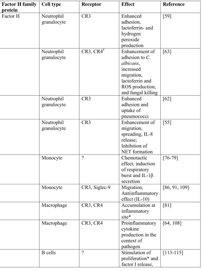
Table 1. Summary of the cellular roles of factor H family proteins Factor H family
protein
Cell type Receptor Effect Reference
Factor H Neutrophil granulocyte
CR3 Enhanced adhesion,
lactoferrin- and hydrogen peroxide production
[59]
Neutrophil granulocyte
CR3, CR4# Enhancement of adhesion to C.
albicans, increased migration, lactoferrin and ROS production, and fungal killing
[63]
Neutrophil granulocyte
CR3 Enhanced adhesion and
uptake of pneumococci
[62]
Neutrophil granulocyte
CR3 Enhancement of
migration, spreading, IL-8 release;
Inhibition of NET formation
[55]
Monocyte ? Chemotactic
effect; induction of respiratory burst and IL-1β secretion
[76-79]
Monocyte CR3, Siglec-9 Migration,
Antiinflammatory effect (IL-10)
[86, 91, 109]
Macrophage CR3, CR4 Accumulation at
inflammatory site*
[81]
Macrophage CR3, CR4 Proinflammatory
cytokine
production in the context of pathogen
[64, 108]
B cells ? Stimulation of
proliferation* and factor I release,
[113-115]
inhibition of immunoglobulin secretion
Epithelial cells CR3 Enhanced
adhesion and uptake of pneumococci
[62]
Platelets GPIIb/IIIa ? (possibly
complement inhibition)
[58]
FHL-1 Epithelial-, melanoma and
fibroblast cell lines
? Spreading, attachment
[119]
Neutrophil granulocyte
CR3, CR4# Adhesion [63]
FHR-1 Neutrophil granulocyte
CR3 Enhancement of
adhesion to C.
albicans, increased migration, lactoferrin and ROS production, and fungal killing
[63]
FHR-3 B cell (FHR-3 binds to
the CR2-ligand C3d)
FHR-3, by binding C3d, blocks co- activation of the cells
[116]
* Described for murine cells
# CR4 is weakly expressed on neutrophils.
Fig. 1.
Fig. 2.
Fig. 3.