Investigation of the molecular background of alternative pathway dysregulation in thrombotic microangiopathies
Ph.D. thesis
Eszter Trojnar, M.D.
Basic and Translational Medicine Doctoral School Semmelweis University
Supervisor: Zoltán Prohászka, M.D., D.Sc.
Official reviewers: Csaba Kálmán Ambrus, M.D., Ph.D.
Judit Müller, M.D., Ph.D.
Head of the Complex Exam Committee: Zoltán Benyó, M.D., D.Sc.
Members of the Complex Exam Committee: Hargita Hegyesi, Ph.D.
Noémi Sándor, Ph.D.
Budapest
2019
Introduction
The complement system is an integral part of the innate immune system that contributes to the elimination of invading pathogens, immune complexes and maintains continuous clearance of redundant cellular debris together with the regulation of the innate immune response.
Complement activation may occur via the simultaneous or distinct initiation of three major pathways, the classical, the lectin and the alternative pathways (CP, LP and AP, respectively).
While each of the activation pathways has its specific triggers of activation and plays a well- defined role in the effector functions of complement, full-blown activation of the individual pathways converges on the level of the C3 molecule, cleavage of which initiates the mutual, terminal pathway of complement and leads to the attack and lysis of the target molecule.
The AP plays a dual role in the activation of complement. On the one hand, it has a baseline, continuous activation, whereas on the other hand the AP may amplify the intensity of complement activation launched through the CP and LP. The significance of AP activation is corroborated by its quantitative role in the augmentation of complement activity. Irrespective of individual pathway initiation, the majority of surface-bound C3b (the major opsonin of the complement system) is generated by the amplification loop of the AP. Since the thus produced C3b may be deposited on all surfaces, including intact host tissues, their protection from complement attack requires the regulation of complement activity.
Restrain of complement activity may take place via multiple mechanisms. Under physiological conditions sufficient control of excessive complement activation is provided by specific fluid phase and surface-bound regulators of the complement system that are capable of complement inhibition as well as discrimination of host tissues from foreign structures.
Regulation may occur via direct inhibition of the activator proteases, substrate-limited restrain of pathway initiation, decay acceleration of the formed C3 convertases and cofactor-mediated cleavage of complement activator proteins. Moreover, inhibition of the terminal pathway or inactivation of the released anaphylatoxins also provides sufficient restrain of complement. In addition to intrinsic complement regulators fluid phase pattern recognition molecules (PRMs) such as pentraxin-3 (PTX3) and C-reactive protein (CRP) may participate in the regulation of the complement system, too.
The impairment of AP regulatory mechanisms can easily lead to complement dysregulation and disease pathogenesis. Reduction of complement regulatory mechanisms may be caused by the dysfunction of the complement regulator proteins (e.g. drop-out of restrain mechanisms or hyperfunction of complement activators) or may be attributed to factor consumption secondary to overactivation of the complement system.
Thrombotic microangiopathies (TMA) are rare but life-threatening disorders characterized by microangiopathic hemolytic anemia and acute thrombocytopenia with or without organ impairment. Endothelial damage and subsequent microvascular thrombosis are key pathogenic factors of TMA that have been associated with complement overactivation in all etiological forms of this disease.
Traditionally atypical hemolytic uremic syndrome (aHUS) was recognized as the first TMA form mediated by complement dysregulation. Atypical HUS is mediated by the dysregulation of the AP, evoked either by pathogenic mutations in the complement genes or by the presence of autoantibodies directed against the complement regulator factor H (FH). Anti-FH antibodies in aHUS have first been reported in a French patient cohort in 2005, and since then multiple independent investigations have shown their functional relevance in disease pathogenesis. Upon binding to FH, autoantibodies interfere with the complement regulatory activity on the surface of host cells that leads to overactivation of the complement AP and subsequent complement-mediated tissue damage. Whereas the functional consequence of the antibody binding to FH is widely studied, detailed structural analysis of where the aHUS- associated FH epitopes are localized at the amino acid level are still lacking.
In addition to aHUS, as a result of intensive research of the recent decades, overactivation of the complement and coagulation systems has been recognized in all etiological forms of TMA. A shared characteristic of the coagulation and complement cascades is their initial activation at local sites of infection or tissue injury. There is a considerable interaction between the activator mechanisms of both cascades, provided by the diverse ligand binding capacity of certain PRMs and activation enzymes of complement, augmented by complement activation through various coagulation factors. Components of each system may cleave and activate one another, thus providing molecular basis for the joint manifestation of pathological thrombosis and complement overactivation in TMA.
Complement dysregulation or consumption in the acute phase of TMA are accompanied by an overdrawn inflammatory response that may induce among others the production of inflammatory proteins, such as CRP and PTX3. Ligand-bound pentraxins were shown to initiate as well as regulate all three pathways of complement in vitro. However, in vivo studies reported inconclusive data on the overall impact of PTX3 on tissue injury and recovery and no study has been designed so far to explore changes in the systemic level of pentraxins in relation to complement overactivation involving human subjects or to analyze the in vivo relation of PTX3 and CRP to complement activity.
Objectives
1. Analysis of the association between systemic pentraxin levels and laboratory signs of disease activity in TMA
Albeit both PTX3 and CRP have been described to regulate complement activation in vitro, no study has been designed so far to explore changes in the systemic level of pentraxins in complement mediated diseases, such as TMAs, in vivo. Therefore we performed a case control study to determine the systemic levels of PTX3 and CRP in patients at the acute phase as well as remission of TMA. We analyzed the relationship between the systemic level of pentraxins and TMA etiology, the clinical outcome of patients and classical laboratory markers of TMA.
2. Investigation of the relation of complement overactivation and systemic pentraxin levels in distinct forms of TMAs
In distinct etiological groups of TMAs including aHUS, Shiga toxin-producing Escherichia coli infection-associated HUS (STEC-HUS), secondary TMA and thrombotic thrombocytopenic purpura (TTP) we further investigated the relation of systemic pentraxin levels to laboratory signs of complement overactivation and consumption in the acute phase of disease. We determined the patients’ complement factor levels, the whole complement CP and AP activities and the systemic levels of complement activation products to reveal any potential association between complement overactivation and the systemic levels of PTX3 and CRP in the acute phase and remission of TMA, as well as in the distinct subgroups of disease. Furthermore we determined the effect of excess PTX3 on the AP activity in vitro to explore the role of PTX3 release in the pathogenesis of TMA.
3. Epitope analysis of anti-FH antibodies in atypical hemolytic uremic syndrome
The functional consequence of the FH-autoantibody interaction (loss of host cell recognition and dysregulation of the AP) has been widely studied, however little is known about the structural basis of FH-blockade and the role of FH-related (FHR) proteins in disease pathogenesis. Based on recent observations we hypothesized that the antibody binding site is located at the C-terminal domains of FH, therefore we aimed for the structural characterization of antibody binding to FH short consensus repeat (SCR) domains 19-20. To localize the aHUS-associated FH epitopes on the amino acid level, we performed fine epitope mapping of the complement regulator using point-mutated FH domains and linear epitope mapping with overlapping synthetic peptides. We further investigated whether aHUS specific linear epitopes are present on FHR1, based on its homology to FH and its described cross-reactivity with the anti-FH antibodies.
Methods
Patient selection, sample collection and study design
171 TMA patients with acute disease flare were enrolled in the case-control study for the analysis of systemic pentraxin levels and complement consumption, whose blood samples were sent to our laboratory for differential diagnostic evaluation between November 2007 and October 2017. Acute phase serum and plasma samples from all enrolled subjects were collected prior to the start of plasma exchange therapy, however in some cases (N=16) fresh frozen plasma (FFP) had been administered to the patients prior to sampling. Diagnosis of TMA was established based on laboratory signs of microangiopathic hemolytic anemia, thrombocytopenia (<150 G/L) and clinical or laboratory signs of organ damage. Patients were included in the study only if all of the above criteria were met. For stratification of TMA patients by disease etiology the following groups were formed: (1) STEC-HUS (N=34): acute gastroenteritis and signs of acute kidney injury with proof of Shiga like-toxin producing Escherichia coli infection, (2) TTP (N=30): ADAMT13 deficiency (activity below 10%) with the presence of ADAMTS13 inhibitors, (3) aHUS (N=44): HUS with presence of anti-FH autoantibodies, or HUS with identified pathogenic or likely pathogenic variations in the complement genes (CFH, CFHR5, CFI, CD46, C3, CFB), THBD or DGKE, or HUS cases without identified likely pathogenic rare variations in these genes, (4) secondary TMA (N=63): evidence of coexisting disease including malignancy, autoimmune disease, sepsis, solid organ transplantation, open heart surgery or malignant hypertension. Exclusion criteria were ongoing plasma exchange or complement inhibitory therapy at the time of sample collection (during the first acute flare), or the lack of available blood sample. Data on the clinical course, blood count and chemistry were collected from the medical charts. Patients were followed-up after hospital discharge and outcome including mortality was registered.
Follow-up samples were available for aHUS (N=31, 15-month median time interval between sampling) and TTP (N=19, 8-month median time interval between sampling) patients in remission. For appropriate comparison age and sex matched healthy individuals were selected, none of whom showed clinical or laboratory signs of TMA or an acute phase reaction that could have influenced the measured laboratory parameters.
For epitope mapping of FH-antibodies, sera of children in the acute phase of autoimmune aHUS were obtained prior to the start of plasmapheresis. Inclusion criteria were as follows:
diagnosis of HUS based on the presence of microangiopathic hemolytic anemia, thrombocytopenia (<150G/L) and renal injury, with an anti-FH antibody level over 110 AU/mL. Exclusion criteria included aHUS in remission or active therapy with either
plasmapheresis, corticosteroids, cyclophosphamide or rituximab as well as the lack of available serum sample. Altogether eight patients were enrolled in this investigation, seven of which had an available remission phase serum sample, too. Remission phase sample collection was performed 6-12 months after the termination of any specific treatment of the subjects. Control sera samples were obtained from leftover serum specimens from children (median age of 9 years) admitted to the 1st Department of Pediatrics at Semmelweis University upon distinct indications, by whom a detailed laboratory analysis (including inflammatory markers) did not reveal any pathological findings. All control children were negative (below cutoff) for anti-FH antibodies.
Both studies were carried out in conformity with the Helsinki Declaration. Written informed consent was obtained from all participants, and the study was approved by the Ethics Committee on Human Clinical Research in Budapest (8361-1/2011-EKU).
Determination of laboratory parameters in acute phase TMA patients
Complement activity-, component-, regulator-, and activation product determinations, CRP and PTX3 measurements were performed in our laboratory from the blood samples of acute phase TMA patients. The AP activity was determined with the commercially available WIESLAB Alternative pathway ELISA kit (EuroDiagnostica, Malmö, Sweden), whereas total complement CP activity was assessed using the sheep-erythrocyte hemolytic titration test.
CRP, C3 and C4 levels were measured by turbidimetry (Beckman Coulter, Brea, CA), while factor B (FB) and factor I (FI) levels were determined by radial immunodiffusion assay. The level of the complement regulators C1q and FH as well as the titer of the anti-FH antibody were measured using in house ELISA techniques. ADAMTS13 activity was evaluated by the application of the fluorogenic substrate FRETS-VWF73. Commercially available kits were used to assess the levels of the complement activation products sC5b-9 and C3a (C3a des-arg) (Quidel, San Diego, CA, USA) and for the measurement of PTX3 (R&D systems Minneapolis, MN, USA).
In vitro assessment of PTX3 effect on AP activation
In vitro effect of recombinant human PTX3 on AP activation was assessed using two assays.
The AP hemolytic activity in the presence of excess PTX3 was determined with a modified C3 nephritic factor hemolytic assay. The assay was performed on washed sheep erythrocytes, where patients’ samples were replaced by normal human serum (NHS) spiked with recombinant human PTX3 (R&D systems Minneapolis, MN, USA) in gradually decreasing concentrations. After a 20 minute incubation of PTX3 with NHS, the solution was added to sheep erythrocytes. The formation of the C3 convertase was allowed within a 10 minute
incubation time at 30oC, and assembly of the terminal pathway membrane-attack complex (C5b-9) was achieved by the addition of undiluted rat serum to the cells. The extent of hemolysis was detected by reading the OD at 412 nm. The AP activity and subsequent assembly of C5b-9 on a plastic surface was measured by the WIESLAB Alternative pathway ELISA kit, using lipopolysaccharide (LPS)-coated ELISA plates with or without excess PTX3. As in the hemolytic assay, patients’ sera were replaced by PTX3 spiked NHS, otherwise the assay was performed according to the manufacturer’s instructions. To allow comparison of data, the AP activities in each experiment were expressed as ratio of the reference (optical density of NHS with buffer control) in percentage.
Peptide synthesis for epitope mapping and determination of antibody binding to the immobilized synthetic peptides of FH and FHR1
Fifteen amino acid-long peptides of FH SCR domains 19–20 and the FHR1 region homolog to the amino acid sequence 1177–1211 on FH (amino acids 276–310 of FHR1) were synthesized by Dr. Katalin Uray at Eötvös Loránd University Budapest, according to Geysen’s method. Peptides overlapping by five amino acid residues were synthesized in duplicates on β‐alanyl‐glycine functionalized polyethylene pins (Mimotopes NCP gears (Clayton, VIC, Australia)) with Fmoc/tBu chemistry.
Serum antibody binding to the overlapping synthetic peptides was determined by a modified ELISA. In brief, after blocking of the non-specific binding sites, pins were incubated with the patients’ sera. Binding of anti-FH antibodies to the immobilized peptides was determined using a peroxidase-labeled rabbit anti-human IgG antibody (Dako, Agilent Technologies, Santa Clara, CA, United States) and the TMB detection system. Pins were used repeatedly after thorough washing by sonication in disruption buffer (PBS, 1% sodium dodecyl sulfate and 0.1% 2-mercaptoethanol). As a negative control peptide, the heat shock protein 60 (HSP60) fragment from amino acids 480–489 was applied. Data were normalized by the following formula: ODsample/ODmin, where ODsample equaled the mean of duplicate OD values of the test samples and ODmin the mean binding to the negative control HSP60 peptide. This ratio represents the fold changes of antibody binding over the background.
Synthesis and antibody binding of recombinant FH 19-20 mutants
Determination of serum antibody binding to the recombinant FH domains 19–20 displaying various single amino acid changes was performed with an ELISA-based method by Dr.
Mihály Józsi and his colleagues at Eötvös Loránd University Budapest. Cloning, expression, and purification of FH 19–20 wild type and mutant protein fragments was performed according to well-established, published protocols of mutagenesis, expression and purification
using the QuikChange® Multi Site-Directed Mutagenesis Kit (Stratagene, La Jolla, CA) and the Pichia-expression system followed by heparin affinity chromatography (HiTrap Heparin, Amersham Bioscience). Antibody binding to each protein fragment was determined by a microtiter plate assay, where wells were coated by the protein fragments and patients’ sera was applied according to their antibody titer (1:50–1:200). Antibody binding was visualized by the application of peroxidase-conjugated anti-human IgG and the TMB detection system.
For standardized comparison of data, the relative binding to the FH mutant peptides compared to wild type FH 19-20 is expressed in percentage, where binding to the wild type peptide is 100%.
Visualization of the linear epitopes and point mutations on the folded FH structure We analyzed the localization of the discovered epitopes on the crystal structure of FH domains 19–20 using the SWISS-PDB Viewer software (http://www.expasy.org/spdbv/).
Statistical analysis
Data analysis in all our investigations was performed using the GraphPad Prism version 6.00 for Windows (GraphPad Software, La Jolla, CA, USA, www.graphpad.com). Since continuous variables of the above studies failed the Shapiro-Wilk’s normality test, non- parametric tests were carried out for group comparisons with two-tailed p-values calculated, and the significance level set at 0.05. The respective statistical method, applied for comparison of datasets is indicated in each figure legend.
Results
1. Analysis of the association between systemic pentraxin levels and laboratory signs of disease activity in TMA
Pentraxin levels in acute phase TMA and their relation to the laboratory markers of disease and clinical characteristics of patients
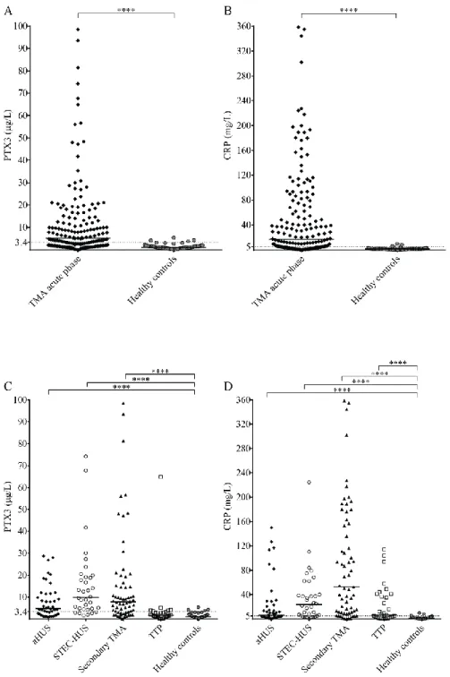
The circulatory PTX3 and CRP levels were significantly elevated in acute phase TMA compared to healthy controls. Elevated PTX3 and CRP levels could be detected in all etiological forms TMA, although PTX3 elevation was exceptional in TTP, despite the elevated CRP level in 53% of the TTP patients (Figure 1). With further subdivision of the study groups, we found that the elevation of both pentraxins was independent of the molecular background in aHUS or the underlying condition in secondary TMA. The calculated cutoff of CRP levels (5.01 mg/L) was equivalent to the upper limit of normal range (5 mg/L) used in our laboratory in frames of diagnostics, whereas the cutoff of PTX3 levels was determined based on the levels measured in the healthy control group and set as 3.40 μg/L.
PTX3 levels were associated with markers of disease activity and organ damage. We observed a significant positive correlation between lactate dehydrogenase (LDH) and PTX3 levels, and a weaker yet significant correlation of the platelet count as well as laboratory sings of kidney damage to PTX3. By contrast, association between CRP and disease activity was not present, except a significant positive correlation to creatinine levels. Furthermore, systemic PTX3 and CRP levels showed a strong positive correlation to each other and to markers of systemic inflammation such as the white blood cell (WBC) and absolute neutrophil counts (ANC) of the patients. Since platelet count is a reliable marker of disease activity in TMA, we grouped the patients according to platelet counts at the time of admission. However, despite the positive correlation, we found that irrespective of the classification, PTX3 and CRP levels of all subgroups remained significantly elevated compared to healthy controls. In order to adjust for confounding variables, the laboratory parameters were also entered into two multiple regression models to explore the relationship between them and the determined pentraxin levels. LDH (standardized regression coefficient beta=0.299) turned out to be a significant predictor of PTX3 in the multivariable model, whereas platelet and kidney function measures did not. For CRP, significant predictors were hemoglobin (beta=0.183), platelet number (beta= -0.179) and creatinine (beta=0.338) levels.
Figure 1. Systemic PTX3 and CRP levels in TMA patients and healthy controls
PTX3 and CRP levels of acute phase TMA patients compared to those of healthy individuals are displayed on panels A-B, whereas pentraxin levels of TMA patients subdivided based on disease etiology are shown on panels C-D. Data points are expressed as mean of technical duplicates, the horizontal line indicates the median of each group and an intermittent line shows the calculated cutoff of each pentraxin, respectively. Statistical analysis was performed with the Kruskal-Wallis test corrected for multiple comparisons using the Dunn’s post hoc test. Statistical significance is indicated by asterisks (****p<0.0001). (aHUS= atypical hemolytic uremic syndrome, CRP= C-reactive protein, PTX3= pentraxin-3, STEC-HUS=
Shiga-like toxin associated HUS, TMA= thrombotic microangiopathy, TTP= thrombotic thrombocytopenic purpura)
Pentraxin levels in disease remission
STEC-HUS and secondary TMA are characterized by one acute episode with no disease recurrence upon the eradication of the underlying cause. On the other hand, relapses are common features of aHUS and TTP with a progressive decline of kidney function and neurological deterioration, respectively. To investigate the regression of pentraxin levels in disease remission, we obtained follow-up samples from 31 aHUS patients and 19 of the TTP patients. In 80% of aHUS both PTX3 and CRP levels decreased compared to the paired acute phase samples, nonetheless the extent of decline did not reach statistical significance in patients with no clarified molecular background of the disease. Although the PTX3 level remained significantly higher in aHUS remission compared to the healthy control group, the systemic CRP levels in remission were similar to those of the healthy controls. The initially low PTX3 level of TTP patients showed no remarkable difference in remission, and the CRP levels also normalized in 85% of the cases.
Association of the systemic pentraxin levels with the acute phase mortality
In-hospital mortality was followed-up during the first month of the acute disease flare and the association of circulatory pentraxin levels to disease mortality was analyzed in the secondary TMA subgroup, where mortality exceeded 30% within a 31-day period. Conversely, only one patient died in the TTP group, whereas no deaths occurred among STEC-HUS or aHUS patients. Elevation of the median PTX3 level was associated with a higher acute phase mortality in the secondary TMA group, whereas the median CRP levels did not differ significantly in secondary TMA patients who survived the first month of the TMA episode compared to those who did not. The optimum PTX3 cut-point was 9 μg/mL to differentiate patients who died during follow up, from those who survived (odd's ratio 3.08 (95% CI 1.02- 9.33)). One-by-one adjustment for key activity indicators showed that high PTX3 levels are hemoglobin and creatinine independent predictors of mortality, whereas dependent on platelet and LDH levels.
2. Investigation of the relation of complement overactivation and systemic pentraxin levels in distinct forms of TMAs
Laboratory signs of complement consumption in acute phase TMA and their association to the systemic pentraxin levels
Complement consumption has been described in all etiological forms of TMA. Nearly 50% of our patients also presented with decreased C3 levels indicative of complement overactivation, while only 8% of the patients (14/171) showed no signs of complement alteration (with C3,
C4, FH, FI, FB levels, CP and AP activities together with the complement activation product levels within the laboratory normal range). Furthermore, we had a notable number of patients with low FH level, originating from all disease subgroups, indicative of complement dysregulation in all investigated forms of TMA.
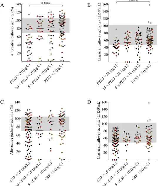
To assess whether elevated pentraxin levels were associated with complement consumption in the acute disease flare, we grouped the patients upon PTX3 and CRP levels and observed a strong linkage between the gradual increase in PTX3 and sings of complement AP and CP consumption (Figure 2 A-B). Complement CP and AP activities were significantly lower in patients with PTX3 above 20 μg/L compared to those with PTX3 below 5 μg/L. Moreover, patients with a PTX3 level exceeding 20 μg/L had a median AP and CP activity below the normal range indicating explicit complement consumption. As a result of complement overactivation and factor consumption, both C3 and C4 levels were significantly lower in patients with PTX3 above 20 μg/L compared to those below 5 μg/L (Figure 3 A-B). However, the gradual increase of PTX3 was not accompanied by a decrease in the FH and FB levels and the level of the complement activation product sC5b-9 was elevated in all groups regardless of the systemic PTX3 level. By contrast, CRP levels showed no association to any of the measured complement activity parameters (Figures 2-3, panels C-D). Since a considerable number of patients presented with a CRP level exceeding 20mg/L, we analyzed the association between CRP levels and laboratory signs of complement consumption in four additional patient subgroups (20mg/L < CRP < 40mg/L; 40mg/L < CRP < 60mg/L; 60mg/L <
CRP < 80mg/L; 80mg/L < CRP), yet we found no association between CRP elevation and laboratory markers of pathological complement activation in TMA.
Figure 2. Association of the systemic PTX3 and CRP levels to the activity of the alternative and classical pathways.
TMA patients are subdivided based on the measured systemic PTX3 (μg/L) or CRP (mg/L) levels indicated on the x axis, with their AP and CP activities shown on the y axis, respectively. The color of each data point indicates the specific form of TMA (brown= aHUS, red= STEC-HUS, black= secondary TMA, grey= TTP). Data are expressed as mean of technical duplicates, the horizontal lines indicate the median of each group, whereas the laboratory normal range in shown with grey shading. Statistical analysis was performed with the Kruskal-Wallis test corrected for multiple comparisons using the Dunn’s post hoc test.
Statistical significance is indicated by asterisks (****p<0.0001). (aHUS= atypical hemolytic uremic syndrome, CRP= C-reactive protein, PTX3= pentraxin-3, STEC-HUS= Shiga-like toxin associated HUS, TMA= thrombotic microangiopathy, TTP= thrombotic thrombocytopenic purpura)
C D
A B
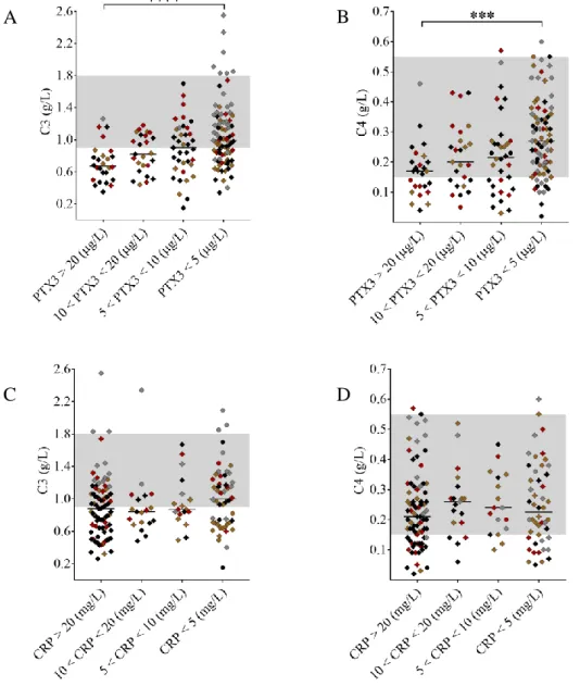
Figure 3. Association of the systemic PTX3 and CRP levels to the complement factor levels C3 and C4
TMA patients are subdivided based on the measured systemic PTX3 (μg/L) or CRP (mg/L) levels indicated on the x axis, with their C3 and C4 levels shown on the y axis, respectively.
The color of each data point indicates the specific form of TMA (brown= aHUS, red= STEC- HUS, black= secondary TMA, grey= TTP). Data are expressed as mean of technical duplicates, the horizontal lines indicate the median of each group, whereas the laboratory normal range in shown with grey shading. Statistical analysis was performed with the Kruskal-Wallis test corrected for multiple comparisons using the Dunn’s post hoc test.
Statistical significance is indicated by asterisks (****p<0.0001). (aHUS= atypical hemolytic uremic syndrome, CRP= C-reactive protein, PTX3= pentraxin-3, STEC-HUS= Shiga-like toxin associated HUS, TMA= thrombotic microangiopathy, TTP= thrombotic thrombocytopenic purpura)
C D
A B
Impact of excess PTX3 on AP activity in vitro
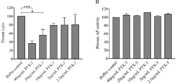
Since in vivo complement consumption was accompanied by a gradual increase in the systemic PTX3 level in our acute phase TMA patients, to explore the functional relevance of this phenomenon, we tested whether PTX3 attenuates or stimulates the AP activity on the cellular surface in vitro. In a modified hemolytic assay allowing for activation of solely the AP, we built up the AP C3-convertase on sheep erythrocytes and determined the hemolytic activity of NHS with the addition of recombinant human PTX3 or buffer control, respectively (Figure 4A). We found that addition of PTX3 dose-dependently decreased the AP hemolytic activity on sheep red blood cells. Conversely, addition of PTX3 to NHS did not influence the AP activity on the surface of ELISA plates and thus resulted in no remarkable change in C5b- 9 deposition through LPS-induced activation of the AP (Figure 4B).
Figure 4. Effect of PTX3 on alternative pathway activity in vitro
The effect of recombinant human PTX3 on AP hemolytic activity (A) and lipopolysaccharide induced complement C5b-9 deposition (B) is shown in the respective figure panels. Data are expressed in percent, compared to buffer control added to pooled serum of healthy individuals (100%). Data represent mean of three times repeated experiments with technical duplicates, error bars indicate the standard error of the mean. Statistical analysis was performed with the Kruskal-Wallis test corrected for multiple comparisons using the Dunn’s post hoc test.
Statistical significance is indicated by asterisks (*p<0.05; ***p<0.001). (AP=complement alternative pathway, PTX3=pentraxin-3)
B A
3. Epitope analysis of anti-FH antibodies in atypical hemolytic uremic syndrome Clinical and laboratory characteristics of patients with autoimmune aHUS
Sera of children with the diagnosis of autoimmune aHUS were obtained for the epitope analysis of FH antibodies. For accurate confinement of the antibody binding epitopes on FH, only sera of treatment-naive patients were applied in the epitope mapping experiments in the acute phase of aHUS. Hence, serum samples of the eight aHUS patients were taken before the initiation of plasmapheresis during the first acute episode of the disease, however two of the study subjects received FFP prior to sampling. The median age of the children at the time of diagnosis was 9 years, therefore sera of age-matched control individuals (N=10) were applied in the epitope analysis experiments. We obtained remission phase sera samples from seven of the patients, who were followed-up on a monthly basis after the termination of aHUS-specific therapy. This included plasmapheresis, immunosuppression with corticosteroids or cyclophosphamide, and rituximab, respectively. The remission phase samples were collected after a minimum follow-up period of 6 months in clinical remission, during which the patients received no immunomodulation or any forms of plasma therapy. Even though in the acute phase the anti-FH antibody level of all patients was higher than the internationally established upper-normal limit, there was a notable difference in the initial antibody titers of the patients ranging from 209-10067 AU/mL. Furthermore, albeit the follow-up periods elapsed devoid of relapse in all of the aHUS patients, the level of free anti-FH IgG remained low-titer positive in three out of seven cases.
Localization of the linear autoantibody epitopes on FH in the acute phase and remission of aHUS
Autoantibody binding to the synthetic peptides was investigated by ELISA. Peptides with a significantly increased relative autoantibody binding in the acute disease flare (patients versus control, Mann-Whitney test) included amino acids 1157-1171 on FH domain 19, and amino acids 1177-1191 and 1207-1226 on FH domain 20 (Figure 5A). The highest average binding was observed to peptides covering the amino acid residues 1212-1226. In disease remission, the epitope recognition pattern of the autoantibodies remained similar compared to what we had observed at the acute disease onset, although with weaker signals. The decline of antibody binding reached statistical significance by one of the epitopes (peptide 1177-1191) although in case of all epitopes a decrease of at least 25% was measured at the level of group mean.
Comparison of autoantibody binding to linear epitopes on FH and FHR1
The C-terminal domains of FH and FHR1 share a 99% homology and differ only in two amino acids. Since seven of our patients carried a homozygous, and one a heterozygous deletion of the gene encoding FHR1 (CFHR1), we also synthesized overlapping peptides of the region containing the two residues which are different in FH and FHR1. We identified significantly increased autoantibody binding to peptide 276-290 of FHR1 compared to controls in the acute phase of aHUS, which covered the exact same location as the previously identified autoantibody epitope on FH (peptide 1177-1191). Even though in remission antibody binding decreased significantly to the homologous epitopes of FH and FHR1, the decline only reached statistical significance by the FH peptide. The extent of autoantibody binding however, was comparable to each of the homologous peptides, suggesting that the serine-leucine exchange did not influence the autoantibody binding to the complement proteins.
Fine epitope mapping of recombinant FH domains 19-20 expressing aHUS-associated point mutations by anti-FH autoantibodies of aHUS patients
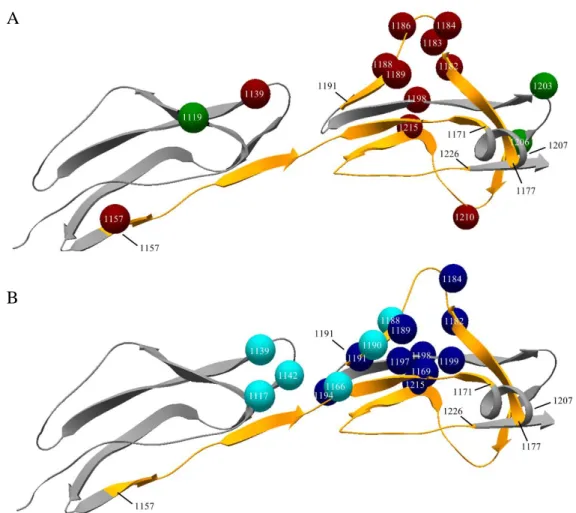
Pathogenic mutations associated with aHUS are most frequently located on the C-terminal SCR domains of FH and their presence results in the loss of host recognition and C3b binding by the complement regulator. Since antibody binding to FH also arrests these functions of the protein, we applied various recombinant FH19-20 constructs displaying single amino acid changes associated with aHUS to locate those that are involved in antibody binding, and to validate the results of the linear epitope mapping on the folded FH domains. For this purpose 14 different constructs were tested in ELISA using serum samples of three patients. On the folded FH domains a distinct antibody binding epitope appeared between amino acids 1183- 1198, with a symmetric gradual decline in antibody binding towards amino acid 1188. We observed the highest decrease in antibody binding to the FH 19-20 construct with a point mutation at amino acid position 1188. Additional positions, where an amino-acid switch significantly decreased the anti-FH antibody binding were detected on domain 19 (amino acids 1139 and 1157) and the C-terminal end of domain 20 (amino acids 1210 and 1215) concordant to the location of the identified linear epitopes (Figure 5A).
Position of the linear FH epitopes and aHUS associated point mutations on the crystal structure of FH
To verify the observations of the distinct approaches of epitope mapping we visualized the location of the linear epitopes and single amino acid-substitutions in the tertiary structure of FH obtained from the Protein Data Bank (ID: pdb2g7i) (Figure 5A). We also positioned the linear epitopes on the amino acids participating in C3b and sialic acid binding (Figure 5B), to
18
explore the potential functional relevance of FH blockade by autoantibodies. In the steric conformation, epitope 1157-1171 appears as a linear segment in the hinge region between FH domains 19 and 20, while epitopes 1177-1191 and especially 1207-1226 are in close sterical proximity with the C-terminal end of peptide 1157-1171 (Figure 5A).
A
B
Figure 5. Linear epitopes, point mutations and ligand binding sites of Factor H
Ribbon model of the folded structure of FH domains 19-20 obtained from the Protein Data Bank (ID: pdb2g7i). Linear epitopes of the anti-FH autoantibodies are highlighted with orange (amino acids 1157-1171, 1177-1191, 1207-1221), arrowheads point towards the C- terminal end of the protein and the initial and final amino acid of each segment is indicated with black numbers. (A) The location of the generated amino acid switches is displayed as colorful spheres on the backbone of the protein, with white numbers pinpointing their location and colors representing their effect on autoantibody binding by FH (red: significantly decreased binding when the amino acid substitutions is present, green: no significant effect on binding). (B) Colorful spheres refer to amino acids forming the C3b (light blue) and sialic acid (dark blue) binding sites of the molecule with numbers within the spheres showing their amino acid position on FH. (anti-FH antibodies = anti-Factor H antibodies FH = Factor H)
19
Conclusions
In frames of our investigations we explored the role of pentraxins in TMA and the molecular background of complement dysregulation in autoimmune aHUS. We studied the role of PTX3 and CRP in association with complement consumption in the acute phase of TMA. In frames of this we provide a detailed description of acute phase-TMA patients’ complement profile linked to changes in the systemic pentraxin levels. We report that PTX3 elevation is present in the acute phase of STEC-HUS, aHUS and secondary TMA but is exceptional in TTP, whereas an elevation of the systemic CRP level was detected regardless of disease etiology during the acute disease flare. We observed the highest acute phase mortality in secondary TMA patients, which was associated with high PTX3 but not CRP levels. Furthermore, laboratory signs of complement activation and consumption were detected in the majority of our patients, regardless of the etiological background of TMA. We show for the first time that AP and CP consumption are associated with elevated PTX3 in the acute phase of the disease, and we confirmed in vitro that PTX3 limits AP activity on the surface of red blood cells, with no effect on terminal pathway assembly during LPS-induced AP activation on ELISA plates.
This is the first study where the association of PTX3 and CRP elevation has been investigated in a complement mediated disease in vivo, and thus our results provide a missing link between the numerous in vitro observations that described the interaction of PTX3 with the complement system under defined experimental conditions.
Through the epitope analysis of anti-FH antibodies we identified three linear, extended autoantibody binding epitopes on FH and one on FHR1. These epitopes were recognized by both acute phase and remission phase serum autoantibodies of aHUS patients and their close sterical proximity on the folded FH domains suggests that they form a joint antibody binding site in the tertiary structure. The identified epitopes overlap with the reported clustering of aHUS-associated FH mutations, as well as with the FH fractions necessary for ligand binding, which provides molecular basis for the reported dysfunction of autoantibody-bound FH, ultimately leading to the dysregulation of the complement AP.
In conclusion, we aimed for the investigation of specific molecular aspects of complement dysregulation in aHUS and TMA and our results hopefully add to the better understanding of the complex molecular mechanisms that contribute to the pathogenesis of these conditions.
20
Bibliography of the candidate’s publications
Publications related to the Ph.D. dissertation of the candidate
I. Trojnár E, Józsi M, Szabó Zs, Réti M, Farkas P, Kelen K, Reusz GS, Szabó AJ, Garam N, Mikes B, Sinkovits G, Mező B, Csuka D, Prohászka Z. Elevated systemic pentraxin-3 is associated with complement consumption in the acute phase of thrombotic microangiopathies. Front Immunol. 2019 Feb 25;10:240.
II. Trojnár E, Szilágyi Á, Mikes B, Csuka D, Sinkovits Gy, Prohászka Z. Role of complement in the pathogenesis of thrombotic microangiopathies. Magazine of European Medical Oncology 2018 September, 11;3:227–234.
III. Trojnár E, Józsi M, Uray K, Csuka D, Szilágyi Á, Milosevic D, Stojanović VD, Spasojević B, Rusai K, Müller T, Arbeiter K, Kelen K, Szabó AJ, Reusz GS, Hyvärinen S, Jokiranta TS, Prohászka Z. Analysis of Linear Antibody Epitopes on Factor H and CFHR1 Using Sera of Patients with Autoimmune Atypical Hemolytic Uremic Syndrome.
Front Immunol. 2017 Mar 30;8:302.
Publications independent of the Ph.D. dissertation of the candidate
IV. Petro CD, Trojnar E, Sinclair J, Liu ZM, Smith M, O'Brien AD, Melton-Celsa A. Shiga toxin (Stx) type 1a reduces the toxicity of the more potent Stx2a in vivo and in vitro.
Infect Immun. 2019 Mar 25;87(4). pii: e00787-18.
V. Bhattacharjee A, Reuter S, Trojnár E, Kolodziejczyk R, Seeberger H, Hyvärinen S, Uzonyi B, Szilágyi Á, Prohászka Z, Goldman A, Józsi M, Jokiranta TS. The major autoantibody epitope on Factor H in atypical Hemolytic Uremic Syndrome is structurally different from its homologous site in Factor H related protein 1 supporting a novel model for induction of autoimmunity in this disease. J Biol Chem. 2015 Apr 10;290(15):9500- 10.