Tumor distribution and efficacy of antiangiogenic receptor tyrosine kinase inhibitors
Ph.D. Dissertation
Szilvia Török
Semmelweis University Clinical Medicine Ph.D. School
Consultants: Dr. Balázs Döme, MD, PhD , head of department Dr. György Marko-Varga, PhD, professor
Official reviewers:Dr. Anna Sebestyén, PhD, senior research fellow Dr. Zsolt Horváth, MD, PhD, associate professor
Head of the final Examination Committee:
Dr. András Matolcsy, DSc, professor Members of the Final Examination Committee:
Dr. Béla Szende, DSc, professor emeritus
Dr. Krisztina Vellainé Takács, PhD, associate professor
Budapest
2016
2 1. TABLE OF CONTENTS
1. TABLE OF CONTENTS 2
2. ABBREVIATIONS 4
3. INTRODUCTION 8
3.1 Angiogenesis 8
3.2 Tumor-induced angiogenesis 10
3.2.1 Main receptor families and signalization pathways in
tumor-induced angiogenesis 12
3.2.1.1 VEGFR family 15
3.2.1.2 PDGFR family 17
3.2.1.3 FGFR family 19
3.2.1.4 TIE receptor family 21
3.2.1.5 TGFβR family 23
3.2.1.6 Notch receptor family 24
3.2.2 Mechanisms of tumor-induced angiogenesis 26
3.2.2.1 Sprouting angiogenesis 26
3.2.2.2 Vessel incorporation or co-option 27 3.2.2.3 Intussusceptive microvascular growth 27
3.2.2.4 Glomeruloid angiogenesis 28
3.2.2.5 Postnatal vasculogenesis 28
3.2.2.6 Vessel-like structures, formed by tumor cells 29 3.2.3 Characteristics of tumor blood vessels 30 3.2.4 Inhibition of the tumor vasculature 32 3.2.4.1 Conventional chemotherapeutic agents 32
3.2.4.2 Vacular disrupting agents 32
3.2.4.3 Vasoactive agents 33
3.2.4.4 Angiogenesis inhibitors 34
3.2.5 Resistance to antiangiogenic tyrosine kinase inhibitor therapy 40
3.3 TKI imaging 41
4. AIMS 44
3
5. METHODS 45
5.1In vivo tumor models and treatments 45
5.1.1 Tumor models 45
5.1.2 Drugs 45
5.1.3 In vivo treatment 47
5.2 Analysis of vascular parameters and target receptors 47 5.3 Detection of the compounds and analysis of drug distribution
and intensity 49
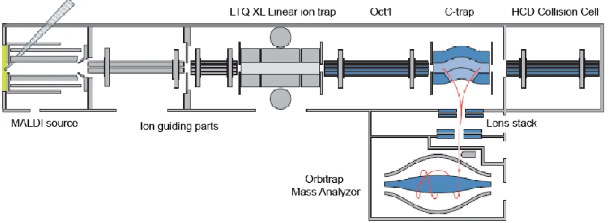
5.3.1 Ionization technique and instrumentation 49
5.3.2 Compound characterization 56
5.3.3 Compound detection in the blood 57
5.3.4 Tissue imaging of antiangiogenic RTKIs 57
5.3.5 Quantification of the compounds 58
5.4 Statistical analysis 58
6. RESULTS 60
6.1 Tumor growth inhibititon 60
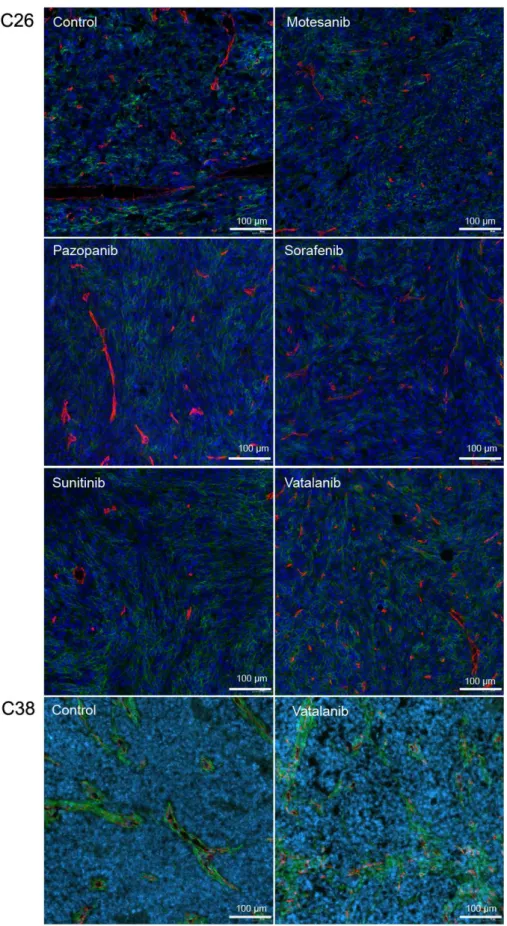
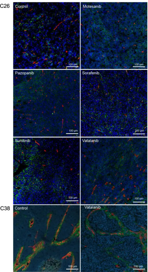
6.2 Immunohistochemical analysis 61
6.3 Mass spectrometric analysis 77
6.3.1 Compound characterization 77
6.3.2 Precursor compound and metabolite detection in the blood 79 6.3.3 Tissue imaging of antiangiogenic RTKIs 87
7. DISCUSSION 95
8. CONCLUSIONS 99
9. SUMMARY 100
10. ÖSSZEFOGLALÁS 101
11. REFERENCES 102
12. PUBLICATIONS 147
13. ACKNOWLEDGEMENT 149
4 2. ABBREVIATIONS
AC: alternate current ACN: acetonitrile
ADME: adsorption, distribution, metabolism and elimination AGC: automatic gain control
AI: angiogenesis inhibitor
AKT: protein kinase with transforming capabilities developed in the Ak strain of mice ALK: activin receptor-like kinase
Ang: angiopoietin
ANOVA: analysis of variance
ARNT: aryl hydrocarbon receptor nuclear translocator ARRIVE: animal research: reporting of rn vivo experiments BAMBI: BMP and activin membrane-bound inhibitor homologue BCR-ABL: break point cluster- abelson
BM: basement membrane
BMP: bone morphogenic proteins CA4P: combretastatin A-4 phosphate CSC: cancer stem cells
CBP: CREBB binding proein
c-FMS: colony-stimulating factor-1 receptor CHCA: α-cyano-4-hydroxycinnamic acid CID: collision-induced dissociation
c-KIT: cellular homolog of the feline sarcoma viral oncogene v-kit c-MYC: cellular myelocytomatosis transcription factor
COX: cyclooxygenase CRC: colorectal cancer
CSF-1R: colony stimulating factor 1 receptor CSL: CBF1/Su(H)/Lag-1) transcription factor C-Trap: curved linear trap
CXCR4: SDF receptor DC: direct current
5 DFG motif: Asp-Phe-Gly motif
APE motif: Ala–Pro–Glu motif DHB: 2,5-dihydroxybenzoic acid DLL: delta-like ligand
DTC: differentiated thyroid carcinoma EC: endothelial cell
ECM: extracellular matrix
EGF/EGFR: epidermal growth factor/ epidermal growth factor receptor EMEA: European Medicines Agency
EPAS: endothelial PAS domain-containing protein EPC: endothelial progenitor cell
ErbB: erythroblastic leukemia viral oncogene homologue ERK: extracellular signal-regulated kinase
FAK: focal adhesion kinase
FDA: Food and Drug Administration
FGF/FGFR: fibroblast growth factor/ fibroblast growth factor receptor FLK: fetal liver kinase
FLT: fms-like tyrosine kinase
FT-MS: Fourier transform-mass spectrometry GDF: growth and differentiation factor GIST: gastrointestinal stromal tumor HCC: hepatocellular carcinoma
HCD: high energy collisional dissociation HE: haematoxylin&eosin
HER: human epidermal growth factor receptor HGF: hepatocyte growth factor
HIF: hypoxia-inducible factor
HPLC: high-peformance liquid chromatography HRE: hypoxia-response element
HSP27: heat shock protein 27
IC50: the half maximal inhibitory concentration
IGF/IGFR: insuline-like growth factor/ insuline-like growth factor receptor
6 IL: interleukin
ITK: interleukin-2-inducible T-cell kinase JNK: c-Jun N-terminal kinase
KDR: kinase domain-containing receptor
LCK: lymphocyte-specific protein tyrosine kinase LC-MS: liquid chromatography-mass spectrometry LYN: Lck/Yes novel tyrosine kinase
LTQ: linear trap quadrupole mAB: monoclonal antibody
MALDI: matrix-assisted laser desorption ionization MAPK: mitogen-activated protein kinase
MEK: MAPK/ERK Kinase
MIS: mullerian inhibitory substance MMP: matrix metalloproteinases MS: mass spectrometry
MSI: mass spectrometry
mTOR: mammalian target of rapamycin MVD: microvessel density
m/z: mass to charge ratio
NCE: normalized collision energy
NF-κB: nuclear factor kappa-light-chain-enhancer of activated B cells NICD: Notch intracellular domain
NRP: neuropilin
NSCLC: non-small cell lung cancer
PA/PAI: plasminogen activator /plasminogen activator inhibitor
PDGF/PDGFR: platelet-derived growth factor/ platelet-derived growth factor receptor PHD: prolyl-hydroxylase domain-containing protein
PI3K: phosphatidilinositole 3-kinase
PIP2: phosphatidylinositol 4,5-bisphosphate PKB: protein kinase B
PKC: protein kinase C
PLCγ: phospholipase C gamma
7 PlGF: placental growth factor
pNET: pancreatic neuroendocrine tumors PTEN: phosphatase and tensin homolog
RAS: rat sarcoma viral oncogene homolog gene RAF: rapidly accelerated fibrosarcoma
RCC: renal cell cancer
RET: rearranged during transfection
RTK/RTKI: receptor tyrosine kinase/ receptor tyrosine kinase inhibitors SAPK: stress-activated kinase
s.c.: subcutan
SDF: stromal derived factor
SH2-domain: Src homology 2 domain SEM: secondary electron multiplyer SEM: standard error of the mean SMA: smooth muscle actin
SNP: single nucleotid polymorphism Src: sarcoma
STAT: signal transducer and activator of transcription TFA: trifluoroacetic acid
TGF: transforming growth factor TIC: total ion current
TIE: tunica intima endothelial receptor TNF: tumor necrosis factor
TOF: time of flight
uPA/uPAR: urokinase plasminogen activator/ urokinase plasminogen activator receptor VDA: vascular disrupting agents
VEGF/VEGFR: vascular endothelial growth factor/ vascular endothelial growth factor receptor
VHL: von Hippel-Lindau
VSMC: vascular smooth muscle cell
8 3. INTRODUCTION
3.1 Angiogenesis
The word “angiogenesis” was first mentioned in 1787 in the work of John Hunter, an English surgeon who studied the process in the growing antlers of deers (1). In contrast to vasculogenesis, which means the development of the vascular system during embryogenesis, angiogenesis is the process when new blood vessels are formed from preexisting ones.
Under physiological conditions, angiogenesis is activated in response to low oxygen level. The process is regulated by the HIF (hypoxia-inducible factor) complex. HIFs are basic helix-loop-helix DNA binding transcription factor proteins of the Per-Arnt-Sim family, and function as heterodimers consisting of an oxygen-regulated alpha subunit and a stably expressed beta subunit (2). The alpha subunits are encoded by three genes in mammals: HIF1α, HIF2α (EPAS1: Endothelial PAS domain-containing protein 1) and HIF3α (2-4). Also three forms of the beta subunit have been identified until now, the HIF1β (aryl hydrocarbon receptor nuclear translocator: ARNT), ARNT2 and ARNT3 (5,6). The structure, regulation and function of all HIFs seem to be similar, but compared to their homologs (HIF1α and ARNT) the members of the HIF2 and -3 family have been reported to have a more restricted pattern of expression and thus may play more specialized roles in oxygen delivery than the HIF1 subunits (6,7).
Under normal oxygen tension, the alpha subunits are hydroxylated at the prolyl residues, a process that is catalyzed by the prolyl-hydroxylase domain-containing protein (PHD). This hydroxylation promotes interaction with the von Hippel-Lindau (VHL) ubiquitin ligase and, consequently, they undergo proteasomal degradation (8,9).
Thus the half-life of alpha subunits is measurable in minutes and the protein is hardly detectable at all (10). The degradation process is regulated not only by the VHL protein, but p53 as well (11).
In case of hypoxia, the HIF alpha and beta subunits form a complex, recruits its co- activator, p300/CBP (CREBB Binding Protein) and binds to the Hypoxia Responsive Element (HRE) of the target genes, thus modifying their transcription (12). Several of these target genes are responsible for inducing angiogenesis in order to increase the oxygen delivery of the tissue, such as vascular endothelial growth factor (VEGF),
9
VEGF receptor 1, tunica intima endothelial receptor 2 (TIE2), angiopoietin 2 (Ang2), erythropoietin, Insulin-like growth factor 2 (IGF2), Transforming Growth Factor β3 (TGFβ3), c-MET, adrenomedullin, NO Synthase 2, plasminogen activator inhibitor-1 (PAI1) (3,13-21). Regulation of oxygenation via the HIF molecule is depicted in Figure 1.
Figure 1. Regulation of oxygenation via the HIF molecule (22). Degradation of HIFα via ubiquitination in normoxia (A.) and angiogenesis stimulation in hypoxia (B.).
VEGF is considered to be the key hypoxia dependent cytokine for endothelial sprouting (23). Besides, many other factors are also able to positively influence the process of angiogenesis such as platelet-derived growth factors (PDGFs) (24-26), fibroblast growth factors (FGFs) (27,28), placental growth factor (PlGF) (29), angiopoietins (30), Jagged (31), epidermal growth factor (EGF) (32), hepatocyte growth factor (HGF) (33) and interleukin-8 (IL-8) (34). These ligands bind to their receptors on the surface of endothelial cells and act in an autocrin or paracrin manner. Binding the growth factor leads to the activation of signaling cascades, influencing the survival, proliferation and migration of endothelial cells and thus the maintenance of existing- and development of new vessels. Moreover, these pathways regulate further processes involved in angiogenesis, such as secretion of additional growth factors (35), upregulation of angiogenic receptors (36), alterations in cell-cell and cell-matrix interactions by matrix metalloproteinases (MMPs) (37), and activation of the members of endothelial cell (EC) adhesion molecule family (38). After the new capillary is formed by ECs, PDGF-BB and bFGF secreted by the endothelium recruits PDGFRβ or FGFR expressing pericytes and vascular smooth muscle cells (VSMCs) (36,39). These supporting cells provide
10
structural stability for the new blood vessel, promote EC survival and regulate blood flow via influencing vasoconstriction and -dilatation. Moreover, via the secretion of VEGF pericytes also promote EC sprouting and survival (40). By binding to their receptor, TGFß1 (41), Ang1 (42) and sphingosine-1-phosphate (43) stabilize the interaction between mural cells and ECs.
The healthy body controls angiogenesis by balancing a series of angiogenesis stimulating and inhibiting factors. Thus, in a healthy adult ECs have long half-life, and angiogenesis is only activated under special conditions, such as the reproducible processes of women or wound healing.
If tissues cannot produce adequate amounts of angiogenic growth factors, blood vessel growth becomes inadequate leading to improper circulation and eventually to necrosis.
Insufficient angiogenesis occurs in diseases such as coronary artery disease, stroke and chronic wounds. In these pathological features therapeutic angiogenesis is aimed to stimulate the sprouting of new vessels with growth factors being developed to treat these conditions.
On the other hand, by overexpressing the angiogenesis stimulating factors, blood vessel growth becomes highly intensive in some pathological conditions, such as endometriosis, atherosclerosis, psoriasis, rheumatoid arthritis, inflammation, ischaemia, ocular neovascularization and cancer (44).
3.2 Tumor-induced angiogenesis
After the experiments of Hunter, more than a century had passed until an interest in the angiogenesis of tumors was piqued. Until the 1930-40s these investigations were restricted mostly to the observation of the morphology of tumor blood vessels (45).
Later the process of neovascularization was also observed, but it had not become the focus of research until 1971, when Judah Folkman postulated that above a certain size (about 1-2 mm diameter) intratumoral diffusion is no longer sufficient and tumors are incapable of growing and metastatizing, unless they develop their own blood supply to ensure the necessary oxygen and nutrient level of the cells. He also assumed that tumor cells secrete growth factors to facilitate the process of angiogenesis, ie. capillary growth is induced by the communication between tumor blood vessels and the tumor tissue (46).
11
This communication is mediated by the HIFα proteins, which are stabilized in hypoxic tumor cells, and thus facilitate the expression of angiogenic proteins. Moreover, the expression level of HIF1α is controlled not only by oxygen tension, but also by reactive oxygen species, that are secreted in response to carcinogens (47,48).
Moreover, oncogenes, such as activated EGFR (49), ErbB-2/Her2 (50) mutant RASv12 (51), mTOR (52) and Src (53) also augments HIF mediated gene expression. On the other hand, some tumor suppressor genes also play an important role in the regulation of HIF1α, therefore loss of function of these gene products may influence angiogenesis in tumor tissues. The product of the tumor suppressor gene PTEN attenuates hypoxia mediated HIF1-α stabilization through the inhibition of AKT (54), while pVHL (9) and p53 (11) regulate the ubiquitination and degradation of the HIF complex, thus mediating the expression of angiogenic molecules.
Furthermore, solid tumors and their microenvironment secrete and often overexpress a range of growth factors, cytokines and hormones that coordinate the complex series of events of new capillary growth. Among them ie. VEGFA (55), IL1β, insuline (56), heregulin (50), EGF (57), IGF1,-2 (13) TGFβ (58), Tissue Necrosis Factor α (TNFα) (59) enhance HIF expression, activating a vicious circle in the angiogenic process.
HIF independent regulation of the VEGF pathway also occurs in tumors, as some oncogenes and tumor suppressor genes (cSrc (60), BCR-ABL (61), RAS (62), p53 (63)); cellular receptors (activated EGFR (64), IGF-1R (65) and overexpressed HER2 (66)); and cytokines (COX-2 (67), PDGF-AA (68)) can also influence VEGF production. These changes in the HIF-VEGF protein system drive the activity of angiogenesis in tumors, which can be further modulated by the expression of other angiogenic growth factors.
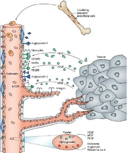
The schematic process of tumor-induced angiogenesis is depicted in Figure 2.
12
Figure 2. The process of tumor-induced angiogenesis (69). Main angiogenic growth factors (VEGF, PDGF, bFGF) are secreted by tumor cells or platelets and bound to their transmembrane receptors on ECs. The process is further regulated by integrins, MMPs and intrinsic angiogenesis inhibitors, such as endostatin, angiostatin or platelet factor-4.
3.2.1 Main receptor families and signalization pathways in tumor-induced angiogenesis
Kinases are phosphotransferase enzymes that transmit the terminal phosphate group of a high-energy donor molecule, such as adenosine triphosphate (ATP), to their substrate.
Protein kinases can be divided into a larger group of serin/threonine kinases and a smaller tyrosine kinase group, based on the amino acid they target. Besides, atypical kinases also exist.
13
Another classification is based on the localization of the kinase. Receptor kinases are membrane bound and essential for the transduction of extracellular signals into the cell.
Non-receptor kinases are responsible for intracellular communication. Angiogenesis is mainly regulated by receptor tyrosine kinases (RTKs).
RTKs have a variable extracellular part for ligand binding, a transmembrane region to anchore the molecule to the cell membrane, and they all share a conserved intracellular secondary structure. This consists of (i) a bi-lobed catalytic core, responsible for binding ATP in a deep cleft, located between the lobes (hinge region); (ii) a substrate binding site, where interacting proteins can bind and (iii) an activation loop, containing Asp-Phe-Gly (DFG) and Ala–Pro–Glu (APE) motifs at the N- and C-terminal part of the loop, respectively.
The ATP-binding site is divided into the following subregions (Figure 3.): adenine region, sugar region and phosphate binding region, based on the part of the ATP it binds. The sugar- and phosphate binding regions form a hydrophilic channel. Besides, a hydrophobic pocket (selectivity pocket) is also formed near the ATP binding region by the activation loop in the inactive conformation. This is not used by ATP, but ensures selectivity of receptor tyrosine kinase inhibitors. Moreover, ATP does not use the hydrophobic channel either, which provides a slot to open for solvents and it can be exploited to gain binding affinity.
Figure 3. Binding of ATP to receptor tyrosine kinases (70). The ATP-binding site is divided into the following subregions: adenine region, sugar region and phosphate binding region, based on the part of the ATP it binds. The sugar- and phosphate binding regions form a hydrophilic channel. Note that the hydrophobic pocket and the hydrophobic channel are not used by ATP, but ensures binding affinity and selectivity of RTKIs.
14
The activation loop can form a number of conformations. In the "out" conformation it creates a hydrophobic pocket near the ATP-binding cleft, thus blocking the accessibility of the receptor for ATP. Ligand binding of the extracellular domain triggers receptor dimerization, resulting in transphosphorylation of specific tyrosine residues in the activation loop, juxtamembrane- and C terminal regions. As a result, the receptor turns to the "in" conformation, which opens the hinge region for ATP. ATP binds in the cleft via the adenine ring, which forms 2 hydrogen bonds with the kinase ‘hinge’. The ribose and triphosphate group of ATP bind in a hydrophilic channel that extends to the substrate binding site, which is essential to the catalysis of autophosphorylation.
After autophosphorylation, the receptor recruits interacting proteins that bind to certain phosphorylation sites, which subsequently phosphorylate other proteins. These activation signaling pathways eventually lead to biological responses, such as cell activation, proliferation, differentiation, migration, survival and vascular permeability, (70,71). Activation of RTKs is depicted in Figure 4.
Figure 4. Activation of RTKs (71). Ligand binding of the extracellular domain triggers receptor dimerization, resulting in transphosphorylation of specific tyrosine residues in the activation loop, juxtamembrane- and C terminal regions. As a result, the receptor turns to the "in" conformation, which opens the hinge region for ATP.
The most important angiogenic RTKs are VEGFRs, PDGFRs, FGFRs, and the TIE receptors. Beside tyrosine kinases, serine-threonine kinases also regulate the process of neovascularization. These act similar to RTKs. Main examples are the members of TGF receptor family. Other receptors exert their effect not by phosphorylating partner molecules, but by regulating potential transcriptional activators. Of these, the Notch receptor family is the predominant member of angiogenic signalling.
15 3.2.1.1 VEGFR family
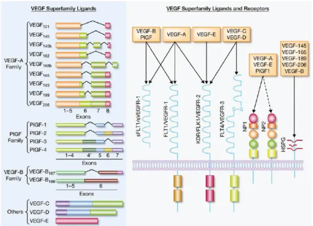
The VEGF family consists of the following growth factors in mammals: VEGFA (VEGF), VEGFB, VEGFC, VEGFD and PlGF. They are homodimeric polypeptides, although naturally occurring heterodimers of VEGFA and PlGF have also been described (72,73). Splicing and processing regulates the ability of the growth factors to bind to the appropriate receptors or co-receptors. Ligands can bind to three different receptors, VEGFR1-3 (FLT1: fms-like tyrosyl kinase-1, FLK1/KDR: Fetal liver kinase- 1/Kinase Domain-containing Receptor and FLT4 respectively) (74). The receptors have a ligand-binding extracellular domain consisting of seven immunoglobulin-like loops, a transmembrane domain, a juxtamembrane domain, and a split tyrosine kinase domain followed by a C-terminal tail (75). As co-receptors neuropilin 1-2 (NRP1, -2) and heparan sulphate play a role in the regulation of angiogenesis by increasing the binding affinity of specific ligands to the receptors (76-78).
VEGFA is the ligand of both VEGFR1 and VEGFR2, but while VEGFR1 has much higher affinity for binding the growth factor, than VEGFR2, its kinase activity is weakly induced (79). Moreover, beside being highly expressed on immune cells, hematopoietic cells, vascular endothelial and -smooth muscle cells and several tumor cells (79-84), this receptor exists partly in a soluble form, thus it rather acts as a „trap receptor” for the angiogenic growth factors (85). As VEGFB and PlGF bind just to VEGFR1, their proangiogenic role is limited (86).
VEGFR2 binds VEGFA and proteolitically processed VEGFC, -D (87-89). VEGFR2 is highly expressed not only on lymphatic and vascular endothelial cells but on hematopoietic and tumor cells as well (90-94). Soluble antiangiogenic form of the receptor also exists, but as its binding affinity is much smaller than that of VEGFR1, its trap function is also less important (95).
VEGFC binds with a higher affinity to VEGFR3 than to VEGFR2, while VEGFD has similar affinity for both receptors in human, but does not bind to VEGFR2 in mouse (96,97). VEGFR3 is expressed in the venous endothelial cells in the cardial vein during the later stages of embryogenesis, which gives rise to lymphatics, thus in adults VEGFR3 can be found mainly in lymphatic endothelium and to a lesser extent in vascular endothelial cells (98). It is disputed, whether it can be found on tumor cells (99). Soluble VEGFR3 suppresses lymphangiogenesis and lymphatic metastasis in
16
different cancer types (100,101). Consequently, the VEGFA-VEGFR2 axis is the predominant mediator of tumor-induced angiogenesis.
Alternative mRNA splicing of VEGFA gives rise to several distinct isoforms. The splice variants are noted as VEGFxxx (xxx means the number of amino acids present in the proteins without the signal peptide) (102). These isoforms differ not only in their expression pattern but their biochemical and thus biological properties as well. Also exon 8b containing less angiogenic or antiangiogenic variants of VEGFxxx are documented (103), and seem to be able to inhibit neovascularization. Moreover, they might be responsible for the failure of antiangiogenic therapies targeting VEGFs (104).
Figure 5. shows the main ligands and receptors of the VEGF superfamily.
Figure 5. VEGF Superfamily Ligands and receptors (22).
VEGFs can either bind to the receptors freely or be presented by co-receptors. Ligand binding results in receptor homo-, or heterodimerization, followed by changes in the intracellular domain conformation. This leads to the liberalization of the ATP binding site of the receptor and binding of ATP results in the auto- or transphosphorilation of tyrosine residues on the receptor and downstream signal transducers (105).
17
The VEGF ligands are produced by most parenchymal cells, and paracrine VEGF signaling is essential for the angiogenic cascade, proliferation, survival, permeability responses and endothelial differentiation. Moreover, autocrine VEGF signaling also exists, but only conveys survival signals (106).
After ligand binding the receptor dimerizes and activates the PLCγ/Ras/Raf/MEK/ERK pathway, leading to the regulation of gene expression and cell proliferation (89). With PI3K activation they modulate AKT signaling and thus the survival of receptor expressing cells (107). Via the elevation of intracellular Ca2+ or NO level, induced by PI3K or PLC they can also enhance vascular permeability (108). Cell migration is regulated by the PKC/p38/MAPK2-3/HSP27 pathway and the FAK or the STAT cascade (109). Signalization pathways activated by VEGFR2 are shown in Figure 6.
Figure 6. Signalization pathways of VEGFR2 (110).
Overexpression, mutation of the receptors and elevation of VEGF have also been related to enhanced angiogenesis and tumor development (111-113).
3.2.1.2 PDGFR family
The PDGF family consists of four ligand chains, PDGF-A, PDGF-B, PDGF-C and PDGF-D, which create five disulphide bonded dimeric isomorphs, PDGF-AA, PDGF-
18
BB, PDGF-AB, PDGF-CC and PDGF-DD (114). The ligands can bind to three membrane bound dimeric receptors, PDGFRαα, PDGFRββ, and PDGFRαβ (115).
Moreover, low density lipoprotein receptor-related protein and NRP1 seem to mediate PDGFR signaling as co-receptors (116,117).
PDGF synthesis is enhanced in response to low oxygen tension (118), thrombin (119), or other growth factors and cytokines (120). A number of cells secrete PDGFs, ie. cells associated with reproductive processes (121,122), vascular functions (123,124), cells of the nervus system (125-127), immune cells (128) and some other cell types (129,130).
PDGF isoforms are synthesized as precursor molecules and after dimerization they are proteolytically processed to the active forms that bind to their receptors. PDGF-AA binds only to PDGFRαα, while PDGF-BB can bind to all three dimeric receptors.
PDGF-AB and PDGF-CC can activate dimeric receptors containing at least one PDGFRα monomer, while PDGF-DD those, containing at least one PDGFRβ monomer.
By binding to PDGF receptors, PDGFs target a broad spectrum of mesoderm-derived cells, like fibroblasts, smooth muscle cells, pericytes, mesangial- and glia cells (131- 135). Soluble form of both PDGFRα and -β are detected and compete with cell- associated PDGF receptors for ligand binding, antagonizing the effects of their membrane bound counterparts (136,137).
The extracellular parts of the PDGF receptors contain 5 Ig-like domains, of which domain 2 and 3 are responsible for ligand binding, while domain 4 stabilizes the dimer by a direct receptor-receptor interaction. The intracellular parts contain split tyrosine kinase domains (138). Ligand-induced dimerization of the receptors is followed by autophosphorylation, which activates their kinase activity and creates docking sites for SH2-domain-containing signaling molecules (139).
The PDGFs play crucial roles during development, and they are essential in wound healing and inflammation (140,141). Increased PDGF activity has been linked with several disorders and pathological conditions, such as atherosclerosis, fibrosis and malignant diseases (142,143). Most solid tumors secrete PDGF and express their receptors on tumor-, endothelial- or perivascular cells (144,145). Both overexpression, and different mutations of either the ligands or the receptors is associated with tumorigenesis (146-148). Moreover, all PDGF ligands are documented to be involved in the process of angiogenesis by activating signaling pathways triggering endothelial cell
19
proliferation (24-26). On the other hand, as pericytes and VSMCs express PDGFRβ, processes driven by this receptor, such as mural cell recruitment to capillaries stabilize the vasculature, thus inhibiting tumor cell extravasation and metastatization (149).
Due to the high structural similarities with VEGFRs - all have a split kinase domain - they induce similar signaling cascades. PI3K and PLC mobilizes Ca2+ from intracellular stores, regulating vascular permeability (150,151). The RAS/RAF/MEK pathway is implicated in the stimulation of cell proliferation, migration, and differentiation (152).
By enhancing the serine/threonine kinase AKT/PKB pathway, it mediates antiapoptotic effects (153). PDGFRs are also involved in the activation of the JNK/SAPK pathway, thus regulating cell migration (154). Binding of PDGF isoforms to PDGF receptors, with the subsequent signalization is depicted in Figure 7.
Figure 7. Binding of PDGF isoforms to PDGF receptors, and subsequent signalization (155).
3.2.1.3 FGFR family
The FGF family consists of 23 ligands, of which 18 can be secreted and bound to 4 high-afinity cell surface receptors, FGFR1-4 in mammals (156). Four FGFs do not bind
20
to FGF receptors, while one ligand is only expressed in mice (157). Despite most ligands act in an autocrine or paracrine manner, endocrine members of the family have also been identified (158). FGFs share a central core of 140 amino acids that is highly homologous between all family members (159,160). The ligands have strong affinity for the glycosaminoglycan side-chains of not only cell surface proteoglycans, but heparin sulphate proteoglycans and heparin-like glycosaminoglycans as well (161,162). They may protect ligands from degradation and stabilise the FGF ligand–receptor complex. A specific FGF ligand can bind to more FGFRs, although it may have a preference for a particular one (163). Ligands are produced in either epithelial or mesenchymal cells and usually activate receptors of the opposite tissue specificity.
The extracellular ligand-binding part of the receptors contains three immunoglobulin- like domains, which are important in receptor dimerisation. Different isoforms of FGFR1–3 are generated by alternative expression of the IgIII domain. Upon the presence of exon 8 or 9 they either express the "b or c" splice variant of domain III.
Different isoforms are expressed in different tissues and have distinct binding specificities. FGFR4 has only one possible form (164). Truncated receptor splice variants coded by IgXa (X refers to the Ig domain being shortened by alternative splicing) are also secreted and may function as an inhibitor for FGFs (165). Structure and splicing of FGFRs are shown in Figure 8.
Figure 8. Structure (A.) and splicing (B.) of FGFR (166). The extracellular ligand-binding part of the receptors contain three immunoglobulin-like domains, which are important in receptor dimerisation.
Different isoforms of FGFR1–3 are generated by alternative expression of the IgIII domain. Upon the presence of exon 8 or 9 they either express the "b or c" splice variant of domain III.
The FGFR signaling pathway plays a key role in embryonic development, wound healing and angiogenesis. Deregulated FGFR signalling contributes to pathological
21
conditions, including different malignancies. Both the receptors and the ligands are expressed by different tumor cell types (167-170). Beside translocation of FGFRs in hematological malignancies, amplification, overexpression, activating mutation or SNP of the receptors can also lead to tumorigenesis (156). Moreover, abnormalities of different FGFs are also linked to tumor progression (171,172).
Binding of bFGF (FGF2) to FGFR1IIIc is considered to be the major regulator of FGF induced angiogenesis, being the most intensively expressed both on ECs and smooth muscle cells (173). Thus it has also a diverse role, by triggering EC proliferation and stabilizing vessel walls.
Ligand binding leads to the activation of signaling cascades similar to the ones of VEGFRs and PDGFRs because of the resemblance of their kinase domain. Receptor dimerization results in the recruitment of adaptor proteins to activate RAS/RAF/MEK pathways, that regulate gene expression and cell proliferation (174). Recruitment of PI3K activates an AKT and mTOR dependent antiapoptotic pathway (175). Hydrolysis of phosphatidylinositol 4,5-bisphosphate (PIP2) by PLCγ triggers the release of intracellular Ca2+ (176), enhancing vascular permeability. The p38/MAPK cascade is responsible for the translocation of FGF1 to the nucleus (177), while activation of the JAK/STAT pathway, or the p70 S6 kinase also plays important roles in the regulation of FGFR signaling by mediating immunity, proliferation and apoptosis (178,179).
3.2.1.4 TIE receptor family
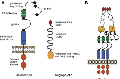
The family consists of two tyrosine kinase receptors, the TIE receptor 1 and 2 which can bind four ligands, Angiopoietin 1-4 (Ang1-4). The ligands are secreted glycoproteins, composed of an N-terminal superclustering domain, responsible for the creation of the higher order multimers of the ligands; a coiled-coil domain, responsible for ligand homo-oligomerization, which is essential for receptor activation and a C-terminal fibrinogen-like domain, responsible for receptor binding. The laters are separated by the linker region (180,181). The extracellular part of TIE1 and TIE2 consists of three Ig- like domains that are flanked by three EGF-like cysteine repeats followed by three fibronectin type III domains. The intracellular part has a split kinase domain. Ligands bind to the Ig- and EGF-like domains of the TIE receptors (182). The structure and binding of Angs and TIE receptors are shown in Figure 9.
22
Figure 9. Structure (A.) and binding (B.) of TIE receptors and angiopoietins (183).
Ang1 is primarily secreted by mesenchymal cells, such as perivascular cells and fibroblasts, and acts in a paracrine manner on the endothelium (184,185). Ang2 is expressed by ECs (186) and retinal neurons (187). Following cytokine activation of the endothelium, it is rapidly released and acts in an autocrine manner on ECs (188). Ang1 and Ang2 bind to TIE2 with similar affinity (182). Ang1 acts as an agonist of the TIE2 receptor, activating EC survival, integrity and vessel maturation (189), whereas Ang2 is thought to be an antagonist, displacing the more active ligand Ang1. However, Ang2 has also been reported to induce receptor phosphorylation in a context-dependent manner (190). The molecular basis for these contradictory functions has not been unraveled. However, a number of factors have been implicated in controlling agonistic versus antagonistic functions (191,192). When functioning as a TIE2 antagonist, Ang2 enhances pericyte dissociation from the vessels and increases vascular permeability (193). In the absence of VEGF these unstable vessels die, but in the presence of VEGF they migrate or proliferate, depending on whether they are located at the sprouting tip or the stalk part of a newly forming blood vessel (194). Ang3 and -4 are counterparts of the same gene locus found in mouse and human, respectively, but with divergent functions. Moreover, while Ang3 is secreted by a number of mouse tissues, Ang4 is expressed to a high level only in the human lung (195).
The full length or proteolytically cleaved orphan receptor, TIE1 heterodimerize with TIE2 (196), and thus act as a co-receptor. Both TIE1 and -2 are expressed by
23
(lymph)endothelial cells (197,198), but TIE2 is present on hematopoietic cells and endothelial precursor cells as well (199,200). In addition to TIE receptors, angiopoietins have been found to bind integrins as well (201). Following context dependent binding of the ligands, the receptors are autophosphorylated, and intracellular signaling pathways are activated, mediating endothelial survival (202), migration, and permeability (203). Ang1 and TIE2 are shown to be essential in the recruitment of pericytes and their interaction with endothelial cells (204).
The Ang-TIE system plays a key role during vessel maturation, stabilization and remodeling, thus abnormalities of either member of the family results in diseases manifested in vascular malformations. Both Ang1 and Ang2 can be expressed by tumor cells and tumor endothelial cells (205). Despite pericyte coverage of the tumor vasculature is massively increased and thereby stabilized in response to Ang1 overexpression (206), the role of Ang1 in tumor-associated angiogenesis and metastasis remains controversial (207). Angiogenesis enhancing functions of Ang2 during VEGF or FGF induced angiogenesis has been demonstrated (208). On the other hand, an agonistic effect of Ang2 on TIE2 has also been documented (190). Furthermore, both TIE1 and -2 are present on both tumor cells and tumor ECs (209), and their expression is upregulated during angiogenesis (210,211). These findings suggest that tumor growth promoting or inhibiting functions of angiopoietins are possibly dependent on the tumor cell type and the balance between both angiopoietins and TIE1/TIE2 (190).
3.2.1.5 TGFβR family
The TGFβR family includes TGFβ ligands: TGF-β1-3, bone morphogenic proteins (BMPs), growth and differentiation factors (GDFs), activins, inhibins, leftys, nodal and the mullerian inhibitory substance (MIS). Ligands can be secreted by tumor or a number of stromal cells (212). The receptors of the superfamily include the Type I receptors:
Alk (activin receptor-like kinase) receptors, and Type II receptors: TGFβ RII, BMP RII, Act RII (activin type II receptor) and RIIB, and MIS RII. Co-receptors of the family include TGFβRIII/betaglycan, endoglin, BAMBI (BMP and activin membrane-bound inhibitor homologue) Crypto and Cryptic (213).
TGFβ and its receptor, TGFβR are key molecules in cancer development and progression. On one hand, TGFβ can inhibit tumorigenesis by suppressing cell cycle
24
progression and stimulating apoptosis in early stages of cancer (214). On the other hand, it can also promote cancer development and spreading by the suppression of anti-tumor immunity (215), modulation of cell invasion via modification of the microenvironment (216), induction of epithelial-mesenchymal transition (217) and degradation of the extracellular matrix (ECM) (218).
ECM and basement membrane (BM) degradation is a critical step in tumor invasion and metastasis. TGFβ plays an important role in ECM degradation via upregulating MMP2 expression in different tumor types (219), and facilitates tumor cell infiltration by degrading basement membrane components via the regulation of protease expression (220). TGFβ also plays a crucial role in angiogenesis by promoting endothelial cell proliferation and migration through signalling via ALK1 and by causing vessel maturation through ALK5 (41). Highly expressed ligands of the other family members, such as endoglin, can antagonize the inhibitory effects of TGFβ and contribute to proliferation, migration, and capillary formation of endothelial cells (218).
3.2.1.6 Notch receptor family
Four receptors (Notch1-4), and five ligands, Jagged 1-2 and delta-like ligand (DLL) 1, 3-4 of the Notch family have been described in mammals. The receptors and the ligands are both type I transmembrane proteins, and activation requires contact of the sender and the receiver cell. The activation of Notch signaling involves three proteolytic events, resulting in the release of Notch intracellular domain (NICD) into the cytoplasm and from there it translocates to the nucleus (221). With the binding of NICD, the CSL family is converted into a transcriptional activator by displacing corepressors and by recruiting coactivators to induce transcription of Notch target genes (222).
Notch signaling plays an important role in cell proliferation, survival, apoptosis and differentiation, as their targets include proteins and factors involved in the control of cell cycle and survival (p21, cyclin D3), transcription factors (c-MYC, NF-κB), growth factor receptors (HER/ErbB, IGF1-R) and regulators of apoptosis (survivin) (223-229).
Recently, non-canonical Notch signaling that does not involve CSL or require cleavage has also been identified. This induces development via interaction with other molecules, such as members of the the IL6/JAK/STAT-, the Wnt pathway, the uPA/uPAR axis or BCR-ABL (230-233). Activation of target genes via Notch is shown in Figure 10.
25
Figure 10. Activation of the Notch signaling pathway (234). Three proteolytic events result in the release of NICD into the cytoplasm and from there it translocates to the nucleus. With the binding of NICD, the CSL family is converted into a transcriptional activator to facilitate transcription of Notch target genes.
Although classically known for its role in embryonic development, the Notch pathway have been shown to mediate tumorigenesis (235) by regulating the formation of cancer stem cells (236), and epithelial- or endothelial-mesenchymal transition (237,238).
Aberrant activation of Notch signaling is implicated in various neoplastic processes.
Somatic gain-of-function mutations in Notch receptors have been identified first in T- cell acute lymphoblastic leukemia and later in other malignancies as well (239).
Functional studies have demonstrated the key role of Notch signaling in vascular development and postnatal angiogenesis (240), which is emphasized by the involvement of both ligands and receptors in vascular diseases. The cerebral autosomal dominant arteriopathy with subcortical infarcts and leukoencephalopathy (CADASIL) and the Alagille syndrome can be traced back to mutations in Notch3 and Jagged1, respectively (241,242). In the endothelium, as well as in VSMCs, both Notch receptors and ligands are highly expressed (243-250). DLL4 is generally considered as a marker of arterial and tip cell phenotype (244), moreover, it is more robustly expressed in tumor ECs, compared to the neighburing normal vessels (251). Thus it is the main ligand expressed in tumor neoangiogenesis (252).
The “tip” cell is located on the leading edge of an angiogenic sprout. It is highly migratory because of the presence of filopodia and expresses high levels of VEGFR2
26
and -3 (253,254). Stalk cells are located adjacent to the tip cell, extend fewer filopodia but form lumen, and proliferate to support sprout elongation. The sprouting process is maintained until proangiogenic signals decrease, and quiescence is reestablished (255).
It has been proposed that in response to VEGF, DLL4 is induced on tip cells. High DLL4 expression on tip cells lead and guide new sprouts, and is thought to activate Notch and suppress the tip phenotype on adjacent (stalk) ECs. As a result, Notch activity is high in stalk cells but low in tip cells, and a VEGF feedback loop is activated, whereby Notch activation causes reduction of VEGFR2 and VEGFR3 and induction of VEGFR1 on stalk cells. The absence of Notch signaling in the tip cells results in high VEGFR2 and VEGFR3 expression, causing enhanced sensitivity to external VEGF, while decreased VEGF receptor expression on the stalk cells limit sprouting and ensures the proper number of tip cells at the angiogenic front (254,256-258). Inactivation of DLL4-Notch leads to the formation of a highly branched and dense vascular network with excessive filopodia. Moreover, these vessels are often not fully lumenized, thus are unable to deliver the necessary amount of oxygen and nutrients to the tissue (252). In contrast, upregulation of DLL4 inhibits VEGF-induced endothelial cell proliferation (259) and downregulates VEGFR2 expression (260).
3.2.2 Mechanisms of tumor-induced angiogenesis 3.2.2.1 Sprouting angiogenesis
The first identified form of angiogenesis is endothelial sprouting. It was originally described by Ausprunk and Folkmann in 1977. According to their hypothesis, in response to the NO mediated VEGF signal, the postcapillary venules become dilatated, cell-cell interactions get lost, the basement membrane (BM) degrades, and thus the vessel becomes fenestrated. Endothelial cells lose their polarity and migrate to the connective tissue. Then a tube is formed from the ECs, which is followed by lumen formation, synthetization of a new BM and recruitment of pericytes (261). This hypothesis has been developed further by Paku and Paweletz in 1991. According to their model, in response to Ang2, tumor cells secrete MMPs and plasminogen activators, which digest the pericytes on the vessel wall. Consequently, the electron density of the mother vessel is changed. A gel-sol transition occurs in the BM, this is maintained by MMP2, which binds αvβ3 integrin. Growth factors are liberated from the
27
BM, such as bFGF and VEGF, thus inducing migration of ECs with a maintained polarity, lumen and except from tip cells, a maintained BM. As the sprouts grow the BM on the tip is continuously synthesized, pericytes are recruited in response to bFGF and PDGF and the interaction between ECs and mural cells is stabilized by TGF-β1 and Ang1 (262). This hypothesis provides explanations missing from the former model of Ausprunk and Folkmann. These include the losening and then regaining ECs polarity, the fact that there is de- and redifferentiation in the same process and that the lumen is formed before the synthesis of the BM, although formation of BM is known to be the facilitator for lumen formation.
3.2.2.2 Vessel incorporation or co-option
In 1987 WD Thompson raised the possibility that tumors acquire their vasculature by vessel incorporation, instead of vessel ingrowth (263). This theory was proven by Josephine Holash in 1999 (264,265). Angiogenesis by vessel incorporation usually occurs,when tumors grow or metastatize into well vascularized tissues, such as lung, liver or skin. Tumor cells grow along the preexisting, well developed vessels of the host tissue, thus annexing its vasculature. The process is faster than sprouting angiogenesis, as it does not require EC proliferation. Moreover, on the periphery of the tumor these vessels provide surface for sprouting angiogenesis. Meanwhile, in response to the locally predominant antiangiogenic factors, ECs can undergo apoptosis in the centre.
This in one hand causes necrosis of the tumor mass, but on the other hand it also triggers the extravasation and metastatization of tumor cells. Maintenance of incorporated vessels is secured by the interaction of Ang1-TIE2.
3.2.2.3 Intussusceptive microvascular growth
Intussusceptive microvascular growth was first described by Sybill Patan in 1996 in a human colon adenocarcinoma model (266). This type of vessel growth is characterized by the incorporation of peritumoral capillaries, which are then separeted by connective tissue pillars. The original model of Caduff has been redrawn by Paku and his collegues in 2011. After the formation of an intraluminal endothelial bridge the BM is locally degraded, thus the EC can attach to a collagene bundle from the underlying collagene layer. The actin cytoskeleton of the EC exerts pulling force to the collagene bundle,
28
which in turn is transported through the vessel lumen. Finally, connective tissue cells are immigrated to the pillars and new collagenous connective tissue is deposited (267), thus the process results in lots of vessels with big lumens, providing surface for EC sprouting. In the absence of EC proliferation, the process is faster than sprouting and does not involve permeability of the vessel wall. This suggests and some experimental data also supports, that intussusception does not rely on VEGF signalization, but instead is regulated by physiologic stimuli and cytokines, that lead to vessel maturation, such as bFGF (268), PDGF-BB (269), Ang1-TIE2 complex (270), ephrin-ephrin receptor (271).
However, the role of VEGF in the process is still contested (269,272,273).
Moreover, it is also shown, that in response to some antiangiogenic treatment, such as vatalanib or the mTOR inhibitor sirolimus, experimental tumors switch from endothelial sprouting to intussusceptive angiogenesis (274,275).
3.2.2.4 Glomeruloid angiogenesis
In 1992 Hauro Ohtani described coiled vascular structures in human gastrointestinal carcinoma. As they resemble renal glomeruli, they were called glomeruloid structures (276). These are characterized by many, tightly associated capillary loops with different thickness of BMs. The molecular mechanism behind the process is still not clear. On one hand, Sundberg et al. suggests that VEGF is a key mediator of inducing glomeruloid body formation and maintaining these vessels (277). On the other hand, it was also shown that the glomeruloid structure is created by proliferating and migrating tumor cells, which pull the capillaries and their branching points into the tumor cell nests. Thus, this type of vascular growth cannot be termed as true angiogenesis, but rather a reorganization of tumor blood vessels, which does not require EC proliferation (278). Moreover, these vessels seem to be able to provide enough oxygen and nutrients to the cells, as no necrosis was observed in tumors developing them. As a result, glomeruloid bodies are not only diagnostic markers of glioblastoma, but also poor prognostic factors in many cancer types (279).
3.2.2.5 Postnatal vasculogenesis
Postnatal vasculogenesis refers to the process, in which bone marrow derived VEGFR2+, TIE2+, AC133+, CD34+ endothelial progenitor cells (EPCs) incorporate
29
into the EC layer of the tumor capillary network in response to tumor derived VEGF (280) and other proangiogenic factors. After incorporation into the EC layer, EPCs are differentiated to mature ECs. Thereafter, by producing pro-angiogenic factors, such as VEGF and PlGF, they mediate the attraction of additional EPCs to the tumor vasculature. VEGF mobilizes these cells from the bone marrow via the stromal derived factor (SDF) and its receptor, CXCR4. The process of postnatal vasculogenesis was described by Takayuki Asahara and his group both in physiologic and pathologic conditions at the end of the 1990s (281).
3.2.2.6 Vessel-like structures, formed by tumor cells
It has been shown, that not only ECs, but aggressive tumor cells can also form vessel- like structures, which facilitate tumor perfusion.
In 1941 Béla Kellner described tumor sinusoids in soft tissue sarcomas (282). These are lumens, which are covered exclusively by tumor cells, and are responsible for the transport of blood cells within the tissue.
It is also possible that tumor cells form a lumen together with ECs, without expressing endothelial or embrional markers. These structures are called mosaic vessels and were also first described in the 1940s (283). The genesis of these types of vessels is still not well understood. They are considered to be formed either by the apoptosis of incorporated ECs, which is followed by the occupacy of the lumen or invasion of the vessel by tumor cells. The process is thus thought to be mediated via Ang2 signalling.
Vasculogenic mimicry refers to the process, when tumor cells express differential markers to completely resemble BM covered ECs. The process was first described by the group of Mary J Hendrix in uveal melanoma, where highly agressive melanoma cells formed vessel-like channels and upregulated endothelial genes and genes involved in microvascular channel formation. Meanwhile, these cells downregulate classical melanoma markers. None of the main angiogenic cytokines, such as VEGF, PDGF, bFGF, TGF-β, Ang, Notch, TNF-α seem to induce the formation of these channels (284).
Different types of vascularization mechanisms in cancer are shown in Figure 11.
30 Sprouting angiogenesis
Glomeruloid agiogenesis
Vessel incorporation Intussusceptive angiogenesis
Postnatal vasculogenesis
Vasculogenic mimicry
Alternative vascularization
mechanisms
Figure 11. Alternative vascularization mechanisms in cancer (285).
3.2.3 Characteristics of tumor blood vessels
Because of the dominance of proangiogenic factors in malignant tissues, tumor blood vessels differ from the normal ones both in function and structure. In a healthy adult, ECs have a long half-life with a well differentiated BM (286), which is stabilized by pericytes and VSMCs, to influence permeability and contractility of the vessel wall.
(287). Moreover, they secrete a minor amount of survival factors for the ECs, such as VEGF and Ang1. In case of Ang2 and MMP effect pericytes and VSMCs get detached from the BM and after their apoptosis, ECs also get prepared for cell death (288). In a healthy adult the endothelial tube is connected with cell-cell junctions. Maintenance of the vessel is supported by autocrin factors and signals launched by the oxygen sensors, such as the PHD2, which secures the appropriate blood flow (289).
In contrast, tumor blood vessels usually have deficient pericyte and VSMC coverage, which are not tightly attached to the ECs. They are often immature, less contractile and their shape is abnormal (290,291). The BM is often degraded and loosely attached to the EC, or it is thick and thus hinder contraction and perfusion (292). The morphology of ECs are changed and cell-cell connections are often lost (293). As a result, vessels get
31
dilated negatively impacting the ratio of vessel surface, which supplies the tumor tissue with oxygen and nutrients. Tumor cells can press in the instable vessel wall, thus vessel diameter becomes uneven (294). Because of this and the altered EC morphology, vessels get curved and form serpentine-like structures, and may also create high amount of anastomosis within the vessel wall (291). This is further supported by the impaired cell junctions, resulting in the loss of the signals of oxygen sensors, eventually leading to abnormal flow in the tumor vasculature (295). Because of the degraded BM, ECs lose their polarity, thus get detached from the endothelial layer and may form a plug in the lumen resulting in thrombosis and further injury of the vessel wall. This leads to enhanced metastatization capacity and the creation of mosaic blood vessels (296).
Moreover, degraded mural cells and BM leads to fenestrated vessel walls, and thus not only metastatizing tumor cells may intravasate to the lumen (297), but vessels get hyperpermeabilized as well, causing an increase in the interstitial fluid pressure, which cannot be restored because of the decreased lymphatic function (298,299). Thus, the oxygen supply of the tumor decreases, resulting in an increased glycolitic activity to provide energy and the necessary building material for the growing tumor (300). The altered acidic microenvironment and abnormal morphology of tumor vessels increase the invasion and metastatization ability of malignus cells, creating a vicious circle in tumor progression. This microenvironment selects for hypoxia resistant, aggressive tumor cells (301). Because of the high intratumoral pressure, intersticial fluid, often containing tumor cells may infiltrate to lymphatic vessels, thus enhancing lymphatic tumor spread (302). Furthermore, some immune cells also promote invasion in these circumstances (303). Unlike in tumors, in case of physiological angiogenesis, these processes are balanced by the generation of antiangiogenic factors. Moreover, as a result of the inadequate blood flow, the resulting hypoxia and/or the tightened BM, efficacy of conventional anticancer therapies are inadequate due to limited penetration.
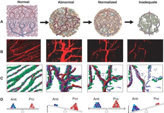
These abnormalities of tumor blood vessels and the resulting aggressiveness of cancers, accompanied by decreased efficacy of conventional treatment led Rakes Jain to the elaboration of the vessel normalization theory (304). Accordingly, the inhibition of the signal of proangiogenic factors and the subsequent normalization of tumor blood vessels has become a critical step in cancer therapy. Proposed structures of normal, tumorous, normalized and inadequate blood vessels are shown in Figure 12.
32
Figure 12. Proposed structures of normal, tumorous, normalized and inadequate blood vessels. (A.) Schematic structures of the vascular system. (B.) Two-photon images of normal blood vessels in skeletal muscle and subsequent images showing human colon carcinoma vasculature in mice at day 0, day 3, and day 5 after administration of VEGFR2-specific antibody. (C.) Diagrams depicting the changes in pericyte (green) and basement membrane (blue) coverage. (D.) Changes in the balance of pro-, and antiangiogenic factors in the tissue (305).
3.2.4 Inhibition of the tumor vasculature
In the complex processes outlined above, several differently acting agents are capable of blocking the blood supply of a tumor. It can be suppressed in distinct cells and also in diverse levels and modes.
3.2.4.1 Conventional chemotherapeutic agents
Conventional therapeutic agents have been found to have antiangiogenic functions as
"side effect". These include microtubule targeting agents, such as Vinca-alkaloids or Taxanes, that block endothelial cell proliferation (306). Beside other drugs, thalidomide and lenalidomide also have complex antiangiogenic functions (307,308).
3.2.4.2 Vacular disrupting agents
Vascular disrupting agents (VDAs) selectively target tumor vessels, causing fast and dynamic effects (309). They destroy rapidly dividing endothelial cells in the tumor
33
tissue by targeting the colchicine binding site of tubulins (tubulin-binding agents) (310) or induce vascular collapse through TNF-α (flavonoid-type VDAs) (311). As a result, vascular supply shuts down, causing necrotization in response to the insufficient oxygen and nutrient delivery (312,313). By this strategy both preexisting and newly formed vessels can be targeted, with the inhibition of the metastatic potential as well. VDAs mostly act on advanced tumors, which are resistant for conventional therapy. However, their effect is very short, and can cause serious cardial toxicities (314). Moreover, vessel density is higher at the edge of tumors, so targeting it with VDAs is not effective enough. Consequently, often there is no visible tumor shrinkage following VDA therapy. Furthermore, as tumors can also be fed by diffusion from nearby tissues, the monotherapy can leave a surviving rim at the edge of the tumor, which allows rapid tumor regrowth (315). Combretastatin A-4 Phosphate (CA4P), 5,6 dimethylxanthenone- 4-acetic acid (DMXAA) and NPI-2358 are the most investigated VDAs.
3.2.4.3 Vasoactive agents
Vasoactive agents also combinatorially block existing vessels and suppress the formation of new ones. Moreover, they do not target preferentially the larger vessels in the tumor center, but also the small ones in the periphery, causing hyperabnormalization and hyperpermeability of the vessels. This allows chemotherapeutic agents better access to the tumor. However, these drugs are highly toxic when administered systemically, thus local, small dose and metronomic application is preferable.
The most often used vasoactive agents are inflammatory modulators. Among them the most important are the followings:
IL-2, a cytokine, that induces T cells, augments natural killer cell activity and demonstrates vasopermeability activity (316).
TNF-α, an inflammatory cytokine, that is principally produced by activated macrophages and monocytes and has direct effects on tumor cells. After exposure to low-dose TNF-α, hyperpermeability, hemorrhagic necrosis, extravasation of erythrocytes, edema and vessel congestion were observed (317), leading to an increased intratumoral chemotherapeutic drug concentration or effect of radiation (318). In the clinic combination therapy of melphalan and TNF-α is a popular approach to treat patients with unresectable advanced sarcoma and advanced melanoma (319).
34
Histamine is an inflammatory modulator that causes edema in small vessels by locally increasing the lymph flow into the extracellular space and by promoting hyperpermeability of the endothelium (320).
3.2.4.4 Angiogenesis inhibitors
Angiogenesis inhibitors (AIs) block the formation of new vessels from preexisting ones, but do not affect already established vasculature. Despite the fact that they are used in advanced tumors in the clinic, they are tought to mainly act on small vessels of the tumor edge, mostly at the early stage of tumorigenesis or metastatization.
As discussed above, endothelial sprouting is activated in physiological angiogenesis in response to hypoxia, thus, suppression of HIF1 to bind to the HRE of proangiogenic molecules is one of the main options to block the process. In the absence of selective HIF inhibitors (321) and the presence of a number of factors that also regulate angiogenic growth factor expression in tumors, the common way is to target molecules downstream of HIF by the blockade of receptor - ligand communication.
Growth factors can be targeted with either monoclonal antibodies (mABs) or soluble
„trap/decoy” receptors. mABs are produced from a common germ cell and have affinity for a specific antigen, thus inhibiting their binding to the corresponding receptors (322).
Although having just one target, their effectiveness may be broad, as angiogenic growth factors usually bind to a number of isoforms of their target receptors. mABs are usually given intravenously, and because of their high molecular weight, their half-life is long (weeks). The resulting more prolonged inhibition allows less frequent dosing. The high molecular weight of mABs reduces diffusional capacity, thus renal filtration is limited, but their penetration into the brain is also impeded. Antiangiogenic antibodies as single agents exhibit only limited clinical activity, thus they are usually applied in combination that significantly improves their therapeutic effect.
Decoy receptors are soluble proteins that compete with their membrane bound counterparts with high affinity for their ligands. In the absence of transmembrane and intracellular domains, they fail to mediate signal, thus function as a trap for angiogenic growth factors (323). These traps can either be physiologically present, as a result of proteolytic cleavage of their transmembrane counterparts (eg. soluble VEGFR1), or designed and added as external therapy (324,325).
35
Receptors on ECs can be targeted at their extracellular part by mABs, thus blocking ligand binding, or by RTKIs at the tyrosine kinase domain, which serve as a docking site for molecules mediating the angiogenic signal (326,327). Anti-receptor mABs can usually target only one of the receptors, thus do not mediate widespread effect.
RTKIs can be divided to five subgroups:
Type I kinase inhibitors recognize the active conformation of the receptor. They typically consist of a heterocyclic ring system that occupies the purine binding site of the enzyme, and form one to three hydrogen bonds by that part of the inhibitor, which mimick the purine ring of the adenine moiety, thus actively competing with ATP.
Extra interactions may also be formed at hydrophobic regions adjacent to the hinge region. The hydrophilic region of the enzyme can be exploited for maximizing the solubility of the compounds. Since the targeted ATP pocket is conserved through the kinome, Type I inhibitors usually have low kinase selectivity, thereby enhancing the potential for off-target side effects. Examples of type I tyrosine kinase inhibitors targeting the VEGF pathway are sunitinib and pazopanib.
Type II inhibitors recognize the inactive, unphosphorilated conformation of the kinase and indirectly compete with ATP by occupying the hydrophobic pocket, which is created by the DFG-out conformation of the activation loop. It is also known as the allosteric site, thus type II inhibitors can modulate kinase activity in an allosteric way.
The DFG-out conformation is unique to all receptors, thus the hydrophobic interactions with the DFG pocket confer a high degree of selectivity. Some type II inhibitors are able to form a hydrogen bond directly to the ATP-binding site, but this is not necessary for their functionality. Sorafenib is a type II kinase inhibitor (328).
Type III or allosteric inhibitors bind outside the catalytic domain of the kinase, in regions that are involved in the regulatory activity of the enzyme. They block the binding of ATP by modulating the conformation of the receptor. As they exploit the binding sites and regulatory mechanisms that are unique to the target, a high degree of kinase selectivity is exhibited. Additionally, allosteric modulators can provide delicate regulation of kinases, which is not easily performed with ATP-competitors.
Type IV kinase inhibitors also act allosterically by forming a reversible interaction outside the ATP binding pocket, in the kinase substrate binding site, thus are not