A new player in the alternative complement pathway: MASP-1 is essential for LPS-induced but not for zymosan-induced alternative pathway
activation
Katalin Paréj*1, Andrea Kocsis*1, Csenge Enyingi*, Ráhel Dani*, Gábor Oroszlán*, László Beinrohr*, József Dobó*, Péter Závodszky*, Gábor Pál†, Péter Gál*
1 K.P. and A.K. contributed equally to this work.
*Institute of Enzymology, Research Centre for Natural Sciences, Hungarian Academy of Sciences, Magyar tudósok körútja 2, H-1117, Budapest, Hungary.
†Department of Biochemistry, Eötvös Loránd University, Pázmány Péter sétány 1/C, H-1117, Budapest, Hungary.
Running title: MASP-1 plays a role in alternative pathway activation
Abstract
The complement system is a sophisticated network of proteases. Here, we describe an unexpected link between two “linear” activation routes of the complement system: the lectin (LP) and the alternative pathways (AP). MASP-1 is known to be the initiator protease of the LP. Using a specific and potent inhibitor of MASP-1, SGMI-1, and also other MASP-1 inhibitors with different mechanism of action, we demonstrated that in addition to its
functions in the LP, MASP-1 is also essential for bacterial LPS-induced AP activation, while it has little effect on zymosan-induced AP activation. We have shown that MASP-1 inhibition does not only prevent AP activation, but it also attenuates an already initiated AP activity on LPS-surface. This newly recognized function of MASP-1 can be important for the defense against certain bacterial infections. Our results also emphasize that the mechanism of AP activation depends on the activator surface.
Introduction
Complement is a proteolytic cascade, which can be activated via three different routes: the classical (CP), the lectin (LP) and the alternative pathways (AP). Ca2+-dependent complexes consisting of pattern recognition molecules (PRMs) and associated serine proteases initiate the CP and the LP. The most abundant protease of the LP is MASP-1. When the LP is
triggered, MASP-1 autoactivates first, then it cleaves MASP-2 in normal human serum (NHS) (1). MASP-2 cleaves C4, while C2 is cleaved by both MASP-1 and MASP-2. The LP
generates the same type of C3 convertase (C4b2a) as the CP. The mechanism of AP activation is quite different, since there is no known PRM of the AP. Properdin, the positive regulator of the AP, was suggested to act as a PRM, but this issue is controversial (2). AP is initiated when C3b or C3b-like molecules emerge near a surface. C3b can covalently bind to the surface and it binds factor B (FB). C3b bound FB is cleaved by factor D (FD), a serine protease that circulates predominantly in the active form. The resulting C3bBb is the AP C3 convertase, which converts more C3 into C3b providing a positive feedback. Properdin, the only known positive regulator of complement, stabilizes the AP C3 convertase (3). The AP can be initiated independently of the CP or the LP. According to the prevailing “tickover”
hypothesis, the reactive thioester bond of C3 slowly hydrolyzes in the fluid phase, and the emerging C3b-like molecule, C3(H2O), binds FB. After having been cleaved by FD the resulting C3(H2O)Bb complex acts as a C3 convertase. If this fluid-phase convertase is generated near a surface, the nascent C3b molecules can bind there covalently and initiate the amplification process. Endogenous cells are protected from excessive complement activation by cell-surface-anchored and fluid-phase inhibitors. The most important AP inhibitor is factor H (FH), which can bind to self-surface-deposited C3b and destroy it by recruiting the serine protease, factor I (FI).
The three activation pathways had been considered as independent activation routes, however, evidence has begun to accumulate suggesting various cross talks. Recently an important connection between the LP and the AP has been demonstrated. Activated MASP-3 is a protease responsible for activating pro-FD in the circulation (4-6), and in fact it is the only protease doing so in resting human blood (7).
Here, we describe an unexpected link between the LP and the AP demonstrating a novel function of MASP-1. On certain activator surfaces MASP-1 has been found to be indispensable for AP activation in the absence of Ca2+. This phenomenon might be of great
importance for the protection against invading microorganisms, but it also might contribute to uncontrolled complement activation leading to severe symptoms.
Materials and methods Reagents
All commonly used reagents were purchased from Sigma-Aldrich or Merck unless otherwise indicated. Human FB was purchased from Merck (Calbiochem brand). Human C1 inhibitor (Berinert P) was from CSL Behring. LPS from three different Gram-negative bacterial strains, such as Salmonella typhimurium (L6511), Escherichia coli (L8274) and Pseudomonas aeruginosa (L9143) was purchased from Sigma-Aldrich. Saccharomyces cerevisiae (sc- 258367A) Zymosan A was from Santa Cruz Biotechnology.
Human serum, anti-MASP-1 antibody and purified proteins
NHS collected from 10 healthy volunteers was pooled and stored at -80 °C. Production of antibody (αM1-SP) against the serine protease domain (SP) of MASP-1 is described in (6).
MASP-1-depleted serum was prepared by running NHS through an anti-MASP-1 column (αM1-SP-Sepharose) twice. The catalytic fragment of human MASP-1 (rMASP-1cf) was purified as described (8). The N-terminal fragment of MASP-1 (M1_D1-3) was purified as described (9). MASP-1-specific inhibitor (SGMI-1) and MASP-2-specific inhibitor (SGMI-2) were produced as described (10). The MASP-3-specific inhibitor (TFMI-3) was prepared as described (7). Recombinant FD was produced as described (5). The recombinant serpin domain of human C1 inhibitor was produced as described (11). C3 was purified from human EDTA plasma (12). C3b was generated from C3 using trypsin (T8003, Sigma-Aldrich), which was later chromatographically removed from C3b.
C3 deposition in Mg2+- EGTA buffer
Microtiter plates were coated with 10 µg/ml mannan, 10 µg/ml LPS or 100 µg/ml zymosan in 15 mM Na2CO3, 35 mM NaHCO3, pH 9.6. After overnight incubation at 4°C, the wells were blocked with 1% BSA in TBS buffer (50 mM Tris, 150 mM NaCl, pH 7.4) for 1 h at 37 °C, then washed three times with 0.1 % Tween-20 in TBS buffer. NHS was diluted 6-fold in 10 mM HEPES, 150 mM NaCl, 10 mM EGTA, 4 mM MgCl2, 0.1% Tween-20, pH 7.4 and if required it was pre-incubated in microcentrifuge tubes with inhibitors for 30 mins at 25 °C.
The samples were added to the microtiter plates and further incubated for 45 mins at 37 °C.
The plates were washed; then 5000-fold diluted anti-human C3c (A0062, DakoCytomation) and 40 000-fold diluted anti-rabbit IgG HRP conjugate (A1949, Sigma-Aldrich) were applied in 1% BSA-wash buffer. Ortho-phenylene-diamine (OPD) in 50 mM citrate buffer (pH 5.0) was used for detection. The absorbance was read at 490 nm.
FB cleavage assay
1.23 µM C3b and 1.07 µM FB were incubated with or without recombinant FD (0.25 nM); or with recombinant FD (0.25 nM) and SGMI-1 (20.5 µM); or with rMASP-1cf (50 nM) in 100 mM NaCl, 50 mM Tris-HCl, 5 mM MgCl2, 5 mM CaCl2, pH 7.4 at 37 °C. Samples were taken at 0 and 1.5 hours. Reactions were stopped by 2-fold dilution with SDS-PAGE sample buffer and heating for 3 mins at 95 °C. Samples were then analyzed by 10% SDS-PAGE under reducing conditions.
Inhibition assays
The activity of the AP was measured in the presence of MASP-1-specific inhibitors (SGMI- 1), 1.25-20 µM; anti-MASP-1 antibody (αM1-SP), 2.1-133 nM; N-terminal fragment of MASP-1 (M1_D1-3), 0.06-7.3 µM; specific inhibitors of other complement proteases (SGMI- 2, 1.25-20 µM; TFMI-3, 0.29-30 µM); full length C1 inhibitor, 0.35-11.3 µM; and serpin domain of C1 inhibitor, 1.14-9.1 µM. The effect of the above-mentioned molecules was followed through the decrease in C3 deposition.
Inhibition of ongoing C3 deposition
6-fold diluted NHS was incubated in the separate wells of LPS-coated microtiter plates for 45 mins at 37 °C. During this period at different time points same volume (4.8 µl) of SGMI-1 or FUT-175 inhibitors were added to the individual reactions to reach a final concentration of 20 µM and 100 µM, respectively. Same volume of serum dilution buffer was also added to some wells as negative control. After a total of 45 mins serum was discarded, the microtiter plate was washed and the deposited C3 fragments were measured similarly as described above.
Statistical analysis
Statistical analysis was performed with Origin 6.0 (OriginLab Corporation, Northampton, Massachusetts) software. Student’s t-test was used to determine significant difference
between the control and treated samples. Two levels of significance were used: *p<0.05 and
** p<5x10⁻⁹.
Results and discussion
MASP-1-specific inhibitor blocks AP activation on LPS-coated but not on zymosan-coated surface
To reveal the individual roles of MASPs in complement activation we have previously
developed selective and potent inhibitors against MASP-1 (SGMI-1), MASP-2 (SGMI-2) and MASP-3 (TFMI-3) (7, 10). In this study, we used these inhibitors to monitor activation of the AP using 6-fold diluted NHS in Mg2+-EGTA buffer. We set up ELISA assays for measuring C3 deposition. For measuring AP activation on plates two activators are used routinely:
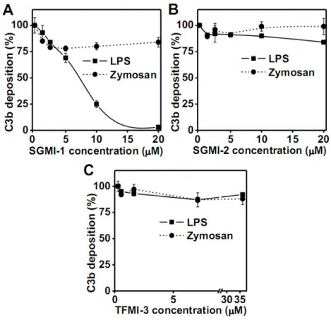
bacterial LPS and yeast zymosan. To our great surprise the MASP-1-specific inhibitor (SGMI-1), at a concentration of 10 µM, efficiently inhibited C3 deposition and caused near complete abrogation at 20 µM on LPS-coated surface (IC50=6.2 µM) (Fig. 1A). Contrary to that, only partial (~20%) inhibitory effect was detected on zymosan-coated surface even at the highest inhibitor concentration (20 µM) (Fig. 1A). SGMI-2, the MASP-2-specific inhibitor, caused only marginal inhibition of C3 deposition on LPS-coated surface and had no effect on zymosan-coated one (Fig. 1B). Inhibition of MASP-3 using TFMI-3 did not influence either the LPS- or the zymosan-induced AP activation (Fig. 1C). These results imply that MASP-1 has a key role in the AP activation on LPS-coated surface. The IC50 value for the inhibition of LPS-induced AP activation by SGMI-1 (6.2 µM) is much higher than the value (20 nM) calculated by considering only the 7 nM Kd value of the enzyme/inhibitor complex and the molar concentration of MASP-1 (25 nM) in the assay. We suggest that the complexity of the serum, and the multistep nature of the ELISA assay we used, results in an IC50 value
significantly higher than expected.
MASP-1 inhibitors having different mechanisms of action elicit the same effect on AP activation
Since the inhibitory effect of SGMI-1 on AP activation was detected only at relatively high inhibitor concentration, we had to rule out the possibility, that the observed effect of SGMI-1 is exerted through its weak, nonspecific inhibition of a protease or proteases other than
MASP-1. To this end, we applied MASP-1 inhibitors having different mechanism of action. A
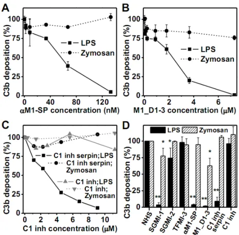
polyclonal antibody developed against the SP domain of human MASP-1 (αM1-SP) showed a concentration-dependent inhibitory effect on C3 deposition on LPS-coated surface, while it had only marginal effect on zymosan-coated surface (Fig. 2A). At 130 nM concentration, it completely prevented LPS-induced AP activation. This antibody is also an efficient LP inhibitor (Supplemental Fig. 1).
The N-terminal fragment of MASP-1 is responsible for dimerization and for binding to the PRMs. Therefore, we investigated the effect of the N-terminal fragment of MASP-1 (M1_D1- 3) on the AP activity. It has already been shown that M1_D1-3 is able to attenuate LP
activation in NHS through displacing the MASPs from the PRMs (9). M1_D1-3 behaved similarly to SGMI-1 in the AP assay system: it inhibited AP activation on the LPS-coated surface in a concentration dependent manner, whereas it showed only limited effect on the zymosan-coated plate (Fig. 2B). This result suggests that binding of MASP-1 via its N- terminal region to a PRM or to other unknown molecules plays an important role in AP activation triggered by LPS. The binding molecule cannot be MBL, since MBL-binding to carbohydrates is strictly Ca2+-dependent. It is very likely that the PRM-MASPs complexes are largely intact at physiological salt concentration in the presence of Mg2+-EGTA (13, 14). We tried to demonstrate MASP-1-binding to LPS-coated wells using the MASP-1-specific antibody, but we could not detect any signal (data not shown). This could mean that the binding is weak or transient, but it is still enough to support AP activation on the LPS surface.
It should be noted, that M1_D1-3 is not strictly MASP-1-specific: it displaces all MASPs and MAPs from the PRMs.
The natural inhibitors of MASPs are serpins. C1 inhibitor is an efficient inhibitor of the LP in NHS (15). We tried to prevent MASP-1-mediated AP activation using C1 inhibitor. We did not get any inhibition of C3 deposition on LPS-coated plates using natural C1 inhibitor isolated from human blood (Fig. 2C). Contrary to that, we detected concentration dependent inhibition using the recombinant serpin domain of C1 inhibitor (Fig. 2C). C1 inhibitor is an atypical serpin, since besides of the serpin domain it possesses a heavily glycosylated N- terminal domain as well. Our results indicate that in the LPS-induced AP-activation system the N-terminal domain somehow prevents the interaction between MASP-1 and C1 inhibitor.
On the other hand, the single globular serpin domain of C1 inhibitor has a free access to active MASP-1 under these conditions. Similar observations were reported earlier using StcE- treated C1 inhibitor. StcE is a metalloprotease secreted by E. coli O157:H7 that can remove the N-terminal domain of C1 inhibitor. The resulting truncated C1 inhibitor efficiently
inhibits endothelial cell-bound kallikrein, whereas the full-length C1 inhibitor fails to do so (16). This suggests that the N-terminal domain of C1 inhibitor might have an important regulatory function: it hinders the rapid and efficient interaction between the serpin and the target protease on a surface of a pathogen promoting complement activation, while the full- length molecule can successfully prevent spontaneous activation of the complement proteases in the fluid phase. On the zymosan-coated surface neither the full-length C1 inhibitor nor the serpin domain could attenuate AP activation. Fig. 2D summarizes the results of 3 independent measurements with each inhibitor on LPS and zymosan surface.
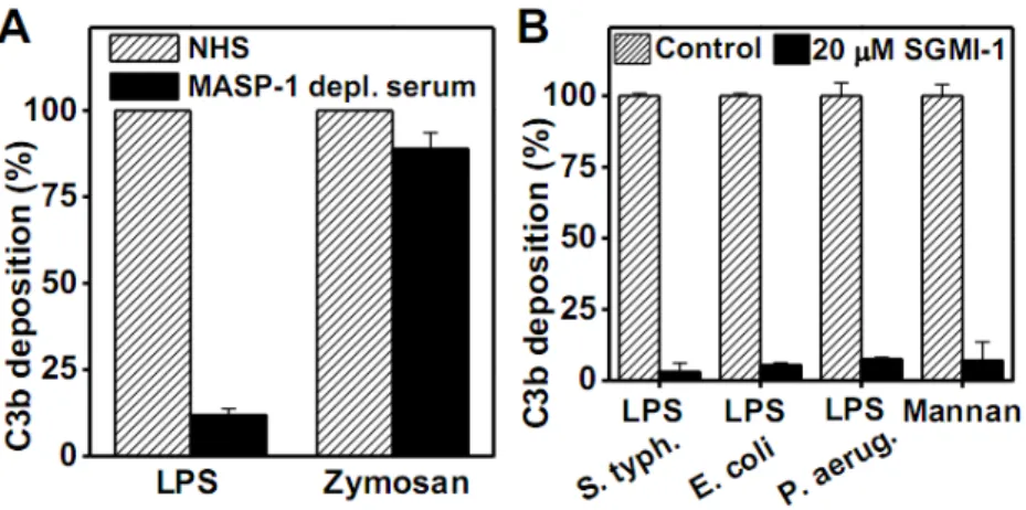
To corroborate the results obtained by using various inhibitors in NHS, we checked the ability of MASP-1-depleted serum to activate the AP in our assay system. In accordance with the above results, the MASP-1-depleted serum showed very low AP activity on LPS- coated surface, while the AP activity was only partially compromised on zymosan-coated surface (Fig. 3A). Taken together, these results show that MASP-1 has a central role in LPS- induced AP activation.
Surface dependence of the MASP-1-mediated AP activation
To refine our knowledge about the surface requirement of MASP-1-mediated AP activation we tested various AP activators. Besides the LPS type (S. typhimurium) that we used in the experiments above, we checked LPS preparations from other bacteria as well. Both the E. coli and the P. aeruginosa LPS preparations gave the same results: high level C3 deposition that could be almost completely inhibited by 20 µM SGMI-1 (Fig. 3B). Selander et al. (17) demonstrated that mannan-coated surface induces efficient AP activation in Mg2+-EGTA buffer. Using the same activation surface, we showed that the mannan-induced AP activation can also be blocked by our MASP-1-specific inhibitor (Fig. 3B). Finally, we tested the lysis of rabbit erythrocytes, a frequently used method to measure AP activity. In this assay system 18- fold diluted human serum readily lysed rabbit erythrocytes, and the lysis could not be
attenuated by SGMI-1 even at high concentration (20 µM) (data not shown). Our results are in line with those of Degn et al. (18), who used MASP-1/3-deficient serum form a patient having 3MC syndrome and measured efficient lysis of rabbit erythrocytes.
Inhibition of MASP-1 attenuates C3 deposition in progress
Theoretically, AP activation can be divided into distinct parts: the first part is the initiation lag phase, when the first few C3b molecules deposit on the activation surface, and the second part
is the amplification phase, when the surface deposited C3b molecules serve as a base for generating additional C3 convertase complexes building up the positive feed-back loop. We also studied to which phase of the AP activation MASP-1 contributes. To this end we
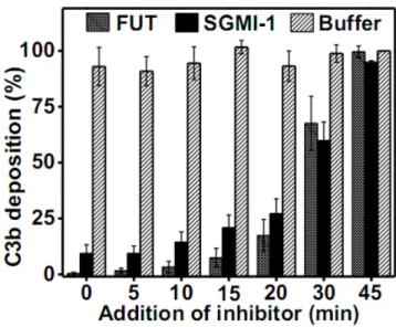
launched parallel AP activation experiments incubating NHS on LPS-coated surface for different time periods to let the C3b deposition reach a certain level, and then we added the same volume of SGMI-1 or FUT-175 or buffer to the individual reactions. FUT-175 is a broad-spectrum serine protease inhibitor which inhibits all complement proteases. In
concordance with previous results (19), the time course of C3 deposition shows an initial lag phase (5-6 min) followed by a rapid amplification phase (Fig. 4). SGMI-1 reduced C3 deposition at any phase of the AP activation. Even in the presence of surface-deposited C3b, SGMI-1 attenuated additional deposition of C3b, though not as efficiently as the broad- spectrum serine protease inhibitor FUT-175. These results suggest that MASP-1 is necessary for maintaining an efficient amplification process of LPS-induced AP activation. However, it does not necessarily preclude that it is also important for the initiation.
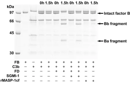
MASP-1 does not cleave FB
We have assessed the direct proteolytic action of MASP-1 on components of the AP.
Previously we studied the ability of MASP-1 to cleave C3 and pro-FD (5, 20). Here we checked whether MASP-1 can cleave FB. We mixed FB with C3b and rMASP-1cf, incubated the mixture at 37 °C and followed the digestion on SDS-PAGE (Fig. 5). We found that 50 nM MASP-1 was not able to cleave C3b-bound FB. Although after 1.5h some Bb fragment appeared, but the band was not significantly stronger than in the case of the negative control.
Under similar conditions 0.25 nM FD cleaved FB with high efficiency. We also demonstrated that SGMI-1 does not inhibit FD-mediated FB cleavage significantly (Fig. 5). Taken together, it seems unlikely that MASP-1 exerts its proteolytic effect directly on the known components of the AP. Instead, it may cleave a still unidentified factor and thereby promote AP activation.
Concluding remarks
Our observations are in accordance with previous results from the literature. The finding that the mechanism of AP activation depends on the nature of the activator was reported by Kimura et al (21). They found that properdin was indispensable for LPS-induced AP activation in mouse serum whereas properdin deficiency impaired only minimally the zymosan-induced AP activation. The reason for the differential requirement of properdin by different AP activators is unknown but it is intriguing that we got the same surface-
dependence for MASP-1-supported AP activation using NHS. It is plausible to assume that the extent of AP activation on certain surfaces depends on the balance between properdin- and MASP-1-mediated promotion and FH-dependent inhibition. It is very likely that FH binds to LPS more strongly than to zymosan. It is possible that there is a connection between the requirement of properdin and MASP-1 for the activity of the AP on certain activation surfaces, but further experiments are needed to investigate this possibility.
Although anti-LPS antibodies may play a role in AP activation, removal of antibodies from the serum did not influence the MASP-1 dependence of LPS-induced AP activation (data not shown).
Selander et al. (17) studied the contribution of MBL to AP activation. Although they concluded that MBL alone (without the contribution of MASPs) triggers C3 deposition on solid-phase mannan or mannan-rich LPS in C2 deficient serum, their experimental data clearly indicated that MASP-1 has a role in the C2-bypass activation. In their experiments serum fractions containing MBL/MASP-1 complexes were able to activate C3 in the absence of C2, while other MBL/MASP complexes failed to do so. The extent of the C3 deposition depended solely on the MASP-1 content of the MBL/MASP complexes. We also observed vigorous C3 deposition on mannan-coated surface in the presence of Mg2+-EGTA, which was entirely MASP-1 dependent. MBL cannot bind to mannan under these conditions; however other PRMs may retain this ability in the absence of Ca2+ (22). It is possible that in the
presence of Ca2+, where MBL binds to its target, the AP promoting activity of PRM-MASP-1 complexes are even higher and may significantly contribute to the defense against Gram- negative bacteria. Selander et al. (17) speculate that MASP-1 may act on C3 indirectly by recruiting other serum proteases that cleave C3. Our results are also reconcilable with this hypothesis especially, as we demonstrated earlier, that MASP-1 is an atypical complement serine protease with relatively broad substrate specificity (23).
The fact, that MASP-1 is necessary for LPS-induced AP activation, but not for zymosan- induced one, suggests that the mechanism of AP activation differs in the case of the two different activators. Further studies are required to clarify the exact molecular mechanism of complement activation on these activator surfaces.
In conclusion, we discovered a novel role of MASP-1 in complement activation. Although it was suggested earlier by several authors that MASP-1 might contribute to AP activation, no solid evidence has been presented. The activator-specific requirement of MASP-1 in AP
activation indicates a tight interaction between the complement activation routes, which were previously considered independent.
Disclosure
The authors have no conflict of interest to report.
References
1. Héja, D., A. Kocsis, J. Dobó, K. Szilágyi, R. Szász, P. Závodszky, G. Pál, and P. Gál. 2012.
Revised mechanism of complement lectin-pathway activation revealing the role of serine protease MASP-1 as the exclusive activator of MASP-2. Proc. Natl. Acad. Sci. USA, 109:
10498-10503.
2. Harboe, M., C. Johnson, S. Nymo, K. Ekholt, C. Schjalm, J. K. Lindstad, A. Pharo, B. C.
Hellerud, K. Nilsson Ekdahl, T. E. Mollnes, and P. H. Nilsson. 2017. Properdin binding to complement activating surfaces depends on initial C3b deposition. Proc Natl Acad Sci U S A.
114: E534-E539.
3. Fearon, D. T., and K. F. Austen. 1975. Properdin: binding to C3b and stabilization of the C3b-dependent C3 convertase. J Exp Med. 142: 856-863.
4. Iwaki, D., K. Kanno, M. Takahashi, Y. Endo, M. Matsushita, and T. Fujita. 2011. The role of mannose-binding lectin-associated serine protease-3 in activation of the alternative
complement pathway. J. Immunol. 187: 3751-3758.
5. Oroszlán, G. , E. Kortvely, D. Szakács, A. Kocsis, S. Dammeier, A. Zeck, M. Ueffing, P.
Závodszky, G. Pál, P. Gál, and J. Dobó. 2016. MASP-1 and MASP-2 Do Not Activate Pro- Factor D in Resting Human Blood, whereas MASP-3 Is a Potential Activator: Kinetic Analysis Involving Specific MASP-1 and MASP-2 Inhibitors. J. Immunol. 196: 857-865.
6. Oroszlán, G., R. Dani, A. Szilágyi, P. Závodszky, S. Thiel, P. Gál, and J. Dobó. 2017.
Extensive Basal Level Activation of Complement Mannose-Binding Lectin-Associated Serine Protease-3: Kinetic Modeling of Lectin Pathway Activation Provides Possible Mechanism.
Front. Immunol. 8: 1821
7. Dobó, J., D. Szakács, G. Oroszlán, E. Kortvely, B. Kiss, E. Boros, R. Szász, P. Závodszky, P. Gál, and G. Pál. 2016. MASP-3 is the exclusive pro-factor D activator in resting blood: the lectin and the alternative complement pathways are fundamentally linked. Sci. Rep. 6:31877.
8. Dobó, J., V. Harmat, E. Sebestyén, L. Beinrohr, P. Závodszky, and P. Gál. 2008.
Purification, crystallization and preliminary X-ray analysis of human mannose-binding lectin- associated serine protease-1 (MASP-1) catalytic region. Acta Crystallographica Section F 64:
781-784.
9. Paréj, K., A. Hermann, N. Donáth, P. Závodszky, P. Gál, and J. Dobó. 2014. Dissociation and re-association studies on the interaction domains of mannan-binding lectin (MBL)- associated serine proteases, MASP-1 and MASP-2, provide evidence for heterodimer formation. Mol. Immunol. 59: 1-9.
10. Héja, D., V. Harmat, K. Fodor, M. Wilmanns, J. Dobó, K. A. Kékesi, P. Závodszky, P.
Gál, and G. Pál. 2012. Monospecific inhibitors show that both mannan-binding lectin- associated serine protease (MASP)-1 and -2 are essential for lectin pathway activation and reveal structural plasticity of MASP-2. J. Biol. Chem. 287: 20290-20300.
11. Beinrohr, L., V. Harmat, J. Dobó, Zs. Lőrincz, P. Gál, and P. Závodszky. 2007. C1 inhibitor serpin domain structure reveals the likely mechanism of heparin potentiation and conformational disease. J. Biol. Chem. 282: 21100-21109.
12. Ruseva, M. M., and M. Heurich. 2014. Purification and characterization of human and mouse complement C3. Methods. Mol. Biol. 1100: 75-91.
13. Thiel, S., S. V. Petersen, T. Vorup-Jensen, M. Matsushita, T. Fujita, C. M. Stover, W. J.
Schwaeble, and J. C. Jensenius. 2000. Interaction of C1q and mannan-binding lectin (MBL) with C1r, C1s, MBL-associated serine proteases 1 and 2, and the MBL-associated protein MAp19. J. Immunol. 165: 878-887.
14. Tan, S. M., M. C. Chung, O. L. Kon, S. Thiel, S.H. Lee, and J. Lu. 1996. Improvements on the purification of mannan-binding lectin and demonstration of its Ca(2+)-independent association with a C1s-like serine protease. Biochem J. 319: 329-332.
15. Paréj, K., J. Dobó, P. Závodszky, and P. Gál. 2013. The control of the complement lectin pathway activation revisited: Both C1-inhibitor and antithrombin are likely physiological inhibitors, while α2-macroglobulin is not. Mol. Immunol. 54: 415-422.
16. Ravindran, S., T. E. Grys, R. A. Welch, M. Schapira, and P. A. Patston. 2004. Inhibition of plasma kallikrein by C1-inhibitor: role of endothelial cells and the amino-terminal domain of C1-inhibitor. Thromb. Haemost. 92: 1277-1283.
17. Selander, B., U. Mårtensson, A. Weintraub, E. Holmström, M. Matsushita, S. Thiel, J. C.
Jensenius, L. Truedsson, and A. G. Sjöholm. 2006. Mannan-binding lectin activates C3 and the alternative complement pathway without involvement of C2. J. Clin. Invest. 116: 1425- 1434.
18. Degn, S. E., L. Jensen, A.G. Hansen, D. Duman, M. Tekin, J.C. Jensenius, and S. Thiel.
2012. Mannan-binding lectin-associated serine protease (MASP)-1 is crucial for lectin pathway activation in human serum, whereas neither MASP-1 nor MASP-3 is required for alternative pathway function. J. Immunol. 189: 3957-3969.
19. Pangburn, M. K., R. D. Schreiber, and H. J. Müller-Eberhard. 1983. C3b deposition during activation of the alternative complement pathway and the effect of deposition on the activating surface. J. Immunol. 131: 1930-1935.
20. Ambrus, G., P. Gál, M. Kojima, K. Szilágyi, J. Balczer, J. Antal, L. Gráf, A. Laich, B. E.
Moffat, W. Schwaeble, R. B. Sim, and P. Závodszky. 2003. Natural substrates and inhibitors of mannan-binding lectin-associated serine protease 1 and 2: A study on recombinant catalytic fragments. J. Immunol. 170: 1374-1382.
21. Kimura, Y., T. Miwa, L. Zhou, and W. C. Song. 2008. Activator-specific requirement of properdin in the initiation and amplification of the alternative pathway complement. Blood.
111: 732-740.
22. Gout, E., V. Garlatti, D. F. Smith, M. Lacroix, C. Dumestre-Pérard, T. Lunardi, L. Martin, J. Y. Cesbron, G. J. Arlaud, C. Gaboriaud, and N. M. Thielens. 2010. Carbohydrate
recognition properties of human ficolins: glycan array screening reveals the sialic acid binding specificity of M-ficolin. J. Biol. Chem. 285: 6612-6622.
23. Dobó, J., V. Harmat, L. Beinrohr, E. Sebestyén, P. Závodszky, and P. Gál. 2009. MASP- 1, a promiscuous complement protease: structure of its catalytic region reveals the basis of its broad specificity. J. Immunol. 183: 1207-1214.
Footnotes
1 The study was supported by the National Research, Development and Innovation Office OTKA grants K108642, K119374, K119386, and the MedInProt program of the Hungarian Academy of Sciences.
2 Address correspondence and reprint request to P. Gál, Institute of Enzymology, Research Centre for Natural Sciences, Hungarian Academy of Sciences, Magyar Tudósok krt. 2, H- 1117, Budapest, Hungary, e-mail: gal.peter@ttk.mta.hu
3 Abbreviations used in this paper:
AP, alternative pathway; CP, classical pathway; LP, lectin pathway; FB, complement factor B; FD, complement factor D; FH, complement factor H; FI, complement factor I; αM1-SP, MASP-1-SP specific antibody; M1_D1-3, N-terminal fragment of MASP-1; MBL, mannose- binding lectin; MASP, MBL-associated serine protease; MAP, MBL-associated protein; CCP, complement control protein domain; SP, serine protease domain; cf, catalytic fragment
Figure 1. Effect of inhibition of MASPs on LPS- and zymosan-induced AP complement activation.
The AP activation on different activation surfaces was followed by the detection of C3
deposition. The experiments were performed using 6-fold diluted NHS in Mg2+-EGTA buffer.
The plates were coated with bacterial LPS (S. typhimurium) or zymosan (S. cerevisiae) and incubated with the serum containing different inhibitors. The MASP-1-specific inhibitor (SGMI-1) completely inhibited AP activation on LPS-coated surface (A), whereas the MASP- 2-specific (B) and the MASP-3-specific inhibitors (C) showed little or no effect. C3b
deposition reflects deposition of various C3 fragments both here and in the subsequent figures. Results are representative of at least three independent experiments.
Figure 2. MASP-1 inhibitors having different mechanism of action inhibit LPS-induced AP activation.
(A) Polyclonal antibody raised against the serine protease domain of MASP-1 (αM1-SP) inhibited LPS-induced AP activation. (B) The N-terminal fragment of MASP-1 (M1_D1-3) inhibited AP activation in a concentration dependent manner on LPS-coated surface. (C) The serpin domain of C1 inhibitor (C1 inh) showed concentration dependent inhibitory effect on LPS-induced AP activation, while the full-length C1 inhibitor molecule had no effect. The AP activation was followed by the detection of C3 deposition. Results are representative of at least three independent experiments. (D) Summarizing the effects of different inhibitors on the AP activation on LPS- and zymosan-coated surfaces. The inhibitors were applied in the following concentrations: 20 µM SGMI-1, 20 µM SGMI-2, 30 µM TFMI-3, 133 nM αM1-SP, 7.3 µM M1_D1-3, 9.1 µM C1 inhibitor serpin domain, 11.3 µM C1 inhibitor. The results shown are the mean ± SD of at least three independent experiments. Student’s t-test was used to test significant differences between NHS and treated groups; * = p<0.05; **=p<5x10⁻⁹.
Figure 3. C3 deposition in the presence of MASP-1-depleted serum and C3 deposition on different surfaces
(A) MASP-1 depletion severely reduced C3 deposition in the case of LPS-induced AP activation while it had only limited effect on zymosan-induced AP activation. MASP-1- depleted serum was applied on LPS and zymosan-coated surface. Results are representative of at least three independent experiments. (B) Inhibition of MASP-1 proteolytic activity blocks the AP activation on different surfaces. The inhibitory effect of SGMI-1 (20 µM) was tested on AP activation triggered by various activation surfaces. The microtiter wells were coated by bacterial LPS isolated from different strains (S. typhimurium, E. coli, P. aeruginosa). SGMI-1 effectively blocked AP activation in all cases. The AP activation induced by mannan was also inhibited by SGMI-1. Results are representative of at least three independent experiments.
Figure 4. Inhibition of MASP-1 attenuates the amplification phase of LPS-induced AP activation.
6-fold diluted NHS (in Mg2+-EGTA buffer) was transferred onto LPS-coated wells, incubated at 37 °C, and at different time points MASP-1-specific inhibitor (SGMI-1; 20 µM), or broad specificity serine protease inhibitor (FUT-175; 100 µM) or buffer was added and C3
deposition was measured. SGMI-1 and FUT-175 prevented further C3 deposition even when the surface was partially covered with C3b. Results are representative of at least three
independent experiments.
Figure 5. The effect of MASP-1 and SGMI-1 on the cleavage of C3b-bound factor B.
C3b (1.23 µM) was mixed with FB (1.07 µM), and the resulting pro-convertase complex was incubated with rMASP-1cf (50 nM), or with FD (0.25 nM) in the presence or absence of SGMI-1 (20.5 µM). After incubation at 37 °C for 1,5 hours, the reactions were stopped, and the cleavage of FB was analyzed on reducing 10% SDS-PAGE. rMASP-1cf was unable to cleave FB, while under the same condition, FD was very efficient. FD mediated FB cleavage could not be significantly prevented by SGMI-1.
Supplement
Supplementary Figure 1. Polyclonal anti-MASP-1 antibody inhibits LP activation Polyclonal antibody raised against the serine protease domain of MASP-1 (αM1-SP)
efficiently inhibited lectin pathway activation, similarly to SGMI-1, while the control rabbit IgG did not affect LP activation. The LP activation was followed by detecting C3 deposition.
Results are representative of at least three independent experiments.