of September 2, 2018.
This information is current as 3
and Factor H-Related Protein 1 to Pentraxin Autoantibodies Impair Binding of Factor H Syndrome-Associated Variants and
Prohászka, Margarita López-Trascasa and Mihály Józsi Rodríguez de Córdoba, Pilar Sánchez-Corral, Zoltán Anne Kopp, Stefanie Strobel, Agustín Tortajada, Santiago
http://www.jimmunol.org/content/189/4/1858 doi: 10.4049/jimmunol.1200357
July 2012;
2012; 189:1858-1867; Prepublished online 11 J Immunol
Material Supplementary
7.DC1
http://www.jimmunol.org/content/suppl/2012/07/11/jimmunol.120035
References
http://www.jimmunol.org/content/189/4/1858.full#ref-list-1
, 26 of which you can access for free at:
cites 55 articles This article
average
*
4 weeks from acceptance to publication Fast Publication!
•
Every submission reviewed by practicing scientists No Triage!
•
from submission to initial decision Rapid Reviews! 30 days*
•
Submit online.
? The JI Why
Subscription
http://jimmunol.org/subscription
is online at:
The Journal of Immunology Information about subscribing to
Permissions
http://www.aai.org/About/Publications/JI/copyright.html Submit copyright permission requests at:
Email Alerts
http://jimmunol.org/alerts
Receive free email-alerts when new articles cite this article. Sign up at:
Print ISSN: 0022-1767 Online ISSN: 1550-6606.
Immunologists, Inc. All rights reserved.
Copyright © 2012 by The American Association of 1451 Rockville Pike, Suite 650, Rockville, MD 20852 The American Association of Immunologists, Inc.,
is published twice each month by The Journal of Immunology
by guest on September 2, 2018http://www.jimmunol.org/Downloaded from by guest on September 2, 2018http://www.jimmunol.org/Downloaded from
The Journal of Immunology
Atypical Hemolytic Uremic Syndrome-Associated Variants and Autoantibodies Impair Binding of Factor H and Factor H-Related Protein 1 to Pentraxin 3
Anne Kopp,* Stefanie Strobel,* Agustı´n Tortajada,
†,‡Santiago Rodrı´guez de Co´rdoba,
†,‡Pilar Sa´nchez-Corral,
‡,xZolta´n Proha´szka,
{Margarita Lo´pez-Trascasa,
‡,‖and Miha´ly Jo´zsi*
Atypical hemolytic uremic syndrome (aHUS) is a renal disease associated with complement alternative pathway dysregulation and is characterized by endothelial injury. Pentraxin 3 (PTX3) is a soluble pattern recognition molecule expressed by endothelial cells and upregulated under inflammatory conditions. PTX3 activates complement, but it also binds the complement inhibitor factor H. In this study, we show that native factor H, factor H-like protein 1, and factor H-related protein 1 (CFHR1) bind to PTX3 and that PTX3-bound factor H and factor H-like protein 1 maintain their complement regulatory activities. PTX3, when bound to extra- cellular matrix, recruited functionally active factor H. Residues within short consensus repeat 20 of factor H that are relevant for PTX3 binding were identified using a peptide array. aHUS-associated factor H mutations within this binding site caused a reduced factor H binding to PTX3. Similarly, seven of nine analyzed anti-factor H autoantibodies isolated from aHUS patients inhibited the interaction between factor H and PTX3, and five autoantibodies also inhibited PTX3 binding to CFHR1. Moreover, the aHUS- associated CFHR1*B variant showed reduced binding to PTX3 in comparison with CFHR1*A. Thus, the interactions of PTX3 with complement regulators are impaired by certain mutations and autoantibodies affecting factor H and CFHR1, which could result in an enhanced local complement-mediated inflammation, endothelial cell activation, and damage in aHUS. The Journal of Immunology, 2012, 189: 1858–1867.
P
entraxin 3 (PTX3) belongs to the family of pentraxins; it consists of a C-terminal domain homologous to the short pentraxins C-reactive protein and serum amyloid P com- ponent and an unrelated N-terminal domain (1). PTX3 is a 45-kDa glycoprotein that forms asymmetric octamers and has a plasma concentration∼2 ng/ml (1–3). PTX3 was originally identified as a molecule induced by TNF in fibroblasts and by IL-1 in endothelial cells (4, 5), but it is expressed by several additional cell types, including monocytes, macrophages, myeloid dendritic cells, and neutrophil granulocytes (3). During inflammation or infection, theexpression of PTX3 is strongly enhanced, reaching plasma levels of up to 0.8mg/ml and, thus, can be used as a molecular marker for local inflammation (1). PTX3 levels are elevated in certain in- flammatory and immune diseases, such as cardiovascular disorders, rheumatoid arthritis, and chronic kidney disease (6–8).
PTX3 is considered a soluble pattern recognition molecule and a functional ancestor of Abs with the capacity to opsonize target surfaces and to initiate complement activation (3). PTX3 recognizes certain pathogens and influences their opsonization (9). In addi- tion, it binds to several host ligands, including extracellular matrix components, late apoptotic cells, and proteins of the complement system (10–12). PTX3 binds C1q, mannose-binding lectin, as well as L-ficolin and M-ficolin, and it activates the classical and the lectin complement pathways, thus modulating opsonization (12–
15). PTX3 also binds the complement regulators factor H (CFH) and C4b-binding protein. These regulatory proteins, in turn, may inhibit excessive complement activation (16, 17).
CFH is a 150-kDa glycoprotein mainly produced in the liver, and it has a plasma concentration∼250mg/ml. CFH is a complement regulator of the alternative pathway in plasma, but it also binds to cellular surfaces, such as endothelial cells and late apoptotic cells, and is thought to be responsible for complement inhibition when attached to basement membranes (18, 19). CFH is composed of 20 short consensus repeat (SCR) domains. The C-terminal SCRs 19–20 are responsible for cell attachment (20, 21). SCRs 1–4 mediate the complement inhibitory activity of CFH, such as the decay-accelerat- ing activity for the disassembly of the alternative pathway C3 and C5 convertases, as well as cofactor activity for factor I-mediated cleavage of C3b (22). SCR7 and SCRs 19–20 were determined to be the main PTX3-binding domains in CFH (16). The factor H-like protein 1 (CFHL1), which is generated through alternative splicing from theCFHgene, is composed of SCRs 1–7 of CFH plus four amino acids at the C terminus, and it shares the complement reg- ulatory activity with CFH (23). CFH mutations and deficiencies are
*Junior Research Group Cellular Immunobiology, Leibniz Institute for Natural Prod- uct Research and Infection Biology, Hans Kno¨ll Institute, 07745 Jena, Germany;
†Departamento Medicina Celular y Molecular, Centro de Investigaciones Biolo´gicas, 28040 Madrid, Spain;‡Centro de Investigacio´n Biome´dica en Red de Enfermedades Raras, 28040 Madrid, Spain;xUnidad de Investigacio´n, Hospital Universitario La Paz, IdiPAZ, 28046 Madrid, Spain;{Research Laboratory, 3rd Department of Internal Medicine, Semmelweis University, 1125 Budapest, Hungary; and‖Unidad de Inmu- nologı´a, Hospital Universitario La Paz, IdiPAZ, 28046 Madrid, Spain
Received for publication January 27, 2012. Accepted for publication June 12, 2012.
This work was supported in part by the Jena School for Microbial Communication, a graduate school of the Friedrich Schiller University Jena, and by the Deutsche Forschungsgemeinschaft (JO 144/1-1 to M.J.). S.R.d.C. and A.T. are funded by the Spanish Ministerio de Ciencia e Innovacion (SAF2008-00226), the Ciber de Enfer- medades Raras, and the Fundacio´n Renal In˜igo Alvarez de Toledo. P.S.-C. is sup- ported by the Spanish Ministerio de Ciencia e Innovacio´n (FIS 06/0625 and PS 09/00268). Z.P. is supported by the Hungarian Research Fund OTKA100687.
Address correspondence and reprint requests to Dr. Miha´ly Jo´zsi, Junior Research Group Cellular Immunobiology, Leibniz Institute for Natural Product Research and Infection Biology, Hans Kno¨ll Institute, Beutenbergstrasse 11a, D-07745 Jena, Ger- many. E-mail addresses: mihaly.jozsi@gmx.net and mihaly.jozsi@hki-jena.de The online version of this article contains supplemental material.
Abbreviations used in this article: aHUS, atypical hemolytic uremic syndrome; CFH, factor H; CFHL1, factor H-like protein 1; CFHR1, factor H-related protein 1;
CFHR4A, factor H-related protein 4 long isoform; DPBS, Dulbecco’s PBS; ECM, extracellular matrix; NHP, normal human plasma; PTX3, pentraxin 3; SCR, short consensus repeat.
CopyrightÓ2012 by The American Association of Immunologists, Inc. 0022-1767/12/$16.00
www.jimmunol.org/cgi/doi/10.4049/jimmunol.1200357
by guest on September 2, 2018http://www.jimmunol.org/Downloaded from
associated with several diseases, including kidney disorders asso- ciated with alternative pathway dysregulation (24, 25).
Atypical hemolytic uremic syndrome (aHUS) is a rare kidney disease with an incidence∼1 or 2 cases/1,000,000/year (26). It is characterized by thrombocytopenia, microangiopathic hemolytic anemia, and acute renal failure, and it has a poor prognosis. aHUS is associated with dysregulation of the alternative complement path- way, with several predisposing mutations, copy number variations, or polymorphisms in complement genes. Endothelial injury is a key event in aHUS pathology, and it may result in the exposure of the subendothelial matrix, which becomes a target for complement ac- tivation. CFH bound from plasma is thought to play a role in down- regulating complement activation on extracellular matrix (ECM) and on endothelial cells (21). Mutations in theCFHgene occur in
∼15–20% of aHUS patients, and autoantibodies against CFH are detected in∼10% of the patients (26–28). Most described muta- tions and autoantibodies affect SCRs 19–20 of CFH and disturb the physiological interaction of CFH with its ligands, in particular with C3b and endothelial cells (29–31).
Five factor H-related genes (CFHR1toCFHR5) are located ad- jacent to theCFHgene on the long arm of chromosome 1 (25). This gene cluster is prone to rearrangements because of sequence homo- logies, and such genomic rearrangements may lead to hybrid genes or deletion ofCFHR1,CFHR3, orCFHR4, all of which have been associated with aHUS (32–35). The deletion ofCFHR1is strongly associated with the development of autoantibodies in aHUS (28, 34–36). Factor H-related protein 1 (CFHR1) was shown to bind C3b and C5 and to regulate the C5 convertase and the terminal complement pathway (37). The CFHR1 protein is composed of five SCRs and is detected as two differently glycosylated forms (aand b) in plasma. In addition, two CFHR1 isoforms exist based on amino acid sequence differences from CFH. The acidic isoform CFHR1*A has three differences in SCR3 (H157, L159, and E175) and two in SCR5 (L290 and A296) from the corresponding ho- mologous domains SCR18 and SCR20 of CFH. SCR3 of the basic CFHR1*B isoform is identical in sequence to SCR18 of CFH, and SCR5 differs in the same two amino acids (L290 and A296) from SCR20 of CFH, as in CFHR1*A. It was shown that the CFHR1*B variant is associated with an increased risk for aHUS (34).
The aim of this study was to further characterize the interaction of PTX3 with CFH, as well as to investigate factor H family proteins, particularly CFHL1 and CFHR1, as potential ligands of PTX3. Moreover, we addressed the role of aHUS-associated CFH mutations and CFHR1 variants, as well as CFH autoantibodies, in these interactions.
Materials and Methods
This study was performed in accordance with the Declaration of Helsinki and was approved by the ethics committee of the Medical Faculty of Friedrich Schiller University (Jena, Germany; control number 2269-04/08).
Biological materials were collected upon informed consent.
Proteins and Abs
Recombinant human PTX3 and biotinylated goat anti-human PTX3 Ab were purchased from R&D Systems (Wiesbaden-Nordenstadt, Germany).
Human CFH, goat anti-human CFH antiserum, factor B, factor D, C5, goat anti-human factor B Ab, and HRP-conjugated streptavidin were purchased from Merck (Darmstadt, Germany). Factor I, C3b, C1q, properdin, and goat anti-human C3 antiserum were obtained from Complement Tech- nology (Tyler, TX). Purified human C4b-binding protein was purchased from Hyphen BioMed (Neuville-sur-Oise, France). The anti-human CFH mAb C18 was purchased from Alexis Biochemicals (Lo¨rrach, Germany).
HRP-conjugated rabbit anti-goat IgG and rabbit anti-mouse IgG were obtained from DakoCytomation (Hamburg, Germany). The anti-SCR1 mAb 90X was obtained from Quidel (TECOMedical, Bu¨nde, Germany).
Codon-optimized CFHL1, CFHR1, CFH SCRs 1–4, and CFH SCRs 15–
20 were synthetized (GenScript, Piscataway, NJ), cloned into the pBSV-
8His baculovirus vector, expressed in insect cells, and purified using nickel- affinity chromatography (29, 38). Single amino acid mutations in SCRs 15–
20 were generated using the QuikChange II XL site-directed mutagenesis kit (Stratagene, Amsterdam, The Netherlands). Recombinant factor H-related protein 4 long isoform (CFHR4A) was provided by Dr. Mario Hebecker (Leibniz Institute for Natural Product Research and Infection Biology).
CFHR1 was purified from whole plasma of healthy individuals homo- zygous for either the CFHR1*A or the CFHR1*B allotype by affinity chromatography using an anti-human CFHR1 mAb (in house). CFHR1 allotypes were eluted from the column using 100 mM glycine (pH 2.5) and immediately neutralized with 2 M Tris-HCl (pH 8). Fractions containing CFHR1 were identified by SDS-PAGE, pooled, and dialyzed extensively against 20 mM Tris-HCl (pH 7.4), 50 mM NaCl. CFHR1 preparations were further purified using a MonoQ 5/50 GL column (GE Healthcare) with a 50–350 mM NaCl gradient in an Ettan LC HPLC system (GE Healthcare).
Fractions containing CFHR1 proteins were identified by SDS-PAGE, pooled, and dialyzed with 10 mM HEPES, 50 mM NaCl.
IgG fractions from control and patient plasma, which were pretreated with 6 M urea in Dulbecco’s PBS (DPBS)–0.05% Tween-20 for 2 h at 37˚C to dissociate immune complexes, were purified as described (29).
Microtiter plate binding assays
Costar microtiter plates (Corning, NY) were coated with 20mg/ml purified CFH, recombinant CFHR1, CFHL1, or CFH SCRs 15–20 diluted in TBS (10 mM Tris, 140 mM NaCl, 2 mM CaCl2, 1 mM MgCl2 [pH 7.4]) overnight at 4˚C. All binding assays were performed in 25ml volume in TBS, unless indicated otherwise. The wells were washed after each step with TBS containing 0.05% Tween-20. After blocking with 4% dry milk in TBS for 2 h at 37˚C, 5mg/ml PTX3 diluted in TBS was added for 1 h at 37˚C. Bound PTX3 was detected with a biotinylated anti-PTX3 Ab, fol- lowed by HRP-conjugated streptavidin. TMB PLUS substrate (Kem-En- Tec Diagnostics, Taastrup, Denmark) was used to visualize binding, and the absorbance was measured at 450 nm. Calcium dependence of PTX3 binding was analyzed as described (17). For inhibition assays, immobilized CFH and CFHR1 were incubated with the respective Ab for 1 h at 20˚C before incubation with PTX3.
To analyze binding of CFH family proteins from human plasma to PTX3, serial dilutions of normal human plasma (NHP) or C3-depleted plasma (Sigma-Aldrich, Taufkirchen, Germany) were added to PTX3- (5mg/ml) or gelatin-coated wells for 1 h at 37˚C. Bound CFH was detected using CFH antiserum. To identify CFH family proteins, bound proteins were eluted from the PTX3-coated wells with SDS sample buffer (60 mM Tris base, 1% SDS, 10% glycerol, bromophenol blue) subsequently to incubation with NHP. Eluted proteins were separated on a 10% SDS-PAGE gel and analyzed by Western blot using CFH antiserum or mAb 90X.
Cofactor assays for C3b inactivation
The influence of PTX3 on the cofactor activity of CFH and CFHL1 in the factor I-mediated cleavage of C3b in the fluid phase was tested by incu- bating factor I (10mg/ml), C3b (5mg/ml), and CFH (10mg/ml) or CFHL1 (20mg/ml), preincubated or not with 5–20mg/ml PTX3, for 30 min at 37˚C. To analyze the impact of PTX3 on the cofactor activity of CFH and CFHL1 in the solid phase, 50mg/ml CFH was added to wells coated with 10mg/ml PTX3 or gelatin for 1 h at 37˚C, followed by the addition of 20mg/ml factor I and 5mg/ml C3b for 1.5 h at 37˚C. For CFHL1 activity, recombinant CFHL1 (100mg/ml) was added, followed by 20mg/ml factor I and 5 mg/ml C3b for 2.5 h at 37˚C. After incubation, samples were subjected to SDS-PAGE under reducing conditions. Western blotting was used to detect C3b cleavage products using a C3 antiserum and a corre- sponding secondary Ab.
C3 convertase assay
The C3 convertase of the alternative complement pathway was assembled on microtiter plates in DPBS containing 4% BSA, 0.1% Tween-20, and 2 mM NiCl2. Wells were coated with 5mg/ml C3b at 4˚C overnight. After washing, factor B (1mg/ml), factor D (250 ng/ml), and properdin (500 ng/
ml) were added for 2 h at 37˚C. To analyze the influence of PTX3 on the decay-accelerating activity, CFH (1 mg/ml) or CFHL1 (2.5mg/ml) was preincubated with 5mg/ml PTX3 for 2 h at 20˚C and then added to the solid-phase C3 convertase. The intact convertase, as remaining C3bBb, was detected using anti-factor B Ab.
ECM assays
To study the binding of PTX3 to human ECM, fibroblast cell culture-derived human ECM (MaxGel; Sigma-Aldrich), diluted 1:50 in TBS, was immo-
by guest on September 2, 2018http://www.jimmunol.org/Downloaded from
bilized on microtiter plate wells overnight at 4˚C and used for subsequent binding assays (as described above). ECM of endothelial cells was prepared, as described by Hindmarsh and Marks (39), by culturing HUVECs (ATCC;
LGC Promochem, Wesel, Germany) on gelatin-coated 96-well tissue culture plates (0.2% gelatin) in DMEM medium supplemented with 10% FCS, 1%
L-glutamine, and 50mg/ml gentamicin sulfate for 7 d at 37˚C in a humidified atmosphere containing 5% CO2. Cells were washed and detached from the surface by incubation in DPBS containing 10 mM EDTA at 37˚C. Removal of the cells was monitored microscopically. Cell-free ECM was washed with TBS and used immediately for subsequent binding assays, as described above. The production of ECM by endothelial cells was monitored by detecting ECM components after cell detachment, using Abs against lam- inin, collagen type IV, and von Willebrand factor (Sigma-Aldrich).
Peptide array
To localize the residues responsible for PTX3 binding within the CFH mol- ecule, a peptide scan was designed using peptides, each 13 aa in length with overlapping 10 aa, which cover the sequences of SCR7, SCR19, and SCR20 of CFH. Peptide membranes were purchased from JPT Peptide Technologies (Berlin, Germany) and were treated according to the manufacturer’s in- structions. After blocking with 4% dry milk in TBS, the peptide array was incubated or not with 5mg/ml PTX3. PTX3-specific spots were detected by further incubation with a biotinylated polyclonal anti-PTX3 Ab, followed by HRP-conjugated streptavidin. PTX3 binding was visualized using a chemi- luminescence detection kit for HRP (Applichem, Darmstadt, Germany).
In silico analysis
The three-dimensional model of SCR domains 19–20 of CFH was con- structed with the Chimera software at the University of California, San
Francisco (http://www.cgl.ucsf.edu/chimera) using structural information from the Research Collaboratory for Structural Bioinformatics protein data bank (http://www.pdb.org) PDB entry 2G7I (40).
Statistical analysis
Statistical analysis was performed using the Studentttest. Apvalue,0.05 was considered statistically significant.
Results
Interaction of PTX3 with the factor H family proteins CFH, CFHL1, and CFHR1
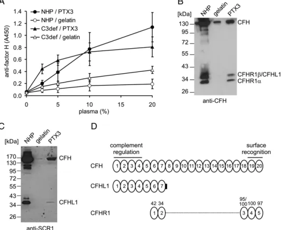
In agreement with the results of Deban et al. (16), we found a dose-dependent binding of PTX3 to immobilized purified CFH in ELISA, and also in the reverse setting (data not shown). Because CFH belongs to a family of related proteins (25), several of which have been implicated in complement regulation, we also investi- gated whether PTX3 interacts with additional CFH family pro- teins. The binding of CFH and CFHR proteins to immobilized PTX3 was analyzed using various dilutions of human plasma as a source of native factor H family proteins, as well as a polyclonal anti-CFH Ab, which cross-reacts with several CFH-related pro- teins. Specific anti-CFH signal was detected both from NHP and from C3-deficient plasma, indicating that PTX3 binding by CFH family proteins was independent of complement activation and was not mediated by C3 fragments (Fig. 1A). To visualize the
FIGURE 1. Binding of factor H family proteins to PTX3. (A) Immobilized PTX3 and gelatin, used as control, were incubated with increasing con- centrations of NHP or C3-deficient serum (C3def). Binding of native CFH was detected by a CFH antiserum. The results are expressed as mean6SD derived from three experiments. To detect binding of CFH family proteins, microplate wells were coated with PTX3 and incubated with 25% NHP. Bound proteins were eluted with SDS buffer, separated on 10% SDS-PAGE, and transferred to nitrocellulose membranes. PTX3-bound CFH family proteins were detected by Western blot using CFH antiserum (B) or anti-SCR1 mAb (C). The relative mobilities of CFH, CFHR1, and CFHL1 are indicated. The blots are rep- resentative of three experiments. (D) Schematic figure showing the homology of SCRs of CFHL1 and CFHR1 with those of CFH. The complement reg- ulatory activities of CFH are mediated by SCRs 1–4, whereas SCRs 19–20 are important for binding of CFH to surface-bound C3b and to host cell surfaces via glycosaminoglycans. CFHL1 shares SCRs 1–7 with CFH, and it contains an additional 4 aa at the C terminus. The numbers above the CFHR1 domains indicate percentage identity with the amino acid sequence of the corresponding CFH domain. The two CFHR1 isoforms CFHR1*A and CFHR1*B differ in SCR3, and this domain has 95% sequence identity to SCR18 of CFH in CFHR1*A and 100% sequence identity to SCR18 of CFH in CFHR1*B (34).
1860 PTX3 INTERACTIONS WITH FACTOR H AND CFHR1 IN aHUS
by guest on September 2, 2018http://www.jimmunol.org/Downloaded from
PTX3-bound proteins directly, the bound proteins were eluted from the PTX3-coated wells, or gelatin-coated wells used as controls, and analyzed by Western blot using a CFH antiserum and an anti- SCR1 mAb. This approach revealed that, in addition to CFH, the CFH splice variant CFHL1 and the two differently glycosylated isoforms of CFHR1 bound to PTX3 (Fig. 1B, 1C). Thus, these assays identified two CFH family proteins with complement regu- latory functions, CFHL1 and CFHR1, as novel ligands of PTX3.
Furthermore, the binding of these CFH family proteins is indicative of at least two PTX3 binding sites within CFH, one in the N ter- minus shared with CFHL1 and the other in the C terminus shared with CFHR1 (Fig. 1D), in agreement with the CFH binding sites reported by Deban et al. (16).
To confirm binding of these proteins to PTX3, recombinant CFHL1 and CFHR1 were used in binding studies. CFH and recom- binant CFHL1 and CFHR1 were immobilized in equimolar con- centration, and their capacity to bind PTX3 was compared. Similarly to the native proteins, recombinant CFHL1 and CFHR1 bound to PTX3, and the binding was weaker compared with that of purified CFH (Fig. 2A). Because PTX3 binds to CFH in a calcium-depen- dent manner (16, 17), we tested the binding of recombinant CFHL1 and CFHR1 under calcium-free conditions. Similarly to the PTX3 binding to CFH, PTX3 binding to CFHL1 and CFHR1 was strongly reduced in calcium-free buffer (Fig. 2B).
Increased binding of CFH and CFHL1 to PTX3 at reduced pH Local infection and inflammatory sites are characterized by de- creased extracellular pH (5.5–7.0) and mild hypocalcemia (41).
Such changes in local conditions can affect different molecular interactions (e.g., the interaction of C-reactive protein with CFH was shown to be influenced by the pH) (42). Therefore, we inves- tigated how the interactions of PTX3 with CFH family proteins are affected by decreased pH. The binding of PTX3 to CFH, CFHL1, and CFHR1 was compared in buffers with pH 7.4, 6.5, and 5.5.
PTX3 binding to CFH and CFHL1 was significantly enhanced at lower pH (Fig. 2C). The binding of CFHR1 and of the classical pathway regulator C4b-binding protein was slightly increased, whereas that of C1q was not increased at lower pH.
CFH maintains its complement regulatory activities when bound to PTX3
Next, the effect of PTX3 on the complement regulatory functions of CFH was analyzed. In a fluid-phase cofactor assay, the cofactor activity of CFH for the factor I-mediated cleavage of C3b, mediated by SCRs 1–4 of CFH, was not influenced by increasing PTX3 concentrations (Fig. 3A). In a solid-phase cofactor assay, CFH, when bound to PTX3, maintained its activity and promoted C3b cleavage by factor I (Fig. 3B), in agreement with the results of Deban et al.
(16). We also measured whether PTX3 influences the decay-accel- erating activity of CFH. The activity of CFH for the disassembly of the alternative pathway C3 convertase was not influenced by increasing concentrations of PTX3 in a solid-phase assay (Fig.
3C, 3D). Thus, CFH remains fully active as a complement regulator when interacting with PTX3. CFHL1 possesses the same comple- ment regulatory activities as does CFH because of the shared SCRs 1–4 (23). Similar to CFH, the cofactor and decay-accelerating ac- tivities of CFHL1 were not influenced by PTX3 (Supplemental Fig.
1). Because CFHR1 was shown to bind to C3b and C5 (37), we tested whether PTX3 influences these interactions. PTX3 did not inhibit the binding of CFHR1 to C3b and C5 (data not shown).
PTX3 recruits functionally active CFH to ECM
Because the ECM becomes exposed during endothelial injury and both PTX3 and CFH were shown to interact with certain components
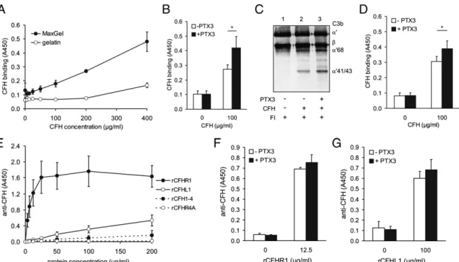
of the ECM (10, 17, 43), we analyzed the interaction of PTX3 and CFH on human ECM. To this end, we used fibroblast-derived ECM (MaxGel). CFH bound to MaxGel in a dose-dependent manner, and a significant binding was achieved at physiological CFH concen- trations (Fig. 4A, 4B). PTX3, when attached to the ECM, signifi- cantly enhanced the binding of CFH (∼85% increase in specific binding) (Fig. 4B). In contrast to this, CFH did not influence the binding of PTX3 to the ECM (data not shown).
Next, the activity of CFH on ECM was analyzed. When bound to the ECM, CFH was able to function as a cofactor for factor I (Fig.
4C, lane 2). When the ECM was preincubated with PTX3, the increased binding of CFH under these conditions resulted in an enhanced inactivation rate of C3b (Fig. 4C, lane 3). In addition, FIGURE 2. Interaction of PTX3 with recombinant CFHL1 and CFHR1 and dependence of binding on calcium concentration and pH. (A) Binding of PTX3 to recombinant CFHR1 and CFHL1 was compared with that of CFH. The three CFH family proteins were immobilized in equimolar concentrations (200 nM), and 5mg/ml recombinant PTX3 was added for 1 h at 37˚C. The values were normalized for CFH binding and show mean + SD derived from three independent experiments. (B) The binding of PTX3 to immobilized CFH, CFHL1, and CFHR1 was compared in DPBS (pH 7.4) with (black bars) and without (white bars) 1 mM Ca2+. The data are normalized to binding in the presence of Ca2+and represent mean + SD from three experiments. (C) The binding of PTX3 to CFH family proteins was compared in TBS with pH 7.4, 6.5, and 5.5. For comparison, the binding of C1q, which activates complement when bound to PTX3, and the binding of C4b-binding protein, a regulator of complement activation, were also measured under these buffer conditions. The data shown are normalized to values obtained with TBS pH 7.4 and represent mean + SD from five experiments. Note that the binding affinity of the individual proteins to PTX3 is different, and C1q and C4BP bind more strongly to PTX3 than does CFH (12, 16, 17). *p,0.05, **p,0.01, Studentttest.
by guest on September 2, 2018http://www.jimmunol.org/Downloaded from
we analyzed the interaction of PTX3 with CFH on ECM produced by human endothelial cells in vitro. HUVECs were cultured in 96- well plates for 7 d and then removed by PBS containing 10 mM EDTA. The resulting cell-free ECM was used for binding assays.
CFH bound to HUVEC-ECM, similar to the binding on MaxGel, and PTX3 enhanced the binding of CFH to endothelial cell-derived ECM by 36% (Fig. 4D).
CFHL1 and CFHR1 also bound to the ECM in a dose-dependent manner, and the binding of recombinant CFHR1 was much stronger than was that of CFH or CFHL1 (Fig. 4E). Recombinant CFHR4A and the CFH SCRs 1–4 fragment, used as control proteins, showed no specific binding to ECM. The binding of CFHR1 and CFHL1 to the ECM was slightly enhanced by PTX3 (Fig. 4F, 4G).
Localization of the PTX3 binding site within SCR20 of CFH The study of Deban et al. (16) indicated two binding sites for PTX3 on CFH, within SCR7 and SCRs 19–20. To identify residues re- sponsible for the interaction of CFH family proteins with PTX3, a peptide array was generated using overlapping peptides that cover SCR7, SCR19, and SCR20 of CFH. These domains are shared by CFHL1 (SCR7) and CFHR1 (SCRs 19–20). Compared with back- ground binding of the detection Ab, PTX3-specific peptides could be identified within SCR20 (Fig. 5A). The consensus sequence from these peptides indicated that the CFH residues 1180-ALRWTAK- 1186 and 1198-EFVCKRG-1204 are involved in PTX3 binding.
The first sequence is located in the hypervariable loop on SCR20, and both are surface exposed (Fig. 5B). With this approach, no
specific PTX3-binding residues were found in SCR7 and SCR19 (data not shown).
Competition assays were performed to confirm the identified PTX3 binding site in CFH. The mAb C18, which binds in SCR20 of CFH to R1192–R1203 (44), strongly and dose dependently inhibited PTX3 binding (Fig. 5C). Moreover, the reduced recruit- ment of CFH by PTX3 resulted in a strongly reduced cofactor activity in the presence of the mAb C18 but not in the presence of a control mouse IgG (Fig. 5D).
CFH mutations and autoantibodies associated with aHUS impair CFH binding to PTX3
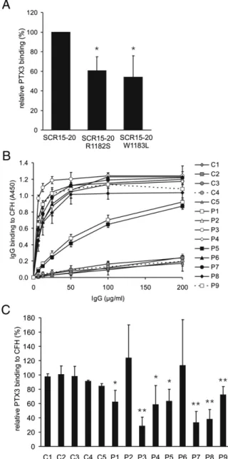
Some of the identified putative PTX3-binding residues have been found mutated in aHUS patients. We generated the R1182S and W1183L substitutions on a CFH SCRs 15–20 background to analyze the possible effect of these mutations on the CFH–PTX3 interaction.
In comparison with the wild-type SCRs 15–20, these mutations resulted in a significantly reduced PTX3 binding (Fig. 6A).
Most anti-CFH autoantibodies in aHUS patients bind to SCR20 of CFH and share a binding site with the mAb C18 (29). Therefore, we investigated whether aHUS-associated CFH autoantibodies influence the binding of CFH to PTX3. To this end, IgG fractions from the plasma of nine patients were isolated after urea treatment of the serum samples to dissociate CFH–IgG complexes. The patient- derived IgGs, in contrast to control IgGs isolated from healthy donors, showed a strong and dose-dependent binding to immobi- lized CFH, although there were differences in the autoantibody FIGURE 3. PTX3 does not influence the complement regulatory activities of CFH. (A) Fluid-phase CFH cofactor assay. C3b was incubated with factor I (FI), CFH, and 5–20mg/ml PTX3 in the indicated combinations, and the cleavage of C3b was detected from reduced samples by Western blot using a C3 antiserum. The C3b chains and thea9-chain cleavage product (a941/43) are indicated on the right. One representative of three experiments is shown. (B) Solid-phase cofactor activity of CFH. CFH was added to wells coated with gelatin or recombinant PTX3. After washing, C3b and factor I were added, and C3b cleavage was analyzed from the supernatants after incubation at 37˚C by Western blot, as described above. A representative experiment of three performed is shown. (C) Solid-phase decay-accelerating activity of CFH was measured on the alternative pathway C3 convertase (C3bBb) assembled on the microtiter plate surface. CFH alone or CFH preincubated with PTX3 was added, and the remaining intact convertase was detected after the indicated time points by ELISA using an anti-factor B Ab. Spontaneous decay is shown in the absence of CFH (buffer). (D) The C3bBb convertase decay is not influenced by PTX3 (s), and increasing concentrations of PTX3 do not influence the decay-accelerating activity of CFH (d). The convertase decay was measured at 60 min, as described above. In (C) and (D), data are mean6SD from three experiments.
1862 PTX3 INTERACTIONS WITH FACTOR H AND CFHR1 IN aHUS
by guest on September 2, 2018http://www.jimmunol.org/Downloaded from
titers (Fig. 6B). The patient-derived autoantibodies bound to SCR20 of CFH, as indicated by their binding only to CFH fragments containing SCR20 and inhibition of binding by mAb C18, deter- mined as described previously (29). None of the tested autoanti- bodies bound to CFHL1 (data not shown). Of the nine samples analyzed, three IgGs strongly inhibited PTX3 binding to CFH (∼30% of binding), four showed a moderate inhibition (∼60% of binding), and two samples did not inhibit this interaction (Fig.
6C). The inhibitory effect of the IgGs apparently did not correlate well with the free autoantibody titers, revealing subtle differences in the exact binding sites of the various autoantibodies on CFH.
Interaction of PTX3 with CFHR1
The mAb C18 also recognizes CFHR1 (44); in our assays, it inhibited PTX3 binding to recombinant CFHR1 (Fig. 7A). The C-terminal SCR5 domain of both CFHR1*A and CFHR1*B differs from SCR20 of CFH by only 2 aa, and the identified PTX3-binding sequences are present in both proteins. However, CFHR1*A and CFHR1*B show 3 aa differences in their SCR3. SCR3 in CFHR1*A displays H157, L159, and E175, whereas SCR3 in CFHR1*B dis- plays Y157, V159, and Q175, the latter being identical with the amino acid sequence of SCR18 of CFH (Fig. 7B). Because the CFHR1*B isoform was shown to be associated with an increased risk for aHUS (34), we analyzed whether the two CFHR1 isoforms bound differently to PTX3. To this end, CFHR1*A and CFHR1*B
were isolated from individuals homozygous for the respective iso- forms. PTX3 showed a dose-dependent binding to both isoforms, but the binding to the disease-associated CFHR1*B variant was significantly weaker (Fig. 7C). Most aHUS-associated anti-CFH autoantibodies were shown to cross-react with CFHR1 (35, 45).
IgG fractions isolated from autoantibody-positive patients showed similar binding to both CFH and recombinant CFHR1 (Fig. 7D). Of the nine tested autoantibodies, five also inhibited the interaction of PTX3 with recombinant CFHR1 (32–62% binding) (Fig. 7E).
Discussion
aHUS is a severe kidney disease associated with dysregulation of the alternative complement pathway and characterized by endo- thelial injury. In this study, we analyzed the interaction of PTX3, a soluble pattern recognition molecule associated with endothelial and vascular damage, with complement regulators of the CFH protein family in the context of aHUS.
Our results confirm the binding of the alternative pathway regulator CFH to PTX3, as reported previously (16), and identify CFHL1, as well as the terminal complement pathway regulator CFHR1, as additional PTX3 ligands (Figs. 1, 2). Direct binding of native CFH, CFHL1, and CFHR1 was detected from human plasma. A recent study that analyzed circulating PTX3 complexes in sepsis using a proteomics approach also detected CFHR1 (46). These data fur- ther support the presence of two PTX3 binding sites within CFH:
FIGURE 4. PTX3 recruits functionally active CFH to human fibroblast- and endothelial cell-derived ECM. (A) Dose-dependent binding of CFH to human fibroblast-derived ECM (MaxGel). Wells were coated with MaxGel or gelatin as control, and CFH was added at the indicated concentrations. CFH binding was detected using a CFH antiserum. Data represent mean6SD from three experiments. (B) Binding of purified CFH to MaxGel, preincubated (black bars) or not (white bars) with 20mg/ml PTX3, was measured by ELISA. Data are mean values + SD of three experiments. (C) Cofactor assay for C3b cleavage on MaxGel. C3b and factor I (FI) were added to MaxGel (lane 1, control) and to MaxGel preincubated with 100mg/ml CFH (lane2) or first with 20mg/ml PTX3 and then CFH (lane3). After incubation at 37˚C for 2 h, the supernatants were separated on 10% SDS-PAGE and subjected to Western blotting. The blot was developed using a C3 antiserum. The C3b fragments are indicated on the right. One representative of three experiments is shown. (D) HUVECs were cultured for 7 d in gelatin-coated 96-well plates; the cells were detached, and binding of CFH to the cell-free ECM was analyzed by ELISA.
Gelatin-coated wells incubated with cell culture medium were used as negative controls. Binding of 100mg/ml CFH to HUVEC-derived ECM preincubated (black bars) or not (white bars) with 20mg/ml PTX3 was measured. Data shown are mean + SD derived from three independent experiments. (E) CFHR1 and CFHL1 bind to the ECM. Binding of recombinant CFHR1 and CFHL1, as well as CFH SCRs 1–4 fragment (rCFH1–4) and the CFHR4A protein used as controls, was measured to MaxGel as in (A). Data are corrected for the background and represent mean6SD from three experiments. To analyze the effect of PTX3, recombinant CFHR1 (F) and CFHL1 (G) were added, at the indicated concentrations in TBS, to microtiter plate wells coated with MaxGel and preincubated (black bars) or not (white bars) with 15mg/ml recombinant PTX3. The binding of CFHR1 and CFHL1 was measured using CFH an- tiserum. Because of the stronger binding of CFHR1 to the ECM, less CFHR1 than CFHL1 was used in the assay. Data are mean values + SD of three experiments. *p,0.05, Studentttest.
by guest on September 2, 2018http://www.jimmunol.org/Downloaded from
one at the N terminus and shared with CFHL1 and another at the C terminus and shared with CFHR1. This is also reflected in the stronger binding of PTX3 to CFH compared with CFHL1 and CFHR1, both of which likely have only one PTX3 binding site.
The physiological presence of calcium seems to be a requirement for an efficient interaction of PTX3 with these complement inhibitors (Fig. 2), in contrast to the binding of PTX3 to the com- plement activator C1q (16, 17).
The main PTX3 binding sites within CFH and CFHR1 were localized to the C termini of both proteins. Using a peptide array, the residues A1180–K1186 and E1198–G1204 in SCR20 of CFH were found to mediate PTX3 binding. These residues are surface exposed and, thus, are accessible for ligand interactions (Fig. 5).
These sequences are also contained in SCR5 of both CFHR1 isoforms. The C-terminal PTX3 binding site was confirmed using the mAb C18, which recognizes both CFH and CFHR1 by binding to SCR20 and SCR5, respectively. The binding site of this mAb in CFH SCR20 was previously mapped to R1192–R1203 (44) (i.e., in the direct vicinity and partly overlapping with the identified PTX3- binding residues). Almost complete inhibition of PTX3 binding to CFH and CFHR1 was achieved by mAb C18 (Figs. 5, 7), indicating that the major PTX3 binding site is located in SCR20 and SCR5 of CFH and CFHR1, respectively. Likewise, the mAb C18 strongly inhibited the complement regulatory activity of CFH in the solid phase by preventing CFH binding to PTX3 (Fig. 5D). However, the differential binding of PTX3 to the CFHR1 sequence variants indicates that additional residues and domains (e.g., SCR18) are likely involved in a physiological PTX3–CFH and PTX3–CFHR1 interaction (Fig. 7C). The putative N-terminal PTX3 binding site in CFH and CFHL1 needs to be determined in future studies.
Importantly, the fluid phase and solid phase cofactor activity and the decay-accelerating activity for surface-bound C3 convertase of CFH and CFHL1 were not affected by PTX3. This is explained by the fact that the identified PTX3-interaction sites are located outside of the regulatory domains (SCRs 1–4). Likewise, interactions of CFHR1 with its ligands C3b and C5 were not inhibited by PTX3.
Soluble complement regulators bind to host ligands and surfaces and prevent excessive complement activation, inflammation, and tissue damage (47). In contrast to cells, basal membranes and the subendothelial ECM have no integral complement regulators, with the exception of some shed CD55 and CD59 (48). Therefore, CFH attachment to surfaces, such as the Bruch’s membrane in the eye or the glomerular basement membrane, is thought to provide protec- tion from complement activation-mediated damage. It was dem- onstrated that factor H binds to the Bruch’s membrane (49) and is associated with aortic ECM (50). Furthermore, CFH was shown to bind to certain ECM components, such as fibromodulin and short leucine-rich glycoproteins (43). In this study, we show that CFH binds to both fibroblast- and endothelial cell-derived ECM and is functionally active when bound to ECM (Fig. 4). PTX3 also binds to components of the ECM (10), and we previously demonstrated that PTX3 binds to subendothelial ECM in vitro and modulates complement activation (17). Binding of PTX3 resulted in activa- tion of the classical pathway and enhanced C3 deposition on ECM.
However, the terminal pathway activity, measured as C5a genera- tion and C5b-9 deposition, was not increased, indicating efficient regulation (17). We showed that C4b-binding protein binds to human fibroblast- and endothelial cell-derived ECM and is additionally recruited to the ECM by PTX3. ECM- and PTX3-bound C4b- binding protein promotes C4b inactivation (17). In this study, FIGURE 5. Identification of the PTX3 binding site in the C termini of CFH and CFHR1. (A) A peptide array was generated using overlapping peptides (13-mers, with 10 aa overlaps) that cover SCR20 of CFH. The membrane was incubated (lower panel) or not (upper panel) with 5mg/ml PTX3 and then developed with anti-PTX3 Ab. The sequences of the identified PTX3 binding peptides are shown below. (B) The CFH SCRs 19–20 model indicates surface localization of the identified PTX3-binding amino acids (shown in black) 1180-ALRWTAK-1186 and 1198-EFVCKRG-1204. (C) Binding of PTX3 to immobilized CFH was inhibited by mAb C18 (binding to SCR20) added in the indicated concentrations, whereas there was no inhibition by a control IgG.
Values are normalized to samples without Ab present and represent means + SD from three experiments. (D) The cofactor activity of PTX3-bound CFH was measured as described in Fig. 4. C3b and factor I were incubated alone (lane 1), with CFH (lane 2), with CFH preincubated with 100mg/ml mAb C18 (lane 3), or with CFH preincubated with 100mg/ml control Ab (lane 4). C3b fragments were detected by Western blot. The mAb C18, but not the control Ab, inhibited CFH binding and, thus, cofactor activity on PTX3. A representative of three independent experiments is shown.
1864 PTX3 INTERACTIONS WITH FACTOR H AND CFHR1 IN aHUS
by guest on September 2, 2018http://www.jimmunol.org/Downloaded from
we found that ECM-bound PTX3 provides additional CFH bind- ing sites and significantly enhances the amounts of CFH bound to this surface. Importantly, CFH maintains its complement regula- tory activity when bound on the ECM and/or to PTX3 (Fig. 4).
Although the binding of CFH to ECM was not of high affinity under our experimental conditions, it is likely relevant in vivo (49,
50). One also has to consider that a relatively modest change in the concentration of regulators can have a decisive effect on comple- ment activation because of the exponential characteristic of the activation steps. Altogether, these data suggest that a concerted action of regulators of the classical/lectin pathways (C4b-binding protein), the alternative pathway (CFH and CFHL1), and the ter- minal pathway (CFHR1) is important to effectively control com- plement activation on surfaces that bind PTX3. The regulation of the alternative pathway by CFH is particularly important, because its activation also occurs secondarily through the classical/lectin pathway C3 convertase (“amplification loop”).
PTX3 is a soluble pattern recognition molecule that interacts with the complement system and binds both complement activatory (C1q, M-ficolin, L-ficolin, mannan-binding lectin) and inhibitory (C4b-binding protein, CFH, CFHL1, CFHR1) molecules. The bal- ance between activators and inhibitors under the given circumstances determines the degree of complement activation and opsonization induced by PTX3 locally.
PTX3 is emerging as a protective molecule that is quickly up- regulated after endothelial injury (6). It has a role in the regulation of inflammation and neutrophil migration because it binds to P- selectin and, thus, reduces neutrophil recruitment at inflammatory sites (51). The plasma concentration of PTX3 during inflammation reaches up to 0.8 mg/ml, which is not derived systemically but mainly from sites of inflammation, where it can thus reach much higher local concentrations, especially on surfaces. In addition, inflammatory cells migrating to the site, such as neutrophils and macrophages, can produce and locally release PTX3 (52). Al- though the local increase in PTX3 concentrations may appear detrimental, because PTX3 could potentially enhance the local inflammatory response by activating complement and allowing the generation of more C5a, bound complement regulators attenuate this activation (16, 17). Importantly, we found that the binding of the potent alternative pathway inhibitors CFH and CFHL1, but not of the complement activator C1q, is enhanced at reduced pH, which is characteristic of sites of inflammation (Fig. 2C). This is in accordance with a previous study showing pH-independent binding of C1q to PTX3 (53).
aHUS is associated with alternative complement pathway dys- regulation. However, there are individual differences in the exact pathomechanism among aHUS patients; in individual patients, a combination of predisposing factors may contribute to the disease.
aHUS is associated with CFH mutations or autoantibodies against CFH in up to 40% of patients. It was demonstrated previously that C-terminal CFH mutations and autoantibodies disturb the physi- ological interaction of CFH with C3b, causing reduced protection of host cells from complement-mediated lysis (29–31, 54, 55). In this study, we show that mutations and autoantibodies also impair the interaction of CFH with PTX3 (Fig. 6). Thus, certain muta- tions and autoantibodies can influence multiple interactions and functions of CFH.
In addition, the CFHR1*B variant was found to be associated with aHUS in a Spanish patient cohort (34). It was suggested that CFHR1*B, which shows a higher similarity to CFH than does the CFHR1*A isoform, could more efficiently compete with CFH for binding to C3b and host cells and, thus, reduce complement control by CFH. In the current study, we identified CFHR1 as an interac- tion partner for PTX3 and showed that the aHUS-associated CFHR1 variant has lower affinity for PTX3. This could potentially result in a reduced local terminal pathway control by CFHR1*B, which would provide an additional explanation for its association with this disease. Moreover, anti-CFH autoantibodies, which were shown to cross-react with CFHR1 (45), also impaired the PTX3–CFHR1 interaction (Fig. 7).
FIGURE 6. The interaction of PTX3 with CFH is impaired by aHUS- associated SCR20 mutations and by autoantibodies. (A) To test the effect of aHUS-associated CFH mutations within the identified PTX3 binding site, the two mutants R1182S and W1183L were generated by site-directed mutagenesis in SCRs 15–20. The proteins were immobilized in equimolar amounts, and binding of PTX3 was measured by ELISA. Values are normalized to PTX3 binding to wild-type CFH SCRs 15–20 and represent mean + SD from three experiments. (B) Dose-dependent binding of IgG isolated from healthy individuals (C1–C5) and IgG purified from aHUS patients with anti-CFH autoantibodies (P1–P9) to CFH. Wells coated with CFH were incubated with IgG in the indicated concentrations, and IgG binding was determined by ELISA, as described (29). Data represent mean 6SD from three independent experiments. (C) To analyze the effects of anti-CFH autoantibodies on PTX3–CFH interaction, CFH was immobilized in microtiter plate wells and incubated with IgGs. After washing, the binding of PTX3 was detected by ELISA. The data are normalized to the control wells without IgG and represent mean + SD from at least three experiments. *p,0.05, **p,0.01, Studentttest.
by guest on September 2, 2018http://www.jimmunol.org/Downloaded from
It was suggested that PTX3 may direct CFH regulatory activity to specific sites, such as the surface of apoptotic cells, to control ex- cessive local complement activation (16). Our data show that PTX3 recruits functional CFH to the ECM; however, this interaction is impaired by certain mutations and autoantibodies affecting SCR20 of CFH. This could amplify local complement-mediated inflam- mation, as well as endothelial cell activation and damage in aHUS.
Thus, the impaired CFH–PTX3 and CFHR1–PTX3 interactions may exacerbate renal pathology in some aHUS patients.
Acknowledgments
We thank the Department of Infection Biology (Hans Kno¨ll Institute) for access to equipment and Mario Hebecker for expression and purification of CFHR4A. We thank the patients, their parents, and Gyo¨rgy Reusz (Semmel- weis University, Budapest, Hungary) for providing anti-CFH positive sam- ples for this study.
Disclosures
The authors have no financial conflicts of interest.
References
1. Mantovani, A., C. Garlanda, A. Doni, and B. Bottazzi. 2008. Pentraxins in innate immunity: from C-reactive protein to the long pentraxin PTX3.J. Clin. Immunol.
28: 1–13.
2. Yamasaki, K., M. Kurimura, T. Kasai, M. Sagara, T. Kodama, and K. Inoue.
2009. Determination of physiological plasma pentraxin 3 (PTX3) levels in healthy populations.Clin. Chem. Lab. Med.47: 471–477.
3. Bottazzi, B., A. Doni, C. Garlanda, and A. Mantovani. 2010. An integrated view of humoral innate immunity: pentraxins as a paradigm.Annu. Rev. Immunol.28:
157–183.
4. Lee, T. H., G. W. Lee, E. B. Ziff, and J. Vilcek. 1990. Isolation and character- ization of eight tumor necrosis factor-induced gene sequences from human fibroblasts.Mol. Cell. Biol.10: 1982–1988.
5. Breviario, F., E. M. d’Aniello, J. Golay, G. Peri, B. Bottazzi, A. Bairoch, S. Saccone, R. Marzella, V. Predazzi, M. Rocchi, et al. 1992. Interleukin-1- inducible genes in endothelial cells. Cloning of a new gene related to C-reactive protein and serum amyloid P component.J. Biol. Chem.267: 22190–22197.
6. Norata, G. D., C. Garlanda, and A. L. Catapano. 2010. The long pentraxin PTX3:
a modulator of the immunoinflammatory response in atherosclerosis and car- diovascular diseases.Trends Cardiovasc. Med.20: 35–40.
7. Luchetti, M. M., G. Piccinini, A. Mantovani, G. Peri, C. Matteucci, G. Pomponio, M. Fratini, P. Fraticelli, P. Sambo, C. Di Loreto, et al. 2000.
Expression and production of the long pentraxin PTX3 in rheumatoid arthritis (RA).Clin. Exp. Immunol.119: 196–202.
8. Tong, M., J. J. Carrero, A. R. Qureshi, B. Anderstam, O. Heimbu¨rger, P. Ba´ra´ny, J. Axelsson, A. Alvestrand, P. Stenvinkel, B. Lindholm, and M. E. Suliman.
2007. Plasma pentraxin 3 in patients with chronic kidney disease: associations with renal function, protein-energy wasting, cardiovascular disease, and mor- tality.Clin. J. Am. Soc. Nephrol.2: 889–897.
9. Moalli, F., S. Jaillon, A. Inforzato, M. Sironi, B. Bottazzi, A. Mantovani, and C. Garlanda. 2011. Pathogen recognition by the long pentraxin PTX3.J. Biomed.
Biotechnol.2011: 830421.
10. Scarchilli, L., A. Camaioni, B. Bottazzi, V. Negri, A. Doni, L. Deban, A. Bastone, G. Salvatori, A. Mantovani, G. Siracusa, and A. Salustri. 2007.
PTX3 interacts with inter-alpha-trypsin inhibitor: implications for hyaluronan organization and cumulus oophorus expansion.J. Biol. Chem.282: 30161–
30170.
11. Rovere, P., G. Peri, F. Fazzini, B. Bottazzi, A. Doni, A. Bondanza, V. S. Zimmermann, C. Garlanda, U. Fascio, M. G. Sabbadini, et al. 2000. The long pentraxin PTX3 binds to apoptotic cells and regulates their clearance by antigen-presenting dendritic cells.Blood96: 4300–4306.
12. Bottazzi, B., V. Vouret-Craviari, A. Bastone, L. De Gioia, C. Matteucci, G. Peri, F. Spreafico, M. Pausa, C. D’Ettorre, E. Gianazza, et al. 1997. Multimer for- FIGURE 7. The aHUS-associated CFHR1*B variant and anti-CFH autoantibodies cause an impaired PTX3–CFHR1 interaction. (A) Binding of PTX3 to immobilized CFHR1 was inhibited by mAb C18 but not by a control Ab (both added at 10mg/ml). The data are normalized to the control wells without mAb and represent mean + SD of data derived from three experiments. (B) Schematic representation of the most C-terminal SCRs of the CFHR1 isoforms and their similarity to CFH. The differences in amino acid sequence between domains of CFHR1 and those of CFH are indicated. SCR3 of CFHR1*A differs from the corresponding domains in CFHR1*B and CFH (highlighted). (C) Purified CFHR1*A and CFHR1*B, or gelatin as negative control, were immobilized in equimolar amounts, and binding of PTX3 was measured as in Fig. 2. Data represent mean6SD derived from three experiments. (D) Microtiter plate wells were coated with 5mg/ml purified CFH or 10mg/ml recombinant CFHR1. The wells were then incubated with 100mg/ml IgG purified from healthy donors (C1–C5) or from aHUS patients with CFH autoantibodies (P1–P9). IgG binding was determined by ELISA. Data represent mean + SD from three independent experiments. (E) To assay the effects of anti-CFH autoantibodies on the binding of PTX3 to CFHR1, recombinant CFHR1 was immobilized in microtiter plate wells and then incubated with IgG fractions derived from controls (C1–C5) and from aHUS patients (P1–P9).
After washing, the binding of PTX3 was detected by ELISA. The data are normalized to the control wells without IgG and represent mean + SD from three experiments. *p,0.05, **p,0.01, Studentttest.
1866 PTX3 INTERACTIONS WITH FACTOR H AND CFHR1 IN aHUS
by guest on September 2, 2018http://www.jimmunol.org/Downloaded from