Investigation of the molecular background of alternative pathway dysregulation in thrombotic microangiopathies
Ph.D. thesis
Eszter Trojnar, MD
Basic and Translational Medicine Doctoral School Semmelweis University
Supervisor: Zoltán Prohászka, MD, D.Sc Official reviewers: Csaba Kálmán Ambrus, MD, PhD Judit Müller, MD, PhD
Head of the Complex Exam Committee: Zoltán Benyó, MD, D.Sc Members of the Complex Exam Committee: Hargita Hegyesi, Ph.D
Noémi Sándor, Ph.D
Budapest
2019
TABLE OF CONTENTS
1. LIST OF ABBREVIATIONS 6
2. INTRODUCTION 10
2.1. Pathophysiology of the complement system 10 2.1.1. Physiological activation of the complement system
and significance of the alternative pathway 10 2.1.2. Regulation of the complement system 13 2.1.3. Regulation of the alternative pathway 15 2.1.4. Significance of Factor H in the regulation
of the alternative pathway 16
2.1.5. Role of pentraxins in complement regulation 18 2.1.6. Dysregulation of the complement alternative pathway 22
2.2. Thrombotic microangiopathies 25
2.2.1. Classification and pathogenesis of thrombotic microangiopathies 25 2.2.2. Thrombotic thrombocytopenic purpura 25 2.2.3. Typical hemolytic uremic syndrome 26 2.2.4. Atypical hemolytic uremic syndrome 26 2.2.5. Secondary thrombotic microangiopathies 28 2.2.6. Pathological complement activation
in thrombotic microangiopathies 28
2.2.7. Alternative pathway dysregulation associated with
anti-Factor H antibodies in atypical hemolytic uremic syndrome 32 2.2.8. Factor H-related proteins and their genetic-linkage
to anti-Factor H antibodies 33
3. SPECIFIC AIMS 36
3.1. Analysis of the association between systemic pentraxin levels
and laboratory signs of disease activity in thrombotic microangiopathies 36 3.2. Investigation of the relation of complement overactivation and systemic
pentraxin levels in distinct forms of thrombotic microangiopathies 36 3.3. Epitope analysis of anti-Factor H antibodies
in atypical hemolytic uremic syndrome 36
4. METHODS 38 4.1. Patient selection, sample collection and study design 38
4.1.1. Sample collection from patients at the acute phase of
thrombotic microangiopathy 38
4.1.2. Sample collection from autoimmune
atypical hemolytic uremic syndrome patients 41 4.2. Determination of laboratory parameters in patients
at the acute phase of thrombotic microangiopathy 43 4.3. In vitro assessment of pentraxin-3 effect on
alternative pathway activation 43
4.4. Determination of anti-Factor H antibody levels 44 4.5. Peptide synthesis for epitope mapping 44 4.6. Antibody binding to the immobilized synthetic peptides of Factor H
and Factor H-related protein 1 46
4.7. Synthesis and antibody binding of recombinant Factor H 19-20 mutants 47 4.8. Visualization of the linear epitopes and point mutations
on the folded Factor H structure 47
4.9. Statistical analysis 48
5. RESULTS 49
5.1. Analysis of the association between systemic pentraxin levels and
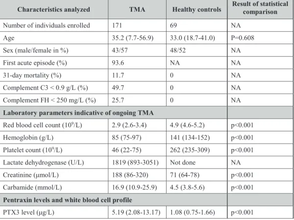
laboratory signs of disease activity in thrombotic microangiopathies 49 5.1.1. Characteristics of patients with acute phase thrombotic
microangiopathy selected for the determination of
the systemic pentraxin levels 49
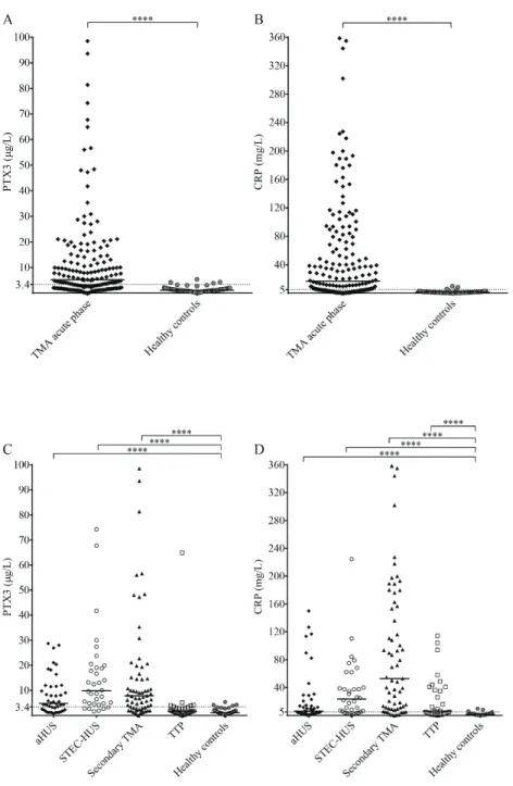
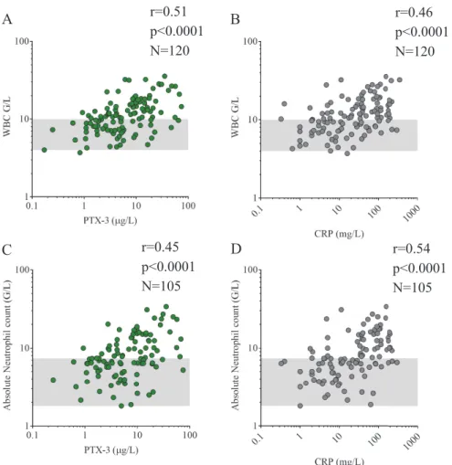
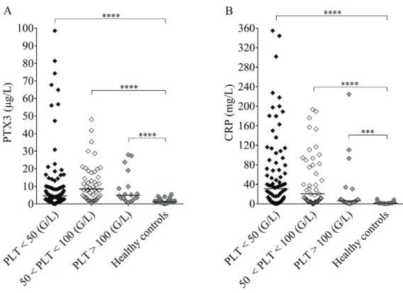
5.1.2. Pentraxin levels in acute phase thrombotic microangiopathy and their relation to the laboratory markers of disease
and clinical characteristics of patients 52 5.1.3. Association of the systemic pentraxin levels
with the acute phase mortality 58
5.1.4. Pentraxin levels in disease remission 58 5.2. Investigation of the relation of complement overactivation and systemic
pentraxin levels in distinct forms of thrombotic microangiopathies 63
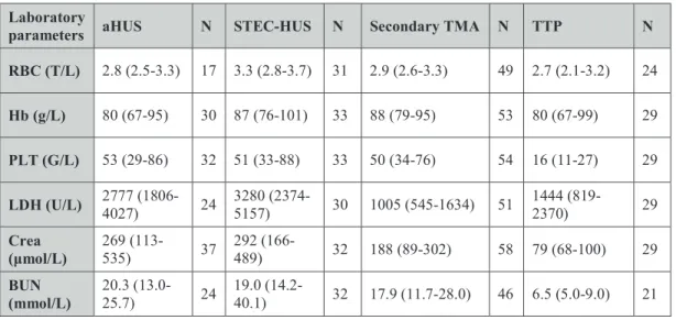
5.2.1. Laboratory signs of complement consumption in acute phase thrombotic microangiopathy and their association to the systemic
pentraxin levels 63
5.2.2. Impact of excess pentraxin-3 on
alternative pathway activity in vitro 70 5.3. Epitope analysis of anti-Factor H antibodies
in atypical hemolytic uremic syndrome 72 5.3.1. Clinical and laboratory characteristics of patients with autoimmune
atypical hemolytic uremic syndrome 72
5.3.2. Localization of the linear autoantibody epitopes on Factor H in the acute phase and remission of atypical hemolytic uremic syndrome 74 5.3.3. Comparison of Factor H autoantibody binding to linear epitopes on
Factor H and Factor H-related protein 1 74 5.3.4. Fine epitope mapping of recombinant Factor H domains 19-20
expressing atypical hemolytic uremic syndrome-associated point mutations by anti-Factor H autoantibodies of
atypical hemolytic uremic syndrome patients 81 5.3.5. Position of the linear Factor H epitopes and atypical hemolytic
uremic syndrome-associated point mutations on
the crystal structure of Factor H 83
6. DISCUSSION 86
6.1. Elevation of the systemic pentraxin levels in the acute phase of thrombotic microangiopathies and their association to complement
overactivation and consumption 86
6.2. Epitope analysis of anti-Factor H antibodies 90
7. CONCLUSIONS 95
8. SUMMARY 96
9. ÖSSZEFOGLALÁS 97
10. BIBLIOGRAPHY 98
11. LIST OF PUBLICATIONS 120
11.1 Publications related to the Ph.D. dissertation of the candidate 120 11.2 Publications independent of the Ph.D. dissertation of the candidate 120
12. ACKNOWLEDGEMENT 121
1.LIST OF ABBREVIATIONS
Acm – acetamidomethyl
ANC – absolute neutrophil count
ADAMTS13 – a disintegrin and metalloproteinase with a thrombospondin type 1 motif member 13
ADAMTS13 – the gene encoding a disintegrin and metalloproteinase with a thrombospondin type 1 motif member 13
aHUS – atypical hemolytic uremic syndrome AMD – Age related macular degeneration Anti-FH antibody – anti-Factor H antibody AP – alternative pathway
Arg – arginine Asp – aspartic acid
Boc – tert-butyloxycarbonyl C1-INH – C1-inhibitor
C1q – complement component 1q C3 – complement component 3
C3 – the gene encoding the complement component 3 C3a – complement component 3a
C3b – complement component 3b
C3bBbP – Alternative pathway C3 convertase stabilized by properdin
C3c – complement component 3c, cleavage product of the complement component 3b C3d – complement component 3d, cleavage product of the complement component 3b C3dg - complement component 3dg, cleavage product of the complement component 3b C3G – C3 glomerulopathies
C3Nef - C3 Nephritic Factor C4 – complement component 4 C4BP – C4b binding protein
C4c – complement component 4c, cleavage product of the complement component 4b C4d – complement component 4d, cleavage product of the complement component 4b CD46 – the gene encoding the cluster of differentiation 46 or membrane cofactor protein
CD59 – cluster of differentiation 59
CFB – the gene encoding the complement protein Factor B CFH – the gene encoding the complement protein Factor H CFHR1-5 – the genes encoding Factor H-related proteins 1-5 CFI – the gene encoding the complement protein Factor I CP – classical pathway
CR – complement receptor
CRIg – complement receptor of the immunoglobulin family CRP – C-reactive protein
Cys – cysteine
DAF – decay accelerating factor
DAMP – danger associated molecular pattern
DGKE – the gene encoding diacylglycerol kinase epsilon DMF – Dimethylformamide
ELISA – enzyme-linked immunosorbent assay FB – complement Factor B
FFP – fresh frozen plasma FH – Factor H
FHR 1-5– Factor H-related proteins 1-5 FI – complement Factor I
Fmoc/tBu - fluorenylmethyloxycarbonyl group/ tertiary butyl group
FRETS-VWF73 – Fluorescence-Quenching Substrate for a disintegrin and metalloproteinase with a thrombospondin type 1 motif member 13
GAG – glycosaminoglycan Glu – glutamic acid
GPI – glycosylphosphatidylinositol His – histidine
HSP60 – heat shock protein 60 HUS – hemolytic uremic syndrome
iC3b – inactive cleavage product of the complement component 3b iC4b – inactive cleavage product of the complement component 4b IgA – Immunoglobulin A
IgG – Immunoglobulin G
IgG-HRP - Immunoglobulin G conjugated to horseradish peroxidase IgM – Immunoglobulin M
LDH – lactate dehydrogenase LPS – lipopolysaccharide Lys – lysine
MAC – membrane attack complex
MAp19 – Map19 protein, smaller alternative splice product of the gene encoding mannose-binding lectin-associated serine protease-2
MAp44 – mannose-binding lectin-associated protein of 44 kDa
MASP-1, -2, -3 – mannose-binding lectin-associated serine proteases 1, 2 and 3 MBL – mannose-binding lectin
MCP – membrane cofactor protein
MLPA – multiplex ligation-dependent probe amplification
MMACHC – the gene encoding methylmalonic aciduria and homocystinuria type C protein
MMACHC-TMA – thrombotic microangiopathy associated to methylmalonic aciduria and homocystinuria, cobalamin C complementation type
NET – neutrophil extracellular trap NHS – normal human serum
NLRP3 – NACHT, LRR and PYD domains-containing protein 3 NĮ-Fmoc – N-(9-Fluorenylmethoxycarbonyl)
OD – optical density OtBu – tertiary butyl ester
PAMP – pathogen associated molecular pattern
Pbf – 2,2,4,6,7-pentamethyldihydrobenzofuran-5-sulfonyl PBS - phosphate-buffered saline
PEX – plasma exchange therapy
PIGA –phosphatidylinositol glycan anchor biosynthesis class A gene Pmc – 2,2,5,7,8-pentamethyl-chroman-6-sulphonyl
PRM – pattern recognition molecule PTX3 – pentraxin-3
RCA – regulators of complement activation
sC5b-9 – soluble form of the membrane attack complex, consisting of complement components 5b to 9
SCR – short consensus repeat Ser – serine
SFTL – a unique amino acid sequence on the C-terminal end of the Factor H-like protein 1 consisting of serine, phenylalanine, threonine and leucine
STEC – Shiga toxin-producing Escherichia coli
STEC-HUS – Shiga toxin-producing Escherichia coli-associated hemolytic uremic syndrome
Stx – Shiga-toxin
tBu – tertiary butyl group
Tert-butyl ether – tertiary butyl ether
THBD – the gene encoding thrombomodulin THBD – thrombomodulin
Thr – threonine
TMA – thrombotic microangiopathy TMB - 3,3ƍ,5,5ƍ-tetramethylbenzidine
TTP – thrombotic thrombocytopenic purpura Tyr – tyrosine
ULVWF – ultra-large form of von Willebrand Factor WBC – white blood cell count
2.INTRODUCTION
2.1. Pathophysiology of the complement system
2.1.1. Physiological activation of the complement system and significance of the alternative pathway
The complement system is an integral part of the innate immune system that contributes to the elimination of invading pathogens, immune complexes and maintains continuous clearance of redundant cellular debris together with the regulation of the innate immune response (Kavanagh et al. 2013). Complement activation is triggered by the recognition of molecular patterns characteristic to microorganisms or modified host tissues (pathogen and danger associated molecular patterns, PAMPs and DAMPs, respectively). Binding of such activators induces the initiation of the proteolytic cascade of complement that ultimately leads to the elimination of the activator either through opsonization or direct lysis, in addition to the simultaneous alarm of both innate and adaptive immunity (Ricklin et al. 2010, Bajic et al. 2015). Complement activation may occur via the simultaneous or distinct initiation of three major pathways, the classical, the lectin and the alternative pathways (CP, LP and AP, respectively). While each of the activation pathways has its specific triggers of activation and plays a well-defined role in the effector functions of complement, full-blown activation of the individual pathways converges on the level of the C3 molecule, cleavage of which initiates the mutual, terminal pathway of complement and leads to the attack and lysis of the target molecule.
The CP of complement is initiated via the surface binding of the C1 complex, a flexible assembly formed by the pattern recognition molecule (PRM) C1q (Wallis et al. 2010) and a heterotetramer of the serine proteases C1r2s2 (Gaboriaud et al. 2014). Target recognition of C1q, either through direct binding of PAMPs and DAMPs, or via the recognition of surface-bound PRMs including immunoglobulins and pentraxins (Ricklin et al. 2010), triggers autoactivation of C1r and thus cleavage of C1s (Gaboriaud et al.
2004, Mortensen et al. 2017). Activated C1s in turn will be capable of the cleavage of the C4 and C2 molecules. The subsequent generation of C4a and C4b exposes a previously hidden thioester bond on C4 that allows for the covalent surface attachment of C4b (Ricklin et al. 2010), hence opsonization of the target tissue, whereas cleavage
of C4b-bound C2 molecules results in the formation of the CP C3 convertase C4bC2a (Mortensen et al. 2017).
The activation of the lectin pathway is initiated in a similar manner as that of the CP.
Even though, primary activation triggers of the LP are surface glycoprotein and glycolipid patterns characteristic to invading microorganisms (Bajic et al. 2015), recognition of host cell organelles, e.g. mitochondria (Brinkmann et al. 2013) may also provide an activation trigger of the LP. Target surfaces are recognized by mannose- binding lectin (MBL), collectins and ficolins, specific PRMs of the LP (Ricklin et al.
2010). Target binding of the LP PRMs activates the MBL-associated serine proteases (MASPs) via the intermolecular cleavage of MASP homodimers complexed to distinct PRMs (Mortensen et al. 2017). This leads to the downstream cleavage of complement factors C4 and C2, thus aiding the formation of the LP C3-convertase (C4bC2a). Since activation of MASP-2 is inevitable for the cleavage of both C2 and C4 (Matsushita et al.
2000, Rossi et al. 2001) and MASP-1 is the exclusive activator of MASP-2 (Heja et al.
2012), the two serine proteases have to be positioned in close proximity for the induction of the downstream proteolytic activity of MASP-2. This resembles a major difference between the initiation of the CP and LP. Whereas CP protease activation is induced by conformational changes within the C1 complex, initiation of the LP requires inter-complex protease activity, thus accentuating the role of the glycan pattern in the clustering and correct orientation of the MASP/PRM complexes (Degn et al. 2014, Mortensen et al. 2017).
The AP plays a dual role in the activation of complement. On the one hand, it has a baseline, continuous activation, whereas on the other hand the AP may amplify the intensity of complement activation launched through the CP and LP (Sanchez-Corral et al. 2018). The baseline activation of the AP is initiated by the spontaneous hydrolysis of C3 in the fluid phase. In the end product of hydrolysis (C3(H2O)) the thioester bond of the C3 molecule becomes accessible and allows the binding of Factor B (FB). Then, Factor D-mediated cleavage of FB activates the preformed AP C3 pro-convertase (C3bB or C3(H2O)B, respectively) (Harrison 2018). Following this, the activated AP C3 convertase (C3bBb or C3(H2O)Bb) is able to cleave further C3 molecules, and the resulting cleavage product (C3b) may be deposited to nearby surfaces, thus providing basis for the amplification of complement activity. Moreover, C3 convertase activity of
the CP and LP also results in the generation of surface-bound C3b that may engage additional activated FB molecules and hence induce the assembly of further AP C3 convertases (Bajic et al. 2015). The AP C3 convertase however quickly dissociates upon activation with a half-life of approximately 90 seconds at 37oC, unless it is stabilized by properdin (Bajic et al. 2015). Properdin is the only positive regulator of the AP. It can stabilize the AP C3 convertase up to 60 minutes (Fearon et al. 1975) and it may direct fluid phase C3b or C3(H2O) to complement activator surfaces such as activated platelets, apoptotic or necrotic cells and invading pathogens (Kemper et al.
2008, Cortes et al. 2012, Saggu et al. 2013).
C3b is the major opsonin of the complement system, which similarly to C4b, carries a thioester bond that allows for the covalent attachment to hydroxyl groups of carbohydrates, present on all biological surfaces (Law et al. 1997). Although C3b may be deposited to host and non-host tissues alike, surface binding only occurs in the immediate vicinity of complement activation, provided by the rapid hydrolysis of its thioester bond following C3 cleavage (Law et al. 1997). Opsonization by C3b stimulates phagocytosis of the complement activator including invading pathogens as well as DAMPs generated by endogenous tissues, hence providing a fundamental tool for the homeostatic elimination of cellular debris.
Upon the assembly of a sufficient number of surface-bound C3b, the C3 convertase, regardless of its origin, may gather an additional C3b molecule, thus creating the C5 convertase (C3bBb3b or C4b2a3b, respectively). Through cleavage of C5, the C5 convertase initiates the downstream activation of the terminal pathway. This implies the formation of the membrane attack complex (MAC) via the assembly of C6, C7, C8 and multiple C9 molecules on target membranes of complement attack (Bajic et al. 2015).
Contingent on the intensity of terminal pathway activation, the assembly of MACs may either lead to the lysis of the target cell or modify downstream cell signaling pathways through the assembly of a sublytic number of MACs (Tegla et al. 2011, Laudisi et al.
2013, Triantafilou et al. 2013, Bajic et al. 2015).
In addition to the launch of the succeeding step in the chain reaction of complement activation, cleavage of the complement factors C3 and C5 yields the powerful anaphylatoxins C3a and C5a. C3aR and C5aR1 receptor binding of these cytokines intensifies the pro-inflammatory response via evoking oxidative burst, synthesis and
release of inflammatory mediators and induction of chemotaxis (Klos et al. 2009, Laursen et al. 2012).
PAMPs and DAMPs alike may create an activation trigger of the complement cascade.
However, following the initial activation of the complement pathways, the intensity and amplifying capacity of the AP determines, whether the elicited response will recoil by opsonization and tagging of the elimination target or will induce the full-blown activation of the terminal pathway with the concomitant induction of an inflammatory response (Harboe et al. 2004). The significance of AP activation is corroborated by its quantitative role in the augmentation of complement activity. Irrespective of individual pathway initiation, the majority of surface-bound C3b is generated by the amplification loop of the AP (Harboe et al. 2004, Harboe et al. 2009). Since the produced opsonin may be deposited on all surfaces, including intact host tissues (Harboe et al. 2004, Harboe et al. 2009), their protection from complement attack requires the regulation of complement activity.
2.1.2. Regulation of the complement system
Under physiological conditions sufficient restrain of excessive complement activation is provided by specific fluid phase and surface-bound regulators of the complement system that are capable of complement inhibition as well as discrimination of host tissues from foreign structures. Restrain of complement activity may take place via multiple mechanisms. These include direct inhibition of the activator proteases, substrate-limited restrain of pathway initiation, decay acceleration of the formed C3 convertases together with cofactor-mediated cleavage of complement activator proteins and inhibition of the terminal pathway or inactivation of the released anaphylatoxins.
The initial activation steps of the CP and LP are kept under control by the shared fluid phase regulators C4b binding protein (C4BP) and C1-inhibitor (C1-INH). C1-INH is responsible for the irreversible blockade of both the C1-complex and MASPs via binding C1r and C1s, as well as MASP-1 and MASP-2 (Bajic et al. 2015). On the other hand, C4BP fills a dual function, aiding the dissociation of C2a from the CP C3 convertase (C4bC2a) and acting as a cofactor in the Factor I (FI)-mediated breakdown of surface-bound C4b to iC4b and further to C4c and C4d. Furthermore, two additional members of the MASP protein family (MAp44 and MAp19) (Takahashi et al. 1999,
Degn et al. 2009) may participate in the regulation of the LP by inhibiting the formation of the MBL-MASP co-complexes and thus limiting the transactivation of MASPs (MAp44) (Garred et al. 2016) or via competition with MASP-2 in MBL binding (MAp19) (Iwaki et al. 2006), however the physiological relevance of the latter mechanism is challenged by the reportedly low affinity of MAp19 to MBL (Degn et al.
2011).
Regulation of the assembled C3 convertases (C4bC2a and C3bBb) determines the amplification capacity of complement. Deactivation of C4b and C3b (either tissue- bound or as part of the activated C3 convertases) is regulated via two processes. Decay acceleration aids the dissociation of C2a or Bb from the preformed complexes, whereas surface-attached C3b and C4b are inactivated through FI-mediated cleavage that is substantiated by cofactors (Liszewski et al. 1991). Complement regulator proteins encoded in the regulators of complement activation (RCA) gene cluster may inhibit complement activation through both decay accelerating and cofactor activities. These proteins are encoded on chromosome 1 at q3.2 and consist of amino acid repeating motifs termed short consensus repeats (SCR) (Reid et al. 1986, Mayilyan 2012). The RCA family consists of both fluid phase (C4BP, Factor H (FH)) and host-cell expressed regulators (membrane cofactor protein (MCP), decay accelerating factor (DAF), complement receptor 1 (CR1), complement receptor 2 (CR2)) that are capable of the binding of C3b and C4b (Liszewski et al. 1991).
Effector functions of the terminal pathway are kept under control by soluble as well as membrane-associated complement regulators. The former ones include clusterin that hinders the assembly of the MAC through the prevention of C9 binding to C5b-8 (Jenne et al. 1989, Tschopp et al. 1994) and vitronectin, which inhibits the membrane attachment of the C5b-7 complex, thus creating a soluble form of MAC (Podack et al.
1978, Bajic et al. 2015), while the membrane expressed analogue of clusterin, CD59 blocks the binding of C9 to C5b-8 and thus inhibits the polymerization of the C5b-9 complex (Meri et al. 1990).
Cytokines of the complement system are inactivated by carboxypeptidases that remove the C-terminal arginine residue of C3a and C5a, thus creating C3a-desArg and C5adesArg (Bokisch et al. 1970). Whereas C3a-desArg loses its signaling potential through the C3aR, C5adesArg maintains approximately 10% of the C5a activity.
Therefore, control of C5a activity is also sustained by the decoy receptor C5aR2 that aids the removal of C5a from the circulation, and as recently suggested, may induce the retrieval of helper T-cells by the inhibition of the NACHT, LRR and PYD domains- containing protein 3 (NLRP3) inflammasome (Arbore et al. 2016).
2.1.3 Regulation of the alternative pathway
Restriction of unwanted complement activation on intact host tissues requires tight regulation of the AP, given its significance in the amplification of complement activity.
The rate limiting step of AP activation is the Factor D-mediated cleavage of FB, which is inevitable for the activation of the preformed AP C3 pro-convertase (C3bB or C3(H2O)B, respectively) (Harrison 2018). Since Factor D is present mainly in its active form in the human blood, provided by the physiological activation of its zymogen (pro- Factor D) by MASP-3 (Yamauchi et al. 1994, Iwaki et al. 2011, Oroszlan et al. 2016) or additional serine proteases upon activation of the coagulation and complement cascades, (Yamauchi et al. 1994), pro-convertase activation via Factor D is limited by the accessibility of its substrate, FB. However, the CP regulator C1-IHN may limit the AP convertase assembly via interfering with the binding of FB to C3b, too (Jiang et al.
2001).
Nevertheless, once the AP C3 convertase is activated, its strict regulation is a key component in the restrain of complement amplification. Deactivation of the C3bBb complex occurs via both direct cleavage of C3b and decay acceleration of the active complex. During the initiation of the AP, cleavage of C3 exposes a reactive thioester domain necessary for the covalent attachment of C3b to nearby surfaces and also exposes the molecular regions involved in FB binding. However, repositioning of the thioester domain simultaneously generates an extended surface on C3b that allows for the binding of complement regulators. Thus, attachment of complement regulator proteins to C3b may interfere with FB binding and accelerate the decay of the AP C3 convertase (Alcorlo et al. 2015). Furthermore, it allows the attached regulator to exert its cofactor activity in the FI-mediated degradation of C3b (Alcorlo et al. 2015) (Delvaeye et al. 2009, Kavanagh et al. 2013, Baines et al. 2017). Soluble or membrane- bound cofactors of FI that participate in the regulation of the AP C3 convertase include FH, Factor H-like protein 1 (FHL-1), MCP, CR1, CR2 and THBD. FH and FHL-1
(discussed in detail below) are plasma proteins that may simultaneously bind C3b and host surface elements, and exert both cofactor as well as decay accelerating activities.
MCP is a regulator ubiquitously expressed on all nucleus-bearing cells of the human body (Andrews et al. 1985). It is a glycosylphosphatidylinositol (GPI)-anchored protein that may bind both C3b and C4b. Complement receptors CR1 and CR2 are immune adherence receptors expressed mainly on erythrocytes and leukocytes. Whereas both proteins exert cofactor activities via binding of C3b and C4b or C3dg, respectively (Liszewski et al. 1991), CR1 also accelerates the decay of the active C3 and C5 convertases (Baines et al. 2017). THBD, the major inhibitor of thrombin generation on endothelial cells, has been shown to enhance C3b cleavage via both FH-dependent and independent mechanisms (Heurich et al. 2016, Tateishi et al. 2016). Additionally, DAF inhibits the downstream assembly of both C3 and C5 convertases on the cellular-surface through its binding to surface-attached C3b and C4b (Medof et al. 1984) and substantiates the disassembly of the C3 convertases (Liszewski et al. 1991). Altogether, cleavage of C3b and acceleration of the decay of the preformed AP C3 convertases abrogates complement amplification and reduces the downstream activation of the C5 convertase and the terminal pathway of complement.
Beside the restrain of complex assembly, AP regulation is substantiated by the removal of the activator structures through the enhancement of phagocytosis. Following multiple cleavages of C3b, the emergence of more and more potent C3 degradation products (iC3b, C3c, C3dg) exposes binding sites for complement receptors (CR1-4 and CRIg) of immune effector cells, and stimulates macrophage phagocytic activity together with the initiation of adaptive immune responses (van Lookeren Campagne et al. 2007, Bajic et al. 2015).
2.1.4. Significance of Factor H in the regulation of the alternative pathway
Albeit regulation of the complement AP takes place through the joint interplay of complement regulator proteins, the most potent and abundant regulator of the AP is FH, a 155 kDa glycoprotein (Bajic et al. 2015, Sanchez-Corral et al. 2018), the systemic level of which ranges between 250-880 mg/L (Sanchez-Corral et al. 2018). FH is built up from 20 SCR domains that share a various degree of homology to one another and to SCR domains of other members of the FH protein family (Ripoche et al. 1988, Jozsi et
al. 2014). The N-terminal four SCR domains are responsible for the regulatory activities of FH (Gordon et al. 1995), whereas the C-terminal SCRs 19-20 and the middle SCRs 6-8 are in charge of ligand binding by the regulator (Schmidt et al. 2008, Kopp et al.
2012). These structural characteristics allow for the simultaneous binding of multiple ligands, while maintaining the AP regulatory activity both in the fluid and solid phases.
FH regulates the AP via multiple mechanisms and targets complement activity through ligand-based discrimination of complement activator surfaces from intact host tissues (Meri 2016).
FH may compete with FB in C3b binding, thus inhibiting the assembly of C3 as well as C5 convertases, both in the plasma and on cellular membranes. Furthermore, FH accelerates the decay of the preformed convertases by acting as a cofactor in the FI- mediated degradation of C3b, while it may also anchor to the cell-bound degradation products C3c and C3d (Sanchez-Corral et al. 2018).
FH has specific ligands on intact host tissues and dead or damaged cells targeted for elimination (Sanchez-Corral et al. 2018). Binding of glycosaminoglycans (GAGs), sialic acid and heparin anchors functionally active FH to the extracellular matrix or directly to the cell membrane of intact host tissues, thus preventing further complement activation on non-activator surfaces. In contrast, ligand binding on apoptotic and necrotic tissues including direct attachment to e.g. Annexin-II, deoxyribonucleic acid, histones and malondialdehyde epitopes or recruitment to damaged cells by ligand bound pentraxins (Bottazzi et al. 2010, Leffler et al. 2010, Weismann et al. 2011, Daigo et al.
2016) compensates for the downregulation of membrane-expressed complement regulators and aids the silent elimination of these targets via opsonophagocytosis (Trouw et al. 2007).
Divergence and overlaps in the epitopes of the distinct FH ligands play an important role in the context specific recruitment and correct positioning of FH, thereby localizing the complement inhibitory activity. For instance binding of C3b and its degradation products through both SCR domains 1-4 and 19-20 (Schmidt et al. 2008) provides structural basis for the recognition of C3b on host tissues via the simultaneous binding of host-specific GAGs and sialic acids (Morgan et al. 2011), and also permits the binding of additional C3b molecules attached to the same surface (Schmidt et al. 2008).
Binding of fluid phase PRMs such as pentraxins likewise leaves room for C3b binding,
since the former involves both SCR domains 6-8 and 19-20 (Jarva et al. 1999, Deban et al. 2008, Kopp et al. 2012). In the meantime, the regulator maintains its functional activity exerted through the N-terminal four SCRs, both in the fluid phase and when attached to cellular surfaces (Sanchez-Corral et al. 2018).
These structural and functional characteristics of FH significantly contribute to a targeted but at the same time restricted complement activation and an optimal degree of phagocytosis avoiding the unnecessary amplification of the complement cascade on intact host tissues during homeostatic clearance of cellular debris and also in the context of an infection. It is noteworthy however, that recruitment of functionally active FH is a well-evolved escape mechanism of tumor cells and pathogens from complement mediated killing. The FH binding site of such pathogens is also located on SRC domains 6-7 and 19-20, binding of which may provide molecular mimicry and also interfere with the physiological, host-cell associated functions of FH (Lambris et al.
2008, Jozsi 2017). Since FH is the most important regulator of the continuously active AP, any dysfunction of the regulator or siege of its ligand binding sites may destroy the above described peculiar mechanisms of host-cell defense and lead to the dysregulation of the complement AP with destructive consequences (Kopp et al. 2012).
In summary, the activation status of the AP and therefore that of the whole complement cascade is determined by the combined effect of activator and regulator mechanisms.
Following the local initiation of the distinct complement pathways, the fate of the generated C3b depends on the strictly regulated balance of amplification and regulator factors (Harrison 2018), the dysfunction of which may lead to the dysregulation of the AP and the manifestation of complement-mediated diseases (Baines et al. 2017).
2.1.5. Role of pentraxins in complement regulation
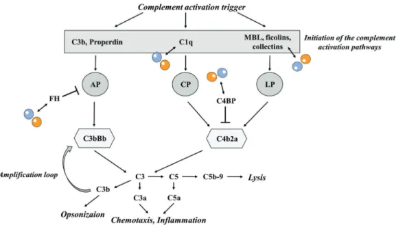
Pentraxin-3 (PTX3) and C-reactive protein (CRP) are fluid PRMs that participate in the innate immune response via the recognition and binding of pathogenic moieties, opsonization of target cells and activation as well as regulation of the complement system (Figure 1) (Daigo et al. 2016). Members of the pentraxin family include short and long pentraxins that share a conserved pentraxin-like domain on their C-terminal ends (Daigo et al. 2016). The primary sequence of the long pentraxin PTX3 is also highly conserved among different animal species, and encodes a 381 amino acid long
protein with a unique N-terminal domain (Inforzato et al. 2013), not related to any known protein structure (Daigo et al. 2016). PTX3 is generated in response to inflammatory stimuli via gene expression induction in innate immune cells, endothelial cells and fibroblasts (Kunes et al. 2012). However, prompt release of PTX3 originates from neutrophil granulocytes (Kunes et al. 2012), where constitutively produced, glycosylated PTX3 monomers are stored in lactoferrin+ intracellular granules (Daigo et al. 2016). Upon neutrophil cell activation and neutrophil extracellular trap (NET) release, PTX3 is secreted from neutrophils and its monomers form octamers through inter-chain disulfide bonds (Daigo et al. 2016).
CRP on the other hand is a member of the short pentraxin protein family (Daigo et al.
2016). Its native, pentameric form is produced and stored in the endoplasmic reticulum of resting hepatocytes (Sproston et al. 2018). Upon inflammatory stimuli, CRP is secreted into the circulation and following the recognition of target cell membranes, its attachment to phosphocholine induces the disassembly of the pentameric structure to CRP monomers in a calcium-dependent fashion (Sproston et al. 2018). CRP is a routinely used biomarker of systemic inflammation in clinical setting, whereas PTX3 was reported to be a prognostic marker of disease activity and severity in an increasing number of diseases (Maekawa et al. 2011, Lech et al. 2013, Speeckaert et al. 2013, Sjoberg et al. 2014, Krzanowski et al. 2017).
Both molecules have been shown to interact with the complement system on multiple levels. PTX3 may initiate the activation of the CP and LP via binding to surface- associated MBL (Ma et al. 2011), ficolins, collectins (Ma et al. 2009) and C1q (Nauta et al. 2003). MBL-bound PTX3 may even recruit C1q molecules to the activator surface thus providing a link between the activation of the LP and CP, whereas the interaction of PTX3 with C1q in the fluid phase inhibits CP activity. Hence, PTX3 restricts unwanted complement activation in the circulation (Nauta et al. 2003, Kunes et al.
2012). Furthermore, ligand binding of PTX3 may prevent AP amplification via the recruitment of the complement regulators FH (Deban et al. 2008) and C4BP (Braunschweig et al. 2011) to the surface of apoptotic cells, thus facilitating phagocytosis of pathogens and clearance of cellular debris without the unnecessary activation of the terminal pathway (Inforzato et al. 2013). CRP also has the ability to activate the complement system. The native form of CRP however, may only attach to
membrane-bound C1q (Biro et al. 2007) with concomitant restrain of the terminal pathway (Thiele et al. 2014). Nevertheless, localized dissociation of pentameric CRP, induced by binding to cell-membrane phosphatidylcholine, allows for the excessive activation of the CP both in vitro and in vivo (Biro et al. 2007, Thiele et al. 2014).
Parallel to complement activation, CRP may also regulate the CP and AP via recruitment of C4BP, FH, but also properdin (Sjoberg et al. 2006, Biro et al. 2007, O'Flynn et al. 2016).
Even though there are differences in their production and specific activities, the expression of both pentraxins is induced in inflammatory conditions. Ligand-bound pentraxins were shown to initiate as well as regulate all three pathways of complement in vitro, while their ligand binding provides basis for cross-talk between the individual pathways (Garred et al. 2016). However, in vivo studies reported inconclusive data on the overall impact of PTX3 on tissue injury and recovery (Souza et al. 2002, Salio et al.
2008, Souza et al. 2009, Lech et al. 2013) and no study has been designed so far to explore changes in the systemic level of pentraxins in relation to complement overactivation in vivo.
Figure 1. Interaction of pentraxins with the complement system
A sketch of the complement activation pathways and the interaction of pentraxin-3 (yellow sphere) and C-reactive protein (blue sphere) with complement activator and regulator proteins is displayed based on the publication of Daigo et al. 2016 with modifications. (AP= alternative pathway, CP= classical pathway, LP= lectin pathway, MBL= mannose-binding lectin)
2.1.6. Dysregulation of the complement alternative pathway
The proper control of AP activation requires a delicate regulatory mechanism, impairment of which can easily lead to complement dysregulation and disease pathogenesis. Dysregulation of the AP may originate from either the drop-out of restrain mechanisms, or from the hyperfunction of complement activators. Reduction of complement regulatory mechanisms may be caused by the dysfunction of the complement regulator proteins or may be attributed to factor consumption secondary to overactivation of the complement system.
Primary mechanisms that lead to the dysregulation of the complement AP include impaired protease activity of FI with or without cofactor dysfunction, reduction of decay mechanisms or increased stability and half-life of the AP C3 convertase. These impairments may be attributed to either genetic mutations in the complement genes causing a quantitative or functional deficiency (or excessive activity) of the expressed protein, or complement factor blockade and complex stabilization by autoantibodies, all of which may induce the manifestation of complement-mediated diseases (Baines et al.
2017). In recent years, deficiencies in complement regulatory processes associated to alterations of the complement genes or autoimmune mechanisms have been described in an increasing number of conditions (Wong et al. 2018).
Dysfunction of FI leads to reduced C3b and C4b cleavage and thus prolongs the inactivation of both the AP and CP C3 convertases, leading to the dysregulation of the AP and CP in the fluid as well as in the solid phases. Whereas genetic mutations in the CFI gene have been linked to disease pathogenesis in atypical hemolytic uremic syndrome (aHUS), autoantibodies against FI were only demonstrated to have a minor contribution to the dysregulation of fluid and solid phase complement regulation, suggesting that their detection is rather an epiphenomenon than a pathogenic factor of disease manifestation (Kavanagh et al. 2012, Kavanagh et al. 2013).
Mutations may also impair the cofactor activity of multiple complement regulator proteins, thus playing a major role in the pathogenesis of complement-mediated diseases. Pathogenic mutations identified in CFH (the most frequently detected genetic alteration in aHUS patients) (Noris et al. 2010, Fremeaux-Bacchi et al. 2013) are usually heterozygous point mutations that cluster at the C-terminal end of the protein.
Hence, they interfere with the membrane-associated regulatory mechanisms and host-
surface discrimination of FH without a quantitative deficiency in the circulatory levels of the protein (Kavanagh et al. 2013). Functional assays have shown that expression of the mutant protein causes a varying degree of impairment in the recognition and binding of heparin and C3b on e.g. endothelial cells and platelets, with subsequent cellular damage and the activation of thrombocytes (Vaziri-Sani et al. 2006, Abarrategui- Garrido et al. 2008, Stahl et al. 2008, Ferreira et al. 2009). Mutations located at the N- terminal regulatory regions of FH, even though are detected less frequently, cause a deficiency of AP regulation both in the fluid and solid phases (Pechtl et al. 2011). In addition to pathogenic point mutations, the regulatory functions of FH may also be altered due to genomic rearrangements in the RCA gene cluster that may yield hybrid genes of CFH and CFHRs, the genes encoding its related proteins (Factor H-related proteins 1-5, FHRs, respectively). Albeit the exact role of FHRs in complement dysregulation remains to date uncertain, mutations in CFHR5 have been associated with disease manifestation in multiple independent cohorts of aHUS and C3 glomerulopathy (C3G) patients (Monteferrante et al. 2007, Maga et al. 2010, Westra et al. 2012, Wong et al. 2018). Autoantibodies against FH have also been described in 4-14% of aHUS patients and 11% of C3G patients, and functional characterization of these antibodies has proven their causative role AP dysregulation and disease pathogenesis, discussed in detail below (Goodship et al. 2012, Zhang et al. 2012, Kavanagh et al. 2013)
Similarly to CFI, mutations affecting CD46, the gene encoding MCP contribute to the dysregulation of both the AP and CP, since under physiological conditions MCP may engage both C3b and C4b. Unlike most of the CFH mutations however, about 75% of the alterations found in CD46 cause a reduced expression of the protein, thus contributing to altered regulation of complement (Kavanagh et al. 2013). In the presence of mutations in the THBD gene, THBD also loses its cofactor activity in the FI- mediated cleavage of C3b, which has been linked to the manifestation of aHUS (Wong et al. 2018).
Mutations the C3 and CFB genes on the other hand belong to the activating mutations of complement. These mutations were reported to either cause altered binding of regulators (e.g. MCP) or an enhanced formation of the AP C3 convertase that is more resistant to decay due to a stronger bond between FB and C3b (Kavanagh et al. 2013).
This leads to increased complement deposition on cellular surfaces and the disruption of
the balance between amplification and regulation (Harrison 2018). However, mutations of C3 have also been described to cause reduced secretion of the protein, the role of which in AP dysregulation and disease pathogenesis remains to date unrevealed (Kavanagh et al. 2013). Increased convertase stability and activity have also been associated with the presence of anti-C3b and anti-FB antibodies (Strobel et al. 2010, Chen et al. 2011, Marinozzi et al. 2017) as well as the production of a C3bBb stabilizing IgG, the C3 Nephritic Factor (C3Nef) (Daha et al. 1976, Daha et al. 1977).
In addition to germline genetic mutations, enhanced AP C3 convertase activity and MAC formation may also be attributed to somatic mutations in the phosphatidylinositol glycan anchor biosynthesis class A (PIGA) gene. The PIGA gene encodes a glycosyl transferase, alterations of which cause clonal deficiency of GPI-anchored proteins including DAF and CD59. Their dysfunction causes dysregulation of complement activity on the surface of red blood cells, clinically manifesting in paroxysmal nocturnal hemoglobinuria, characterized by chronic hemolysis with acute paroxysms and an increased risk of thrombus formation (Baines et al. 2017).
Despite adequate functional evidence on the contribution of the above genetic alterations and autoimmune mechanisms to complement dysregulation, fundamental questions regarding the clinical manifestation of complement mediated diseases remain to date unrevealed. Multiple facts e.g. incomplete penetrance of the genetic alterations, presence of autoantibodies in disease remission and disease manifestation in adulthood suggest that an additional, yet undiscovered trigger event is required to set off the self- amplifying mechanism of AP dysregulation. Furthermore, despite the accumulated knowledge on the structure-function relationships of genetic alterations, to date we still have no definitive explanation for the association of certain mutations to one clinical condition rather than another (Pechtl et al. 2011). Therefore further molecular as well as clinical investigations are required to challenge these controversies and to fill the gaps in our ever increasing knowledge on the pathogenesis of complement mediated diseases.
2.2. Thrombotic microangiopathies
2.2.1. Classification and pathogenesis of thrombotic microangiopathies
Thrombotic microangiopathies (TMA) are rare but life-threatening disorders characterized by microangiopathic hemolytic anemia and acute thrombocytopenia with or without organ impairment (e.g. signs of neurological or kidney injury). The bed-side diagnosis of TMAs is based on this triad of clinical symptoms and routine laboratory results, however, intensive research of the recent decades revealed that TMAs cover distinct subgroups of diseases. Their etiology-based classification discriminates between acquired or inherited primary TMAs with known molecular etiology, secondary TMAs as complication of underlying diseases or conditions, and specific infection-associated TMAs. TMAs are severe conditions that require immediate attention and diagnostic workup for complement abnormalities. Their expeditious differential diagnostics is especially important, since the identification of the exact subgroup of TMA has an impact on the determination of the optimal therapeutic choice immediately from the onset of the acute phase.
2.2.2. Thrombotic thrombocytopenic purpura
Thrombotic thrombocytopenic purpura (TTP), covers a TMA subgroups, where beyond the clinical and laboratory triad of TMA, patients usually present with critical thrombocytopenia (as low as 4-5G/L) and neurological symptoms, whereas signs of kidney injury are less frequent clinical features of disease. This form of TMA is caused by the deficiency of a disintegrin and metalloproteinase with a thrombospondin type 1 motif member 13 (ADAMTS13), either due to its blockade by antibodies or mutations in the ADAMTS13 gene. Under physiological conditions this enzyme cleaves the ultra- large form of von Willebrand Factor (ULVWF), which is secreted by activated endothelial cells, and can spontaneously bind and activate platelets, thus contributing to thrombus formation at the site of activation. Although ADAMTS13 deficiency is accountable for TTP, it may also be present in disease remission, which supports the hypothesis that the onset of TTP requires a trigger event (such as pregnancy or infections), which leads to endothelial activation and therefore an enhanced expression of ULVWF, hence causing the imbalance of pro- and anti-thrombotic factors in the microvasculature.
2.2.3. Typical hemolytic uremic syndrome
Hemolytic uremic syndrome (HUS) on the other hand covers distinct forms of primary TMAs, which usually present with the triad of hemolytic anemia, thrombocytopenia and acute renal failure, while neurologic symptoms have only been reported in a minority of patients. In about 90% of the cases, HUS is caused by Shiga toxin-producing Escherichia coli (STEC), defined as typical or STEC-HUS (Wong et al. 2018) that represents the most frequent cause of acute kidney failure in children. It is induced by a gastrointestinal infection, where Shiga-toxin (Stx) is released to the circulation from mucus-adhered bacteria and causes microangiopathy through the blockade of protein synthesis and subsequent activation of the endothelial cells, red blood cells and platelets (Karpman et al. 2006, Orth-Holler et al. 2014, Arvidsson et al. 2015, Jokiranta 2017).
2.2.4. Atypical hemolytic uremic syndrome
Atypical HUS on the other hand represents a rare form of TMA (Fakhouri et al. 2017) with an incidence rate of approximately 0.23-0.42 per 106 per year (Fakhouri et al.
2017). Atypical HUS is mediated by the dysregulation of the AP, evoked either by pathogenic mutations in the complement genes or by the presence of autoantibodies directed against the complement regulator FH (discussed in detail below). Mutation associated forms of aHUS usually manifest in either very early childhood, often in infants younger than 1 year of age, or in women at the postpartum period, whereas autoimmune aHUS is a disease of adolescence. Atypical HUS has a relapsing-remitting disease course and therefore requires long-term patient management. Just like in the case of STEC-HUS, prodromal symptoms of aHUS may include gastrointestinal complaints and signs that could be misleading at the initial presentation. However, since the endothelial injury in aHUS is caused by the dysregulation of the AP, detection of laboratory signs of complement AP consumption may aid the differential diagnostics of aHUS together with the lack of proof for an STEC infection and clinical improvement of patients. Definite diagnosis of aHUS however, requires the functional and genetic testing of the patients’ complement profile that reveals the underlying molecular mechanism of disease pathogenesis in nearly two third of the cases. Likely pathogenic mutations are identified by 50-60% of aHUS patients, with a combination of mutations identified in 10-25% of the cases (Kavanagh et al. 2013). Approximately 25% of the
pathogenic mutations are found in the CFH gene (Noris et al. 2010, Fremeaux-Bacchi et al. 2013), whereas in addition to mutations in the complement genes, genetic alterations of THBD and diacylglycerol kinase epsilon (DGKE) have been associated with aHUS, too (Delvaeye et al. 2009, Lemaire et al. 2013, Azukaitis et al. 2017). Along likely pathogenic mutations that have established functional consequences, haplotypes and a multitude of variations with an unknown significance have been linked to an increased risk of aHUS development. Even though these have not been shown to directly contribute to disease pathogenesis, in combination with other trigger mechanisms they might induce the manifestation of disease.
Unlike the genetically determined forms, autoimmune aHUS usually manifests in older children and adolescents following a preceding minor infection of the respiratory or gastrointestinal tracts (Dragon-Durey et al. 2010). It accounts for 4-14% of all aHUS cases (Skerka et al. 2009, Hofer et al. 2013, Kavanagh et al. 2013, Nester et al. 2015), although an Indian cohort reported an incidence rate of 56% with an unexplained reason behind the high frequency of autoantibody positivity (Sinha et al. 2014). Autoimmune aHUS has a high relapse rate with the risk of developing end stage renal disease (Noris et al. 2009, Dragon-Durey et al. 2010, Dragon-Durey et al. 2010, Noris et al. 2015).
Laboratory diagnostics of autoimmune aHUS involve the detection of anti-Factor H (anti-FH) antibodies that often goes together with low levels of FH due to immune complex formation (Blanc et al. 2012), the levels of which were shown to correlate with disease severity (Blanc et al. 2012). Further characteristic laboratory findings in autoimmune aHUS are low C3 and FB levels indicative of the overactivation and subsequent factor consumption of the AP (Blanc et al. 2012), frequently accompanied by a decreased activity or deficiency in the whole complement AP activity. The genetic linkage of autoimmune aHUS to deficiency of CFHR1 justifies testing for copy number variations in the CFHR genes via multiplex ligation-dependent probe amplification (MLPA) analysis. However, a negative finding in this regard does not exclude the presence of autoimmune aHUS, since the lack of CFHR1 is not a prerequisite for the development of autoantibodies, indicated by a considerable proportion of autoimmune aHUS patients with no genetic alterations in the CFHR genes (Dragon-Durey et al.
2009, Moore et al. 2010, Strobel et al. 2011, Geerdink et al. 2012).
2.2.5. Secondary thrombotic microangiopathies
Secondary forms of TMA encompass a heterogeneous group of disorders all of which are evoked by the progression of a preexisting condition. Secondary TMA may be associated among others with glomerular diseases, severe infections and septic conditions, allogenic hematopoietic stem cell or solid organ transplantation, systemic autoimmune diseases, pregnancy, tumor progression and antitumor therapy as well as malignant hypertension. Despite their distinct etiologies, shared features of secondary TMAs include decreased ADAMTS13 activity and overactivation of multiple complement pathways, leading to laboratory signs of complement consumption.
2.2.6. Pathological complement activation in thrombotic microangiopathies
Endothelial damage and subsequent microvascular thrombosis are key pathogenic factors in TMA. Albeit traditionally aHUS was recognized as a TMA form mediated by complement dysregulation, by now vascular injury has been associated with complement overactivation in all etiological forms of this disease (Table 1) (Noris et al.
2012, Reti et al. 2012, Baines et al. 2017, Farkas et al. 2017).
A shared characteristic of the coagulation and complement cascades is their initial activation at local sites of infection or tissue injury (Baines et al. 2017). There is a considerable interaction between the activator mechanisms of both cascades (Oikonomopoulou et al. 2012, Kenawy et al. 2015, Foley 2016), provided by the diverse ligand binding capacity of certain PRMs and activation enzymes of complement (Bossi et al. 2011, Takahashi et al. 2011, La Bonte et al. 2012), augmented by complement activation through thrombin and additional coagulation factors (Huber-Lang et al. 2006, Amara et al. 2010, Krisinger et al. 2012, Berends et al. 2014). Components of each system may cleave and activate one another (Baines et al. 2017), thus providing molecular basis for the joint manifestation of pathological thrombosis and complement overactivation in distinct etiological forms of TMA (Noris et al. 2012, Reti et al. 2012, Baines et al. 2017, Farkas et al. 2017).
Atypical HUS is a prototypic disease of AP dysregulation attributed to the above discussed genetic alterations of complement components and autoimmune mechanisms (Wong et al. 2018). STEC-HUS on the other hand is primarily not a complement- mediated disorder. Nevertheless, increased levels of complement degradation products
(C3a(desArg) and C3d), Bb and the properdin-stabilized AP C3 convertase (C3bBbP) together with the elevation of sC5b-9 have been detected in the circulation of STEC- HUS patients (Thurman et al. 2009, Stahl et al. 2011, Westra et al. 2017), indicative of complement overactivation in the acute phase of disease. Intensified complement activity in STEC-HUS is supposedly due to the combined effect of Stx and bacterial lipopolysaccharide (LPS) on complement activation and coagulation (Stahl et al. 2011, Arvidsson et al. 2015), a hypothesis corroborated by independent in vitro ad in vivo observations. Inter alia, Stx was shown to activate the AP in the fluid phase and to interfere with the regulatory activity of FH on cellular surfaces (Orth et al. 2009), together with the upregulation of P-selectin on microvascular endothelium, thus promoting C3b deposition and thrombus formation (Morigi et al. 2011). Stx together with LPS was also reported to induce the formation of platelet-leukocyte aggregates in vitro (Stahl et al. 2011, Arvidsson et al. 2015), whereas the combined thrombotic effects of Stx and LPS could be diminished by the admission of a C3a receptor antagonist or by the application of FB-deficient mice in animal models of STEC-HUS in vivo (Morigi et al. 2011).
Increased complement activation product levels together with an elevated cytokine response have also been reported in TTP (Reti et al. 2012, Westwood et al. 2014).
Increased complement activity has been detected at both the acute presentation and in remission of TTP, the extent of which was reported to be associated with the level of anti-ADAMTS13 antibodies in the autoimmune form of this disease (Reti et al. 2012, Westwood et al. 2014). The underlying mechanism of the sustained complement response in TTP could be attributed to multiple mechanisms participating in disease pathogenesis. On the one hand, increased expression of P-selectin on activated platelets and endothelial cells may lead to an enhanced C3b deposition and thus substantiate complement activation on the tagged endothelial surface (Del Conde et al. 2005). On the other hand, ULVWF may also initiate the AP and the terminal pathway of complement via direct binding of complement factors (Bettoni et al. 2017). Enhanced generation of the complement activation products C3a, C5a, and sC5b-9 may in turn lead to further activation of endothelial cells with inherently increased ULVWF and P- selectin expression as well as decreased THBD expression, ultimately causing the
uncontrolled activation of coagulation and the dysregulation of complement (Bettoni et al. 2017).
Despite the many different etiologies of secondary TMA, overactivation and subsequent consumption of both the CP and AP have been described in this condition, too (Farkas et al. 2017). Elevated systemic levels of sC5b-9 and C3a have been associated with poor long-term outcome in independent cohorts of secondary TMA patients (Jodele et al.
2013, Farkas et al. 2017, Wong et al. 2018). Although dysregulation of the AP is not a distinguished mediator of disease pathogenesis in secondary TMA, its involvement has been recently suggested in the development of hematopoietic stem cell transplantation- associated TMA, that could be attributed to chemotherapy, radiation, and infection- induced endothelial injury and neutrophil cell activation (Jodele et al. 2013, Yuen et al.
2016, Gloude et al. 2017, Wong et al. 2018).
In conclusion, the acute phase of TMAs are accompanied by complement dysregulation or consumption, and an overdrawn inflammatory response. This may induce among others the production of inflammatory proteins, such as CRP and PTX3. Based on the reported complement regulatory activity of pentraxins, the released proteins could interfere with local complement activity at the site of vascular injury (Jaillon et al.
2007). However, so far only in vitro studies have been published in this regard, whereas no investigation involving human subjects has explored the systemic levels of pentraxins in the acute phase of TMA or the in vivo relation of PTX3 and CRP to complement activity.
Table 1. Role of complement in thrombotic microangiopathies
Distinct forms of thrombotic microangiopathies (TMA) are displayed, grouped based- on the most important, common pathogenic factors and the role of complement in each subgroup of disease. (ADAMTS13 = a disintegrin and metalloproteinase with a thrombospondin type 1 motif member 13, ADAMTS13 = the gene encoding ADAMTS13, aHUS = atypical hemolytic uremic syndrome, AP = alternative pathway, CP = classical pathway, DGKE = diacylglycerol kinase epsilon, IgM = Immunoglobulin M, MMACHC-TMA = thrombotic microangiopathy associated to methylmalonic aciduria and homocystinuria, cblC complementation type, MMACHC = the gene encoding methylmalonic aciduria and homocystinuria type C protein, STEC-HUS =
Shiga toxin-producing Escherichia coli-associated hemolytic uremic syndrome, Stx = Shiga-toxin, TTP = thrombotic thrombocytopenic purpura)
TMA subgroup Key pathogenic factors Role of complement in the pathogenesis Inherited primary TMA
Complement-mediated aHUS
-Endothelial damage due to severe dysregulation of the complement AP
-Genetic alteration in the complement genes and subsequently altered expression or function of their encoded proteins
DGKE-aHUS
-Prothrombotic state on the endothelium due to the loss of DGKE function
-Enhanced terminal pathway activation and therefore increased sC5b-9 levels that may alter podocyte metabolic pathways, the structure and function of the extracellular matrix, membrane lipids and key proteins of the cytoskeleton and the slit-diaphragm MMACHC-TMA
-Impaired intracellular vitamin B12 metabolism due to mutations in the MMACHC gene
-Not known
Inherited TTP (Upshaw-Schulman Syndrome)
-Congenital deficiency of the ADATMS13 metalloproteinase due to mutations in the ADATMS13 gene
-C3b binding and complement activation through activated endothelial cells and platelets
-Direct activation of the complement AP by the ULVWF
Acquired primary TMA Autoimmune aHUS
-Endothelial damage due to severe dysregulation of the complement AP
-Dysfunction of the complement regulator Factor H due to autoantibodies
Acquired TTP -Inhibitory antibodies that block the ADATMS13 metalloproteinase
-C3b binding and complement activation through activated endothelial cells and platelets
-Direct activation of the complement AP by the ULVWF
Infection-associated TMA
STEC-HUS -Endothelial damage caused by the binding and internalization of Stx
-Complement activation through Stx and LPS
-Increased C3b binding to the endothelium due to high P-selectin expression induced by Stx
Streptococcus pneumoniae/Influenza induced HUS
-Neuraminidase production and cleavage of N-acetylneuraminic acid from glycoproteins on the cell membrane of erythrocytes, platelets, glomeruli and hepatocytes, exposure of the Thomsen–Friedenreich antigen
-Interaction of the Thomsen–Friedenreich antigen with preformed IgM initiates excessive complement activation of both the CP and AP
Secondary forms of TMA
Secondary TMA
-Worsening of a known preexisting condition and subsequent coagulopathy with tissue or organ damage involving the endothelium
-Dysregulation of both the CP and AP with severe consumption of the individual complement factors
2.2.7. Alternative pathway dysregulation associated with anti-Factor H antibodies in atypical hemolytic uremic syndrome
Anti-FH antibodies in aHUS have first been reported in a French patient cohort in 2005 (Dragon-Durey et al. 2005), and since then multiple independent investigations have shown their functional relevance in disease pathogenesis. Upon binding to FH, autoantibodies interfere with the complement regulatory activity on the surface of host cells that leads to overactivation of the complement AP and subsequent complement- mediated tissue damage. The majority of the anti-FH antibodies belongs to the IgG subclass, although some patients have been reported to develop IgA antibodies, too (Strobel et al. 2011). Albeit the antibodies may recognize multiple domains on FH (Moore et al. 2010, Blanc et al. 2012), epitope mapping using recombinant FH fragments have shown that most of the antibody binding epitopes cluster on the C- terminal SCRs of FH (Jozsi et al. 2007, Jozsi et al. 2008, Moore et al. 2010, Bhattacharjee et al. 2015). The antibody binding site was reported to overlap with that of C3b and cellular ligands, which explains why anti-FH antibodies inhibit the binding of C3b and its degradation products thus reducing the cofactor and decay accelerating activities of FH. Furthermore, antibody binding perturbs the FH-mediated cell- protection, shown by increased hemolysis of red blood cells in the presence of patient IgG (Jozsi et al. 2007, Blanc et al. 2012). Anti-FH antibodies have also been described in C3G, however their functional characteristics were reported to be substantially different from those identified in aHUS patients (Blanc et al. 2015). Whereas the functional consequence of the antibody binding to FH is widely studied and comprehensive knowledge has been accumulated in regard to the structural exploration of antibody binding to the folded FH domains, detailed structural analysis of where the aHUS-associated FH epitopes are localized at the amino acid level are still lacking.
Furthermore, the immediate trigger that would evoke autoantibody production against FH has not yet been identified. Nevertheless, since a mild infection is often recorded prior to the manifestation of aHUS, the higher prevalence of gastrointestinal pathogens (Togarsimalemath et al. 2018) together with the genetic linkage of the disease (e.g.
deficiency of FHR-1) have been proposed to contribute to the pathological activation of B lymphocytes and trigger the production of anti-FH antibodies (Bhattacharjee et al.
2015).
2.2.8. Factor H-related proteins and their genetic-linkage to anti-Factor H antibodies
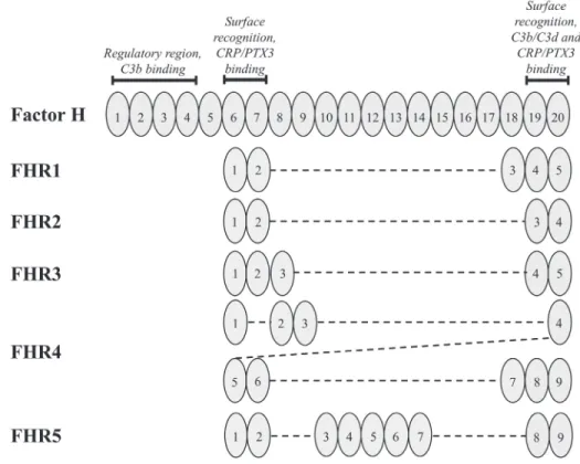
Alongside with FH, there are six additional members of the FH protein family: FHL-1 and the FHRs 1-5 (Figure 2). FHL-1 is a splice variant derived from the CFH gene, whereas FHRs evolved from FH via non-allelic homologous recombination (Jozsi et al.
2014). As a result of this, all members of the FH protein family are composed of SCR domains that share a high sequence homology to each other. Corresponding to their structural similarities, FHL-1 and FHRs were reported to participate in the regulation or sometimes de-regulation of complement activity through multiple mechanisms. FHL-1 carries a unique four amino acid long C-terminus, a so called SFTL tail corresponding to its components: serine, phenylalanine, threonine and leucine (Perez-Caballero et al.
2000), but the rest of the protein is identical to the first 7 SCR domains of FH (Schwaeble et al. 1987). Therefore, FHL-1 shares functional similarities to FH including complement inhibition through C3b binding (Kuhn et al. 1995) and the recognition of common ligands such as pentraxins and malondialdehyde epitopes (Sanchez-Corral et al. 2018). However, apparent contrasts in the conformation of the two proteins and the presence of the C-terminal tail on FHL-1 contribute to considerable differences in the ligand interactions of the two proteins (Sanchez-Corral et al. 2018).
FHRs on the other hand do not carry all the four N-terminal regulatory domains of FH, therefore they have no or only residual cofactor and decay accelerating activities in the regulation of the complement AP. However, their reported binding of C3b and other deposited fragments of C3 (iC3b and C3d) (Goicoechea de Jorge et al. 2013) via their C-terminal domains may induce competition with FH for the C3 binding site, a process referred to as complement de-regulation (Sanchez-Corral et al. 2018). Additional ligand binding similarities include the recognition of host surface ligands and ECM components (Blackmore et al. 1998, Hellwage et al. 1999, McRae et al. 2005, Heinen et al. 2009, Strobel et al. 2011, Csincsi et al. 2015, Rudnick et al. 2018) as well as recruitment of FHRs to apoptotic and necrotic cells via surface-bound PRMs (e.g.
pentraxins), where in contrast to FH some FHRs have been reported to enhance the activation of not only the alternative but also the classical pathways of complement (Mihlan et al. 2009, Hebecker et al. 2010, Hebecker et al. 2012, Csincsi et al. 2015, Csincsi et al. 2017, Rudnick et al. 2018). However, given the relatively low
concentration of FHRs and their lower avidity for C3b binding compared to that of FH (Sanchez-Corral et al. 2018), the complement activator effect of FHRs may be insignificant in the physiological regulation of the AP.
Under pathological AP activation however, locally increased levels and activity of FHRs may contribute to disease pathogenesis. Numerous reports have described the association of genetic alterations in CFHRs to complement-mediated diseases (Hughes et al. 2006, Gharavi et al. 2011, Sanchez-Corral et al. 2018). The autoimmune form of aHUS is linked to the deficiency of CFHR1 and -3 that covers the homozygous deletion of an 80 kb-long genomic region in 82-88% of patients with FH autoantibodies (Zipfel et al. 2007, Jozsi et al. 2008, Dragon-Durey et al. 2010, Geerdink et al. 2012, Hofer et al. 2013). Conversely, the frequency of the heterozygous deletion of CFHR1 is similar in healthy individuals and aHUS patients.
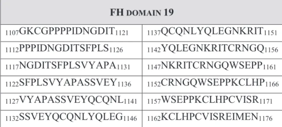
FHR1 shares a high sequence homology with FH. The C-terminal SCRs of the two proteins differ in only two amino acids, namely FH carries a serine at amino acid position 1191 and a valine at position 1197, while FHR1 contains a leucine and an alanine at the corresponding positions (residue 290 and 296), whereas FHR1 domain 4 and FH domain 19 are identical to one another (Abarrategui-Garrido et al. 2009). FHR1 was shown not only to compete with FH in C3b binding (Goicoechea de Jorge et al.
2013), but also to cross react with anti-FH antibodies and to neutralize the antibody- associated enhanced red blood cell lysis in vitro (Strobel et al. 2011, Bhattacharjee et al.
2015). These functional studies together with the high sequence homology of the C- terminal domains of FH and FHR1 suggest an overlap of the antibody binding sites on the two proteins. Genetic and structural analyses have pointed out CFHR1 deficiency as a potential predisposing factor for the development of autoimmune aHUS (Kavanagh et al. 2013, Bhattacharjee et al. 2015) and anti-FH binding of the folded FH and FHR1 C- terminal domains have recently been compared (Bhattacharjee et al. 2015), however no investigation aimed to date for the localization of the antibody binding epitopes on FH as well as FHR1 on the amino acid level, which however, could provide a direct explanation for the reported interference of autoantibody binding with FH function.
Figure 2. Homology between Factor H and the Factor H related proteins
Illustration of Factor H (FH) and its related proteins (FHRs) based on the publication of Sanchez-Corral et al. 2018 with modifications. The proteins are displayed with the sketch of their short consensus repeat (SCR) domains indicated by numbers. SCR domains of the FHRs are aligned vertically to their homologous domains in FH. Ligand binding sites and functional characteristics of FH are indicated above the schematic pictures of the FH domains. (C3b = complement component 3b, C3d = complement component 3d, CRP = C-reactive protein, FHR1-5 = Factor H-related proteins 1-5, PTX3 = pentraxin-3)