ORIGINAL RESEARCHARTICLE
BACKGROUND: Chronic heart failure (HF) is associated with altered signal transduction via β-adrenoceptors and G proteins and with reduced cAMP formation. Nucleoside diphosphate kinases (NDPKs) are enriched at the plasma membrane of patients with end-stage HF, but the functional consequences of this are largely unknown, particularly for NDPK-C. Here, we investigated the potential role of NDPK-C in cardiac cAMP formation and contractility.
METHODS: Real-time polymerase chain reaction, (far) Western blot, immunoprecipitation, and immunocytochemistry were used to study the expression, interaction with G proteins, and localization of NDPKs. cAMP levels were determined with immunoassays or fluorescent resonance energy transfer, and contractility was determined in cardiomyocytes (cell shortening) and in vivo (fractional shortening).
RESULTS: NDPK-C was essential for the formation of an NDPK-B/G protein complex. Protein and mRNA levels of NDPK-C were upregulated in end- stage human HF, in rats after long-term isoprenaline stimulation through osmotic minipumps, and after incubation of rat neonatal cardiomyocytes with isoprenaline. Isoprenaline also promoted translocation of NDPK-C to the plasma membrane. Overexpression of NDPK-C in cardiomyocytes increased cAMP levels and sensitized cardiomyocytes to isoprenaline- induced augmentation of contractility, whereas NDPK-C knockdown decreased cAMP levels. In vivo, depletion of NDPK-C in zebrafish embryos caused cardiac edema and ventricular dysfunction. NDPK-B knockout mice had unaltered NDPK-C expression but showed contractile dysfunction and exacerbated cardiac remodeling during long-term isoprenaline stimulation.
In human end-stage HF, the complex formation between NDPK-C and Gαi2 was increased whereas the NDPK-C/Gαs interaction was decreased, producing a switch that may contribute to an NDPK-C–dependent cAMP reduction in HF.
CONCLUSIONS: Our findings identify NDPK-C as an essential requirement for both the interaction between NDPK isoforms and between NDPK isoforms and G proteins. NDPK-C is a novel critical regulator of β-
adrenoceptor/cAMP signaling and cardiac contractility. By switching from Gαs to Gαi2 activation, NDPK-C may contribute to lower cAMP levels and the related contractile dysfunction in HF.
Nucleoside Diphosphate Kinase-C Suppresses cAMP Formation in Human Heart Failure
© 2016 American Heart Association, Inc.
*Drs Abu-Taha, Heijman, and Hippe contributed equally.
†Drs Dobrev and Wieland share senior authorship.
Correspondence to: Thomas Wieland, PhD, Maybachstrasse14, 68169 Mannheim, Germany or Dobromir Dobrev, MD, Hufelandstrasse 55, 45122 Essen, Germany. E-mail thomas.wieland@
medma.uni-heidelberg.de or dobromir.dobrev@uk-essen.de Sources of Funding, see page 895 Key Words: heart failure
◼ myocardial contraction
◼ receptors, adrenergic, beta
◼ signal transduction Issam H. Abu-Taha, PhD*
Jordi Heijman, PhD*
Hans-Jörg Hippe, MD*
Nadine M. Wolf, PhD Ali El-Armouche, MD Viacheslav O. Nikolaev, PhD Marina Schäfer, MSc Christina M. Würtz, PhD Stefan Neef, MD Niels Voigt, MD
István Baczkó, MD, PhD András Varró, MD, PhD, DSc Marion Müller, MSc
Benjamin Meder, MD Hugo A. Katus, MD Katharina Spiger, PhD Christiane Vettel, PhD Lorenz H. Lehmann, MD Johannes Backs, MD Edward Y. Skolnik, MD Susanne Lutz, PhD Dobromir Dobrev, MD†
Thomas Wieland, PhD†
by guest on February 28, 2017http://circ.ahajournals.org/Downloaded from by guest on February 28, 2017http://circ.ahajournals.org/Downloaded from by guest on February 28, 2017http://circ.ahajournals.org/Downloaded from by guest on February 28, 2017http://circ.ahajournals.org/Downloaded from by guest on February 28, 2017http://circ.ahajournals.org/Downloaded from by guest on February 28, 2017http://circ.ahajournals.org/Downloaded from by guest on February 28, 2017http://circ.ahajournals.org/Downloaded from by guest on February 28, 2017http://circ.ahajournals.org/Downloaded from by guest on February 28, 2017http://circ.ahajournals.org/Downloaded from by guest on February 28, 2017http://circ.ahajournals.org/Downloaded from by guest on February 28, 2017http://circ.ahajournals.org/Downloaded from by guest on February 28, 2017http://circ.ahajournals.org/Downloaded from by guest on February 28, 2017http://circ.ahajournals.org/Downloaded from by guest on February 28, 2017http://circ.ahajournals.org/Downloaded from by guest on February 28, 2017http://circ.ahajournals.org/Downloaded from by guest on February 28, 2017http://circ.ahajournals.org/Downloaded from by guest on February 28, 2017http://circ.ahajournals.org/Downloaded from by guest on February 28, 2017http://circ.ahajournals.org/Downloaded from by guest on February 28, 2017http://circ.ahajournals.org/Downloaded from by guest on February 28, 2017http://circ.ahajournals.org/Downloaded from by guest on February 28, 2017http://circ.ahajournals.org/Downloaded from by guest on February 28, 2017http://circ.ahajournals.org/Downloaded from by guest on February 28, 2017http://circ.ahajournals.org/Downloaded from
H
eart failure (HF) is a common cause of death and disability.1 Altered signal transduction via β-adrenoceptors (βARs) and G proteins is a hall- mark of chronic HF and contributes to impaired cardiac contractility.2,3 The exact molecular pathophysiological mechanisms contributing to contractile dysfunction in patients with HF are incompletely understood, and a better understanding of these processes is expected to foster the development of improved treatment options for patients with HF.3The plasma-membrane content of nucleoside diphos- phate kinases (NDPKs) is increased in patients with end- stage HF,4 pointing to a potential role for NDPKs in HF.
NDPKs represent a family of multifunctional proteins en- coded by 10 human nm23 genes, of which the class I subfamily (consisting of NDPK-A, -B, -C, and -D) exerts enzymatic activity.5 NDPKs form heterohexamers and catalyze the transfer of γ-phosphate between nucleotide triphosphates and nucleotide diphosphates.6,7 NDPK-A and NDPK-B play a role in numerous cellular processes, often as part of larger signaling complexes. For exam-
ple, NDPK-B, but not NDPK-A, forms complexes with Gβγ dimers and acts as a protein histidine kinase that can activate cardiac G proteins in a receptor-independent manner.8–10 Nevertheless, our previous studies indicated that the complex between NDPK-B and G proteins cannot be reconstituted in vitro, suggesting that NDPK-B alone is insufficient to regulate G-protein signaling and that an as-yet unidentified key cofactor is required for the com- plex formation of NDPK-B and G proteins at the plasma membrane.10
NDPK-C exerts enzymatic activity and is able to form heterohexamers with NDPK-A and NDPK-B.11 NDPK-C shares 72% homology with NDPK-A and NDPK-B but has an additional hydrophobic N-terminal domain (Figure I in the online-only Data Supplement), which could serve as a membrane anchor.12,13 NDPK-C is generally less abun- dantly expressed than the major isoforms NDPK-A and NDPK-B11 but is highly enriched at the cardiac plasma membrane of patients with HF.14 Thus, NDPK-C might be the limiting factor targeting NDPK hexamers to membra- nous G proteins and could be the most relevant NDPK isoform for cAMP regulation in the heart and for the pro- gression of HF. However, the function of NDPK-C in the heart is unknown.
To investigate the role of NDPK-C in HF, we performed biochemical studies of NDPK-C and G-protein signaling in human and rat tissue samples, assessed the functional impact of NDPK-C on cAMP levels and cardiac contractil- ity in isolated rat cardiomyocytes, and determined the in vivo effects of NDPKs on contractility in zebrafish and mice. We identify NDPK-C as the critical isoform for the regulation of G-protein function and cAMP levels in the heart, with important consequences for cardiac contrac- tility. Our results show that NDPK-C can interact promiscu- ously with both Gαs and Gαi proteins. The switch to Gαi2- dominant regulation of G proteins by NDPK-C in human HF may contribute to the lower cAMP levels and impaired contractility characteristic of this clinical condition.
METHODS
A detailed overview of all methods is provided in the online-only Data Supplement. Key aspects are summarized below.
Tissue Procurement
Nonfailing human myocardium from the free wall of the left ven- tricle was obtained from organ donors with no apparent heart disease and normal left ventricular function (determined by echocardiography) for whom no suitable heart transplant recip- ients had been identified. Left ventricular myocardial samples of failing human hearts were obtained from patients with HF (New York Heart Association class III–IV) who underwent car- diac transplantation. The experimental protocol was approved by the ethics review board of the University of Szeged Medical Center. Informed written consent was obtained for the use of nondiseased human hearts. All procedures conformed to the Helsinki Declaration of the World Medical Association.
Clinical Perspective
What Is New?
• We show for the first time that the nucleoside diphos- phate kinase (NDPK)-C is required and indispensable for the interaction of NDPKs with both stimulatory Gs and inhibitory Gi proteins.
• NDPK-C–mediated targeting of NDPKs to the plasma membrane is increased in response to β-adrenoceptor stimulation and enhances intracel- lular cAMP levels, cardiomyocyte contractility, and in vivo cardiac function.
• We provide novel mechanistic insights into remod- eling of β-adrenoceptor signaling in heart failure (HF), showing that the HF-related increase in NDPK- C expression may cause constitutive Gi-mediated inhibition of adenylyl cyclases, providing a plausible explanation for the lower cAMP levels in HF.
What Are the Clinical Implications?
• The increased NDPK-C membrane content in human HF could potentially counteract a fading β-adrenoceptor response in the early stages of HF by increasing the amount of Gαs proteins in the plasma membrane. However, by switching to Gαi2 activation, NDPK-C may play a role in HF progres- sion by reducing cAMP levels, typical for end-stage human HF.
• A better understanding of the molecular processes underlying altered G-protein signaling in HF may help to develop new HF therapies. We identify NDPK-C as a novel therapeutic target involved in the regulation of aberrant G-protein signaling and cardiac contrac- tility in HF.
by guest on February 28, 2017http://circ.ahajournals.org/Downloaded from
ORIGINAL RESEARCHARTICLE
Rat and Mouse Models of Isoprenaline
Stimulation and Isolation of Rat Cardiomyocytes
Male Wistar rats received 4 days of either isoprenaline or vehicle administered via osmotic minipumps, as previously described.15 Male C57Bl/6 wild-type and NDPK-B knockout mice received the same treatment but for 7 days. All animals were anesthetized with 2% isoflurane inhalation and received carprofen (5 µg/kg SC) as analgesic. All procedures concern- ing the care and use of animals were in accordance with institu- tional guidelines (Az. G-12\10 Regierungspräsidium Karlsruhe, Germany, and Az. G10\65 LAVES Niedersachsen, Germany).
Generation of Recombinant Adenoviruses
Recombinant adenoviruses were generated as previously described.8,16–18
mRNA Analysis, Transfection and Transduction, Membrane Fractionation, Western Blots,
Immunoprecipitation, Far Western Blotting, and ATP/GTP Hydrolysis Assays
Details are provided in the online-only Data Supplement. The primers that were used are listed in Table I in the online-only Data Supplement.
Visualization of the Subcellular Localization of NDPK-C
Neonatal rat cardiomyocytes (NRCMs) and adult rat cardiomyo- cytes (ARCMs) were cultured with serum-free medium on cov- erslips and infected with Ad-Flag–NDPK-C. Twenty-four hours later, cells were stimulated with solvent or isoprenaline for up to 6 hours. The subcellular localization of NDPK-C was visual- ized by confocal fluorescence microscopy.
Measurement of Intracellular cAMP
cAMP levels in NRCM or zebrafish lysates were assayed with a cAMP immunoassay.8,9,19 For cAMP assays in living cells, ARCMs were isolated and transduced with Epac2-camps adenovirus, as previously described,18 together with a control (LacZ) or NDPK-C–encoding adenovirus. Cells were stimulated with isoprenaline, forskolin, and 3-isobutyl-1-methylxanthine.
Fractional Shortening in ARCMs
ARCMs were infected with an adenovirus encoding enhanced green fluorescent protein (Ad-EGFP) or Ad-Flag–NDPK-C. Sarcomere shortening was assessed during field stimulation with a video- based sarcomere-length detection system at 1.25 mmol/L Ca2+ in the bath solution.20
Zebrafish Maintenance, Morpholino Injection, Measurement of Fractional Shortening, and Histology
Zebrafish danio rerio were maintained under standard con- ditions.21 Embryos at a 1-cell stage were injected with the indicated amounts of the NDPK-B or NDPK-C targeting mor- pholino or a standard control oligonucleotide at the same
concentration. Injections and analyses of fractional shortening, ventricular diameters, immunohistochemistry, and electron microscopy were performed as previously described.19,21
Statistics
Results are presented as mean±SEM. Normality was assessed with the D’Agostino and Pearson omnibus test. The Mann- Whitney test (for nonnormally distributed data or data with too few points to assess normality), an unpaired 2-tailed Student t test, or 1- or 2-way ANOVA followed by a post hoc Bonferroni or the Tukey test was used to compare means between groups.
Values of P<0.05 were considered statistically significant.
RESULTS
Interactions Among NDPK-C, NDPK-B, and G Proteins
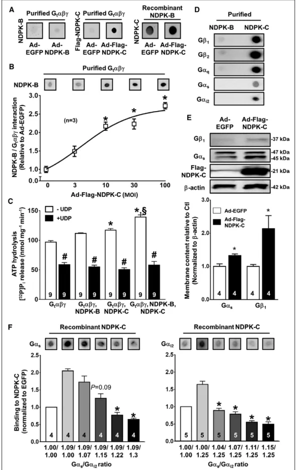
NDPK-C forms hetero-oligomers with NDPK-A and NDPK- B.11 Moreover, NDPK-B/Gβγ complexes regulate G-protein activity and membrane content in cardiomyocytes.8,19,22 Therefore, we assessed whether NDPK-C is involved in the complex formation of NDPKs with G proteins using far Western blotting.10 When the purified heterotrimeric G protein transducin (Gtαβγ)10 was spotted on cellulose membranes and incubated with lysates of control NRCMs or NRCMs with adenovirus-mediated overexpression of NDPK-B, no direct interaction occurred (Figure 1A). In con- trast, purified Gtαβγ showed a strong direct interaction with exogenous Flag–NDPK-C, detected with an anti-Flag antibody, in Flag–NDPK-C overexpressing NRCMs. The pu- tative interaction between NDPK-B and endogenous NDPK- C was confirmed with recombinant NDPK-B spotted on cellulose membranes and incubated with NRCM lysates.
Adenovirus-mediated overexpression of NDPK-C further increased the interaction between NDPK-B and NDPK-C (Figure 1A). Furthermore, raising the relative NDPK-C con- tent in NRCM lysates with a constant overexpression of NDPK-B (multiplicity of infection, 100) by increasing the multiplicity of infection of the Ad-Flag–NDPK-C virus from 0 to 100 produced a concentration-dependent increase in the binding of NDPK-B to Gtαβγ (Figure 1B). These data suggest that NDPK-C is essential for the previously de- tected complex formation between NDPK-B and Gβγ.
ATP/GTP hydrolysis assays were used to analyze the effects of NDPK-B and NDPK-C on the activation of Gtαβγ, as monitored by the GTPase activity of its Gα subunit, which does not hydrolyze ATP. Thus, only in the presence of NDPKs can the radiolabeled phosphate group from [32P]ATP be transferred to GDP, forming [32P]GTP, the ap- propriate Gtα-GTPase substrate. The Gtαβγ preparation exhibited some ATP hydrolysis capacity, as indicated by the inhibitory effect of the NDPK activity–suppressing uridine 5'-diphosphate (UDP; Figure 1C).23 This basal activity can be attributed to the NDPKs copurified in bo- vine Gtαβγ.10 Addition of purified recombinant NDPK-C,
by guest on February 28, 2017http://circ.ahajournals.org/Downloaded from
Figure 1. Nucleoside diphosphate kinase (NDPK)-C interacts directly with NDPK-B and G proteins.
A, Representative far Western blots: The purified heterotrimeric G protein transducin (Gtαβγ) was incubated with lysates of control neonatal rat cardiomyocytes (NRCMs) or NRCMs overexpressing NDPK-B (left) or flag-tagged NDPK-C (Continued )
by guest on February 28, 2017http://circ.ahajournals.org/Downloaded from
ORIGINAL RESEARCHARTICLE but not NDPK-B, significantly increased ATP hydrolysis.
Combining both NDPKs produced a significant additional increase. All increments in ATP hydrolysis were sensi- tive to UDP, confirming the involvement of the enzymatic activity of NDPKs.23 Similar results were obtained when GTP hydrolysis was measured (Figure II in the online-only Data Supplement), supporting an NDPK-C–dependent activation of Gtαβγ.
To investigate the interaction between individual G- protein family members and NDPK-B or NDPK-C, purified NDPK-B and NDPK-C were spotted on cellulose mem- branes and incubated with lysates of mouse embryonic fibroblasts. Binding of G proteins to these NDPKs was subsequently detected with specific antibodies against Gβ1, Gβ2, Gαq, Gαs, and Gαi2. Purified NDPK-B showed no direct interaction with any tested G-protein subunit, whereas NDPK-C strongly interacted with all G-protein subunit compositions tested (Figure 1D). To test whether NDPK-C alone can regulate G proteins, independently of NDPK-B, mouse embryonic fibroblasts of NDPK-A/NDPK- B double-knockout mice were used. Cardiac myocytes of these animals were not available because they die shortly after birth.24 Adenovirus-mediated overexpres- sion of Flag–NDPK-C in NDPK-A/NDPK-B double-knock- out mouse embryonic fibroblasts significantly increased the membrane content of Gαs and Gβ1 (Figure 1E), showing that NDPK-C alone is sufficient to regulate G proteins. We then determined whether Gαs and Gαi2 compete for binding to NDPK-C. Different amounts of NRCM lysates overexpressing Gβ1γ2, Gαs, or Gαi2 (Figure III in the online-only Data Supplement) were combined to obtain specific ratios of Gαi2 to Gαs, and the interaction between recombinant NDPK-C and Gαs or Gαi2 proteins was determined with Far Western blots. As expected,8,9 recombinantly expressed Gαs or Gαi2 in NRCM lysates bound to NDPK-C in the presence of sufficient amounts of recombinantly expressed Gβ1γ2 (Figure 1F). However, increasing the levels of Gαi2 with a fixed amount of Gαs produced a ratio-dependent decrease in the amount of Gαs binding to NDPK-C (Figure 1F, left). Likewise, in-
creasing Gαs levels for a fixed Gαi2 amount produced a ratio-dependent decrease in the amount of Gαi2 binding to NDPK-C (Figure 1F, right). Together, these data strong- ly suggest that NDPK-C is essential for both complex formation of NDPKs with G proteins and the resulting G-protein activation. Moreover, Gαs and Gαi2 proteins di- rectly compete for binding to NDPK-C, and even modest changes in the Gαi2 to Gαs ratio apparently modulate the interaction between NDPK-C and G proteins.
NDPK-C Levels Increase in Response to βAR Stimulation
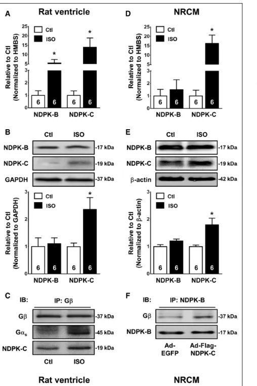
Using human tissue samples from a previous study,14 we found a significantly increased membrane protein con- tent of NDPK-A, NDPK-B, and NDPK-C isoforms in failing versus nonfailing hearts (Figure IV in the online-only Data Supplement), pointing to a role of NDPKs in HF patho- physiology. mRNA levels of NDPK-C, but not NDPK-A and NDPK-B, were significantly increased in HF hearts (Figure IV in the online-only Data Supplement), suggesting a se- lective transcriptional upregulation of NDPK-C in human HF. Patients with HF show a hyperactive sympathetic nervous system with elevated plasma catecholamine levels and subsequent chronic activation of βARs. There- fore, we asked whether chronic βAR stimulation contrib- utes to the NDPK-C upregulation in HF. Compared with saline-infused controls, chronic in vivo βAR stimulation with isoprenaline in rats induced a significant 5- and 14- fold increase in NDPK-B and NDPK-C mRNA content, re- spectively (Figure 2A) but enhanced the protein levels of NDPK-C only (Figure 2B). Thus, the increased expression of NDPK-C in patients with HF could directly result from the chronic activation of βARs.
As shown in Figure 2C, NDPK-C and Gαs could be detected in Gβ precipitates from rat ventricular tissue lysates, indicating that NDPK-C interacts with the hetero- trimeric G protein. Furthermore, the amount of NDPK- C and Gαs coimmunoprecipitating with Gβ apparently increased after long-term isoprenaline treatment, sug-
Figure 1 Continued. (middle). Recombinant NDPK-B was incubated with lysates of control NRCMs (left) or NRCMs overex- pressing flag-tagged NDPK-C. NDPK-C was detected with an antibody against NDPK-C (right). B, Representative far Western blot and quantification of the NDPK-B/Gtαβγ interaction in NRCM lysates infected with Ad-NDPK-B (multiplicity of infection [MOI], 100) and varying combinations of an adenovirus encoding enhanced green fluorescent protein (Ad-EGFP) and Ad-Flag–NDPK-C (total combined MOI, 100). *P<0.05 vs 100-MOI Ad-EGFP (0-MOI Ad-Flag–NDPK-C). C, ATP hydrolysis by Gtαβγ (1 µmol/L) and Gtαβγ plus NDPK-B (1 µmol/L), plus NDPK-C (0.25 µmol/L), or plus NDPK-B and NDPK-C determined by the amount of [32P]
Pi released in the absence or presence of 500 µmol/L uridine 5'-diphosphate (UDP). *P<0.05 vs Gtαβγ. #P<0.05 vs without UDP. §P<0.05 vs Gtαβγ+NDPK-C. D, Far Western blot analysis of purified NDPK-B and NDPK-C incubated with cell lysates from mouse embryonic fibroblasts (MEFs). G-protein subunits were subsequently detected with antibodies against Gβ1, Gβ2, Gαq, Gαs, and Gαi2. E, Representative Western blots and quantification of Gβ1, Gαs, Flag–NDPK-C, and β-actin in membrane fractions of NDPK-A/NDPK-B double-knockout MEFs infected with Ad-EGFP or Ad-Flag–NDPK-C. *P<0.05 vs Ad-EGFP. F. Far Western blot analysis of NDPK-C incubated with mixtures of lysates of NRCMs overexpressing Gβ1γ2, Gαs, or Gαi2 to obtain specific ratios of both G proteins (indicated below the corresponding bars). The interaction between NDPK-C and Gαs or Gαi2 was subsequently quantified (bars). *P<0.05 vs incubation with lysates from NRCMs overexpressing only Gαs plus Gβ1γ2 (left) or Gαi2 plus Gβ1γ2 (right, second bar in each chart).
by guest on February 28, 2017http://circ.ahajournals.org/Downloaded from
gesting that isoprenaline not only increases NDPK-C ex- pression but also may enhance the interaction between NDPK-C and G proteins. To verify that these alterations occur in cardiomyocytes, NRCMs were incubated with isoprenaline or solvent for 7 days in vitro. Consistent with the data obtained in human ventricular tissue samples, both mRNA content and protein expression of NDPK-C were increased by in vitro βAR stimulation, whereas the expression of NDPK-B remained unchanged (Figure 2D and 2E). The interaction between NDPK-B and Gβγ was confirmed in NRCMs and was enhanced by overexpres- sion of NDPK-C (Figure 2F), suggesting that NDPK-C is
critical for the interaction between NDPK-B and Gβγ in cardiomyocytes.
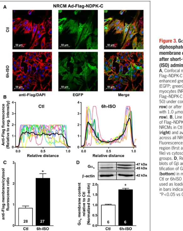
Subcellular Localization of NDPK-C After βAR Stimulation
NDPK-C was detected predominantly in the cytosol of unstimulated NRCMs. In vitro stimulation of NRCMs with isoprenaline for 6 hours significantly increased the pro- tein levels of NDPK-C at the plasma membrane, with a membrane/cytosol fluorescence ratio of 2.26±0.22 (n=27) versus 0.83±0.10 (n=28) under control condi-
Figure 2. Nucleoside diphosphate kinase (NDPK)-C expression is increased after long-term iso- prenaline (ISO) administration.
A and B, NDPK-B and NDPK-C in con- trol rats or rats after 4 days of ISO administration (2.4 mg·kg−1·d−1). A, mRNA content determined by quan- titative polymerase chain reaction in ventricular samples. Hydroxymethyl- bilane synthase (HMBS) was used as housekeeping gene for normalization.
B, Representative Western blots (top) and quantification of protein expres- sion (bottom) in ventricular lysates.
GAPDH was used as loading control.
C, Immunoprecipitation (IP) of Gβ in ventricular lysates of control rats or rats after 4 days of ISO administra- tion. Coimmunoprecipitated proteins were detected with antibodies against Gαs and NDPK-C. IB indicates im- munoblot D and E, Similar to A and B for control neonatal rat cardio- myocytes (NRCMs) or NRCMs after 7 days of incubation with ISO (1.0 μmol/L). Data are shown relative to controls (Ctl). Numbers in bars indi- cate number of samples. *P<0.05 vs corresponding control. F, Immunopre- cipitation of NDPK-B in lysates from NRCMs infected with an adenovirus encoding enhanced green fluorescent protein (Ad-EGFP) or Ad-Flag–NDPK-C, for 48 hours. Coimmunoprecipitated proteins were detected with antibod- ies against NDPK-B and Gβ.
by guest on February 28, 2017http://circ.ahajournals.org/Downloaded from
ORIGINAL RESEARCHARTICLE
tions (P<0.001; Figure 3A–3C), pointing to translocation of NDPK-C to the plasma membrane by isoprenaline in addition to the transcriptional upregulation of NDPK-C observed after 7 days of isoprenaline treatment. The interaction between NDPK-C and Gβγ determined by coimmunoprecipitation (Figure V in the online-only Data Supplement), as well as the membrane content of Gαs (Figure 3D), were also increased. Taken together, these data support the formation of NDPK-C, NDPK-B, and G- protein complexes, resulting in their enrichment at the plasma membrane after isoprenaline stimulation, and point to a potential role of NDPK-C in the regulation of cellular cAMP synthesis.
NDPK-C Modulates cAMP and Cardiomyocyte Contractility
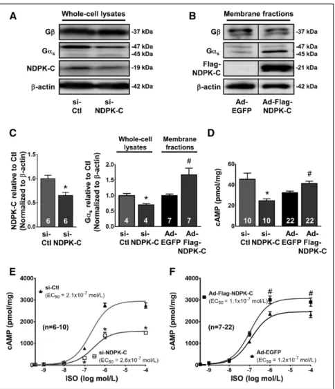
To study the functional consequences of NDPK-C regula- tion on cellular cAMP levels, NDPK-C protein levels were reduced by siRNA-mediated knockdown (si–NDPK-C; Fig- ure 4A) or increased by adenovirus-mediated overexpres- sion (Ad-Flag–NDPK-C; Figure 4B). NDPK-C expression was reduced by 34.8±6.4% with si–NDPK-C compared with si-control (Figure 4C), but the use of Ad-Flag–NDPK- C and corresponding anti-Flag antibody precluded quan- tification of NDPK-C overexpression levels. Modification of NDPK-C protein levels produced parallel changes in
Figure 3. Gαs and nucleoside diphosphate kinase (NDPK)-C membrane content are enhanced after short-term isoprenaline (ISO) administration.
A, Confocal microscopy showing Flag–NDPK-C (red), DAPI (blue), and enhanced green fluorescent protein (EGFP; green) in neonatal rat cardio- myocytes (NRCMs) overexpressing Flag–NDPK-C (multiplicity of infection, 50) under control conditions (Ctl; top row) or after 6 hours of stimulation with 1.0 μmol/L ISO (6h-ISO; bottom row). B, Line profiles (colored lines) of Flag–NDPK-C in 8 representative NRCMs in Ctl (left) or 6h-ISO groups (right) and average line profiles across all NRCMs (black lines). C, Fluorescence intensity in membrane region (first and last 15% of line pro- file) vs cytosol in the Ctl and 6h-ISO groups. D, Representative Western blots of Gβ and Gαs (top) and quan- tification of Gαs-protein expression (bottom) in membrane fractions from Ctl or 6h-ISO NRCMs. β-Actin was used as loading control. Numbers in bars indicate number of samples.
*P<0.05 vs Ctl.
by guest on February 28, 2017http://circ.ahajournals.org/Downloaded from
Gαs, with significantly reduced Gαs levels in lysates of si–NDPK-C NRCMs, and increased Gαs membrane con- tent in Ad-Flag–NDPK-C NRCMs (Figure 4C). Knockdown of NDPK-C expression also significantly reduced cAMP levels, whereas NDPK-C overexpression enhanced cAMP content (Figure 4D). Isoprenaline induced a dose-depen- dent increase in cAMP content with a >50-fold increase in cAMP levels compared with control conditions after maximal stimulation (Figure 4E). The increase in isopren- aline-induced cAMP content was significantly reduced in si–NDPK-C NRCMs (Figure 4E) but further enhanced by overexpression of NDPK-C (Figure 4F). These findings es- tablish a causal relationship between NDPK-C content at the plasma membrane and cellular cAMP levels in NRCMs.
We next determined whether NDPK-C also regulates G proteins and cAMP in ARCMs. NDPK-C levels at the plasma membrane were significantly increased after 6 hours of isoprenaline stimulation in ARCMs (Figure 5A and 5B). To assess the effects of NDPK-C in living ARCMs, we compared cAMP formation and fractional shortening8,20 in ARCMs overexpressing NDPK-C with control ARCMs overexpressing EGFP or LacZ. Isoprenaline-induced cAMP levels were quantified in isolated ARCMs via fluorescent
resonance energy transfer with an Epac-derived cAMP sensor.25,26 Stimulation of control ARCMs with isoprena- line produced 55±3% of the maximal cAMP response achieved by direct stimulation of adenylyl cyclases with forskolin in the presence of the phosphodiesterase inhibi- tor 3-isobutyl-1-methylxanthine (Figure 5C). In NDPK-C–
overexpressing ARCMs, the isoprenaline-induced cAMP response was significantly increased to 70±3% of the maximal cAMP response (Figure 5C).The isoprenaline concentrations producing half-maximal cell shortening were significantly higher in the control (4.5±0.9 nmol/L) than in NDPK-C–overexpressing ARCMs (0.8±0.1 nmol/L;
Figure 5D), suggesting that the NDPK-C–induced increase in cAMP affects cardiomyocyte contraction by sensitizing single-cell shortening to βAR stimulation.
Knockdown of NDPK-C Modulates Contractility in Zebrafish
To analyze whether NDPK-C modulates contractility in vivo in a vertebrate organism, we performed morpholino- mediated knockdown of NDPK-C in zebrafish embryos.
The injection of 300 µmol/L morpholino–NDPK-C caused Figure 4. Nucleoside diphosphate kinase (NDPK-C) modulates cAMP levels.
A and B, Representative Western blots of Gβ, Gαs, and NDPK-C or Flag–NDPK-C in lysates from neonatal rat cardiomyocytes (NRCMs) transfected with siRNA (si)-Ctl or si–NDPK-C (96 hours) and in membrane fractions from NRCMs infected with an adenovirus encoding enhanced green fluorescent protein (Ad-EGFP) or Ad-Flag–
NDPK-C (multiplicity of infection, 500 for 48 hours). β-Actin levels served as load- ing control. C, Quantification of NDPK-C protein levels normalized to β-actin in si-Ctl vs si–NDPK-C (left) or Gαs-protein levels from A and B normalized to β-actin relative to corresponding controls (right).
D, cAMP levels in NRCMs transfected with si-Ctl or si–NDPK-C and NRCMs infected with Ad-EGFP or Ad-Flag–NDPK-C NRCMs in the presence of 1.0 mmol/L 3-isobutyl-1-methylxanthine (IBMX) and 1.0 μmol/L propranolol. E and F, Isoprenaline (ISO)-induced cAMP levels in NRCMs with si-RNA–mediated reduction in NDPK-C (left), or in NRCMs overex- pressing NDPK-C (right) in the presence of 1.0 mmol/L IBMX and the indicated ISO concentration compared with corresponding controls. Numbers in bars indicate number of samples. *P<0.05 vs si-Ctl. #P<0.05 vs Ad-EGFP.
by guest on February 28, 2017http://circ.ahajournals.org/Downloaded from
ORIGINAL RESEARCHARTICLE
a loss of NDPK-C protein levels, resulting in phenotypic alterations, including reduced cardiac pump function and pericardial edema, compared with zebrafish inject- ed with a control morpholino at 72 hours after fertiliza- tion (Figure 6A and Supplementary Videos I and II in the online-only Data Supplement). NDPK-C morphant hearts had a normal morphology of endocardial and myocardial cell layers and a regular expression of atrial and ven- tricular myosin heavy chains (Figure VIA–VID in the online-
only Data Supplement). In addition, electron microscopy revealed no differences in the ultrastructure of sarco- meres, z line, and thick and thin filaments (Figure VIE and VIF in the online-only Data Supplement). Therefore, the structural development of the NDPK-C morphant hearts appears to be unhampered. Functionally, deple- tion of NDPK-C resulted in decreased basal cAMP levels (Figure 6B), similar to that reported previously for zebraf- ish with knockdown of NDPK-B.19 Ventricular fractional Figure 5. Consequences of nucleoside diphosphate kinase (NDPK)-C modulation in adult rat cardiomyocytes (ARCMs).
A, Confocal microscopy showing Flag–NDPK-C (red), DAPI (blue), and enhanced green fluorescent protein (EGFP; green) in ARCMs overexpressing Flag–NDPK-C (multiplicity of infection [MOI], 50) under control conditions (Ctl; top row) or after 6 hours of stimula- tion with ISO (6h-ISO; 1.0 μmol/L; bottom row). B, Line profiles of Flag–NDPK-C in 4 representative ARCMs (colored lines) in Ctl (top left) or 6h-ISO groups (top right) and average line profiles across all ARCMs (black lines). Bar chart shows fluorescent intensity in membrane region (first and last 15% of line profile) vs cytosol in the Ctl and 6h-ISO groups. C, Representative fluores- cent resonance energy transfer (FRET; cyan fluorescent protein/yellow fluorescent protein [CFP/YFP]) ratio traces recorded from control and Ad-Flag–NDPK-C–overexpressing ARCMs (MOI, 300) expressing the cAMP sensor Epac2-camps after stimulation with ISO (100 nmol/L) and subsequent maximal stimulation of cAMP levels by forskolin (10 μmol/L) and 3-isobutyl-1-methylxanthine (IBMX; 100 μmol/L). Decreases in the FRET ratio represent increases in intracellular cAMP. The magnitude of the cAMP response to ISO as a percent of maximal response induced by forskolin plus IBMX in control and NDPK-C–overexpressing ARCMs is shown on the right. n=16 to 17 cells obtained from 2 independent infections. D, Concentration-dependent increase in relative fractional shortening of isolated ARCMs infected with Ad-EGFP or Ad-Flag–NDPK-C (MOI, 300) after short-term stimulation with the indicated concentrations of ISO. n=29 to 42 cells per condition from 3 independent infections. Inset shows the EC50 of ISO-induced aug- mentation of cellular fractional shortening. Numbers in bars indicate number of samples. *P<0.05 vs corresponding control.
by guest on February 28, 2017http://circ.ahajournals.org/Downloaded from
shortening was progressively reduced from 26.0±1.9%
at 48 hours after fertilization to 7.4±1.7% at 72 hours after fertilization in NDPK-C morphants, whereas frac- tional shortening remained stable in morpholino-control zebrafish (Figure 6C). To evaluate synergism between NDPK-C and NDPK-B in vivo, a partial knockdown of NDPK-B and/or NDPK-C was performed with the use of low morpholino concentrations, and ventricular fraction- al shortening was assessed. Morpholino–NDPK-B (125
µmol/L) or morpholino–NDPK-C (150 µmol/L) alone did not significantly impair ventricular function, whereas their combination severely reduced fractional shortening (Figure 6D). To analyze whether an increased NDPK-B expression can substitute for the loss of NDPK-C, we overexpressed NDPK-B, an approach that previously rescued the phenotype caused by morpholino-mediated knockdown of NDPK-B.19 Compared with controls, NDPK- C morphants frequently showed phenotypic abnormali- Figure 6. Nucleoside diphosphate kinase (NDPK)-C knockdown in zebrafish reduces ventricular contractility.
A, Lateral view of representative zebrafish larvae injected with 300 µmol/L control or NDPK-C morpholinos (MO-Ctl and MO–NDPK-C, respectively) 72 hours post fertilization (hpf; left). NDPK-C–depleted embryos develop a pericardial edema (arrow) as a result of cardiac dysfunction. Right, Representative Western blot of NDPK-C and β-actin as loading control. B, cAMP levels in zebrafish injected with control or NDPK-C morpholinos 72 hpf in the presence of 3-isobutyl-1-methylxanthine (IBMX; 1.0 mmol/L). Shown is the average of 2 independent cAMP determinations in fish lysates. Numbers in bars indicate the total number of fish used for the determination of this value. C, In vivo fractional shortening of zebrafish ventricle in zebrafish injected with control or NDPK-C morpholinos at 48, 60, and 72 hpf. *P<0.05 vs control morpholinos (repeated- measures 2-way ANOVA). D, In vivo fractional shortening of zebrafish ventricle in zebrafish injected with control, NDPK-B (125 µmol/L), NDPK-C (150 µmol/L), or both NDPK-B (125 µmol/L) and NDPK-C (150 µmol/L) morpholinos at 72 hpf.
*P<0.05 vs control morpholinos. E, Percentage of zebrafish developing morphological abnormalities under control condi- tions and after knockdown of NDPK-C, knockdown of NDPK-C with NDPK-B overexpression, or NDPK-B overexpression alone. *P<0.05 vs control.
by guest on February 28, 2017http://circ.ahajournals.org/Downloaded from
ORIGINAL RESEARCHARTICLE ties (eg, pericardial edema or impaired cardiac function;
Figure VII in the online-only Data Supplement). NDPK-B overexpression in NDPK-C knockdown embryos did not reduce the incidence of these phenotypic abnormalities (Figure 6E). Thus, in accordance with the in vitro data, NDPK-C is apparently indispensable for the complex for- mation between NDPKs and G proteins, which is involved in cAMP formation and the subsequent alterations in ven- tricular contractility in vivo.
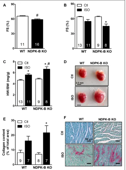
NDPK-B–Deficient Mice Are Prone to Cardiac Dysfunction
To verify the importance of the complex formation of NDPK-C, NDPK-B, and G proteins described above for cardiac function in mammals, we took advantage of NDPK-B knockout mice.27 These mice are viable, without obvious phenotype, and have a normal life span. The
loss of NDPK-B does not alter the expression of NDPK-C in the heart of these mice (Figure VIII in the online-only Data Supplement). Nevertheless, at the age of 5 months, they start to develop cardiac dysfunction, as revealed by decreased fractional shortening (Figure 7A). To test whether the complex formation of NDPK-B with NDPK-C and G proteins is functionally relevant in the response to chronic βAR stimulation in the mammalian heart, we sub- jected these mice to the long-term isoprenaline stimula- tion protocol we used in rats (Figure 2A) and studied car- diac contractility and remodeling. As shown in Figure 7B through 7D, long-term isoprenaline treatment decreased fractional shortening more strongly in NDPK-B knock- out mice than in wild-type controls. In addition, NDPK-B knockout mice were more susceptible to isoprenaline-in- duced cardiac hypertrophy and fibrosis. Taken together, these data indicate that the complex formation of NDPKs with Gαs proteins, which apparently requires the pres-
Figure 7. Ablation of nucleoside diphosphate kinase (NDPK)-B reduces cardiac function in aging mice and aggravates catecholamine-induced cardiac remodeling in young mice.
A, Fractional shortening (FS) of 5-month-old male wild-type (WT) and NDPK-B knockout (NDPK-B KO) mice calculated from echocardiographic analysis in conscious animals. B and C, Two-month-old male WT and NDPK-B KO mice were either subjected to control conditions or treated with isoprenaline (ISO; 30 mg·kg−1·d−1) for 7 days via osmotic minipumps.
B, FS in conscious animals at day 7 of the treatment. C, Quantification of hypertrophic growth by ratios of heart weight (HW) to body weight (BW). D, Representative images of explanted hearts. E, Quantification of fibrotic remodeling by Sirius Red/Fast Green Collagen staining of paraffin-embed- ded heart sections normalized to total tissue area. F, Representative image of collagen-stained (magenta) heart sections. *P<0.05 vs respective control. #P<0.05 versus WT, 2-way ANOVA with Bonferroni correction.
by guest on February 28, 2017http://circ.ahajournals.org/Downloaded from
ence of both NDPK-B and NDPK-C, is beneficial during catecholamine-induced cardiac stress and protects the heart from an early onset of remodeling.
NDPK-C Causes a Switch From Gα
s- to Gα
i- Predominant Signaling in Human HF
The data presented so far identify NDPK-C, by mediating complex formation and membrane targeting of NDPK- B and Gαs, as a regulator with stimulatory effects on cAMP levels and contractility. However, the increased expression of NDPK-C in human HF appears at odds with the reduced contractility observed in patients with HF.
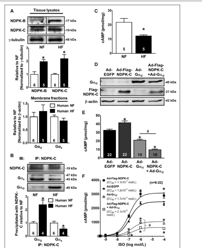
On the other hand, our far Western blots indicate that NDPK-C, but not NDPK-B, interacts with both Gαs and Gαi, and the ratio of these proteins determines which G protein is preferentially bound (Figure 1D and 1F). There- fore, we hypothesized that NDPK-C may switch from Gαs- to Gαi-predominant signaling in human HF, thereby contributing to the lower cAMP levels and reduced car- diomyocyte contractility. To address this hypothesis, we analyzed NDPK-B and NDPK-C expression in ventricular tissue samples from a separate patient cohort. In accor- dance with the data obtained in the previous collective,14 NDPK-B and NDPK-C protein levels were significantly in- creased in tissue lysates of patients with HF compared with healthy donor hearts (Figure 8A). The Gαi2- and Gαs- protein expression in membrane fractions of patients with HF appeared to be slightly increased (20%) and decreased (−15%), respectively, without reaching statis- tical significance (Figure 8A and Figure IXA in the online- only Data Supplement).
Next, we assessed the interactions among NDPK-C, Gαi2, and Gαs in ventricular tissue lysates of control samples and those from patients with HF using coim- munoprecipitation. The NDPK-C content of the Gαi2 pre- cipitates was increased 2.8±0.8-fold in patients with HF (P=0.11), whereas the NDPK-C content in the Gαs pre- cipitates of HF samples was decreased to 0.54±0.14- fold of nonfailing controls (P=0.07; Figure IXB in the online-only Data Supplement). Conversely, the amount of Gαi2 that coprecipitated with NDPK-C was increased 2.39±0.35-fold (P<0.05), whereas the amount of Gαs that coprecipitated with NDPK-C was decreased to 0.55±0.08 of nonfailing controls (P<0.05) in human HF, pointing to a switch in the interaction of NDPK-C from Gαs to Gαi2 (Figure 8B). cAMP levels were signifi- cantly reduced by 42% in patients with HF (Figure 8C), consistent with previous results.28 To determine wheth- er the switch in NDPK/G-protein signaling affects cAMP levels, NRCMs overexpressing EGFP, NDPK-C, Gαi2,, or NDPK-C plus Gαi2 (Figure 8D) were incubated with increasing concentrations of isoprenaline. Overexpres- sion of NDPK-C increased the basal and isoprenaline-in- duced cAMP levels in NRCMs. In contrast, overexpres- sion of Gαi2 reduced the basal cAMP content and the
isoprenaline-induced increase in cAMP levels compared with EGFP-expressing controls (Figure 8E and 8F). It is notable that overexpression of both NDPK-C and Gαi2 further reduced basal cAMP levels and isoprenaline- induced cAMP production compared with Gαi2 overex- pression alone (Figure 8E and 8F). These data indicate that the HF-related increase in NDPK-C and the stronger interaction between Gαi2 and NDPK-C (Figure 8A and 8B) amplify the inhibitory effects of Gαi2 proteins on cardiomyocyte cAMP levels and positions NDPK-C as a novel, potentially critical regulator of Gαi-protein signal- ing and cellular cAMP in human HF.
DISCUSSION
In the present study, we identified NDPK-C as an es- sential and indispensable component of the interaction between NDPKs and G proteins. NDPK-C anchors these complexes at the plasma membrane, thereby being an important regulator of cAMP levels and cardiomyocyte contractility. We also discovered that a switch from pre- dominantly Gαs stimulation to Gαi signaling by NDPK-C in human HF might cause the lower cAMP levels in pa- tients with HF. Together, our findings identify NDPK-C as a novel critical regulator of βAR/cAMP signaling that may contribute to contractile dysfunction in HF.
G-Protein Signaling in HF
HF induces complex remodeling, with changes in G-pro- tein signaling as a hallmark of this remodeling process.
Long-term sympathetic stimulation results in desensiti- zation of βARs, including reduced expression of β1ARs and upregulation of inhibitory G-protein–coupled recep- tor kinases.29,30 In addition, the expression and activity of inhibitory Gαi proteins are increased ≈30% in end-stage HF,31,32 and a shift from a prevalence of Gαs-mediated adenylyl cyclase stimulation to Gαi-mediated inhibition of adenylyl cyclases through β2-ARs in HF has been re- ported.33 The increase in Gαi-protein activity may result partly from transcriptional upregulation of Gαi2 and likely contributes to the impaired βAR responsiveness associ- ated with HF.34 A modest upregulation of Gαi2 was also identified in the rat model with long-term isoprenaline administration in our study,35 which suggests that these effects are an adaptive response to chronic sympathetic stimulation. However, in a chronic setting, these chang- es result in reduced ventricular cardiomyocyte cAMP levels that decrease the activation of protein kinase A in HF. Together with the augmentation of global protein phosphatase activity in HF,36 the restricted cAMP signal- ing results in reduced phosphorylation of key cardiac Ca2+-handling proteins with subsequent decreases in Ca2+-transient amplitude and cellular shortening,37 which are well-established contributors to the reduced ventricu- lar contractility in HF.38
by guest on February 28, 2017http://circ.ahajournals.org/Downloaded from
ORIGINAL RESEARCHARTICLE
Figure 8. Nucleoside diphosphate kinase (NDPK)-C interaction with G proteins shifts toward Gαi2 in heart failure (HF).
A, Representative Western blots (top) and quantification of protein expression (middle) of NDPK-B and NDPK-C in ventricular whole- tissue lysates obtained from explanted hearts from patients with end-stage HF or nonfailing (NF) control donor hearts. γ-Tubulin served as loading control. Bottom, Quantification of Gαi2 and Gαs in ventricular membrane fractions obtained from HF or NF control donor hearts. β-Actin served as loading control. Original blots shown in Figure IX in the online-only Data Supplement. B, Immunopre- cipitation (IP) of NDPK-C in ventricular lysates obtained from patients with HF or NF donor hearts and quantification of the amount of Gαs and Gαi2 immunoprecipitating with NDPK-C relative to NF controls. IB indicates immunoblot. C, cAMP levels in ventricular samples of patients with end-stage HF or NF control donor hearts in the presence of 3-isobutyl-1-methylxanthine (IBMX; 1.0 mmol/L). Numbers in bars indicate number of hearts. From 1 HF sample, not enough lysate could be obtained for immunoprecipitation. *P<0.05 vs NF. D through F, Representative Western blots of Gαi2, Flag–NDPK-C, and β-actin as loading control (D), basal cAMP levels (E), and ISO-induced cAMP levels with EC50 values (F) in neonatal rat cardiomyocytes (NRCMs) infected with an adenovirus encoding enhanced green fluorescent protein (Ad-EGFP, multiplicity of infection [MOI], 500), Ad-Flag–NDPK-C (MOI, 500), Ad-Gαi2 (MOI, 50), or Ad-Flag–
NDPK-C (MOI, 500) plus Ad-Gαi2 (MOI, 50). *P<0.05 vs Ad-EGFP controls. #P<0.05 for the effect of Ad-Flag–NDPK-C plus Ad-Gαi2.
by guest on February 28, 2017http://circ.ahajournals.org/Downloaded from
Previous work has shown that the plasma membrane content of NDPK-A, NDPK-B, and NDPK-C is increased in patients with end-stage HF4 and that long-term ac- tivation of βARs increases anchoring of NDPKs to the plasma membrane.39 Here, we discovered that this in- crease is due at least in part to a transcriptional upregu- lation and that NDPK-C is required and indispensable for membrane localization of NDPKs. The human nm23-H3 gene encoding NDPK-C contains active AP-2 sites and a putative CREBP-1 binding site in its promoter region,40 which are involved in cAMP-dependent regulation of gene transcription41,42 and could contribute to the en- hancement of NDPK-C protein levels after isoprenaline administration or in end-stage human HF with chronic sympathetic stimulation. Further work is needed to di- rectly test these hypotheses.
Our data on the interaction of NDPK-C with NDPK- B and heterotrimeric G proteins also strongly suggest that NDPK-C is an essential prerequisite for the complex formation of NDPK-C/NDPK-B hetero-oligomers with G proteins. In addition, the lipophilic nature of NDPK-C may allow an easier association with the plasma mem- brane43,44 and is therefore likely the limiting factor for the plasma membrane localization of NDPK isoforms.
Novelty and Potential Clinical Implications
We have identified a novel mechanism by which the plasma membrane content of heterotrimeric G pro- teins is regulated in cardiomyocytes through NDPK-C.
In particular, we show for the first time that NDPK-C is required and indispensable for the interaction between NDPKs and both stimulatory Gαs and inhibitory Gαi pro- teins, find that NDPK-C–mediated targeting of NDPKs to the plasma membrane is increased in response to βAR stimulation and enhances intracellular cAMP levels, dem- onstrate that cardiomyocyte contractility in vitro and in vivo is strictly modulated by NDPK-C levels, and provide novel mechanistic insights into cAMP signaling in HF, identifying previously unrecognized molecular targets for the development of new and potentially more effective HF therapy options. Our data put previous studies about regulation of cAMP signaling through NDPKs in a novel and conclusive perspective: NDPK-B can exert its effects on cAMP signaling and contractility only in the presence of NDPK-C, which is responsible for both the membrane targeting and interaction of NDPKs with G proteins. Fur- thermore, our findings provide a potential mechanistic explanation for why even a small increase in Gαi2-protein levels and increased membrane-associated NDPKs in HF may inhibit cAMP synthesis14 and could cause a pro- found negative inotropic effect. It is interesting to note that modest changes in the Gαs/Gαi ratio (Figure 1F), which are in the same range as those reported in HF,31,32 may determine which of the G-protein subtypes is bound to NDPK-C. Thus, in the absence of Gαi upregulation, the
increased membrane content of NDPK-C in human HF is a mechanism that could potentially counteract a fading βAR response in the early stages of HF by increasing the amount of Gαs proteins at the plasma membrane. How- ever, by switching to Gαi2 activation, NDPK-C could play a role in the progression of the disease and the reduced cAMP levels observed in end-stage human HF. Together, these data provide new insights into the complex altera- tions in G-protein signaling that are a hallmark of the vi- cious cycle of chronic sympathetic stimulation in HF and, for the first time, establish increased NDPK-C function as a novel possible molecular facilitator of detrimental Gαi- protein signaling in patients with HF.
Treatment with β-blockers has been the standard therapy for patients with HF. Despite their positive ef- fects on morbidity and mortality, β-blockers are not without limitations. Accordingly, other ways to regulate G-protein signaling and cardiac proteins involved in car- diac contractility are currently being investigated in the treatment of HF.45,46 A better understanding of the mo- lecular processes involved in dysregulation of G-protein signaling in HF is expected to foster the development of improved HF therapies. On the basis of our studies, we identify NDPK-C as a novel potential therapeutic target involved in the regulation of aberrant G-protein signaling and cardiac contractility in HF. Even short-term stimula- tion of cardiomyocytes with isoprenaline increases the plasma membrane content of NDPK-C and strengthens the interaction between NDPK-C and G proteins, thereby increasing cAMP synthesis. The increased expression or plasma membrane content of NDPK-C in human HF might initially allow the heart to compensate a beginning loss in βAR-induced contractile response in the onset of HF.
However, because of the promiscuous nature of NDPK- C, which is able to interact with both Gαs and Gαi, this compensatory mechanism might become detrimental for the heart when the interaction with Gi2 increases over time. Along these lines, we show that if Gαi2 levels are enhanced, an increase in NDPK-C abundance reduces cAMP levels (Figure 8E and 8F), which may contribute to the detrimental cardiomyocyte phenotype of human HF. Thus, inhibition of NDPK-C might represent a novel adjuvant therapy for patients with end-stage HF.
Limitations
The causes of HF are diverse. We used human ventricu- lar tissue samples from a selected group of patients with end-stage HF. Our finding of increased NDPK-C expres- sion may not hold true for all types and stages of HF. In addition, experimental conditions in vitro may not fully re- flect the dynamic regulation of cAMP and contractility in vivo. For example, cAMP levels may change rapidly after tissue excision, and in situ cAMP levels were found to be similar between nonfailing patients and patients with HF, possibly as a result of increased plasma norepinephrine
by guest on February 28, 2017http://circ.ahajournals.org/Downloaded from
ORIGINAL RESEARCHARTICLE levels counteracting intrinsic downregulation of cAMP in
cardiomyocytes.47
Isoprenaline increased the plasma membrane content of the primarily cytosolic NDPK-C within several hours af- ter stimulation. The mechanisms involved could include translocation, increased protein stability at the plasma membrane, or reduced degradation and should be ad- dressed in future studies.
Here, we characterized the role of NDPK-C in mod- ulating cAMP levels. However, as histidine kinases, NDPKs could also directly phosphorylate various tar- gets within the cardiomyocyte, thereby participating in the control of various cardiac functions.48 For example, NDPK-B activates transient-receptor potential vallinoid type-5 channels through phosphorylation of histidine 711, thereby controlling Ca2+ reabsorption in the kid- ney.49 Similarly, NDPK-B activates small-conductance Ca2+-activated K+ channels in vascular smooth muscle cells, controlling neointima formation in carotid arter- ies.50 Subsequent work should test these possibilities in the heart.
Conclusions
NDPK-C is indispensable for the interaction between NDPKs and G proteins and the anchoring of these com- plexes at the plasma membrane, thereby dynamically regulating cAMP levels and cardiomyocyte contractility.
The switch from predominantly Gαs stimulation to Gαi activation by NDPK-C in human HF might cause lower cAMP levels in patients with HF, potentially contributing to the progression of HF. Together, our findings position NDPK-C as a novel critical determinant of βAR/cAMP sig- naling that could contribute to impaired cardiac function and remodeling in human HF.
ACKNOWLEDGMENTS
The authors gratefully acknowledge the expert technical as- sistance from Doris Baltus, Kristina Stephan-Schnatz, Heike Rauscher, Felicia Radtke, Geronimo Heilmann, Annette Kötting- Dorsch, Katrin Kupser, Barbara Langer, Claudia Liebetrau, Al- exandra Müller, Ramona Nagel, and Lisa Walter. The authors also thank Ioan Lascu, University Bordeaux Segalen, for kindly providing purified NDPKs and the custom-made NDPK-C anti- body.
SOURCES OF FUNDING
The authors’ work was supported by grants from the Deutsche Forschungsgemeinschaft (Wi1373/9-3 to Dr Wieland and EL 270/7-1 to Dr El-Armouche), the Fondation Leducq (European North-American Atrial Fibrillation Research Alliance to Dr Do- brev), the European Network for Translational Research in Atrial Fibrillation (No. 261057 to Dr Dobrev), and the German Federal Ministry of Education and Research through the DZHK
(German Center for Cardiovascular Research to Drs Dobrev, Katus, Backs, and Wieland).
DISCLOSURES
None.
AFFILIATIONS
From Institute of Experimental and Clinical Pharmacology and Toxicology, Mannheim Medical Faculty (I.H.A.-T., N.M.W., K.S., C.V., S.L., T.W.), and Department of Internal Medicine III (H.- J.H., N.M.W., M.M., B.M., H.-A.K., L.H.L., J.B.), Heidelberg University, Heidelberg-Mannheim, Germany; Institute of Phar- macology, West German Heart and Vascular Center, University Duisburg-Essen, Essen, Germany (I.H.A.-T., J.H., M.S., N.V., D.D.); Institute of Pharmacology and Toxicology, University Medical Center Göttingen, Germany (A.E.-A., C.M.W., S.L.);
Department of Pharmacology and Toxicology, Medical Faculty Carl Gustav Carus, Dresden University of Technology, Germany (A.E.-A.); Institute of Experimental Cardiovascular Research, University Medical Center Hamburg-Eppendorf, Germany (V.O.N.); Department of Internal Medicine II, University of Re- gensburg, Germany (S.N.); Department of Pharmacology and Pharmacotherapy, Faculty of Medicine, University of Szeged, Hungary (I.B., A.V.); Division of Nephrology, New York Universi- ty Langone Medical Center, New York (E.Y.S.); and DZHK (Ger- man Center for Cardiovascular Research), Partner Site HD/MA, Heidelberg-Mannheim, Germany (B.M., H.A.K., C.V., J.B., T.W.).
The current affiliation for H.-J.H. is the Department of Cardi- ology and Angiology, University Hospital Schleswig-Holstein, Kiel, Germany.
FOOTNOTES
Received April 7, 2016; accepted November 23, 2016.
The online-only Data Supplement is available with this article at http://circ.ahajournals.org/lookup/suppl/doi:10.1161/CIR- CULATIONAHA.116.022852/-/DC1.
Circulation is available at http://circ.ahajournals.org.
REFERENCES
1. Go AS, Mozaffarian D, Roger VL, Benjamin EJ, Berry JD, Blaha MJ, Dai S, Ford ES, Fox CS, Franco S, Fullerton HJ, Gillespie C, Hailpern SM, Heit JA, Howard VJ, Huffman MD, Judd SE, Kis- sela BM, Kittner SJ, Lackland DT, Lichtman JH, Lisabeth LD, Mackey RH, Magid DJ, Marcus GM, Marelli A, Matchar DB, McGuire DK, Mohler ER 3rd, Moy CS, Mussolino ME, Neumar RW, Nichol G, Pandey DK, Paynter NP, Reeves MJ, Sorlie PD, Stein J, Towfighi A, Turan TN, Virani SS, Wong ND, Woo D, Turner MB; American Heart Association Statistics Committee and Stroke Statistics Subcommittee. Heart disease and stroke statistics–2014 update: a report from the American Heart As- sociation. Circulation. 2014;129:e28–e292. doi: 10.1161/01.
cir.0000441139.02102.80.
2. Movsesian MA. Cyclic AMP-mediated signal transduction in heart failure: molecular pathophysiology and therapeutic implications.
J Investig Med. 1997;45:432–440.
by guest on February 28, 2017http://circ.ahajournals.org/Downloaded from