Oxidative stress with altered element content and decreased ATP level of erythrocytes in hepatocellular carcinoma and colorectal liver metastases
La´szlo´ Va´li
a, Oszka´r Hahn
c, Pe´ter Kupcsulik
c, A´gnes Drahos
e, Enik o Sa´rva´ry +
d, Kla´ra Szentmiha´lyi
f, Zsolt Pallai
g, Timea Kurucz
g, Pe´ter Sı´pos
band
Anna Bla´zovics
aOur aim was to study the possible alterations of redox status (enzymatic and nonenzymatic parameters and metal elements) in erythrocytes of patients with hepatocellular carcinoma (HCC), colorectal liver metastases (CRLM) and benign liver neoplasms. The function of redox homeostasis is closely connected to the energy level of erythrocytes, therefore, the ATP level was also determined. Antioxidant parameters, enzyme activities of superoxide dismutase and glutathione peroxidase were estimated in the
erythrocytes of 11 patients with benign tumour, 23 patients with primary malignant and 37 metastatic liver tumour patients and 30 age-matched and sex-matched healthy controls. Element content with inductively coupled plasma optical emission spectrometer and ATP level by the chemiluminometric method were also determined from the samples. Free radical intensity was significantly increased, whereas erythrocyte glutathione peroxidase and
superoxide dismutase activities were significantly decreased in the HCC and CRLM groups versus benign groups and controls. Se, Mn and Zn levels were lowered in HCC and CRLM groups versus benign and control groups.
The content of Cu, Mg, Se and Zn changed significantly between HCC and CRLM groups. Similarly, ATP
concentration decreased in HCC and CRLM versus controls
and benign groups. The lowest levels of ATP and antioxidant enzyme activities were found in the case of CRLM patients. These results reveal an alteration in the ATP level of erythrocytes with concomitant changes in the antioxidant defence system in hepatic cancer patients.
Altered redox homeostasis (oxidative damage) may lead to decreased ATP level and consequently may play an important role in primary carcinogenesis and generation of metastases, as well. Eur J Gastroenterol Hepatol 20:393–398c 2008 Wolters Kluwer Health | Lippincott Williams & Wilkins.
European Journal of Gastroenterology & Hepatology2008,20:393–398
Keywords: ATP, liver neoplasms, metal elements, redox homeostasis
II Departments ofaMedicine,bSurgery,cI Department of Surgery,dDepartment of Transplantation and Surgery, Semmelweis University,eNational Research Institute for Radiobiology and Radiohygiene,fChemical Research Center, Hungarian Academy of Sciences andgDiachem Ltd, Budapest, Hungary
Correspondence to Dr Anna Bla´zovics, II Department of Medicine, Biochemical Research Group, Semmelweis University, H-1088 Budapest, Szentkira´lyi u. 46, Hungary
Tel: + 36705335012; e-mail: blaz@bel2.sote.hu
Received19 July 2007Accepted19 November 2007
Introduction
Hepatic cancer continues to be one of the most frequently diagnosed neoplasms and one of the leading causes of mortality related to cancer. In the pathogenesis of neoplastic and age-related degenerative diseases oxidative stress seems to play a key role [1]. A wide variety of reactive oxygen species (ROS) can cause membrane destruction, protein aggregation and DNA break. Endogenous defences against free radicals include antioxidant enzymes such as the thoroughly studied glutathione peroxidase (GSHPx) and superoxide dismu- tase (SOD). Lower levels of essential antioxidants in circulation were found to be associated with an increased risk of cancer [2–4].
The processes of metastatization are decreased by free radical formation [5]. Tumour cell interactions with microvasculature is a rate-limiting step in metastasis
development. Initial interactions trigger a sequence of activation pathways that involve cytokines, growth factors, bioactive lipids and ROS produced by the cancer cell or the endothelium. On the other hand, induction of endothelial free radicals can be cytotoxic to cancer cells [6].
The presence of activated oncogenes and/or inactivated tumour suppressor genes may result in constitutive activation of multiple transcription factors. This may be especially true in the early stages of tumour develop- ment. At advanced stages, however, uncontrolled tumour growth and the consequent development of a stress microenvironment, such as free radical overproduction, may further alter the activity of these transcription factors. Abnormal activation of and interplay between these factors lead to aberrant expression of multiple metastasis-related proteins and confer a tremendous survival and growth advantage to emerging metastatic
variants [7]. Cytokines of serum, such as IL-6 and TNF-a, proved to be related to oxidative stress in cancer patients [8]. Exposure to chronic stress increased lipid peroxidation level and free radical content of erythrocytes [9].
Therefore, we conducted investigations to evaluate the possible alteration of oxidant/antioxidant and metal element status in the circulation of patients with hepatocellular carcinoma (HCC) and colorectal liver metastases (CRLM) compared with patients with benign neoplasms and healthy volunteers. Metal elements (Cu, Mn, Se, Zn) play a key role in the function of antioxidant enzyme activity of SOD and GSHPx. Determination of these elements may explain biochemical changes owing to the presence of oxidative stress related to tumour.
ATP concentration of erythrocytes was also determined.
A correlation between ATP level, element concentration and redox homeostasis may provide relevant information on supportive or therapeutic perspectives. Our aim was to determine the difference between the prooxidant/anti- oxidant balance (antioxidant parameters, metal elements) of benign tumour and cancer, and to determine whether there is a relationship between redox homeostasis and ATP level.
On the basis of our earlier studies [10], erythrocytes may be considered as markers of oxidative stress caused by tumourous tissues. In addition, genetic and environmen- tal factors (which are hard to interpret in certain cases) in the background of the development of cancer are present.
We presume that the measurement of redox homeostasis and energy level of the body offers new perspectives for the understanding of the biochemical pathways induced in liver neoplasms.
Methods
Patients
Eleven patients with diagnosis of benign liver neoplasm (two hemangiomas, five, focal nodular hyperplasias, four hepatocellular adenomas) (average age: 48.73 ± 10.26 years; between 32 and 62 years), 23 patients with HCC diagnosis (average age: 50.17 ± 8.04 years; between 37 and 70 years) and 37 patients with CRLM diagnosis of the liver (average age: 53.08 ± 8.72 years; between 39 and 72 years) who had not undergone previous treatment for their liver tumour were examined in this study. Patients were recruited from the I. Department of Surgery and from the Department of Transplantation and Surgery, Semmelweis University, Budapest, Hungary. Thirty volunteers matching the age and sex of patients (average age: 54.42 ± 8.15 years; between 36 and 62 years) were selected as controls. Specific exclusion criteria for this study were the following: hepatitis virus infection, diabetes mellitus, heart or renal failure and oral antioxidant supplementation. Some of the patients were heavy drinkers, but none of them had consumed alcohol
48 h before blood collection. Informed consent was obtained from all participants in the study before blood collection.
The diagnosis of hepatic neoplasms was based on computer tomography and ultrasonographic-fine needle aspiration biopsy, and this diagnosis was verified by histopathological examination of their postoperative specimens. The study was approved by the TUKEB Committee of Semmelweis University, Budapest, Hungary (TUKEB No: 15/2004).
Laboratory methods
After an overnight fast, blood samples were drawn from the antecubital vein by venipuncture into tubes contain- ing sodium citrate. Plasma and erythrocyte samples were separated and stored at – 201C until assayed.
Free SH-group concentrations of plasma samples were determined by the Sedlak method on the basis of the Ellmann reaction [11].
The H-donating ability of plasma samples was deter- mined spectrophotometrically at 517 nm in the presence of a 1,1-diphenyl-2-picryl-hydrasyl (DPPH) radical by Blois’s method modified by Bla´zovicset al. [10]. For the characterization of the ability, inhibition was expressed as a percentage of DPPH degradation [12,13].
Oyaizu’s method was adapted for the determination of the reducing power of plasma samples. The change in absorbance was measured, which accompanied Fe3 +-Fe2 + transformation at 700 nm [14]. All spectrophotometric measurements were carried out with a Jasco V-550 instrument (Jasco Europe, Cremella, Italy).
A chemiluminescent assay adapted to a Berthold Lumat 9501 instrument was applied (Berthold Technologies, Bad Wildbad, Germany). The procedure was carried out by the method of Bla´zovicset al.[10]. The volume of each plasma and red blood cell (RBC) sample was 0.100 ml.
Chemiluminescent intensity of the samples was expressed in relative light units (basic chemical reaction means:
H2O2-microperoxidase-luminol reaction).
The level of total antioxidant status from the plasma, and the activity of GSHPx and SOD were determined from the RBC samples with Randox kits. The results were expressed in arbitrary units calculated from the absor- bance detected by spectrophotometry.
Element concentration of erythrocyte lysates was deter- mined in three parallel measurements with an inductively coupled plasma optical emission spectrometer. The type of instrument used was Atom Scan 25 (Thermo Jarrell Ash Co. Franklin, Massachusetts, USA). After digestion
of the RBC samples with a mixture of nitric acid and hydrogen peroxide and dilution with deionized water, the concentration of Ca, Cu, Fe, Mg, Mn, P, S and Zn elements was determined. Three times 3-s integration time, blank subtraction and background correction were applied during the measurements [15].
Se content of erythrocytes was determined with a hanging mercury drop electrode by a cathodic stripping voltametric method (instrument: Trace Lab 50). For element measure- ment, the digested samples were measured in 1 mol/l HCl as supporting electrolyte by preconcentration at – 350 mV.
Electrolysis time was 100 s, potential: from – 300 mV up to – 900 mV, step duration – 50 mV/s.
ATP concentration of erythrocytes was determined by a chemiluminescent technique adapted to a Leader 50 luminometer (Gen-Probe Inc., San Diego, California, USA) based on the luciferin–luciferase method. The method described by Rieger [16] was modified. Hemolyzed RBC samples (stored on – 201C) with hemoglobin content set to 1 g% were diluted (100) with bidistilled water and centrifuged (with 2500g for 10 min at 01C). These supernatants were 0.01 g%. Standard luciferin–luciferase solution: lyophilized luciferin–luciferase was diluted with bidistilled water to 1 mg/ml concentration. ATP was diluted to 10– 6mol/l with bidistilled water to gain a standard solution. Both of these solutions were freshly prepared on the day of the measurement. About 25ml of erythrocyte supernatants were added to the luciferin–
luciferase solution. The number of intervals was five, interval time was 10 s, detection time was 50 s. The results were expressed as a percentage of the control values.
Materials
Luminol, microperoxidase, hydrogen peroxide, DPPH radical, luciferin–luciferase (Cat. No. L 0633) and ATP standard (Cat. No. 8937) were obtained from SIGMA (St Louis, Missouri, USA). Total antioxidant status (NX2332), SOD (SD 125) and GSHPx (RS 506) kits were bought from Randox (Crumlin, UK), the standard solution for ICP measurements was made from Spectars- can (Kolbotn, Norway) and High Purity Standards (Charleston, South Carolina, USA), and other chemical reagents were purchased from Reanal Ltd (Budapest, Hungary).
Statistical analysis
Comparison among the different groups was carried out by analysis of variance tests. The difference between two groups was examined by the Student’s t-probe. The figures show mean ± SD. Significance levels were deter- mined atP< 0.05.
Results
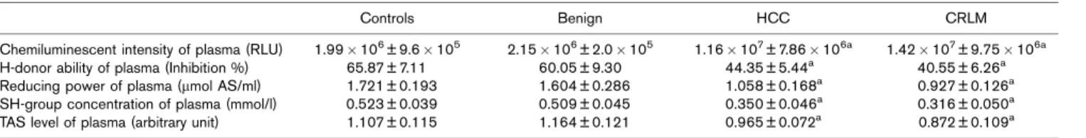
In this study characteristic changes in the redox parameters of blood in cancer patients have been determined. Chemiluminescent activity of the plasma was significantly higher in HCC and CRLM patients versus controls (P< 0.05) and versus patients with benign liver neoplasm (P< 0.05). No significant differ- ence was measured between the plasma relative light unit values of the two cancer groups. Other plasma redox parameters (H-donating ability, reducing power, SH- group concentration) were significantly lower in HCC and CRLM patients versus controls (P< 0.05) and versus patients with benign liver neoplasm (P< 0.05). No significant difference was found between these values of the two cancer groups (Table 1).
Our measurements showed significant increase in free radical content with concomitant significant decrease in GSHPx and CuZn-SOD activities in the erythrocytes of HCC and CRLM patients versus controls (P< 0.05) and versus patients with benign liver neoplasm (P< 0.05) (Figs 1–3). These values were significantly different between HCC and CRLM groups, indicating that processes of metastatization were connected with greater imbalance of redox homeostasis. Cu, Se and Zn (elements crucial for antioxidant enzyme activity) levels were significantly lower in the erythrocytes of cancer patients versus controls (P< 0.05) and also in the benign group (P< 0.05). Alterations in the content of other macro elements (Ca, Mg) may imply that the whole element homeostasis is affected by the oxidative stress of tumourous processes (Table 2).
ATP concentration in the erythrocytes of HCC and CRLM groups decreased significantly compared with the control and benign groups (P< 0.05). In addition, ATP content decreased significantly in the CRLM group versus the HCC group (P< 0.05) (Fig. 4).
Table 1 Redox parameters in the plasma of patients with different liver tumors
Controls Benign HCC CRLM
Chemiluminescent intensity of plasma (RLU) 1.99106± 9.6105 2.15106± 2.0105 1.16107± 7.86106a 1.42107± 9.75106a
H-donor ability of plasma (Inhibition %) 65.87 ± 7.11 60.05 ± 9.30 44.35 ± 5.44a 40.55 ± 6.26a
Reducing power of plasma (mmol AS/ml) 1.721 ± 0.193 1.604 ± 0.286 1.058 ± 0.168a 0.927 ± 0.126a
SH-group concentration of plasma (mmol/l) 0.523 ± 0.039 0.509 ± 0.045 0.350 ± 0.046a 0.316 ± 0.050a
TAS level of plasma (arbitrary unit) 1.107 ± 0.115 1.164 ± 0.121 0.965 ± 0.072a 0.872 ± 0.109a
CRLM, colorectal liver metastases; HCC, hepatocellular carcinoma; RLU, relative light unit; TAS, total antioxidant status.
aSignificant versus control and benign groups,P< 0.05.
On the basis of these findings, redox parameters of erythrocytes seem to be more suitable than parameters of plasma for monitoring the redox status of tumour patients.
Discussion
The relationship between changes in the redox home- ostasis of the body and the development of cancer is not yet completely clear. It is generally believed that generation of free radicals and accelerated lipid peroxida- tion are etiologic factors in cancer development. It is still not clarified, however, whether accelerated ROS forma- tion leads to neoplastic malformation, or pathways of dysplasia induce free radical production, or whether these pathways are part of a circulus vitiosus. The ATP content of the tissues has a close connection with the essential biochemical pathways and plays a key role in the regulation of redox homeostasis [17]; and similarly, ATP content is affected by oxidative stress [18]. Erythrocytes are even susceptible to ‘apoptosis’ following phosphate and ATP depletion [19]. As red blood cells have a close physical connection with the tissues of the whole body, measuring their redox status and ATP concentration may provide relevant information on changes owing to the neoplastic reactions of the liver as well.
We have found elevated free radical content and increased levels of oxidative stress of plasma and erythrocyte samples in HCC and CRLM groups versus controls and benign groups. The worst antioxidant level was found in the CRLM group. This indicates that the progress of neoplastic process may be a key factor of oxidative damage. Lipid peroxidation and oxidative stress are thought to be more prominent in erythrocytes than in the plasma of cancer patients. The erythrocytes are particularly vulnerable to oxidative damage because of exposure to high oxygen tension, high content of polyunsaturated fatty acids and the presence of large amounts of iron [4,20]. In our study, the levels of SOD and GSHPx enzymes along with the content of Cu, Se and Zn elements in the erythrocytes of the HCC and CRLM groups were significantly lower than in the benign group and in healthy controls. As described in the literature, Cu and Zn are essential for the function of SOD enzyme, whereas Se is essential for GSHPx [21,22].
The content of certain elements in erythrocytes changed parallel with the decrease in antioxidant parameters. It has not been clarified, however, or whether these changes are to be considered as primary or secondary, or they cause a circulus vitiosus. Damage of the erythrocyte membranes caused by free radicals may be an explanation for the loss of certain elements and a compensatory increase in the content of others, but there are other potential mechanisms also. In the future, dietary supplementation of these elements may play a key
Fig. 1
8×10⁶
RLU
6×10⁶ 4×10⁶ 2×10⁶ 0
Control Benign HCC a
a
CRLM Chemiluminescent intensity of erythrocytes of patients with different liver tumours (a, significant vs. control and benign groups;P< 0.05).
CRLM, colorectal liver metastases; HCC, hepatocellular carcinoma;
RLU, relative light unit.
Fig. 2
a
b 300
200
Arbitrary unit 100
0
Control Benign HCC CRLM
Glutathione peroxidase activity of erythrocytes of patients with different liver tumours (a, significant vs. control and benign groups; b, significant vs. control, benign and HCC groups;P< 0.05). CRLM, colorectal liver metastases; HCC, hepatocellular carcinoma.
Fig. 3
Arbitrary unit
Control 1
2
0
Benign HCC
a b
CRLM Superoxide dismutase activity of erythrocytes of patients with different liver tumours (a, significant vs. control and benign groups; b, significant vs. control, benign and HCC groups;P< 0.05). CRLM, colorectal liver metastases; HCC, hepatocellular carcinoma.
role in the therapy of diseases related to oxidative stress [21,23,24].
A few hypotheses might be offered to explain the depletion of the antioxidant defence of the plasma: we assume that circulating antioxidant enzymes might be used in an attempt to counteract enhanced free radical generation in the tumour-affected tissue. Another assumption is that the high free radical content is owing to prolonged insufficient power of a depleted antioxidant defence system. Further- more, as GSHPx and SOD are themselves susceptible to oxidation by the oxidative reactive molecules and lipid peroxides, they could be inactivated by their own substrates [25]. Deprivation of trace elements such as Cu, Zn and Se could lead to the inactivation of antioxidant enzymes.
With the lower GSHPx and SOD activity in the cancer groups, ROS (e.g. superoxide anion) were not eliminated completely. Accumulation of these molecules may result in binding and oxidization of DNA, lipids and proteins in numerous organs.
Tumourous tissues with increased ROS generation cause a low energy level of the body. Red cell proteome analysis
demonstrates that several proteins involved in ATP synthesis, folding/chaperone function, redox regulation and red cell metabolism show altered expression because of oxidative damage to proteins [26]. Decreased ATP content may cause deficiencies of membrane pump function and osmolality of RBCs. Therefore, a marked primary change in the content of major elements may occur (e.g. Ca and Mg), which is connected to secondary altered minor element content [27–29], and is deter- mined by this study. Without mitochondria RBCs are, however, sensitive to ATP loss, and energy depletion causes cell shrinkage and ‘programmed cell death’
through Ca2 + influx and PKC activation [30].
The results reveal an alteration in the antioxidant defence system (in plasma and erythrocytes) with concomitant changes in the element content of RBCs in cancer patients. Whether the alterations in the antioxidant status are the cause or consequence of the enhanced ROS generation remains unclear. We presume that an altered prooxidant–antioxidant balance may lead to oxidative damage and play an important role in hepatic carcinogenesis. It seems that oxidative damage is closely connected to alterations of metal element content in erythrocytes. Depletion of elements, which are essential for antioxidant enzyme activities, such as Cu, Se and Zn, leads to a circulus vitiosus. Sulphur content is strongly connected to the activity of enzymes with SH groups.
Imbalance of the redox state and element homeostasis may cause insufficient ATP generation and/or transfer.
Further research should be carried out to find out whether oxidative stress-related parameters could be used as differential diagnostic and prognostic tools in the therapy of benign and malignant liver neoplasms. The presence of the impaired antioxidant status may be a risk factor of both primary and metastatic tumourigenesis of the liver.
On the basis of our findings, redox parameters of erythrocytes seem to be sensitive factors for monitoring the clinical status of tumour patients.
Table 2 Alterations in the element content of erythrocytes in patients with different liver tumors
Controls Benign HCC CRLM
Ca (mg/g) 6.13 ± 2.55 5.51 ± 2.99 7.23 ± 1.98 8.47 ± 2.154
Cu (mg/g) 0.387 ± 0.033 0.376 ± 0.046 0.279 ± 0.061a 0.209 ± 0.077a
Fe (mg/g) 449.1 ± 47.3 464.8 ± 61.6 527.1 ± 87.5 397.2 ± 156.4
Mg (mg/g) 14.30 ± 3.46 13.54 ± 2.56 21.78 ± 5.89a 20.87 ± 4.90a
Mn (mg/g) 0.0725 ± 0.0137 0.0643 ± 0.0074 0.0181 ± 0.0123a 0.0271 ± 0.0159a
P (mg/g) 301.8 ± 39.3 295.4 ± 51.2 357.7 ± 95.0 320.7 ± 83.7
S (mg/g) 1257 ± 64 1297 ± 82 1066 ± 132a 912 ± 159a
Se (mg/kg) 301.3 ± 57.2 313.7 ± 83.7 128.8 ± 58.3a 61.5 ± 39.9b
Zn (mg/g) 6.11 ± 1.10 5.20 ± 1.51 2.82 ± 0.89a 1.95 ± 1.12a
CRLM, colorectal liver metastases; HCC, hepatocellular carcinoma.
aSignificant versus control and benign groups.
bSignificant versus control, benign and HCC groups,P< 0.05.
Fig. 4
Control 120
a
b
%
80
40
0
Benign HCC CRLM
ATP content of erythrocytes of patients with different liver tumours (a, significant vs. control and benign groups; b, significant vs. control, benign and HCC groups;P< 0.05). CRLM, colorectal liver metastases;
HCC, hepatocellular carcinoma.
Acknowledgements
This study was supported by the PhD Programme of the Semmelweis University No. 2/1, ETT 012/2006, NKFP 1A 005/2004 and NKFP1B 047/2004 Projects.
The authors thank Sarolta Ba´rkovits, Edina Pinte´r and Erzse´bet Bı´ro´ for their useful technical assistance.
Conflict of interest: none declared.
References
1 Sasaki Y. Does oxidative stress participate in the development of hepatocellular carcinoma?J Gastroenterol2006;41:1135–1148.
2 Manju V, Balasubramanian V, Nalini N. Oxidative stress and tumor markers in cervical cancer patients.J Biochem Mol Biol Biophys2002;6:387–390.
3 Saygili EI, Akcay T, Konukoglu D, Papilla C. Glutathione and glutathione- related enzymes in colorectal cancer patients.J Toxicol Env Health Part A 2003;66:411–415.
4 Aydin A, Arsova-Sarafinovska Z, Sayal A, Eken A, Erdem O, Erten K,et al.
Oxidative stress and antioxidant status in non-metastatic prostate cancer and benign prostatic hyperplasia.Clin Biochem2006;39:176–179.
5 Xie K, Huang S. Contribution of nitric oxide-mediated apoptosis to cancer metastasis inefficiency.Free Radc Biol Med2003;34:969–986.
6 Orr FW, Wang HH. Tumor cell interactions with the microvasculature: a rate- limiting step in metastasis.Surg Oncol Clin N Am2001;10:357–381.
7 Xie K, Huang S. Regulation of cancer metastasis by stress pathways.
Clin Exp Metastasis2003;20:31–43.
8 Mantovani G, Maccio A, Madeddu C, Mura L, Massa E, Gramignano G,et al.
Reactive oxygen species, antioxidant mechanisms and serum cytokine levels in cancer patients: impact of an antioxidant treatment.J Cell Mol Med2002;
6:570–582.
9 Sahin E, Gumuslu S, Ozturk O, Abidin I, Yargicoglu P, Agar A. Marked changes in erythrocyte antioxidants and lipid peroxidation levels of rats exposed to acute, repeated and chronic restraint stress.Pharmazie2004;
59:961–964.
10 Bla´zovics A, Kova´cs A´, Lugasi A, Hagyma´si K, Bı´ro´ L, Fehe´r J. Antioxidant defence in erythrocytes and plasma of patients with active and quiescent Crohn’s disease and ulcerative colitis: a chemiluminescent study.
Clin Chem1999;45:895–896.
11 Sedlak J, Lindsay RH. Estimation of total protein bound and non protein sulfhydryl groups in tissues with Ellmann’s reagent.Anal Biochem Biophys 1985;25:192–205.
12 Blois MS. Antioxidant determination by the use of stable free radicals.
Nature1958;4617:1999–2000.
13 Hatano T, Kagawa H, Yasuhara T, Okuda T. Two new flavonoids and other constituents in licore root: their relative astringency and radical scavenging effects.Chem Pharm Bull1988;36:2090–2097.
14 Oyaizu M. Studies on products of browning reaction prepared from glucosamine.Jpn J Nutr1986;44:307–315.
15 Szentmiha´lyi K, Bla´zovics A, Kocsis I, Fehe´r E, Lakatos B, Vinkler P.
The effect of fat rich diet and alcohol on ion concentration in bile in rats.
Acta Aliment Hung2000;29:359–366.
16 Rieger D. Batch analysis of the ATP content of bovine sperm, oocytes and early embryos using a scintillation counter to measure the
chemiluminescence by the luciferin-luciferase reaction.Anal Biochem1997;
246:67–70.
17 Okunieff P, Fenton B, Chen Y. Past, present, and future of oxygen in cancer research.Adv Exp Med Biol2005;566:213–222.
18 Korge P, Weiss JN. Redox regulation of endogenous substrate oxidation by cardiac mitochondria.Am J Physiol-Heart C2006;291:436–445.
19 Birka C, Lang PA, Kempe DS, Hoefling L, Tanneur V, Duranton C,et al.
Enhanced susceptibility to erythrocyte ‘apoptosis’ following phosphate depletion.Pflugers Arch2004;448:471–477.
20 Akcil E, Cayalakli F, Akiner M, Kocak M. Trace element concentrations and superoxide dismutase and catalase activities in benign and malignant larynx tumors.Biol Trace Elem Res2004;101:193–201.
21 Bacic-Vrca V, Skreb F, Cepelak I, Mayer L, Kusic Z, Petres B. The effect of antioxidant supplementation on superoxide dismutase activity, Cu and Zn levels, and total antioxidant status in erythrocytes of patients with Graves’
disease.Clin Chem Lab Med2005;43:383–388.
22 Batcioglu K, Ozturk C, Karagozler A, Karatas F. Comparison of the selenium level with GSH-Px activity in the liver of mice treated with 7,12 DMBA.
Cell Biochem Funct2002;20:115–118.
23 Alissa EM, Bahijri SM, Lamb DJ, Ferns GA. The effects of coadministration of dietary copper and zinc supplements on atherosclerosis, antioxidant enzymes and indices of lipid peroxidation in the cholesterol-fed rabbit.
Int J Exp Pathol2004;85:265–275.
24 Maret W, Sandstead HH. Zinc requirements and the risks and benefits of zinc supplementation.J Trace Elem Med Bio2006;20:3–18.
25 Pigeolet E, Corbisier P, Houbion A, Lambert D, Michiels C, Raes M,et al.
Glutathione peroxidase, superoxide dismutase, and catalase inactivation by peroxide and oxygen derived free radical.Mech Ageing Dev1990;
51:283–297.
26 Friedman JS, Lopez MF, Fleming MD, Rivera A, Martin FM, Welsh ML,et al.
SOD2-deficiency anemia: protein oxidation and altered protein expression reveal targets of damage, stress response, and antioxidant responsiveness.
Blood2004;104:2565–2573.
27 Barvitenko NN, Adragna NC, Weber RE. Erythrocyte signal transduction pathways, their oxygenation dependence and functional significance.
Cell Physiol Biochem2005;15:1–18.
28 Kedzierska K, Bober J, Ciechanowski K, Golembiewska E, Kwiatkowska E, Nocen I,et al. Copper modifies the activity of sodium-transporting systems in erythrocyte membrane in patients with essential hypertension.Biol Trace Elem Res2005;107:21–32.
29 Osborn KD, Zaidi A, Urbauer RJ, Michaelis ML, Johnson CK. Single- molecule characterization of the dynamics of calmodulin bound to oxidatively modified plasma-membrane Ca2 + -ATPase.Biochemistry2005;
44:11074–11081.
30 Klarl BA, Lang PA, Kempe DS, Niemoeller OM, Akel A, Sobiesiak M,et al.
Protein kinase C mediates erythrocyte ‘programmed cell death’ following glucose depletion.Am J Physiol-Cell Ph2006;290:C244–253.