CHARACTERIZATION OF THE INFLAMMATORY STATUS OF PRETERM INFANTS
PhD thesis
Florentina Sava
Doctoral School of Clinical Medicine Semmelweis University
Consultant: Gergely Toldi, MD, Ph.D Official reviewers: Katalin Csordás, MD, Ph.D
Tamás Németh, MD, Ph.D
Head of the Final Examination Committee: Edit Buzás, MD, D.Sc
Members of the Final Examination Committee: László Csáthy, MD, Ph.D Zoltán Pós, Ph.D
Budapest
2016
TABLE OF CONTENTS
1. ABBREVIATIONS 4
2. INTRODUCTION 6
2.1. THE NEONATAL IMMUNE SYSTEM 7
2.1.1. The innate immune system of preterm neonates 8 2.1.2. The adaptive immune system of preterm neonates 9
2.2. INFLAMMATORY DISORDERS IN THE NEONATE 11
2.2.1. Bronchopulmonary dysplasia (BPD) 11
2.2.2. Necrotising enterocolitis (NEC) 14
2.2.3. Sepsis 15
2.3. PERINATAL AND POSTNATAL COMPLICATIONS 16
2.3.1. The role of vitamin D levels at birth in preterm infants 16 2.3.2. The impact of preeclampsia in preterm neonates 19 2.3.3. Early and late activation marker expression in T cells of
preterm neonates 21
3. AIMS 24
4. MATERIALS AND METHODS 25
4.1. Patients 25
4.2. Ethical considerations 28
4.3. CBMC isolation 28
4.4. Flow cytometry 29
4.5. Plasma cortisol and vitamin D levels 30
4.6. Plasma cytokine levels 31
4.7. Statistics 31
5. RESULTS 33
5.1. The role of vitamin D levels at birth in preterm infants 33
5.3. Early and late activation marker expression in t cells of
preterm neonates 41
6. DISCUSSION 44
6.1. Plasma vitamin D levels control the inflammatory balance in
preterm infants 44
6.2. Maternal preeclampsia has a major impact on the immune system
of preterm infants 46
6.3. Early and late T lymphocyte activation markers are associated
with perinatal complications in preterm infants 49
6.4. Limitations 51
7. CONCLUSIONS 52
8. SUMMARY 53
9. ÖSSZEFOGLALÁS 55
10. REFERENCE LIST 57
11. PUBLICATIONS 70
12. ACKNOWLEDGEMENTS 72
1. ABBREVIATIONS
ACE angiotensin-converting enzyme ALRI acute lower respiratory infection BPD bronchopulmonary dysplasia CBMC cord blood mononuclear cell CTL cytotoxic T lymphocyte
CTLA-4 cytotoxic T lymphocyte antigen 4
DC dendritic cell
EOS early onset neonatal sepsis
G-CSF granulocyte colony-stimulating factor
GM-CSF granulocyte-macrophage colony-stimulating factor GST glutathione S transferase
HDM house dust mite
HEV high endothelial venules IFN-a Interferon alpha
IFN-g Interferon gamma
iNKT invariant natural killer T cell mDC myeloid dendritic cell
MHC major histocompatibility complex NEC necrotising enterocolitis
NETs neutrophil extracellular traps NICU neonatal intensive care unit
NK natural killer cell
NP normal pregnancy
OVA ovalbumine
PAMPs pathogen-associated molecular patterns on microbes pDC plasmacytoid dendritic cell
PE preeclampsia
PS prenatal steroid
RDS respiratory distress syndrome RSV respiratory syncytial virus TCR T cell receptor
TGF-b tumor growth factor beta TLR9 toll-like receptor 9 TNF tumor necrosis factor
TNF-alpha-238 tumor necrosis factor-alpha-238 Treg regulatory T cell
Th1/Th2 T helper 1/ T helper 2 cells
TTN transitory tachypnea of the neonate VDR vitamin D receptor
VLBW very low birth weight
2. INTRODUCTION
Preterm birth, defined as birth at less than 37 completed weeks of gestation, occurs in around 12% of deliveries worldwide with major implications for the long term health of the child. Mortality rates of preterm infants have decreased substantially over the last few decades due to advances in medical care system. However, morbidity rates, particularly in the very preterm infant (born less than 28 weeks of gestation) have continued to rise [1].
On the long term, preterm infants who survive may suffer from permanent disabilities due to organ damage resulting from either an infection or from the systemic inflammatory response triggered by different factors [2]. In these vulnerable preterm infants, early diagnosis of the inflammatory response is critical and can set the stage for lifelong morbidities [3].
Our research group has a longstanding interest in the investigation of the neonatal and preterm immune response and function. The results obtained in such studies do not only help us better understand the basic principles of the development of the human adaptive immune system, but also play a key role in identifying diagnostic and therapeutic targets that may provide better care for this vulnerable population in the very near future.
2.1. THE NEONATAL IMMUNE SYSTEM
Normal term neonates rely heavily on their immune system at birth to fight infection in the pathogen-prone extrauterine environment, however it is still not fully developed (Figure 1.) [4].
Preterm infants have an under-developed immunoregulatory system, therefore there is the potential for chronic inflammation to develop [5]. For instance, the immune system of preterm infants has a smaller prevalence of monocytes and neutrophils, impaired ability of these cells to kill pathogens, and lower production of cytokines which limits T cell activation and reduces the ability to fight bacteria and detect viruses in cells, compared to term infants. Intrauterine inflammation is a major contributor to preterm birth, and causes premature immune activation and cytokine production. This can induce immune tolerance leading to reduced newborn immune function [6].
Figure 1. Leukocyte development begins in the yolk sac before moving to the liver and finally to the bone marrow. The development and maturation of primary lymphoid organs (blue) and peripheral blood leukocytes (red) occur throughout gestation but is not complete until after birth. The light gray shading shows the gestational ages of
preterm births from the threshold of viability (24 weeks; at which 50% of infants in developed countries survive) to 37 weeks of gestation [7].
Both the innate and adaptive immunity are compromised in preterm neonates, and they also have deficiencies in the interaction between these two systems [7-8].
2.1.1. The innate immune system of preterm neonates
The innate immune response of preterm infants is diminished in its potential to adequately respond to infections due to deficiencies in the soluble protein/peptide and cellular responses to infection.
Phagocytes include neutrophils, monocytes/macrophages and dendritic cells (DCs).
Preterm infants have a reduced prevalence of neutrophils and monocytes, and their precursors, due to reduced granulocyte colony-stimulating factor (G-CSF) and granulocyte-macrophage colony-stimulating factor (GM-CSF) levels [9].
Neutrophils are among the first responders to infection and have an important role in bacterial clearance [10]. Neutrophils of preterm infants may have difficulty in migrating to sites of infection due to a reduction in the expression of adhesion molecules such as L- and E-selectin [11]. Impairment in neutrophil function (phagocytosis, generation of oxygen radicals and intracellular killing of pathogens) is a risk factor for the development of sepsis [12]. Extracellular pathogen killing is also limited in neonates, with reduced ability to produce neutrophil extracellular traps (NETs) [13]. NETs are lattices made of DNA bound to granular and cytoplasmic protein, which are released from neutrophils to trap and kill pathogens [14].
Monocytes are phagocytic blood-borne cells that differentiate into macrophages or DCs in tissue. Monocytes are capable of phagocytosis, have bactericidal mechanisms and are involved in antigen presentation to T cells [10]. The monocytes of preterm infants have reduced cytokine production, but a similar efficiency in phagocytosis and intracellular killing of pathogens as term neonates [15]; however, they may be limited in their ability to activate the adaptive immune response because major histocompatibility complex (MHC) class II expression is reduced on leukocytes from preterm neonates [16], thus
2.1.2. The adaptive immune system of preterm neonates
Adaptive immunity, which involves B and T lymphocytes, is pathogen-specific and requires acquisition of immunological memory.
At birth, lymphocyte subpopulations in term infants are lower when compared to adults.
Moreover, maturation of adaptive immunity occurs mostly after term birth, suggesting that most newborn infants have deficiencies in T cell activation and cytokine production, B cell immunoglobulin production, and interactions between T and B cells, when compared to adults [6, 17]. A few studies showed that lymphocyte subpopulations are reduced in children born preterm at 7 months [18] and 8 years [19] of age, compared to age-matched children born at term.
Cell-mediated immunity involves two main types of T cells, cytotoxic T lymphocytes (CTL; CD8+) and T helper lymphocytes (Th; CD4+). CD8+ CTLs are involved in the eradication of intracellular pathogens such as viruses and are presented antigen by APCs expressing MHC class I. CD4+ T helper cells, activated by MHC II, are further divided into Th1 and Th2 CD4+ cells, defined by their cytokine profile. The Th1 phenotype is often classed as inflammatory, producing the cytokines interferon-γ (IFN-γ), interleukin (IL)-2 and tumor necrosis factor-α (TNF-α). The Th2 phenotype is anti-inflammatory, producing cytokines such as IL-4, IL-5, IL-13, and IL-10 [6].
Recently, two further types of T helper cytokines have been characterized. Th17 cells, producing IL-17, also have strong pro-inflammatory properties and play a role in the development of autoimmunity. On the other hand, regulatory T cells (Tregs) have the potential to suppress the above subsets, commonly known as effector T cells. An important transcription factor for the development of Tregs is FoxP3 [6].
Preterm and term neonates have deficient T cell function as a result of a greater proportion of naïve T cells in the circulation and a low subpopulation of memory T cells [17]. The increased proportion of naive T cells is a result of inefficient DC antigen uptake and presentation, and is contributed to by a reduction in MHC II expression on antigen-presenting cells [8].
Cytokine responses are driven toward a Th2 phenotype during fetal life (Figure 2). The Th2 bias is believed to be a preventative measure against fetal rejection by the maternal immune system, with increased Th1 cytokine production linked to an increased risk of
spontaneous abortion [6]. Preterm and term neonates are believed to be vulnerable to infection due to this bias toward a Th2 CD4+ T cell phenotype and as a result have lower production of cytokines such as IFN-γ in comparison to adults, which can result in deficient viral detection and clearance [4].
Figure 2. Developmental changes occurring in the human immune system early in life. This figure illustrates maturational events occurring in major adaptive and innate immune functions as the human being transitions from a fetal tolerance state and becomes exposed to microorganisms as well as other environmental antigens de novo after birth. Adaptive immune functions [top panel]: Maternal transplacental antibody transfer (IgG) mainly occurs during late gestation, followed by maternal antibody protection (e.g. IgA) acquired through breast-milk after birth. Infants' own antibody response becomes fully mature later during early childhood. Neonatal T cells are largely biased towards helper type II responses and humans display high proportions of T regulatory and natural killer T cells at birth [20]. Innate immune functions [bottom panel]: Pro-inflammatory (IL-1β, IL-6, TNF-α, IL-12, IL-23) and anti-viral (IFN-α) cytokine responses are largely attenuated in preterm infants, whereas production of the
2.2. INFLAMMATORY DISORDERS IN THE NEONATE
Several inflammatory disorders have been described in neonates which lead to an exaggerated release of inflamatory mediators, amongst the most important and common are BPD, NEC and sepsis. These disorders also have a multi-factorial pathogenesis [21].
2.2.1. Bronchopulmonary dysplasia (BPD)
BPD is a disorder of prematurity characterised by the need for assisted ventilation or supplemental oxygen at 36 weeks postmenstrual age and signs of impaired alveolarisation and vasculogenesis in the lungs. BPD occurs in approximately 45% of infants born less than 29 weeks gestation that survive preterm birth [5, 22]. Ongoing lung damage may be caused by the preterm infant's inability to down-regulate and maintain control of the inflammatory immune response, leading to a chronic inflammatory state [5, 22-24]. Figure 3. shows general critical steps and associated mediators in lung inflammation, injury and remodelling that lead to BPD.
Several biomarkers have been described to be involved in the development of BPD, such as chemokines, adhesion molecules, pro-inflammatory cytokines, growth factors and others.
IL-8 (also called CXCL8) is the chemokine that has been investigated most extensively in preterm infants. Increased concentrations of IL-8 precede neutrophil infiltration in tracheal aspirates from preterm infants as well as in infants ventilated with high tidal volumes. Other chemokines associated with the development of BPD include the monocyte chemoattractant proteins, MCP-1 (CCL2), MCP-2 (CCL8) and MCP-3 (CCL7), and macrophage inflammatory proteins, MIP-1a (CCL3) and MIP-1b (CCL4).
In addition to an association with BPD, increased concentrations of MCP-1 in airway secretions have been observed in the presence of tracheal colonisation with Ureaplasma urealyticum, a putative risk factor for BPD [23].
Adhesion molecules include selectins, responsible for transient adhesion and rolling, and integrins and immunoglobulins, responsible for firm adhesion and transmigration.
These proteins appear to be critical in the development of parenchymal damage in
infants with BPD [23].
The common pro-inflammatory cytokines TNF-α, IL-1, IL-6 and IL-8 are the most extensively studied cytokines in this category, and are important biomarkers for the prediction of adverse pulmonary outcomes in preterm infants. Increased concentrations of IL-1, TNF-α, IL-6 and IL-8 correlate with the duration of supplemental oxygen and mechanical ventilation and are increased in infants who develop BPD compared with infants of similar gestational age who do not develop BPD [25].
Recently, genetic susceptibility for BPD has also been reported as a potential factor in the pathogenesis. There have been several studies addressing the association of polymorphisms in genes, such as angiotensin-converting enzyme (ACE), glutathione S transferase (GST) and TNF-alpha 238, with the development of BPD [22, 26-28]. Twin studies have revealed that the BPD status of one twin, even after correcting for contributing factors, is a highly significant predictor (53%) of BPD in the second twin [29].
Figure 3. Diagram representing critical steps and associated mediators in lung inflammation, injury and remodelling that result in BPD [23].
2.2.2. Necrotising enterocolitis (NEC)
NEC is predominantly a disease of prematurity, it is the most common gastrointestinal illness in newborns, and remains a major cause of neonatal morbidity and death (15%–25%) [21, 30-31]. Due to the multifactorial etiology (Figure 4.), early diagnosis and effective treatment of NEC is limited. Consequently, many cases, 20% to 40% of patients, require surgical intervention [32].
Up to date, several in vitro and in vivo experiments have been developed in order to understand the immune mechanisms of NEC. Most of the studies are using animal models due to the limitations of access to human preterm infants’ samples.
Current evidence suggests that NEC is due to the dysfunction of the intestinal mucosal barrier and an inappropriate inflammatory response of the immature gut. As the disease progresses, inflammation in the intestine worsens causing breakdown of the mucosal barrier and an escalating immune cascade leading to sepsis, shock and even death [30- 31]. The risk for developing NEC is strongly influenced by commensal bacteria, which exert metabolic, nutritional and immunological effects on the host [18]. For instance, high TLR-4 expression has been associated with human NEC [33]. Toll-like receptors (TLRs) are membrane spanning receptors that recognize pathogen-associated molecular patterns on microbes (PAMPs) once they have breached the physical barriers, such as the mucosal layer, and therefore play a key role in the innate immune system [34].
2.2.3. Sepsis
Sepsis is a systemic inflammation caused by infection that may occur in infants throughout the whole neonatal period. Globally, sepsis is responsible for approximately 15% of neonatal deaths [36], with rates of infection dependent on the geographic region.
In preterm and term infants, sepsis is classified as either early-onset (<72 h of life) or late-onset (>72 h of life), with the latter being a common complication associated with prolonged admission to the neonatal intensive care unit (NICU). The distinction between the two is of clinical importance, as early-onset sepsis usually results from exposure to bacteria in utero or during delivery and late-onset sepsis is acquired from bacteria in the environment (like nosocomial infections) [37-38]. Therefore, the choice for empirical antibiotic treatment is different in clinical practice.
In the early state of sepsis, the excessive activation of the antigen recognition system and the release of pro-inflammatory mediators lead to serious multisystem dysfunction in the body. During sepsis, microbes or necrotic tissue release high levels of harmful substances, resulting in the activation of the systemic immune response and excessive activation of immune cells. The excessive release of pro-cytokines plays a destructive effect. The uncontrolled inflammatory cascade may lead to endothelial dysfunction, resulting in life-threatening conditions, such as shock and hypotension.
T cells, especially Th1 and Th2 cells, play an important role in the regulation of inflammation. In the pathogenesis of sepsis, the acquired immune response is transformed from the Th1 cell-mediated immune response (characterized by the production of IFN-γ and IL-12) into the Th2 cell-mediated immune response (characterized by the production of IL-4, IL-5, IL-10 and IL- 13), leading to further immune suppression. In addition, the increased rate of apoptosis of lymphocytes and DCs also plays an important role in immune suppression. In contrast, apoptosis of macrophages and neutrophils is decreased rather than increased [39-40], further promoting inflammation. Early T cell-mediated innate immune suppression is reported to reduce the septic damage [41]. There is evidence that sepsis does not only affect the body's immune system but also acts on the body's coagulation system and the autonomic nervous system [42-43].
2.3. PERINATAL AND POSTNATAL COMPLICATIONS
A variety of prenatal factors and events may lead to preterm birth, further modulating and compromising the immune system of the newborn. A better understanding of perinatal complications which affect the newborn's immune status will therefore allow for a further refinement of care leading to a reduction of lifelong adverse immune consequences. Several research groups, including ours, aimed to describe the immunologic effects of perinatal complications that lead to preterm birth and severely affect the immune system of the newborn.
Amongst the most important complications during pregnancy are hypertensive disorders, such as preeclampsia (PE), gestational diabetes, premature rupture of membranes (PROM) and of vitamin D deficiency [44-45]. These perinatal complications seem to have a serious impact on different immune cell subpopulations, cytokines and lymphocyte activation markers in the maternal and fetal immune system.
The use of antenatal steroid prophylaxis for the prevention of surfactant deficiency and respiratory distress syndrome (RDS) may also have long-term consequences on neonatal immunity.
2.3.1. The role of vitamin D levels at birth in preterm infants
Vitamin D (25(OH)D) is a steroid hormone essential for the regulation of a large number of physiological processes such as calcium homeostasis, muscle and bone health. Low serum vitamin D concentrations (that are directly correlated with maternal vitamin D status at birth) are common in preterm infants during birth, hospitalisation and at discharge from the neonatal intensive care unit [46-47]. Different studies and organizations have recommended vitamin D intake of 400 IU/day to achieve a target serum 25 (OH)D concentration above 20 ng/mL for all infants [47-48].
Vitamin D has an important role in the control of inflammatory responses [49].
Recently, it was found that severely low maternal and neonatal 25(OH)D levels are associated with early-onset neonatal sepsis (EOS) [50]. Subclinical vitamin D deficiency in neonates is associated with diminished immune function and increases the
increased risk of acute lower respiratory infections (ALRI) [52]. Low cord blood vitamin D status was also found to be associated with higher risk of milk sensitization during early childhood [53].
Active form of the vitamin (1,25(OH)2D3) regulates the growth and differentiation of multiple immune cell types, and displays immunoregulatory and anti-inflammatory properties (Figure 5.) [54].
Figure 5. Mechanisms by which 25(OH)D and 1,25(OH)2D3 modulate innate and adaptive immune responses. All these cells possess the enzyme (CYP27bB1) for hydroxylation steps to generate 1,25(OH)2D3. Through endocrine, intracrine and paracrine mechanisms, the active form of vitamin D binds to the vitamin D receptor (VDR) to induce a wide range of immunological effects [55-56].
1,25(OH)2D3 inhibits several components of the immune system [57], including DC differentiation and maturation as well as modulation of their activation and survival
leading to T cell hyporesponsiveness (Figure 6.) [58]. 1,25(OH)2D3 was also shown to play an important role in the maintenance of B cell homeostasis and differentiation [59]
and T cell proliferation in response to T cell receptor stimulation [60]. A more recent study [61] demonstrated that vitamin D impairs the capacity of murine and human plasmacytoid dendritic cells (pDCs) to induce T cell proliferation and secretion of the T helper 1 cytokine IFN-γ. The inhibitory effect of vitamin D was found to be dependent on the expression of its receptor (VDR) in DCs. Subsequent human and animal model studies also demonstrated that 1,25(OH)2D3 acts directly on T cells to promote FoxP3+
and IL-10+ Tregs, secretion of the immunomodulatory cytokines IL-10 and TGF-β, and upregulation of the inhibitory molecule cytotoxic T lymphocyte antigen (CTLA)-4 on the cell surface [62].
Figure 6. Overview of immunomodulatory actions of 25(OH)D3 and 1,25(OH)2D3 on monocytes and macrophages, dendritic cells, effector, and memory T and B lymphocytes [55-56].
2.3.2. The impact of preeclampsia in preterm neonates
Balanced immune responses are required for the maintenance of a successful pregnancy in order to avoid adverse pregnancy outcomes, such as preeclampsia (PE) and miscarriage [63]. To date, there is very limited information about the impact of maternal PE on the fetal immune system. PE is a multisystem, highly variable disorder unique to pregnancy and a major cause of maternal and fetal/neonatal morbidity and mortality [64].
PE has been reported with an incidence of 6% to 10% [65]. The majority of cases happen to occur in healthy nulliparous women with a reported incidence of 10%
to 12% [66-67]. The rates are even higher in women with certain risk factors, such as multifetal gestation (25-30%), PE in previous pregnancy (20-50%), women with preexisting chronic hypertension (15-50%), pregestational diabetes mellitus (15-35%), renal disease, or thrombophilia [65, 68].
PE is characterized as either a maternal syndrome (hypertension and proteinuria with or without other multisystem abnormalities), or a fetal syndrome (the inability of the trophoblast to invade the decidual arteries leading to alterations in placental development, placental perfusion, insufficient transport of nutrients and abnormal oxygenation) [68-69].
Activation of the maternal innate and adaptive immune system plays an essential role in the pathophysiology of PE (Figure 7.). Activated neutrophils, monocytes, and natural killer (NK) cells initiate inflammation, which induces endothelial dysfunction, and activated T cells may support inadequate tolerance during pregnancy [70].
In healthy non-pregnant women, there is a balance between Th1 and Th2 responses [71]. However, many authors observed Th1/Th2 immunity alterations with a shift to a predominantly Th2 type immunity during normal pregnancy (NP) [72]. This alteration might be due to the development of the placenta, since its production of progesterone and cytokines may gear the immune cells to a Th2 response [73]. The anti-inflammatory Th2 reaction is crucial for tolerance to the fetus, decidualization and remodelling.
The pro-inflammatory Th1 immune response is responsible for extravillous trophoblast invasion, parturition and host defence in NP [64]. In contrast, several groups reported that PE is associated with pathological Th1 type immunity which also impairs maternal
tolerance to the fetus [74-77]. However, it still remains unclear which factors influence Th1/Th2 imbalance in PE.
Th17 and Treg cells have also been suggested to be of importance in the development of maternal systemic inflammation in PE [78]. The frequency of Th17 cells was found to be elevated in the peripheral blood of PE patients compared to NP women in the third trimester of pregnancy [79-80]. A number of groups, including ours, demonstrated that the prevalence of peripheral Tregs is lower in PE compared to healthy pregnancy [70, 81-84].
Furthermore, it was found that NK cells are also involved in PE. Several groups showed that the numbers of NK cells are either increased or decreased in PE women [85-86]
when compared to NP. The conflicting results may be due to different methods of isolation and the location of sampling (ie. peripheral blood or placenta). Moreover, the NK1/NK2 and NKT1/NKT2 cell ratios were found to be significantly decreased in normal pregnancy compared with non-pregnant and PE women [87].
Several studies have shown that the neonatal outcome of PE mothers is often complicated by severe clinical features, such as neonatal thrombocytopenia [88], BPD [89], persistent pulmonary hypertension (PPHN), respiratory failure and an increased risk for transient tachypnea of the newborn (TTN) [64], as well as cardiovascular diseases and intellectual behavioural problems in later life [90]. Since a high number of PE pregnancies end in preterm delivery of the fetus, it is often challenging to distinguish between the role of prematurity and the effects of PE per se in the above conditions.
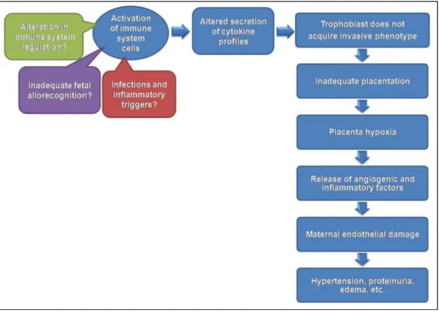
Figure 7. Activation of selected immune cells alters secretion of cytokine profiles that contributes to the maternal syndrome of preeclampsia. Initially these adverse events cause persistent placental hypoxia followed by inadequate placentation. The pathological cytokine profiles may be due to an alteration in immune system regulation, or an inadequate fetal allorecognition, or to inflammatory triggers present during implantation [69].
2.3.3. Early and late activation marker expression in T cells of preterm neonates
Activation of T lymphocytes is a complex, yet finely regulated cascade of events that results in the expression of cytokine receptors, production and secretion of cytokines, upregulation of several cell surface molecules, and activation of direct cell killing. Therefore, it is one of the cornerstones of the implementation of an appropriate immune response for different types of antigens [91].
Preterm birth affects an estimated number of 15 million infants each year globally [92].
The rate of preterm birth ranges from 5% to 18% percent. Premature babies face
numerous acute and chronic complications, including respiratory, cardiovascular, gastrointestinal, and perhaps most importantly, neurodevelopmental problems. Risk factors associated with preterm labour include previous preterm births or miscarriages, multiple pregnancy, in vitro fertilization, cervical or placental insufficiency, smoking, poor nutrition, hypertensive disorders, infections, stress and trauma. Numerous studies to date have shown that preterm deliveries associated with preeclampsia (PE), premature rupture of membranes (PROM), intrauterine infection and respiratory distress syndrome (RDS) are linked with higher levels of neonatal adaptive immune response [93]. However, data on T lymphocyte activation markers of preterm infants is scarce [94].
The most important activation molecules expressed on T lymphocytes can be classified as early activation markers, such as CD69 and CD25, and late activation markers, such as CD62L and HLA-DR. Additionally, very late activation markers, such as VLA-1 have also been described, playing a role in lymphocyte adhesion and extravasation [95].
CD69
CD69 is generally regarded as the earliest activation cell surface marker of both mononuclear umbilical cord and peripheral blood cells induced by a mitogenic stimulus.
The expression of CD69 molecule is not restricted to activated lymphocytes, as activated neutrophils and eosinophils can also express CD69. Moreover, platelets, epidermal Langerhans cells and bone marrow myeloid precursors express CD69 constitutively. The engagement of CD69 can activate NK and T cells, resulting in increased cytotoxic activity and pro-inflammatory cytokine production [96]. CD69 seems to be expressed in higher levels on the surface of activated neonatal cells when compared to adults [97]. Upregulation of CD69 on NK cells was identified as a sensitive marker of neonatal infection [98].
CD25
CD25, or the alpha subunit of the IL-2 receptor, is involved in the early stage of
immune homeostasis. Early work on CD4+ CD25 high+ cells later termed as regulatory T cells showed that their activation via their T cell receptor (TCR) generates suppressor cells that are capable of non-specifically suppressing the activation of any CD4+ or CD8+ T cell [99]. FoxP3+ regulatory T cells, also characterized by high expression of CD25 are present at the fetal-maternal interface and have been reported to be important for the maternal acceptance of the allogeneic fetus [100].
CD62L
CD62L (L-selectin) is considered a late activation marker and a key regulator of T cell trafficking. It acts as a homing receptor for lymphocytes to enter secondary lymphoid tissues via high endothelial venules (HEV). Following activation, CD62L is rapidly downregulated on T cells, which prevents effector T cells from trafficking to lymph nodes through HEV [101]. Activation of CD62L takes place during the first postnatal days in preterm infants with RDS, and this activation is associated with the development of BPD [102]. Furthermore, it was also demonstrated that carriers of the L-selectin 213Ser allele are at increased risk for premature birth and BPD [103].
HLA-DR
HLA-DR molecules are involved in antigen processing and presentation, mediating antigen-specific T cell activation [104]. It is known that low levels of HLA- DR expression on monocytes contributes to impaired neonatal host defence, especially in preterm infants [105]. Decreased expression of HLA-DR molecules in preterm newborns is linked with development of several complications, such as high incidence of bacterial infections and pulmonary morbidity, especially in the presence of RDS [106-107].
3. AIMS
Our aim was to characterise the inflammatory status of preterm infants at birth and during the first week of life and its association with perinatal complications as well as the influence of maternal factors.
The specific aims for our investigations were:
1. To assess the plasma 25(OH)D concentrations from cord blood of 28 preterm infants born before the 30th gestational week and to determine its correlation with cellular and soluble indicators of the inflammatory status. Effects of other factors, such as gestational age and plasma cortisol levels were also assessed.
2. To address the hypothesis that PE impacts the fetal immune system, we analysed the prevalence of distinct lymphocyte subsets and plasma cortisol and cytokine levels in preterm neonates of PE mothers during the first week of life (at birth and on the 1st, 3rd and 7th postnatal days) and compared them to preterm neonates with comparable clinical characteristics born from pregnancies not complicated by PE.
3. To assess the association of gender, gestational and postnatal age, preeclampsia (PE), premature rupture of membranes (PROM) and prenatal steroid treatment (PS) with the frequency of activated T lymphocyte subsets (CD69+, CD25+, CD62L+, HLA-DR) and major T lymphocyte subpopulations (CD4, CD8, Th1, Th2, naïve, memory) in peripheral blood during the first postnatal week in preterm neonates. Since data on the physiological frequency of these cell subsets is challenging to obtain, we aimed to gather preliminary data to describe the dynamic postnatal alteration of these parameters in preterm neonates affected by different perinatal factors.
4. MATERIALS AND METHODS
Blood samples for all three studies were collected at the Neonatal Intensive Care Unit of the 1st Department of Obstetrics and Gynecology, Semmelweis University, Budapest, Hungary. Flow cytometry measurements were performed at the Research Laboratory of the 1st Department of Pediatrics, Semmelweis University, Budapest, Hungary. Some of the patients fulfilling the inclusion criteria for more than one study were overlapping across multiple studies.
4.1. PATIENTS
In the study on vitamin D levels, venous cord blood samples were taken from 28 preterm infants (gestational age: 29 (27-30) weeks, median (range); birth weight: 1080 (920-1550) grams, median (range)). Patient characteristics are summarized in Table 1.
All infants had a highly suspected or proven intrauterine infection based on standard criteria [108-109]. However, no ongoing inflammatory reaction was detected in patients at the time of sampling, as indicated by normal IL-6 levels. Of note, since no suggestive clinical signs of chorioamnionitis were present, placental histology was not performed.
Based on plasma vitamin D levels, patients were divided into two groups (below and above median, 23.3 ng/mL), between which all comparisons were made.
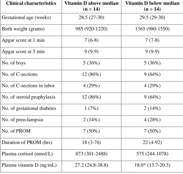
Table 1. Clinical characteristics of participants in the study on vitamin D levels. Data are presented as median (range). * p < 0.05 vs. Above median group.
Clinical characteristics Vitamin D above median (n = 14)
Vitamin D below median (n = 14)
Gestational age (weeks) 28.5 (27-30) 29.5 (29-30) Birth weight (grams) 985 (920-1220) 1365 (980-1550)
Apgar score at 1 min 7 (6-8) 7 (7-8)
Apgar score at 5 min 9 (9-9) 9 (9-9)
No. of boys 5 (36%) 5 (36%)
No. of C-sections 12 (86%) 9 (64%)
No. of C-sections in labor 4 (29%) 4 (29%)
No. of steroid prophylaxis 12 (86%) 9 (64%)
No. of gestational diabetes 1 (7%) 2 (14%)
No. of preeclampsia 2 (14%) 4 (28%)
No. of PROM 7 (50%) 7 (50%)
Duration of PROM (hrs) 18 (3-76) 22 (4-92)
Plasma cortisol (nmol/L) 873 (301-2488) 575 (244-1078) Plasma vitamin D (ng/mL) 27.2 (24.8-38.8) 18.0* (13.7-20.3)
In the study on the effects of preeclampsia, we enrolled 14 preterm neonates born to PE mothers and 14 preterm neonates born to healthy mothers as controls. Median gestational age was 30 weeks in the PE group and 29 weeks in controls, while birthweight was 985 g in the PE group and 1180 g in controls, respectively. All neonates had a highly suspected intrauterine infection based on standard criteria [108-109] and went through partial septic screen, however, CRP and IL-6 levels were within the
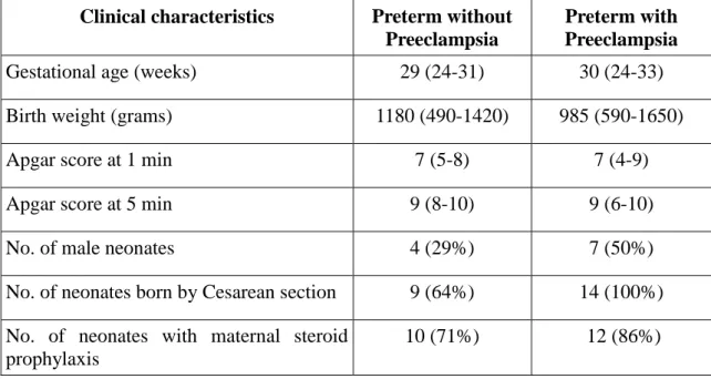
Table 2. Clinical characteristics of preterm neonates enrolled in the study on the effects of PE. Data are expressed as median (range).
Clinical characteristics Preterm without Preeclampsia
Preterm with Preeclampsia
Gestational age (weeks) 29 (24-31) 30 (24-33)
Birth weight (grams) 1180 (490-1420) 985 (590-1650)
Apgar score at 1 min 7 (5-8) 7 (4-9)
Apgar score at 5 min 9 (8-10) 9 (6-10)
No. of male neonates 4 (29%) 7 (50%)
No. of neonates born by Cesarean section 9 (64%) 14 (100%) No. of neonates with maternal steroid
prophylaxis
10 (71%) 12 (86%)
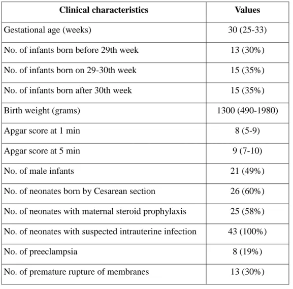
In the study on activation markers, we enrolled 43 preterm infants. Gestational age was 30 (25-33) weeks, while birthweight was 1300 (490-1980) g at birth. The suspected ground for preterm birth was PE in 8 cases, PROM in 13 cases, and could not be settled in 22 cases. PS treatment was applied in 25 cases. All infants had a highly suspected or proven intrauterine infection based on standard criteria [108-109]. PROM cases were coupled with elevated IL-6 levels (256.2 (64.8-2358.9) pg/ml) measured in cord blood, while IL-6 levels in cord blood of infants who had no PROM were normal (11.6 (2.9-45.1) pg/ml). Patient characteristics are summarized in Table 3.
Table 3. Clinical characteristics of preterm neonates enrolled in the study on activation markers. Data are presented as median (range).
Clinical characteristics Values
Gestational age (weeks) 30 (25-33)
No. of infants born before 29th week 13 (30%)
No. of infants born on 29-30th week 15 (35%)
No. of infants born after 30th week 15 (35%)
Birth weight (grams) 1300 (490-1980)
Apgar score at 1 min 8 (5-9)
Apgar score at 5 min 9 (7-10)
No. of male infants 21 (49%)
No. of neonates born by Cesarean section 26 (60%) No. of neonates with maternal steroid prophylaxis 25 (58%) No. of neonates with suspected intrauterine infection 43 (100%)
No. of preeclampsia 8 (19%)
No. of premature rupture of membranes 13 (30%)
4.2. ETHICAL CONSIDERATIONS
Written informed consent was obtained from parents of subjects, and our study was reviewed and approved by an independent ethical committee of the institution (Semmelweis University, Budapest). The study was adhered to the tenets of the most recent revision of the Declaration of Helsinki.
4.3. CBMC ISOLATION
Cord blood mononuclear cells (CBMCs) were separated by a standard density gradient centrifugation (Ficoll Paque, Amersham Biosciences AB, Uppsala, Sweden, 25 minutes, 400 g, 22 °C) from freshly drawn blood collected in lithium heparin-treated tubes (BD Vacutainer, BD Biosciences, San Jose, CA, USA). Peripheral blood samples were taken on the 1st, 3rd, and 7th postnatal days of life. Plasma was handled separately and frozen until further analysis.
4.4. FLOW CYTOMETRY
CBMCs and peripheral whole blood were stained for 30 min at room temperature in the dark with specific monoclonal antibodies.
In the study on vitamin D levels we used the following monoclonal antibodies: PE Cy7-conjugated CD4, APC-Cy7-conjugated CD8, FITC-conjugated CD25, PerCP- conjugated CD62L, APC-conjugated CXCR3, PE-conjugated CCR4, APC-conjugated CD69, PerCP-conjugated HLA-DR, PerCP-conjugated CD3, APC-conjugated CD161, PE-conjugated 6B11, APC-conjugated CD11c, PE-conjugated CD123 and FITC conjugated Lin 1 cocktail in separate tubes, respectively (all from BD Biosciences).
Th1 cells were defined as CD4+ CXCR3+ CCR4-, while Th2 cells were defined as CD4+ CXCR3- CCR4+. NK cells were identified as CD3- CD161+, while NKT cells were identified as CD3+ CD161+ and invariant NKT (iNKT) cells as CD3+ 6B11+. For the determination of DC, pDC and mDC subsets, samples were stained with a lineage cocktail containing antibodies against CD3, CD14, CD16, CD19, CD20, and CD56 (Lin 1). After gating on Lin 1 negative cells, DCS were determined as HLA-DR+, pDCs were determined as the CD123+ HLA-DR+ subset, while mDCs were determined as the CD11c+ HLA-DR+ subset.
After washing, cells were analyzed on a BD FACSAria flow cytometer (BD Biosciences) equipped with 488 nm and 633 nm excitation lasers. Data were processed using the FACSDiVa software.
For the study on the effects of preeclampsia we used the following monoclonal anibodies:
PE Cy7-conjugated CD4, APC-Cy7-conjugated CD8, FITC-conjugated CD25, PerCP- conjugated CD62L, APC-conjugated CXCR3, PE-conjugated CCR4, APC-conjugated CD69, PerCP-conjugated HLA-DR, PerCP-conjugated CD3, APC-conjugated CD161, PE-conjugated 6B11, APC-conjugated CD11c, PE-conjugated CD123 and FITC conjugated Lin 1 cocktail in separate tubes, respectively (all from BD Biosciences).
Th1 cells were defined as CD4+ CXCR3+, while Th2 cells were defined as CD4+
CCR4+. Naïve T cells were defined as CD4+ CD45RA+, while memory T cells were defined as CD4+ CD45RO+. Myeloid dendritic cells (mDCs) were defined as Lin 1- CD11c+, while plasmacytoid dendritic cells (pDCs) were defined as Lin 1- CD123+.
After lysing red blood cells and washing, samples were analyzed on a BD FACSAria flow cytometer (BD Biosciences) equipped with 488 nm and 633 nm excitation lasers.
Data were processed using the FACSDiVa software (BD Biosciences).
In the study on activation markers we used the following monoclonal antibodies:
PE-Cy7-conjugated CD4, APC-Cy7-conjugated CD8, FITC-conjugated CD25, PerCP- conjugated CD62L, APC-conjugated CXCR3, PE-conjugated CCR4, APC-conjugated CD69, PerCP-conjugated HLA-DR, FITC-conjugated CD45RA, PE-conjugated CD45RO in separate tubes, respectively (all from BD Biosciences).
Th1 cells were defined as CD4+ CXCR3+, while Th2 cells were defined as CD4+
CCR4+. Naïve T cells were defined as CD4+ CD45RA+, while memory T cells were defined as CD4+ CD45RO+.
After lysing red blood cells and washing, CBMCs and PBMCs were analyzed on a BD FACSAria flow cytometer (BD Biosciences) equipped with 488 nm and 633 nm excitation lasers. Data were processed using the FACSDiVa software. Figure 8.
demonstrates the gating strategy applied.
4.5. PLASMA CORTISOL AND VITAMIN D LEVELS
Plasma cortisol levels and vitamin D levels were measured with commercially
4.6. PLASMA CYTOKINE LEVELS
Cytokine levels were measured using the Bio-Plex Pro Human Cytokine 17-Plex Panel (M50-00031YV, Bio-Rad, Hercules, CA, USA) following the manufacturer’s instructions. Samples were read using a Bio-Plex reader (Bio-Rad).
4.7. STATISTICS
Data are expressed as median and range. Since Kolmogorov–Smirnoff analysis indicated non-normal distribution of data, the Mann-Whitney test was used to make comparisons between the study groups in the study on the effects of PE. The sample size was estimated to achieve 80% power with 0.45 effect size to detect differences between the patient groups.
The independent effects of gestational and postnatal age, PE, PROM, PS and gender were analyzed using the ‘mixed effect model’ method in the study on activation markers. This is a statistical model containing both fixed effects and random effects. It is particularly used in settings where repeated measurements are made on the same statistical units (ie. longitudinal studies), or where measurements are made on clusters of related statistical units [110].
The mixed effect model was also used to assess the effect of factors other than plasma vitamin D levels on the analyzed inflammatory parameters in the study on vitamin D levels [111]. These factors were plasma cortisol levels as continuous variables and gestational age as categorical variables (27 weeks, 28-29 weeks and 30 weeks).
Statistics were calculated at 5% significance level (p = 0.05) using the GraphPad Prism 5 software (La Jolla, CA, USA) and the SAS software (Cary, NC, USA).
Figure 8. Gating strategy of flow cytometry measurements in the study on the early and late activation markers. Example of a representative sample. FSC – forward scatter, SSC – side scatter.
5. RESULTS
5.1. THE ROLE OF VITAMIN D LEVELS AT BIRTH IN PRETERM INFANTS
In the study on vitamin D levels, we included 28 preterm infants with a median gestational age of 29 weeks at birth. In the majority of them, vitamin D levels were higher than normal in cord blood (23.3 [9.9–45.4] ng/ml (median [range])). Based on vitamin D levels, two patient groups were created: below median (<23.3 ng/ml) and above median (>23.3 ng/ml) (Table 1.).
First, we compared the prevalence of pro- and anti-inflammatory cell subsets and plasma cytokine levels between these groups. Results are demonstrated in Tables 4. and 5., respectively. In infants with vitamin D level below median the prevalence of CD4+
CXCR3+ (Th1) and CD8+ CXCR3+ cell subsets was higher, while the prevalence of CD4+ CCR4+ (Th2), CD8+ CCR4+ and pDC cell subsets was lower than in infants with a plasma vitamin D level above median. No difference was detected in the prevalence of other cell subsets between the two groups, including lymphocyte activation markers (CD25, CD62L, CD69, HLA-DR) and NK cell subtypes.
Of note, no difference was detected in the plasma cytokine levels investigated as shown in Table 5.
Table 4. The prevalence of T cell, NK cell and dendritic cell subsets among lymphocytes and all PBMCs, respectively, determined by flow cytometry. Data are presented as median (range). * p < 0.05 vs. Above median group.
Cell subset Vitamin D above median
Vitamin D below median CD4+ CXCR3+ (Th1 cells) 4.15 (2.68-5.73) 5.86* (3.85-7.32) CD4+ CCR4+ (Th2 cells) 1.56 (1.18-2.01) 1.07* (0.36-1.94)
CD4+ CD25+ 2.34 (2.00-3.66) 2.45 (0.00-0.06)
CD4+ CD62L+ 8.69 (7.74-9.86) 9.68 (7.46-10.60)
CD4+ CD69+ 1.39 (0.87-9.57) 1.68 (1.42-5.15)
CD4+ HLA-DR+ 5.12 (1.60-17.46) 4.05 (2.16-7.09)
CD8+ CXCR3+ 8.65 (6.80-9.02) 9.01* (7.39-10.57)
CD8+ CCR4+ 0.23 (0.09-0.75) 0.12* (0.10-0.23)
CD8+ CD25+ 1.15 (0.86-2.07) 1.29 (0.15-2.78)
CD8+ CD62L+ 7.89 (7.04-8.45) 8.05 (6.96-8.54)
CD8+ CD69+ 3.52 (2.63-6.77) 3.75 (1.06-7.00)
CD8+ HLA-DR+ 0.51 (0.23-4.02) 0.56 (0.14-0.94)
CD3- CD161+ (NK cells) 2.94 (0.57-4.31) 1.81 (0.89-2.66) CD3+ CD161+ (NKT cells) 1.30 (0.22-1.89) 0.69 (0.34-1.05) CD3+ 6B11+ (iNKT cells) 0.02 (0.01-0.06) 0.02 (0.01-0.04) Lin 1- HLA-DR+ (DCs) 6.38 (1.89-8.16) 4.34 (1.48-8.40) Lin 1- HLA-DR+ CD11c+ (mDC) 0.97 (0.28-2.40) 1.03 (0.12-2.29) Lin 1- HLA-DR+ CD123+ (pDC) 1.29 (0.09-2.28) 1.15* (0.64-5.42)
Table 5. Cord blood plasma cytokine levels of the investigated preterm infants (pg/mL). Data are presented as median (range).
Cytokine Vitamin D above median Vitamin D below median
MIP 1b 160 (109-247) 201 (146-223)
MCP1 84 (42-176) 132 (69-182)
IL-17 55 (30-90) 79 (65-86)
IL-13 2.45 (0.43-3.45) 1.61 (0.19-2.97)
IL-12 10.68 (5.22-20.6) 18.85 (9.37-22.39)
IL-10 3.11 (1.63-6.91) 4 (1.99-7.15)
IL-8 38 (19-71) 29 (18-58)
IL-7 0.28 (0.20-1.49) 0.20 (0.20-0.93)
IL-6 11.38 (4.51-14.53) 7.21 (1.89-16.79)
IL-5 0.65 (0.43-1.36) 1.08 (0.13-1.75)
IL-4 0.78 (0.78-0.93) 0.78 (0.78-1.66)
IL-2 1.07 (0.07-2.30) 1.84 (0.07-2.62)
IL-1b 2.82 (0.46-3.56) 1.47 (0.22-4.04)
IFN-g 44.85 (14.85-79.43) 133.1 (47.14-225.1) GM-CSF 136.22 (11.38-198.23) 201.6 (21.54-243.9)
G-CSF 11.13 (2.40-20.90) 8.00 (1.80-30.61)
TNF-a 19.22 (0.95-33.1) 19.22 (0.30-65.23)
Since the inflammatory status might also be influenced by gestational age and plasma cortisol levels, we used the mixed effect model to assess the effects of these factors. According to our analysis, CD4+ CXCR3+ (Th1) lymphocytes were also influenced by both gestational age (at the gestational age of 28–29 weeks) and plasma cortisol levels, while CD8+ CXCR3+ and CD8+ CCR4+ lymphocytes were affected by
gestational age only (in the 28–29 weeks and ≥30 weeks categories, respectively).
Results are shown in Table 6. and Figure 9. Plasmacytoid dendritic cells (pDCs) and CD4+ CCR4+ (Th2) lymphocytes are the only cell subsets from the current study which were only influenced by vitamin D levels.
Table 6. Positive results of mixed effect model analysis for gestational age and plasma cortisol levels. Estimate and % change values demonstrate the difference of the given parameter related to presence of the investigated effect.
Cell subset Effect Effect group p Estimate value % change CD4+ CXCR3+ Gestational age 28-29 weeks 0.0267 1.2030 232
Cortisol level 0.0052 0.3625 43
CD8+ CCR4+ Gestational age 30 weeks 0.0492 -2.1341 -11 CD8+ CXCR3+ Gestational age 28-29 weeks 0.0379 0.6619 93 Gestational age 30 weeks 0.0397 0.5395 69
5.2. THE IMPACT OF PREECLAMPSIA IN PRETERM NEONATES
In the study on the effects of PE, we first compared the prevalence of distinct lymphocyte, NK and dendritic cell subsets between the two groups. Results are summarized in Table 7.
The prevalence of CD4+ T lymphocytes and CD4+HLA-DR+ T cells was significantly lower in preterm neonates of PE mothers on postnatal day 3 (p= 0.0159 and p= 0.0348, respectively) when compared with preterm neonates born to non-PE mothers. In contrast, memory T cells (CD4+CD45RO+) were found to have a significantly higher prevalence in PE on day 7 (p= 0.0308) when compared with the control group.
The prevalence of CD8+CXCR3+ cells was significantly lower in PE on postnatal days 1 and 7 (p= 0.0009 and p= 0.0163, respectively) when compared with control subjects.
CD8+CD69+ T lymphocytes had a lower prevalence on days 0 and 1 (p= 0.0109 and p=
0.0015, respectively) in preterm neonates born to PE mothers. Furthermore, CD8+HLA- DR+ T cells had a significantly lower prevalence in PE on days 0, 3 and 7 (p= 0.0084, p= 0.0308 and p= 0.0019, respectively). mDCs had a lower prevalence on days 1 and 3 (p= 0.0011 and p= 0.0538, respectively) in PE neonates when compared with controls.
No significant difference was detected in the prevalence of other investigated cell subsets between the two groups, including CD3+ T cells, cytotoxic T cells, naïve T cells, CD25+ and CD62L+ activated T cells, Th1 and Th2 cells, NKT, iNKT, NK cells or pDCs.
We also compared plasma cytokine levels between the two groups. Results are shown in Table 8. Interestingly, the indicated cytokine levels are significantly higher in preterm neonates of PE mothers on days 1, 3 and 7 and significantly lower on day 0 when compared to the control group. Of note, monocyte chemotactic protein-1 (MCP-1) and IL-4 had significantly higher levels on all 3 days (1, 3 and 7) (MCP-1: p= 0.0069, p=
0.0089 and p= 0.0178, respectively; IL-4: p= 0.0013, p= 0.0339 and p= 0.0106, respectively) in preterm neonates of PE mothers. Cortisol levels were found to be significantly lower in PE neonates on day 1 and 7 (p= 0.0370 and p= 0.0471, respectively).
Table 7. Significant results of cell prevalence data of the enrolled preterm neonates in the study on the effects of PE, determined by flow cytometry. Data are given as median and (range). * represents significant values, p≤ 0.05.
Cell subset Day Preterm without Preeclampsia
Preterm with Preeclampsia
p value
CD4+ in all cells
0 20.76 (1.57-54.43) 12.59 (0.17-36.72) 0.1322 1 7.95 (0.44-24.63) 3.53 (0.0-15.76) 0.1029 3 10.42 (0.92-32.96) 1.5 (0.01-21.21) 0.0159*
7 10.77 (0.87-58.36) 1.72 (0.07-40.23) 0.1129
CD4+HLA-DR+
in CD4+ cells
0 13.47 (5.17-74.75) 7.42 (0.0-24.18) 0.1063 1 7.02 (1.91-69.60) 6.13 (0.63-19.96) 0.2413 3 7.66 (2.61-17.98) 2.73 (0.97-15.27) 0.0348*
7 7.58 (0.66-15.22) 7.02 (0.43-28.82) 0.9085
CD4+CD45RO+
in CD4+ cells
0 0.91 (0.13-19.15) 1.76 (0.21-3.30) 0.4599 1 0.31 (0.09-2.92) 0.51 (0.0-27.78) 0.2233 3 0.28 (0.01-3.84) 0.42 (0.0-7.41) 0.8903 7 0.6 (0.08-3.84) 1.2 (0.12-6.04) 0.0308*
CD8+CXCR3+ in CD8+ cells
0 83.46 (45.21-97.79) 85.08 (10.67-97.01) 0.9346 1 91.82 (80.88-98.08) 74.23 (50-93.79) 0.0009*
3 90.41 (0.0-98.39) 88.40 (16.30-100) 0.8005 7 94.13 (82.1-97.34) 88.45 (0.0-98.84) 0.0163
CD8+CD69+ in CD8+ cells
0 82.16 (3.39-96.13) 30.1 (0.0-92.42) 0.0109*
1 72.33 (14.23-93.53) 31.22 (0.0-91.56) 0.0015*
3 68.13 (0.0-93.25) 19.14 (0.0-83.53) 0.1235
CD8+HLA-DR+
in CD8+ cells
0 1.51 (0.43-2.67) 0.61 (0.0-1.83) 0.0084*
1 0.80 (0.12-55.51) 0.52 (0.0-26.32) 0.3702 3 0.79 (0.38-5.01) 0.32 (0.0-3.04) 0.0308*
7 1.3 (0.2-2.99) 0.27 (0.0-1.07) 0.0019*
Lin 1- CD11c+ in Lin 1- cells
0 46 (3.90-83.54) 19.06 (5.84-65.22) 0.1514 1 26.84 (3.06-91.42) 10.89 (0.79-44.20) 0.0538*
3 44.42 (0.0-75.39) 11.4 (0.38-39.65) 0.0011*
7 37.42 (5.61-75.84) 19.39 (0.62-65.98) 0.2541
Table 8. Significant results of plasma cytokine and cortisol levels of the enrolled preterm neonates in the study on the effects of PE (pg/mL). Data are given as median and (range). All p values are significant (p≤ 0.05).
Cytokine Day Preterm without Preeclampsia
Preterm with Preeclampsia
p value
MIP-1b 7 118.2 (66.96-183) 1385 (43.41-7579) 0.0106 MCP-1 1 67.05 (12.69-319.5) 267.9 (85.07-2896) 0.0069 MCP-1 3 53.91 (17.18-294.7) 286.4 (17.36-1864) 0.0089 MCP-1 7 81.82 (13.68-200.8) 182.2 (15.10-925.6) 0.0178 IL-17 7 32.85 (10.32-83.83) 104.3 (4.72-4345) 0.0185
IL-13 0 3.78 (0.5-8.290) 1.09 (0.11-5) 0.0131
IL-8 7 22.16 (5.92-79.46) 1475 (1.84-58080) 0.0141 IL-6 7 8.28 (1.99-16.15) 111.9 (3.04-237.1) 0.0035
IL-4 1 0.78 (0.02-1.93) 6.22 (0.78-17.5) 0.0013
IL-4 3 0.78 (0.14-1.38) 1.93 (0.02-4.67) 0.0339
IL-4 7 0.78 (0.02-1.38) 2.45 (0.03-12.9) 0.0106
IL-2 1 0.07 (0.07-1.84) 1.84 (0.07-15.72) 0.0199
IL-2 7 0.07 (0.07-1.84) 2.46 (0.07-15.32) 0.0131
IFN-g 0 157.8 (7.24-775) 22.18 (1.11-133.1) 0.0100 TNF-a 0 68.86 (2.07-334) 15.29 (0.54-61.61) 0.0115 Cortisol 1 845.3 (161.7-2458) 186.1 (41.92-1144) 0.0370 Cortisol 7 381.9 (146.9-792.8) 222.2 (137.3-573.5) 0.0471
5.3. EARLY AND LATE ACTIVATION MARKER EXPRESSION IN T CELLS OF PRETERM NEONATES
The frequency of CD4+ CD25+ and CD8+ CD25+ activated T lymphocytes was higher in case with PROM at all time points. We observed a decrease in the frequency of CD4+ and CD8+ T lymphocytes as well as the CD4+/CD8+ T cell ratio in PE compared to infants not affected by PE at all time points. The frequency of CD4+CD62L+ and CD8+ CD62L+ T lymphocytes was higher in male infants when compared to female infants at all time points. None of the investigated factors had an effect on the expression of the HLA-DR and CD69 activation markers,or the frequency of Th1 (CD4+ CXCR3+), Th2 (CD4+ CCR4+), naïve (CD45RA+) and memory (CD45RO+) T cell subsets. The frequency of Th1 (CD4+ CXCR3+) lymphocytes was higher in infants born before the 29th gestational week compared to those born on the 29-30th gestational week on postnatal days 1 and 3. When we looked at the effect of postnatal age (day 1, 3 and 7 of life) on the frequency of the investigated markers and subsets, we detected several changes. CD4+ T cells have a higher frequency on postnatal days 0 and 3 when compared to day 7. CD4+ CD25+ cells had a lower frequency on postnatal day 0 than on day 7. Of note, Th2 (CD4+ CCR4+) lymphocytes also had a lower frequency on postnatal days 1 and 3 when compared to day 7. Results are summarized in Table 9. and Figure 10.
Table 9. Significant results of mixed effect model analysis for the investigated factors in the study on activation markers. “% change” is expressed vs. Day 7 for postnatal age, vs. PE (present) for preeclampsia, vs. PROM (present) for premature rupture of membranes, vs. Boys for gender, vs. < 29 weeks for gestational age.
T cell subset Effect p Estimate % change
CD4+ Day 0 0.0487 0.04143 4
Day 3 0.0018 0.06641 6
No PE 0.023 0.09089 9
CD8+ No PE 0.0371 0.02683 2
CD4+ CD25+ Day 0 0.0331 -0.1305 -87
No PROM 0.0219 -0.1826 -83
CD8+ CD25+ No PROM 0.0285 -0.1592 -86
CD4+ CD62L+ Boys 0.0572 0.1071 10
CD8+ CD62L+ Boys 0.0309 0.1404 15
CD4+ CXCR3+ 29-30 weeks 0.0291 -0.1256 -88
CD4+ CCR4+ Day 1 0.0341 -0.1342 -87
Day 3 0.024 -0.1431 -86
Figure 10. Box-plots representing frequency values of the investigated cell subsets in different subgroups of preterm infants at birth (Day 0) and on days 1, 3 and 7 of life in the study on activation markers. Horizontal line: median, box: interquartile range, whisker: range. PE – preeclampsia, PROM – premature rupture of membranes. * p <
0.05 vs. No PROM, ** p < 0.05 vs. Male infants, *** p < 0.05 vs. No PE, # p < 0.05 vs.
Day 0, ## p < 0.05 vs. Day 0.
6. DISCUSSION
6.1. PLASMA VITAMIN D LEVELS CONTROL THE INFLAMMATORY BALANCE IN PRETERM INFANTS
Vitamin D can modulate both the innate and adaptive immune responses.
Deficiency in vitamin D is associated with increased autoimmunity and susceptibility to infection. Antigen presenting cells (macrophages and dendritic cells), T cells and B cells have the necessary machinery to synthesize and respond to vitamin D [112]. Stimulation of T cells with vitamin D impairs their proliferation and pro-inflammatory cytokine secretion (IFN-g, IL-17) and promotes the development of Th2 and Treg cells [113]. It was also found that vitamin D inhibits the ability of pDCs to induce T cell activation in mouse and man [114].
pDCs are important players within the immune system, and are the main producers of type I IFN upon TLR9 activation, thus linking the innate and adaptive immune responses. In our study, the prevalence of pDCs was solely influenced by vitamin D levels: infants with a vitamin D level below median had a lower prevalence of pDCs.
Increased susceptibility to viral infections in preterm neonates might be associated with altered pDC function. Recent studies found similar numbers of circulating pDCs in term and preterm newborns and healthy adults. The pDC population seems to appear early in fetal circulation, below 27 weeks. However, pDCs from preterm newborns demonstrate an impaired antiviral cytokine response, in terms of hampered IFN-a production, and also show distinct immature morphological features, such as the lack of TLR9 functionality, leaving preterm newborns vulnerable to viral infections [114]. The other cell subset we found to be influenced by vitamin D levels only in the current study are Th2 cells. The prevalence of this cell subset was also found to be lower in infants with a vitamin D level below median. This positive correlation between plasma vitamin D levels and Th2 cell prevalence is in line with earlier results [113]. Neonatal T cells appear to have a strong bias towards Th2 polarization in vitro [115-116]. Subsequent reports indicated that the skewing to Th2 responses seen in vitro may accurately reflect the activities of neonatal T cells in vivo. For instance, exposure of human fetal T
placenta induce their differentiation into Th2 cells. This Th2 response, comprising IL-4, IL-5, IL-6, IL-9 and IL-13, was found in all newborn infants and seemed to be dominated by high production of IL-10. In later life, normal immune deviation mechanisms redirect these fetal immune responses toward the Th1 cytokine phenotype in non-atopic adult individuals, indicating that the key etiologic factor in atopic disease may not be the initial acquisition of allergen-specific Th2-skewed immunity per se [117]. Besides the influence of vitamin D levels on Th1 and cytotoxic T cell subsets (CD8+ CXCR3+ and CD8+ CCR4+), our study identified that the prevalence of Th1 lymphocytes were also influenced by both gestational age and plasma cortisol levels, while cytotoxic T cells were further influenced by gestational age only. Glucosteroids have a role in inhibiting the IFN-g response in adults, acting directly on T cells or indirectly through IL-12. In this way, an increase in plasma cortisol would induce a decrease in the Th1 products with the imbalance between Th1/Th2 cytokines and a shift to Th2 response [118]. Pinto et al. found an association between increased plasma cortisol levels and a decreased Th1-type response in infants between 6 and 12 months of age with severe RSV (Respiratory Syncytial Virus) infection [119]. On the contrary, our findings indicate that the prevalence of Th1 cells in cord blood of preterm neonates increases with higher plasma cortisol levels. This contradiction might be explained by altered glucosteroid homeostasis and effects in preterm infants. Cortisol is an important physiologic hormone for stress in preterm neonates. Neonates under stress have been shown to have impaired production of cortisol and accumulation of precursors for cortisol [120]. The source of fetal cortisol is also limited because the adrenal cortex does not produce cortisol de novo from cholesterol until around the gestational age of 30 weeks. Although the fetus may produce cortisol earlier in gestation using placental progesterone as a precursor, the preterm delivery will leave the infant with a paucity of the enzymes required for de novo cortisol synthesis [120-121]. CD8+ cytotoxic T cells express relatively high levels of VDR (20). Our current study confirms that vitamin D has an important effect on the prevalence of cytotoxic T cell subsets in preterm infants.
Interestingly, our results show that the prevalence of the CD8+CXCR3+ cell subset was higher, while that of CD8+ CCR4+ cells was lower in the group of infants with a serum vitamin D level below median. The same pattern was observed concerning the prevalence of CD4+ cells expressing these chemokine receptors, which might indicate
that vitamin D may have a direct effect on their cellular expression.
6.2. MATERNAL PREECLAMPSIA HAS A MAJOR IMPACT ON THE IMMUNE SYSTEM OF PRETERM INFANTS
PE is one of the most common complications of pregnancy and it is associated with adverse health outcomes for the mother and her offspring. Dynamic changes are anticipated in the immune system of preterm neonates of PE mothers immediately after birth.
The prevalence of CD4+ T cells was significantly lower on day 3 in neonates of PE mothers when compared with control subjects and a similar trend was observed on the other days. These findings are in line with previous results of Kotiranta-Ainamo et al., showing that neonates born to PE mothers had significantly less CD4+ cells and CD4+CD8+ double-positive cells compared to neonates not affected by PE.
Furthermore, their study also identified a decrease in the CD4/CD8 ratio in PE neonates [93].
The available data on the expression of HLA-DR+ T cells in preterm neonates born to PE mothers is sparse. HLA-DR molecules are involved in antigen processing and presentation, mediating antigen-specific T cell activation [94]. Our study identified a significantly lower prevalence of CD4+HLA-DR+ T lymphocytes in preterm neonates of PE mothers on postnatal day 3 when compared with preterm neonates born to healthy mothers. Furthermore, CD8+HLA-DR+ T cells also had a significantly lower prevalence in PE on days 0, 3 and 7, when compared to control subjects. Previously, it was demonstrated that low levels of HLA-DR expression in preterm newborns is linked with the development of several complications, including high incidence of bacterial infections and pulmonary morbidity, especially in the presence of respiratory distress syndrome (RDS) [106-107]. Several research groups have reported low HLA-DR expression on monocytes on the first days of life (1 and 3) in preterm neonates (<32 weeks) and in very low birth weight (VLBW) neonates. These results seemed to be associated with impaired neonatal host defense, also contributing to the high incidence


![Figure 3. Diagram representing critical steps and associated mediators in lung inflammation, injury and remodelling that result in BPD [23]](https://thumb-eu.123doks.com/thumbv2/9dokorg/1360668.110789/13.892.127.762.126.1007/figure-diagram-representing-critical-associated-mediators-inflammation-remodelling.webp)

![Figure 6. Overview of immunomodulatory actions of 25(OH)D3 and 1,25(OH) 2 D3 on monocytes and macrophages, dendritic cells, effector, and memory T and B lymphocytes [55-56]](https://thumb-eu.123doks.com/thumbv2/9dokorg/1360668.110789/18.892.131.752.501.930/figure-overview-immunomodulatory-monocytes-macrophages-dendritic-effector-lymphocytes.webp)