Novel kinase inhibitor compounds for combination cancer therapy
Ph.D. Thesis book
Pál Gyulavári
Semmelweis University
Doctoral School of Pharmaceutical Sciences
Supervisor: Tibor Vántus Ph.D. senior res. fellow Official rewievers: Tamás Tábi Ph.D. associate professor
Lívius Wunderlich Ph.D. assistant prof.
Chair of exam committee: László Tretter Ph.D., D.Sc. professor Members of exam committee: András Balla Ph.D. associate professor
András Micsonai Ph.D. assistant lecturer Budapest
2019
1 1. Introduction
The various cancerous cell growths are among the most deadly diseases nowadays. Their pivotal, common trait is the uncontrolled multiplication of cells. When cancer cells detach from the originating tissue and invade neighbouring ones -called metastases- they become a serious problem. The function of invaded organs deteriorate and the patient dies soon. Cancers are highly heterogeneous in many aspects but in the last thirty years it became clear, that their genetic variability is the most important one. It is generally agreed today that genetic alterations and the resulted malfunctioning proteins are that stimulate cancer cells to divide ceaselessly. Numerous extrinsic and intrinsic factors can generate these genetic alterations, but the DNA damage repair mechanisms of healthy cells correct them. Provided a critical number of potent alterations evade correction, a cancer cell borns. Surprisingly few genes of the human genome -only about 140- are responsible for the formation of most cancers. These altered genes and the resulting malfunctioning proteins called “drivers”.
Among drivers kinase proteins are overrepresented. Kinases are able to transfer a phosphoryl group (PO3) to their substrates and thus influence their functions. If the substrate of a kinase is also a kinase, a kinase cascade formed. Kinase cascades set up the most important intracellular signalling pathways that eventually regulate all cell functions -also cell proliferation. Effector kinases -like Aurora kinases- directly regulate the functioning macromolecules.
Aurora kinases are crucial regulators of the division of every human cells. The Aurora kinase family constitutes of three paralogue proteins, of which the proteins designated “A” and “B” are the most important ones. Aurora A orchestrates the division of the centrosome and the building up of emanating microtubule spindles. Aurora B has three separate functions: 1) stimulates the condensation of DNA into chromosomes, 2) ensures correct microtubule spindle-chromosome
2
connections and 3) vital for the physical separation of daughter cells, called cytokinesis.
Malfunction (increased or decreased activity) of Aurora kinases result in unequal distribution of chromosomes and so genes into daughter cells. Therefore malfunctioning Aurora kinases facilitate the accumulation of further genetic alterations in cancer cells. Whereas neither Aurora kinase seem to be a driver, their malfunction is common in many types of cancer. Furthermore, their central role in cell proliferation (which is the fundamental property of all cancer cells -as we have seen) and the fact that they are druggable (can be inhibited) by targeted agents make them promising therapeutic targets. According to several experimental results inhibition of Aurora kinases indeed induces apoptosis of cancer cells. However, the question which Aurora paralogue is the best therapeutic target is still open to debate.
About thirty targeted Aurora kinase inhibitors have been developed during the last one and a half decade and all but one failed in clinical trials. There is still no approved Aurora kinase inhibitor anti-cancer drug on the market.
Unfortunately the assortment of targeted agents against drivers is generally skimpy. Moreover, cancer cells easily collect new genetic alterations (for example due to malfunctioning Aurora kinases) and became resistant to a given drug fast. One possible way to bypass this problem is to combine multiple drugs that are synergistic -that is the combination is far more potent than just the summarised effect of monotherapies.
3 2. Aims
As we have seen Aurora kinases are promising therapeutic targets yet, there is no approved inhibitor of them. Therefore we chose the study of Aurora kinase inhibitors as my Ph.D. topic. Previously, a family of benzotiophene-3-carboxamide derivatives were identified in the chemical library of Vichem Ltd. which had some effect on either Aurora kinases. My purpose was to:
I) Characterise the benzotiophene-3-carboxamide compound family in several in vitro and cell-based experiments, corroborate their Aurora kinase inhibitor potency and choose a lead molecule.
II) Test the lead molecule in drug combination experiments.
4 3. Materials and Methods
Cancer cell lines
HCT 116 and HT-29 human colon cancer cell lines were acquired from ATCC and cultured in the recommended media (supplemented with 10% (V/V) foetal bovine serum and antibiotics) in humidified 5%
CO2 incubator.
Inhibitors and recombinant enzymes
All the benzotiophene-3-carboxamide derivatives were designed and synthetized in the Vichem Ltd. Reference Aurora kinase inhibitors VX- 680, MLN8054 and MLN8237 were purchased from Selleck Chemicals (LLC, USA) and Sigma-Aldrich, respectively. All compounds were solved in anhydrous DMSO, stored at room temperature and their purity was verified by HPLC every three months.
Recombinant Aurora A enzyme was purchased from Proteros Biostructures, Aurora B enzyme from SignalChem (lot: E021-1).
Cell viability assay
Viability of compound treated cells was measured using MTT assay on 96 well plates. After removing all media, treated cultures were incubated with 50 µl MTT (3-(4,5-dimethylthiazol-2-yl)-2,5- diphenyltetrazolium bromide) solution (2 mg/ml) for 1.5 hours at 37°C.
MTT solution was carefully removed and crystalline formazan was solubilized with 200 μl detection solution (2-propanol, 1 mM HCl and 10% (V/V) Triton X-100). Absorbance was measured with a Synergy 2 plate reader (BioTek), at wavelengths 570 nm and 635 nm. The 635 nm data (reference wavelength) was subtracted from 570 nm data (test wavelength) and results were used to calculate normalised cell viability data compared to DMSO-treated positive and cell-free negative control wells. Using these raw data IC50 values were determinated with Excel (Microsoft) and XLfit 5.1.0 (IDBS, Surrey, UK) software.
5
In vitro recombinant kinase inhibition assay
Reactions of the purchased kinazes were run in optimized buffers on 384 well plates (Corning 3676). ATP concentration was adjusted to KM[ATP] value in a separate set of experiments. Enzyme activity was arrested and detected by IMAP (Molecular Devices) assay system. IC50 values were calculated with Excel (Microsoft) and XLfit 5.1.0 (IDBS, Surrey, UK) software.
Flow cytometry
Cells were seeded to 24 well plates, treated and supernatants were collected together with trypsinized cells into sample tubes. Samples were stained with propidium-iodide (to visualize DNA content) or propidium-iodide and Annexin V-FLUOS (to assess rate of apoptosis).
The proportion of fluorescent cell populations was detected and quantified with a FACSCalibur flow cytometer using CellQuest Pro software (BD Biosciences). Sample evaluation was performed also with CellQuest Pro and Excel (Microsoft) software.
Western blot analysis
Cells were cultured and treated on 60 mm Petri-dishes then lysed with ice-cold RIPA buffer (50 mM Tris pH 7.4, 150 mM NaCl, 1%
(V/V) NP-40, 0.5% (V/V) sodium deoxycholate, 0.1% (V/V) SDS, 2 mM EDTA, 2 mM EGTA, supplemented right before use with 1 mM DTT, 1 mM sodium orthovanadate, 200 µM PMSF and 0.5% (V/V) protease inhibitor cocktail (Calbiochem)). Samples were prepared as usual and separated with 10% SDS-PAGE, blotted to PVDF membrane and incubated with antibodies (1° -overnight, 4°C / HRP-conjugated 2° - 1 h, room temperature). Antibodies were purchased from CellSignaling Technologies (Danvers, MA, USA) and from Sigma-Aldrich (St. Louis, MO, USA). Proteins of interest were visualized with chemilumi- nescence reagent (Western Lightning Plus-ECL, PerkinElmer) on CL- XPosure Films (Thermo Scientific, MA, USA).
6 Fluorescence microscopy
Cells were seeded to and treated on 96 well Ibidi µ-plates (89626) then fixed with formalin (4% V/V, 10 min, room temperature), permeabilized with 0.1% (V/V) Triton X-100 (10 min, room temperature) and incubated with antibodies (1° -overnight, 4°C / fluorescent dye-conjugated 2° -1 h, room temperature). Nuclei were stained with 1 μg/ml DAPI solution (10 min, room temperature) and cells were observed using Zeiss Axiovert 200M fluorescence microscopeand AxioVision 3.1 software. Merged images were created by FIJI software.
Drug combination experiments
For drug combination studies cell viability was measured with MTT assay as described above. All compounds were applied in monotherapy and also in combination at a constant ratio of 1:1. Serial three-fold dilutions starting from 30 µM were prepared for every treatment. Means of raw cell viability data were transformed to be between 0 and 1 as required by the CompuSyn® v1.0 software (ComboSyn Inc.). The software calculated the CI (combination index) values at the IC50 value of the combination. CI>1 indicates antagonism, CI=1 additive effect and CI<1 synergism, respectively.
Statistical analysis
Most data are the mean values of at least three independent experiments. Recombinant kinase inhibition measurements were evaluated by calculating the Z’ value: Z’=1-((3SDmax+3SDmin)/(AVmax- AVmin)) where SDmax is the standard deviation of the positive, SDmin is of the negative control, AVmax is the mean value of the positive and AVmin is of the negative controls. Only measurements of a Z’ value higher than 0.5 were accepted for evaluation.
Flow cytometry data were analysed by Student’s t-test (two-sided, unpaired) using Excel software. Statistical significance was defined as p
< 0.05.
7 4. Results
I tested the HCT 116 cell viability inhibition properties of 84 small molecules of the Vichem Ltd. with a benzotiophene-3-carboxamide scaffold. There are only minor differences between the 84 compounds that allowed me to make nice conclusions of structure-activity relationship. Eight compounds had the same or better effect than of the reference Aurora kinase inhibitors. As opposed to the HCT 116 colon cancer cells the eight chosen compounds had no considerable effect on primer human fibroblast cells.
All eight compounds inhibited Aurora A and B activity equally to reference Aurora kinase inhibitors in in vitro recombinant kinase activity assay. However I could not observe considerable differences in potency among the compounds.
Appearance of multinucleated cells (that also means elevated DNA content) in the culture is the sure hallmark of Aurora B and cytokinesis inhibition. According to the in vitro kinase inhibition results, all eight selected compounds should have abrogated cytokinesis but in the flow cytometry experiments only five of them did at 100 nM concentration, after 24 hours (similarly to VX-680 reference Aurora kinase inhibitor).
I could observe the same pattern in fluorescence microscopy experiments regarding multiplication of cell nuclei at 100 nM and 1 µM concentration after 24 hours.
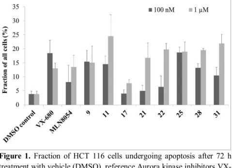
Moreover, exactly the same compounds induced apoptosis at 100 nM which inhibited cytokinesis and created multinucleated cells. On the other hand, at 1 µM concentration all compounds induced apoptosis, also the dedicated Aurora A inhibitor MLN8054 (Figure 1). It is worth to note that 24 or 48 hour treatment was not enough to induce cell death.
Multinuclear cell state triggered apoptosis of cancer cells only after 72 hours.
8
Figure 1. Fraction of HCT 116 cells undergoing apoptosis after 72 h treatment with vehicle (DMSO), reference Aurora kinase inhibitors VX- 680 and MLN8054 and eight selected benzotiophene-3-carboxamide compounds. Error bars represent standard deviation (SD).
On the basis of cell viability and apoptosis induction data I declared compound 25 as the lead molecule of benzotiophene-3-carboxamides (Figure 2). 25 required only 100 nM to create multinuclear cells and induce apoptosis while -for example- 21 needed 1 µM concentration to achieve that effect. Next I also investigated the effect of both compounds on Aurora A and B activity in living HCT 116 cells using western blot. Whereas 21 inhibited phosphorylation (and activity) of Aurora A at 500 nM, did only partially of Aurora B at 1 µM. On the contrary, 25 -in concordance with flow cytometry results- inhibited both Aurora A and B kinase activity already at 100 nM.
Finally, my last goal was to test the potency of 25 in concurrent treatment. Therefore I treated HCT 116 and HT-29 cancer cells with 25 or the reference Aurora kinase inhibitor VX-680 in combination with
9
several targeted agents. The following six drug partners had already been demonstrated to synergise with Aurora kinase inhibitors:
- GSK2126458, a PI3K/mTOR inhibitor - Erlotinib, an EGFR inhibitor
- Trichostatin A, a Class I and II histone deacetylase inhibitor - Dasatinib, a BCR/Abl and Src family KI
- Lonafarnib, a farnesyltransferase inhibitor - Carfilzomib, a proteasome inhibitor
Further two compounds had no such precedent:
- Crizotinib, the ALK, ROS1 and c-Met inhibitor.
- Compound 34, an in-house EGFR–c-Met dual inhibitor
Just like VX-680 25 showed synergism in most compound combinations. Furthermore, both Aurora kinase inhibitors had a synergistic effect with the c-Met inhibitor compounds.
Figure 2. Chemical structure of compounds 21 (A) and 25 (B).
10 5. Conclusions
• The completely novel benzothiophene-3-carboxamide scaffold is a promising structure for the further development of AKIs. Many benzothiophene-3-carboxamide derivatives inhibit Aurora A and B kinase function in in vitro assays, abrogate viability and induce apoptosis of human colon cancer cells at concentrations comparable to reference compounds.
• Inhibition of Aurora B kinase and the resulting cytokinesis disruption and multinuclear cell state is sufficient and necessary to induce apoptosis in HCT 116 cells. Compounds selective to Aurora A need higher concentration to inhibit also Aurora B and induce apoptosis.
• Compound 25 is a drug-like multi-kinase inhibitor with strong AKI properties and qualifies as the lead molecule of the benzotiophene- 3-carboxamide derivative compounds of Vichem Ltd.
• In combination with various targeted agents 25 behaves like a true AKI. I demonstrated the first time that the combination of a c-Met inhibitor and an AKI can be synergistic in some circumstances.
11 6. List of the candidate’s publications
Publications related to the Thesis:
1. Pál Gyulavári, Bálint Szokol, István Szabadkai, Diána Brauswetter, Péter Bánhegyi, Attila Varga, Péter Markó, Sándor Boros, Eszter Illyés, Csaba Szántai-Kis, Marcell Krekó, Zsófia Czudor, László Őrfi (2018) Discovery and optimization of novel benzothiophene- 3-carboxamides as highly potent inhibitors of Aurora kinases A and B. Bioorganic & Medicinal Chemistry Letters 28(19), 3265-3270.
DOI: 10.1016/j.bmcl.2018.05.064.
IF: 2,442
2. Bálint Szokol, Pál Gyulavári, Ibolya Kurkó, Ferenc Baska, Csaba Szántai-Kis, Zoltán Greff, Zoltán Őrfi, István Peták, Kinga Pénzes, Robert Torka, Axel Ullrich, László Őrfi, Tibor Vántus and György Kéri (2014) Discovery and biological evaluation of novel dual EGFR/c-Met inhibitors. ACS Medicinal Chemistry Letter 5(4), 298- 303.
DOI: 0.1021/ml4003309 IF: 3,120
3. Bálint Szokol, Pál Gyulavári, Ferenc Baska, Ibolya Kurkó, Zoltán Greff, Csaba Szántai-Kis, Zoltán Őrfi, István Peták, Axel Ullrich, Tibor Vántus, György Kéri and László Őrfi (2013) Development and biochemical characterization of EGFR/c-Met dual inhibitors.
Acta pharmaceutica Hungarica 83(4), 121-33.
12 Further publications:
4. Margita Márton, Nikolett Tihanyi, Pál Gyulavári, Gábor Bánhegyi, Orsolya Kapuy (2018) NRF2-regulated cell cycle arrest at early stage of oxidative stress response mechanism. PLoS One 13(11), e0207949
DOI: 10.1371/journal.pone.0207949 IF: 2,766
5. István Szabadkai, Robert Torka, Rita Garamvölgyi, Ferenc Baska, Pál Gyulavári, Sándor Boros, Eszter Illyes, Axel Choidas, Axel Ullrich and László Őrfi (2018) Discovery of N-[4-(Quinolin-4- yloxy)phenyl]benzenesulfonamides as Novel AXL Kinase Inhibitors. Journal of Medicinal Chemistry 61(14), 6277-6292.
DOI: 10.1021/acs.jmedchem.8b00672.
IF: 6,253
6. József Murányi, Attila Varga, Bianka Gurbi, Pál Gyulavári, Gábor Mező, Tibor Vántus (2017) In Vitro Imaging and Quantification of the Drug Targeting Efficiency of Fluorescently Labeled GnRH Analogues. Journal of Visualized Experiments 121 Paper: e55529.
DOI: 10.3791/55529.
IF: 1,184
7. József Murányi, Pál Gyulavári, Attila Varga, Györgyi Bökönyi, Henriette Tanai, Tibor Vántus, Domonkos Pap, Krisztina Ludányi, Gábor Mező and György Kéri (2016) Synthesis, characterization and systematic comparison of FITC-labelled GNRH-I, -II and -III analogues on various tumour cells. Journal of Peptide Science 22(8), 552-60.
DOI 10.1002/psc.2904 IF: 1,972
13
8. Attila Varga, Pál Gyulavári, Zoltán Greff, Krisztina Futosi, Tamás Németh, Laura Simon-Szabó, Krisztina Kerekes, Csaba Szántai- Kis, Diána Brauswetter, Márton Kokas, Gábor Borbély, Anna Erdei, Attila Mócsai, György Kéri, Tibor Vántus (2015) Targeting vascular endothelial growth factor receptor 2 and protein kinase D1 related pathways by a multiple kinase inhibitor in angiogenesis and inflammation related processes in vitro. PLoS One 10(4), e0124234.
DOI: 10.1371/journal.pone.0124234.
IF: 3,057