CHANGES IN mRNA EXPRESSION OF MEMBERS OF TGFB1-ASSOCIATED PATHWAYS IN HUMAN
LEUKOCYTES DURING EBV INFECTION
ELENA NIKOLAEVNAFILATOVA*, NIKOLAYALEKSANDROVICHSAKHARNOV, DMITRYIGOREVICHKNYAZEV and OLEG VLADIMIROVICH UTKIN Laboratory of Molecular Biology and Biotechnology, Blokhina Scientific Research Institute of Epidemiology and Microbiology of Nizhny Novgorod, Nizhny Novgorod,
Russian Federation
(Received: 5 September 2018; accepted: 24 October 2018)
Transforming growth factorβ1 (TGFB1) likely contributes to the pathogenesis of Epstein-Barr virus (EBV)-mediated cancer. A microarray containing 59 probes for detecting mRNA of members of TGFB1-associated pathways was developed. mRNA expression of TGFB1 receptors and members of connected pathways were examined in peripheral blood leukocytes of patients during acute EBV infection and after recovery.TGFB1andTGFBR2mRNA expression was increased in patients with EBV infection. Similarly, mRNA expression of protein kinase C (PRKCB), MAP3K7, PDLIM7, and other members of TGFB1 and NF-κB signaling pathways increased.
A shift of mRNA transcript variant expression of some key members (TGFBR2, PRKCB, and NFKBIB) of involved signaling pathways was detected. After the patients’recovery, most of the altered mRNA expression has been normalized. We speculate that in patients with EBV infection, members of TGFB1-associated path- ways contribute to the suppression of proapoptotic and induction of pro-survival factors in leukocytes. The modulation of TGFB1-associated pathways may be considered as a potential risk factor in the development of EBV-associated tumors in patients with acute EBV infection.
Keywords:EBV, TGFB1, NF-κB, PRKCB, apoptosis, proliferation, leukocytes Introduction
Epstein-Barr virus (EBV, Human gammaherpesvirus 4) is a ubiquitous human oncogenic virus that persists in over 90% of the world’s population. It is known that EBV makes a significant contribution to several human cancers, such as B-cell, T-cell, and NK-cell lymphomas [1].
*Corresponding author; E-mail:filatova@nniiem.ru
Transforming growth factorβ1 (TGFB1) is a multifunctional polypeptide that plays a critical role in the regulation of cell proliferation, differentiation, and growth [2]. It induces EBV lytic infection by activating the expression of EBV’s latent-lytic switchBZLF1gene through the canonical SMAD pathway [3]. TGFB1 might provide a significant contribution to the transforming potential of EBV. In patients with EBV- associated nasopharyngeal carcinomas, high serum level of TGFB1 is an unfavorable prognostic [4], for it can enhance tumor progression by stimulating epithelial- mesenchymal transition [5]. On the other hand, in EBV-infected gastric epithelial cell lines, TGFB1 mediates growth inhibition and apoptosis [6].
The dual nature of TGFB1 is determined by its ability to crosstalk with MAPK8 and NF-κB signal pathways (hereafter official full names of genes are provided in Supplementary Table I; available at https://cloud.nniiem.ru/s/
1FdiA0LgZEOrNtV) [7–9]. Regulatory molecules like protein kinase C PRKCB are capable of preventing TGFB1-dependent apoptosis in tumor cells by suppres- sion of activity of transcription modulator SMAD3 [10] and activation of MAPK8 and NF-κB signaling pathway, thereby promoting cell survival [11–13]. In addition, for some members of TGFB1-associated pathways, multiple mRNA transcript variants with different functions are identified.
All of the above make it difficult to assess the contribution of TGFB1 to the development of EBV-associated pathology. Expression pattern of members of TGFB1-connected pathways in blood cells of patients with non-oncological EBV infection is currently poorly understood. Simultaneously, this knowledge will improve our understanding of the mechanisms of transformation of infected cells and will offer new strategies for the therapy of EBV-associated cancer.
The aim of this study was to examine the expression pattern of members of TGFB1-associated pathways in leukocytes of patients with acute EBV infection.
Materials and Methods
A microarray design was developed using “Splice variants microarray design pipeline”algorithm [14]. A total of 59 target probes were selected including 23 gene“total mRNA”probes (designed to detect all mRNA transcript variants of one gene) and 36 “mRNA transcript variant” probes (designed to detect one specific mRNA transcript variant of one gene; Supplementary Table I). Seventy
“negative control” probes, design based on Rhizobium rubi genome, were included in each microarray. Microarray design was adapted for probe synthesis using Layout Designer software (CustomArray Inc., WA, USA). Probe synthesis was conducted in situ on the 12K microarray slides by the B3 Synthesizer (CustomArray Inc.). The synthesis and post-synthetic slide deprotection were
carried out in accordance with the manufacturer’s protocols using recommended reagents.
Blood samples were obtained from 7- to 18-year-old (median=12) patients diagnosed with infectious mononucleosis. Acute EBV infection was confirmed by clinical symptoms and laboratory tests (ELISA and PCR). Blood samples were taken in acute phase of infection (EBVinf, n=6) before the beginning of symptomatic drug therapy, which included, by prescription, ibuprofen, acetamin- ophen, and xylometazoline, and 2 months after apparent clinical recovery (EBVrec,n=5). Due to the high similarity of the pathogenesis mechanism and symptoms of the disease, children of 8–17 (median=11) years old with confirmed acute Human herpesvirus 6 infection (HHV-6inf, n=7 and HHV-6rec, n=7) were used as the comparison group. Another group comprised healthy volunteers of 8–17 (median=11) years old without clinical and laboratory signs of infection (NORM,n=17). All procedures performed in studies involving human partici- pants were in accordance with the ethical standards of the institutional and national research committee and with the 1964 Helsinki Declaration and its later amend- ments or comparable ethical standards. Informed consent was obtained from all individual participants’ parents or legal guardians included in the study.
Blood samples were treated with a hemolytic solution (CRIE, Russia) to remove red blood cells. From the obtained leukocyte fraction, total RNA was isolated and proceeded to biotin-labeled antisense RNA according to common protocols using commercial kits and reagents (Evrogen, Russia; DNA synthesis, Russia; Thermo Scientific, EU). Hybridization of the labeled RNA on the microchip and measurement of the amperometric signal were carried out using a commercial set of ElectraSense reagents and equipment (CustomArray Inc.), according to the manufacturer’s recommendations.
All further calculations were performed using R software (RStudio version 1.1.453, RStudio, Inc., Boston, MA, USA). Data were normalized using the subset quantile normalization based on the non-specific control features [15]. To estimate fold change, sample means of NORM and EBV were calculated for each target probe. Fold change (%) was calculated as [(EBV sample mean×100/NORM sample mean)−100]. For each target probe, NORM sample versus EBVinf sample were tested using t-test; p=0.05 was selected as threshold value. The same procedures were performed using EBVrec, HHV-6inf, and HHV-6rec samples.
Results
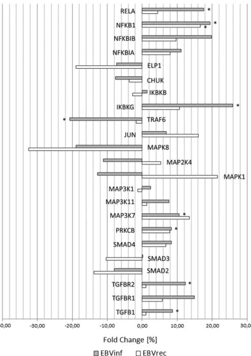
It was shown that EBV infection was followed by change of expression pattern of TGFB1 and TGFB1-connected pathways in human leukocytes
Figure 1.Fold change of total mRNA expression of members of TGFB1-connected pathways in leukocytes of patients with acute EBV infection (EBV) and after apparent clinical recovery (EBVrec) in comparison with healthy volunteers. *Difference is statistically significant (p<0.05). For mRNA
transcript variants, see Supplementary Table I
(Figure 1, Supplementary Table I, available at https://cloud.nniiem.ru/s/
1FdiA0LgZEOrNtV). TGFB1 total mRNA expression and mRNA transcript variant 1 expression increased in EBVinf compared with NORM. After the recovery, TGFB1expression has been normalized.
Expression of some of the members of SMAD-mediated TGFB1 signaling pathway was increased in EBVinf compared with healthy volunteers (NORM). In EBVinf leukocytesTGFBR2total mRNA expression and mRNA transcript variant 2 expression,SMAD2mRNA transcript variant 2 expression increased.TGFBR1, SMAD3, andSMAD4mRNA expression did not change in EBVinf compared with NORM. In EBVrec, expression of mRNA of members of SMAD-mediated TGFB1 signaling pathway did not differ compared with NORM, except for SMAD2 mRNA transcript variant 2 expression, which remained elevated. In EBVinf,PRKCBtotal mRNA expression and mRNA transcript variant 2 expres- sion decreased compared with NORM. Apparent clinical recovery was accompa- nied by normalization of PRKCBmRNA expression.
In EBVinf, MAP3K7 total mRNA expression increased compared with NORM. This indicator returned to normal after clinical recovery. EBV infection was not accompanied by the change in the mRNA expression of other participants of MAPK8 signaling pathways (MAP3K11, MAP3K1, MAPK1, MAP2K4, MAPK8, and JUN). TRAF6 total mRNA expression and expression of both TRAF6 mRNA transcript variants decreased in EBVinf compared with NORM.
After recovery, theTRAF6 mRNA expression did not differ from NORM.
mRNA expression of some members of NF-κB signaling pathway was modulated in leukocytes of patients with EBV infection. Among members of IKK complex, CHUK, IKBKB, and ELP1 mRNA expression did not change, but IKBKGtotal mRNA expression and mRNA transcript variants 3 and 4 expression increased in EBVinf compared with NORM. After clinical recovery, IKBKG mRNA transcript variant 4 expression remained elevated. Among NF-κB inhibitors, NFKBIA mRNA expression did not change in patients with EBV.
NFKBIBtotal mRNA expression also did not change in EBVinf compared with NORM, but NFKBIB mRNA transcript variants 1 and 2 expression increased, whereas NFKBIBnon-coding mRNA transcript variant 3 expression decreased.
NFKBIB mRNA transcript variants 1 and 2 expression remained elevated in EBVrec, andNFKBIBnon-coding mRNA transcript variant expression normal- ized after recovery. Among NF-κB subunits, NFKB1 total mRNA expression, NFKB1mRNA transcript variant 1 expression, andRELAtotal mRNA expression increased, whereas NFKB1mRNA transcript variant 3 expression decreased in EBVinf compared with NORM. NFKB1 total mRNA expression and NFKB1 mRNA transcript variant 1 expression remained elevated after apparent clinical recovery.
We did not reveal similar mRNA expression pattern in children with acute HHV-6 infection (Supplementary Table I).
Discussion
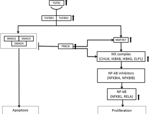
We speculate that revealed changes of mRNA expression pattern in leuko- cytes of patients with EBV were caused by acute EBV infection. Our results also suggest that members of TGFB1-associated pathways play significant role in viral pathogenesis. The revealed changes might be associated with suppression of apoptosis and promotion of leukocytes’proliferation through activation of protein kinase C PRKCB (Figure 2) or/and modulation of mRNA transcript variants expression of some key members (TGFBR2,PRKCB, andNFKBIB) of involved signaling pathways.
Figure 2.mRNA expression of members of TGFB1-associated pathways in leukocytes of patients with acute EBV infection. Thick arrows show changes in total mRNA expression of some members of TGFB1-associated pathways in infected patients compared with healthy volunteers. Detected
mRNA expression pattern’s changes might result in inhibiting TGFB1-dependent apoptosis, activation of NF-κB, and stimulation of cell proliferation
Specifically, TGFBR2 mRNA transcript variant 2, which was highly expressed in EBVinf leukocytes, is typically detected in leukemia cells where it is associated with impaired cell differentiation and proliferation, as well as suppression of apoptosis [16]. Expression ofTGFBR2mRNA transcript variant 1, which is able to stimulate TGBF1-dependent suppression of proliferation and apoptosis in tumor cell lines [17], did not change in EBVinf. In this study,PRKCB mRNA transcript variant 2 expression decreased in patients with EBV, and similar results were observed in colorectal cancer cells in whichPRKCBmRNA transcript variant 2 isozyme reverses cell transformation. In patients with colorectal cancer, low levels ofPRKCBmRNA transcript variant 2 expression serve as a predictor for poor survival outcome [18]. In addition, according to ourfindings, expression ofPRKCBmRNA transcript variant 1, which is directly involved in the MAPK and NF-κB activation and cell proliferation, increased in EBVinf blood, although it was not statistically significant. Again, in EBVinf leukocytes,NFKBIBmRNA expression pattern changed upward, since protein coding mRNA transcript variant expression increased, whereas non-coding transcript variant expression decreased.
All the above contribute to the suppression of proapoptotic and induction of pro-survival factors in leukocytes. The results allow us to consider the modulation of TGFB1-associated pathways as a potential risk factor in the development of EBV-associated tumors in patients with acute EBV infection.
The data sets generated and analyzed during this study are available from the corresponding author on reasonable request.
Conflict of Interest No conflict of interest was declared by the authors.
References
1. Dojcinov, S. D., Fend, F., Quintanilla-Martinez, L.: EBV-positive lymphoproliferations of B- T- and NK-cell derivation in non-immunocompromised hosts. Pathogens7, 28 (2018).
2. Yan, X., Xiong, X., Chen, Y.-G.: Feedback regulation of TGF-βsignaling. Acta Biochim Biophys Sin50, 37–50 (2018).
3. Iempridee, T., Das, S., Xu, I., Mertz, J. E.: Transforming growth factor β-induced reactivation of Epstein-Barr virus involves multiple Smad-binding elements cooperative- ly activating expression of the latent-lytic switchBZLF1gene. J Virol85, 7836–7848 (2011).
4. Xu, J., Menezes, J., Prasad, U., Ahmad, A.: Elevated serum levels of transforming growth factorβ1 in Epstein-Barr virus-associated nasopharyngeal carcinoma patients. Int J Cancer 84, 396–399 (1999).
5. Xie, L., Law, B. K., Chytil, A. M., Brown, K. A., Aakre, M. E., Moses, H. L.: Activation of the Erk pathway is required for TGF-β1-induced EMTin vitro. Neoplasia (New York, NY) 6, 603–610 (2004).
6. Fukuda, M., Kurosaki, W., Yanagihara, K., Kuratsune, H., Sairenji, T.: A mechanism in Epstein-Barr virus oncogenesis: Inhibition of transforming growth factor-β1-mediated induction of MAPK/p21 by LMP1. Virology 302, 310–320 (2002).
7. Al-Azayzih, A., Gao, F., Goc, A., Somanath, P. R.: TGFβ1 induces apoptosis in invasive prostate cancer and bladder cancer cells via Akt-independent, p38 MAPK and JNK/SAPK- mediated activation of caspases. Biochem Biophys Res Commun427, 165–170 (2012).
8. Bailey, K. L., Agarwal, E., Chowdhury, S., Luo, J., Brattain, M. G., Black, J. D., Wang, J.:
TGFβ/Smad3 regulates proliferation and apoptosis through IRS-1 inhibition in colon cancer cells. PLoS One12, e0176096 (2017).
9. Freudlsperger, C., Bian, Y., Contag Wise, S., Burnett, J., Coupar, J.: TGF-βand NF-κB signal pathway cross-talk is mediated through TAK1 and SMAD7 in a subset of head and neck cancers. Oncogene32, 1549–1559 (2013).
10. Yakymovych, I., Ten Dijke, P., Heldin, C.H., Souchelnytskyi, S.: Regulation of Smad signaling by protein kinase C. FASEB J15, 553–555 (2001).
11. Kawakami, T., Kawakami, Y., Kitaura, J.: Protein kinase C beta (PKC beta): Normal functions and diseases. J Biochem (Tokyo)132, 677–682 (2002).
12. Krappmann, D., Patke, A., Heissmeyer, V., Scheidereit, C.: B-cell receptor- and phorbol ester-induced NF-κB and c-Jun N-terminal kinase activation in B cells requires novel protein kinase C’s. Mol Cell Biol21, 6640–6650 (2001).
13. Uemura, N., Kajino, T., Sanjo, H., Sato, S., Akira, S., Matsumoto, K., Ninomiya-Tsuji, J.:
Tak1 is a component of the Epstein-Barr virus Lmp1 complex and is essential for activation of JNK but not of NF-κB. J Biol Chem281, 7863–7872 (2006).
14. Solntsev, L. A., Starikova, V. D., Sakharnov, N. A., Knyazev, D. I., Utkin, O. V.: Strategy of probe selection for studying mRNAs that participate in receptor-mediated apoptosis signaling. Mol Biol49, 457–465 (2015).
15. Wu, Z., Aryee, M. J.: Subset quantile normalization using negative control features.
J Comput Biol17, 1385–1395 (2010).
16. Wu, Y., Su, M., Zhang, S., Cheng, Y., Liao, X. Y., Lin, B. Y., Chen, Y. Z.: Abnormal expression of TGF-beta type II receptor isoforms contributes to acute myeloid leukemia.
Oncotarget8, 10037–10049 (2016).
17. Wan, J., Sun, L., Mendoza, J. W., Chui, Y. L., Huang, D. P.: Elucidation of the c-Jun N-terminal kinase pathway mediated by Epstein-Barr virus-encoded latent membrane protein 1. Mol Cell Biol24, 192–199 (2004).
18. Dowling, C. M., Phelan, J., Callender, J. A., Cathcart, M. C., Mehigan, B.: Protein kinase C beta II suppresses colorectal cancer by regulating IGF-1 mediated cell survival. Oncotarget 7, 20919–20933 (2016).