Oral Epithelial Cells Distinguish between Candida Species with High or Low Pathogenic Potential through MicroRNA
Regulation
Márton Horváth,aGábor Nagy,bNóra Zsindely,aLászló Bodai,bPéter Horváth,d,eCsaba Vágvölgyi,aJoshua D. Nosanchuk,f,g Renáta Tóth,a Attila Gácsera,c
aDepartment of Microbiology, University of Szeged, Szeged, Hungary
bDepartment of Biochemistry and Molecular Biology, University of Szeged, Szeged, Hungary
cMTA-SZTE Lendület Mycobiome Research Group, University of Szeged, Szeged, Hungary
dSynthetic and Systems Biology Unit, Biological Research Centre (BRC), Szeged, Hungary
eInstitute for Molecular Medicine Finland, University of Helsinki, Helsinki, Finland
fDepartment of Medicine (Infectious Diseases), Albert Einstein College of Medicine, Bronx, New York, USA
gDepartment of Microbiology and Immunology, Albert Einstein College of Medicine, Bronx, New York, USA Renáta Tóth and Attila Gácser share last authorship.
ABSTRACT Oral epithelial cells monitor microbiome composition and initiate immune response upon dysbiosis, as in the case of Candidaimbalances.Candidaspecies, such as C. albicans and C. parapsilosis, are the most prevalent yeasts in the oral cavity.
Comparison of healthy oral epithelial cell responses revealed that while C. albicans infection robustly activated inflammation cascades, C. parapsilosis primarily activated various inflammation-independent pathways. In posttranscriptional regulatory proc- esses, several miRNAs were altered by both species. For C. parapsilosis, the dose of yeast cells directly correlated with changes in transcriptomic responses with higher fun- gal burdens inducing significantly different and broader changes. MicroRNAs (miRNAs) associated with carbohydrate metabolism-, hypoxia-, and vascular development-related responses dominated withC. parapsilosisinfection, whereasC. albicansaltered miRNAs linked to inflammatory responses. Subsequent analyses of hypoxia-inducible factor 1a (HIF1-a) and hepatic stellate cell (HSC) activation pathways predicted target genes through which miRNA-dependent regulation of yeast-specific functions may occur, which also supported the observed species-specific responses. Our findings suggest thatC. parapsilosisis recognized as a commensal at low doses by the oral epithelium;
however, increased fungal burden activates different pathways, some of which overlap with the inflammatory processes robustly induced byC. albicans.
IMPORTANCEA relatively new topic within the field of immunology involves the role of miRNAs in innate as well as adaptive immune response regulation. In recent years, posttranscriptional regulation of host-pathogenic fungal interactions through miRNAs was also suggested. Our study reveals that the distinct nature of human oral epithelial cell responses towardC. parapsilosisandC. albicansis possibly due to spe- cies-specific fine-tuning of host miRNA regulatory processes. The findings of this study also shed new light on the nature of early host cell transcriptional responses to the presence of C. parapsilosisand highlight the species’potential inflammation- independent host activation processes. These findings contribute to our better understanding of how miRNA deregulation at the oral immunological barrier, in non- canonical immune cells, may discriminate between fungal species, particularly Candidaspecies with high or low pathogenic potential.
KEYWORDS Candida, oral epithelial cell, host-pathogen interaction, miRNA regulation
CitationHorváth M, Nagy G, Zsindely N, Bodai L, Horváth P, Vágvölgyi C, Nosanchuk JD, Tóth R, Gácser A. 2021. Oral epithelial cells distinguish betweenCandidaspecies with high or low pathogenic potential through microRNA regulation. mSystems 6:e00163-21.
https://doi.org/10.1128/mSystems.00163-21.
EditorTamia A. Harris-Tryon, UT Southwestern Medical Center at Dallas
Copyright© 2021 Horváth et al. This is an open-access article distributed under the terms of theCreative Commons Attribution 4.0 International license.
Address correspondence to Attila Gácser, gacsera@bio.u-szeged.hu.
Received11 February 2021 Accepted14 April 2021 Published11 May 2021
T
he barrier function of epithelium is of paramount importance in maintaining home- ostasis and protecting hosts against an array of injuries, including from microbes.Besides providing physical protection, cells of the epithelium produce and secrete vari- ous enzymes (e.g., lysozymes), peptides (e.g., defensins), and other small molecules (e.g., free oxygen radicals) that inhibit or kill diverse microbes (1). Epithelial cells also actively contribute to innate immune responses (2). Opportunistic pathogenicCandida species are members of the normal human mucosal microflora of the oral cavity, airways, intestinal tract, and genitals (3). These species primarily cause infections in immunosup- pressed patients or individuals with disrupted barrier functions (4). WhenCandidacells are able to avoid or subvert host responses, serious and persistent local or systemic infec- tions can arise (collectively also referred to as candidiasis), which includes life-threatening invasive infections (5). The most common species associated with systemic invasive candi- diasis isCandida albicans, although the occurrence of non-albicans Candida(NAC) species has risen sharply in recent years, and invasive infections from NAC species are more fre- quent thanC. albicansin many geographical regions (6, 7).
One of the most common forms of candidiasis is oral candidiasis, which is primarily caused byC. albicans, followed byC. glabrata,C. parapsilosis,C. tropicalis, andC. pseu- dotropicalis(8). All of these species may be present in the healthy oral mycobiome;
however, their amount and diversity increase upon dysbiosis due to inflammation or cancer (9, 10). These conditions also significantly increase the risk of developing oral candidiasis. Recent cohort studies suggest that oral candidiasis occurs in ;32% of organ transplant patients (11),;36% of diabetic patients (12), 55% of patients with radiation-induced stomatitis (13), and;3% to 88% of individuals infected with HIV (depending on their immune status), depending on the geographical location (14).
Interactions between oral epithelial cells (ECs) andC. albicansare widely studied. In C. albicans, the most important step of the commensal-to-pathogen conversion is the yeast-to-hypha morphology shift. Hypha-associated proteins enable the fungus to ac- quire trace elements (e.g., iron) from ECs, attach to host cells, and invade through the epithelial barrier via induced endocytosis or active penetration (15, 16). Once adhered to the host’s surface, fungal cells are recognized mainly by Toll-like receptors and C- type lectin receptors, which activate various signaling pathways (NF-κB and mitogen- activated protein kinase [MAPK] signaling). Epithelial damage also occurs, due to the secretion of various fungal enzymes or toxins, including candidalysin (17). As a result, a shift in the host biphasic MAPK signaling occurs, which discriminates between the commensal and pathogenic states of C. albicans (18). In contrast, relatively little is known about EC responses to NAC species, such asC. parapsilosis. This is important as the pathobiologies of these two species are extremely different. For example,C. albi- canselicits an almost immediate and vigorous proinflammatory host responses, while the response evoked byC. parapsilosisis mild and delayed (19).
The milieu of the colonization site also seems to greatly influence the host response toward these species, given the following previousfindings. (i) In contrast withC. albi- cans,C. parapsilosisis a common natural commensal of the human skin (20, 21). (ii)C.
parapsilosisinfrequently causes oral candidiasis (22, 23). One possible explanation for the markedly different host responses may be due to differences in posttranscriptional regulatory processes. MicroRNAs (miRNAs) are important players in fine-tuning the expression of genetic information. Recent studies demonstrate several pathogen-asso- ciated molecular pattern (PAMP)-inducible miRNAs as well as miRNAs activated by Toll- like receptor (TLR) signaling, such as miR-155, miR-132, miR-125b, or miR-146a (24), that exhibited altered expression upon bacterial or viral induction (25). Despite their confirmed relevance in host-pathogen interactions, only a few studies have analyzed miRNA profiles of host cells followingC. albicansexposure. According to these investi- gations, miR-146 expression was significantly increased following b-glucan (a cell wall component ofC. albicanscells) treatment in THP-1 cells, which resulted in the inhibi- tion of the proinflammatory response (26). Heat-killedC. albicanscells were found to increase expression offive miRNAs in macrophages, including miR-155 and miR-146a,
and the changes were induced by the activation of NF-κB signaling (27). In terms of epithelial barriers, the presence ofCandida-reactive miRNAs has also been reported in airway ECs where several miRNA species associated with, for example, cell division, ap- optosis, and differentiation processes, were identified (28).
In this study, we aimed to investigate how healthy oral ECs discriminate betweenC.
albicans and C. parapsilosis and to dissect the potential underlying discriminatory mechanisms of the detected host responses. We further sought to examine whether species-specific posttranscriptional regulatory processes controlled the phenomenon by performing in-depthin silicoanalyses of both transcriptomic and miRNA sequenc- ing data.
RESULTS
Robust antifungal humoral response is triggered by C. albicans, but not C.
parapsilosisin oral epithelial cells.In contrast with other innate immune cells, direct cellular responses, such as pathogen internalization and subsequent killing, are not a major function of ECs. The function of ECs manifests in the activation of professional phagocytic cells through the secretion of chemokines, cytokines, or other signaling molecules. Additional responses include the secretion of antimicrobial peptides, such as beta-defensins, another route to effectively combat invading pathogens (29, 30). To investigate the nature of healthy oral epithelial humoral responses toC. albicansandC.
parapsilosis, OKF6/TERT2 cells were used. Wefirst examined the host cell damaging capacity of both yeast species (Fig. 1A). For subsequent analyses, infection doses were selected that did not exceed 25% of host cell damage. ForC. albicans, the multiplicity of infection (MOI) of 1:1 met this criterion (22.09%63.23%), while none of the applied C. parapsilosis doses resulted in a more than 10% of host cell loss. Therefore, we
FIG 1Epithelial response activation byC. albicansandC. parapsilosis. OKFT6/TERT2 cells were cultured withC. albicans(Ca) orC. parapsilosis (Cp) in different infection ratios to investigate their host cell damaging capacity and EC responses after 6 h of coincubation. (A) Host cell damage was assessed by lactate dehydrogenase (LDH) measurement. (B and C) Relative normalized expression of cytokine- and chemokine- encoding genes (B) and the human b-defensin 2 (hBD2)-encoding gene (C) were determined by quantitative PCR (qPCR). The depicted significance defines the differences between the two fungal treatments. Data were normalized to the uninfected control values (set at equal to 1). Data were obtained from three independent experiments (n= 3) and analyzed by unpairedttests. Statistical significance is indicated by asterisks as follows:*,P,0.05;**,P,0.01;***,P,0.001;****,P,0.0001.
selected the highest infection dose (MOI of 5:1), which is in accordance with the litera- ture (31–33).
Next, we investigated the expression of proinflammatory (tumor necrosis factor alpha [TNF-a], interleukin 1a/b [IL-1a/b], and IL-6) and immunoregulatory (granulo- cyte-macrophage colony-stimulating factor [GM-CSF]) cytokines, chemokines (IL-8, ccl2, and ccl20) and an antimicrobial peptide (human b-defensin 2, or hBD-2).
Remarkably,C. albicanselevated the expression of all examined chemokines and cyto- kines (from 7 to 840 times higher expression) relative to the untreated control (Fig. 1B).
Coculture withC. parapsilosisalso resulted in statistically significant differences in cyto- kine/chemokine responses; however, compared to the exuberant immune response evoked byC. albicans, these changes were modest (Fig. 1B). ForC. parapsilosis, these included IL-1a(2.2160.65,P= 0.097), IL-1b (1.6560.11,P,0.01), ccl2 (0.5260.1, P,0.01), and CSF2 (3.1260.53,P,0.05) relative to the untreated sample’s normal- ized value of 1. Although not significant, the expression of hBD-2 increased in the pres- ence ofC. albicansonly (Fig. 1C). Hence, we found marked differences in the immune response triggered by the twoCandidaspecies. Next, we aimed to examine whether these distinctive responses were due to alterations in regulatory processes during stim- ulation with these fungi.
Species-specific host gene and miRNA expression profiles detected for C.
parapsilosis and C. albicans.Transcriptome and miRNA profile analyses were per- formed to further examine the distinctive responses of the ECs to the two yeast spe- cies. To obtain analysis-ready count data from the raw sequencing resultfiles, we fol- lowed the pipeline detailed in Fig. 2A. In addition to the above-mentioned doses for the two species (MOI of 1:1 forC. albicansand 5:1 forC. parapsilosis), we also applied the 1:1 dose forC. parapsilosisfor the subcellular analyses in order to have an equiva- lent ratio of host and pathogen for comparisons. Transcripts were analyzed both after an early (1-h) and a later (6-h) time point of fungal exposure to further examine both inflammatory, as well as potentially activated non-inflammation-related EC responses.
These data were compared to those of the uninfected controls. Our results indicate that the majority of host cell responses to both species occurred 6 h after the start of the coculture, rather than shortly following the initial interactions (Fig. 2B). When com- paring the MOI 1:1 infection doses of both species at 1 h of coculture, 50 differently expressed genes (DEGs) were identified with 46 (20 downregulated and 26 upregu- lated genes) occurring inC. albicansand 4 (all downregulated) inC. parapsilosis-treated cells. At the 6-h time point, 648 DEGs (348 downregulated and 259 upregulated genes) were identified in the setting of ECs withC. albicansand 79 (23 downregulated and 56 upregulated genes) withC. parapsilosis. Thus, at both time points,C. parapsilosistreat- ment effected the expression of a significantly lower number of genes at the MOI of 1:1.
Once the fungal load was increased however (MOI of 5:1), significantly more DEGs were identified at both times (83 DEGs at 1 h: 23 downregulated and 60 upregulated genes; 262 DEGs at 6 h: 55 downregulated and 207 upregulated), which exceeded the number of DEGs identified afterC. albicansstimulus at 1 h. During the transcriptome analysis, we identified genes with species-specific expression and identical genes with similar or opposite expression patterns when comparingC. albicansand both ratios of C. parapsilosis(Fig. 2B). The identified DEGs under the different conditions are listed in Table S1 in the supplemental material.
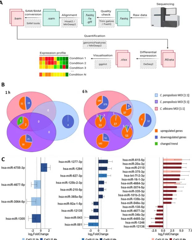
Next, we examined the ECs’miRNA profile. miRNA analysis results revealed several miRNAs that were specifically expressed not only in the presence of the two species but also specific to the applied doses ofC. parapsilosis. We identified 2, 2, and 3 mature miRNA transcripts at 1 h and 2, 8, and 16 at 6 h ofC. parapsilosisMOI 1:1, MOI 5:1, and C. albicans MOI 1:1 stimulus, respectively (Fig. 2C). While the majority of miRNAs showed a condition-specific altered expression, miR-4464-3p showed a significantly increased expression at 1 and 6 h ofC. albicanstreatment compared to the untreated control. Of the differentially expressed miRNAs, 1, 2, and 2 target mRNAs were found at 1 h, while 12, 56, and 185 target mRNAs were identified at 6 h ofC. parapsilosisMOI 1:1,
MOI 5:1, andC. albicansMOI 1:1 stimulus, respectively (Tables S2 to S4). These results suggest that species-specific, and in the case ofC. parapsilosis, dose-specific posttran- scriptional regulatory mechanisms regulate host responses under the applied condi- tions, which could explain the altered transcriptomic responses.
FIG 2 Differentially expressed genes (DEGs) and dysregulated miRNAs in host responses following fungal stimuli. Host transcriptomic and miRNA responses were examined with next generation (NGS) sequencing methods (Illumina). (A) Workflow of raw data analysis, where the obtained sequences were processed alongside the above detailed bioinformatical pipeline via command line (perl- and java-based) bioinformatical tools (gray boxes), through the listed intermediatefiles (green/red boxes).
Adapted from“Next Generation Sequencing Data Processing”by BioRender.com (2021). Retrieved fromhttps://app.biorender.com/
biorender-templates. (B) Venn diagrams of host genes identified at 1 and 6 h under each applied condition. The numbers of condition-specific genes as well as genes regulated by multiple conditions are shown. The term“changed trend”(green) refers to genes regulated by more than one condition, but the fold change was positive under at least one condition and negative in another. (C) Differently expressed host miRNA profiles after the applied conditions.
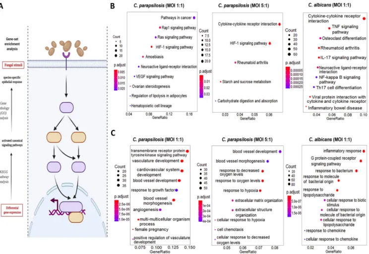
Carbohydrate metabolism-, hypoxia-, and cardiovascular development-related responses dominate afterC. parapsilosisstimulus, whileC. albicanspredominantly induces inflammation responses.Next, we aimed to categorize the identified tran- scripts and characterize host responses based on the activated host signaling path- ways. We employed different gene set enrichment analyses (GSEA) and overrepresen- tation analyses (ORA) methods (Fig. 3A) to examine the modified canonical pathways (KEGG’s pathway analysis [Fig. 3B]) and biological functions based on gene ontologies (gene ontology [GO] term analysis [Fig. 3C]). At 1 h after fungal exposure, the responses detected did not allow for a more in-depth analysis. Therefore, we focused on the 6-h data set. WithC. albicans-treated ECs, both biological pathways and functions were clearly dominated by inflammatory responses as shown by the 10 most activated path- ways and functions in Fig. 3B and C. Some of the most significantly regulated pathways were the cytokine-cytokine receptor interaction, tumor necrosis factor signaling, and IL-17 signaling pathways, while activated biological functions included inflammatory responses, responses to bacteria, to molecules of bacterial origin (e.g., lipopolysaccha- ride [LPS]), and to chemokines (Table S5). In contrast, C. parapsilosis 1:1 infection resulted in the activation of routes involved in vascular development and, interestingly, pathways frequently associated with carcinogenesis (e.g., Rap1 and Ras signaling path- ways, HIF1 and vascular endothelial growth factor [VEGF] signaling pathways). The affected biological pathways dominantly clustered around vascular development
FIG 3Results of the KEGG pathways and GO term analyses. (A) The“enrichKEGG”and“enrichGO”functions (provided within the R-package DOSE) and
“CNA”and “URA”analyses (provided within Ingenuity Pathway Analyses methods) were used to analyze the significantly up/downregulated pathways, functions, and upstream regulatory networks, respectively. (B and C) List of the 10 most activated pathways and functions after each stimuli (B). The KEGG results were labeled with their respective interest-to-background ratio (xaxis on thefigures) within the pathways, their significance (color coding) and with a corresponding“count,”which refers to the number of DEGs within a specific pathway. Ten (or less) significant pathways with the biggest“Gene Ratio” were visualized as dotplots via the “enrichplot”package. (C) The GO term results were visualized similarly for all the applied conditions. (Created with BioRender.com.)
(e.g., vasculature development or angiogenesis) (Table S6). Similar pathways were also activated by theC. parapsilosisMOI 5:1 coculture, although these were also comple- mented with activations of carbohydrate metabolic pathways (e.g., starch and glucose metabolism pathways) and hypoxia-related response routes (e.g., HIF1-a signaling pathway). Coculture of ECs withC. parapsilosisat an MOI of 5:1 also induced the activa- tion of a few inflammation-related pathways (e.g., cytokine-cytokine-R interaction pathway) (Table S7). Thus, whileC. albicanstriggered multiple inflammation pathways, C. parapsilosisevoked a variety of mainly inflammation-independent host responses that have not been previously associated with this species.
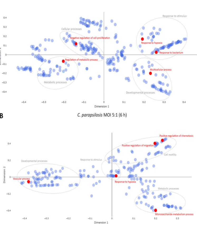
Potential effects of species-specific miRNA responses on host cell function.To examine whether the identified miRNAs could affect the evoked transcriptomic responses, wefirst overlapped the targets of the obtained miRNAs and the correspond- ing altered transcriptomic profiles in each condition. Then, using cluster analyses of GO overrepresentation tests, we analyzed functions that the potential target mRNAs (or the target genes of all identified miRNAs per condition) could affect under each condi- tion. As the early transcriptional responses under all conditions and the later transcrip- tional responses after C. parapsilosisMOI 1:1 treatment were only mild, none of the predicted functions passed the set P value threshold. Thus, only the differentially expressed target mRNAs derived fromC. parapsilosisMOI 5:1 andC. albicansMOI 1:1 stimuli were analyzed (Fig. 4A and B). According to the GO term analyses, the topfive target mRNA functions of target mRNA functions after C. albicans challenge were
“response to bacteria,” “regulation of metabolic processes,” “negative regulation of cell proliferation,” “response to hypoxia,”and“multicellular processes’(P,0.0001) (Fig. 4A and Table S8). These functions were clustered in the following categories:“response to stimulus,” “metabolic processes,” “cellular processes,”and“developmental processes,” respectively. WithC. parapsilosiscoculture,“response to hypoxia,” “positive regulation of chemotaxis,” “vascular processes,” “positive regulation of migration,”and“monosac- charide metabolic processes”were listed as the most significantly enriched functions (P,0.0001), within the major identified clusters of“response to stimulus,” “cell motil- ity, “developmental processes,” and “metabolic processes” (Fig. 4B and Table S9).
These data suggest that the identified miRNAs could actively regulate the identified species-specific transcriptomic responses.
HIF1-a pathway activation results in disrupted glucose metabolism after C.
parapsilosisstimulus and in survival promotion afterC. albicansinfection.In addi- tion to the several species-specifically activated signaling pathways, we found two— HIF1-aand hepatic stellate cell (HSC) activation signaling pathways—that were signifi- cantly regulated in all three experimental setups at 6 h. Therefore, we examined these pathways in more depth. In the HIF1-apathway, theC. parapsilosisMOI 1:1, MOI 5:1, andC. albicansMOI 1:1 stimuli resulted in the significant up- or downregulation of 7, 11, and 15 genes, respectively. In each case, we found treatment-specific activated genes as well as genes whose expression altered under at least two conditions (Fig. 5).
The HIF1-asignaling pathway was significantly activated during all three types of stim- uli compared to the“basal expression”level of the unstimulated cells. The effect was statistically most significant after theC. parapsilosis5:1 treatment (P= 3.32e208), fol- lowed byC. parapsilosisMOI 1:1 (P= 3.99e207) andC. albicansMOI 1:1 (P= 1.19e206) (Fig. 6A). We next examined potential functions that could be altered with HIF1apath- way deregulation. A similar activation pattern was observed in three of these biological processes: cell survival, migration, and angiogenesis.C. albicansclearly activated these processes (z-scores: 3.781, 3.185, and 3.481 of cell survival, migration, and angiogene- sis, respectively), while theC. parapsilosisMOI 5:1 treatment resulted in a similar effect, but activation occurred to a lesser extent (z-scores: 3.060, 2.789, and 3.879, respec- tively). TheC. parapsilosisMOI 1:1 stimulus led to only mild activation or even inhibi- tion (z-scores of survival, 1.601, of angiogenesis, 0.860, and of migration, 20.19).
Furthermore, extracellular matrix (ECM) synthesis inhibition was a characteristic ofC.
albicanstreatment, while activation of glucose uptake and metabolism was a unique effect of the twoC. parapsilosisstimuli (Fig. 6B).
FIG 4Multidimensional scaling plot of target mRNA functions. Potential effects of condition-specific miRNAs on transcriptomic responses. The potential functions of target mRNA were analyzed using cluster analyses of GO overrepresentation tests. Topfive functions (shown in red) of target mRNAs at 6-hC.
albicansMOI 1:1 treatment (A) andC. parapsilosisMOI 5:1 challenge (B). Gray circles represent the corresponding clusters of each highlighted function.
FIG 5Pathway explorer results on HIF1-asignaling in Ingenuity Pathway Analysis (IPA). Significantly down- or overexpressed genes in each condition were visualized within the canonical HIF1-asignal transduction network using pathway explorer and designer tools within IPA. The individual genes affected by each treatment were marked by the corresponding colors of each condition: blue forC. parapsilosisat an MOI of 1:1, purple for an MOI of 5:1, and red forC. albicansat an MOI of 1:1.
Next, we aimed to examine potential correlations between the results of the transcrip- tome and miRNA analyses by identifying miRNA-target mRNA pairs. Such pairs were iden- tified after bothC. albicansandC. parapsilosisMOI 5:1 coculture, but none were found for theC. parapsilosisMOI 1:1 condition (Fig. 6C). ForC. albicans-treated ECs, miR-34b (down- regulation, logarithmic fold change [LFC] =21.87) and its potential target mRNA, TGF-a (transforming growth factor alpha; upregulation, LFC = 1.758)—a known regulator of cell proliferation and survival—was identified. Another miRNA-mRNA pair included miR-2110 (LFC = 5.00) and PIK3R3 (phosphoinositide-3-kinase regulatory subunit 3; LFC =22.53). In the case ofC. parapsilosisMOI 5:1-treated ECs, miR-210 (LFC = 1.98) and its potential tar- get CAMK2B (calcium-dependent protein kinase 2 beta; LFC = 22.36) and another miRNA, miR-92a (LFC =21.85), were identified. The latter’s potential HIF1-apathway tar- get elements include EDN1 (endothelin-1 precursor; LFC = 2.11) and two glucose trans- porters SLC2A1 (GLUT1; LFC = 2.552) and SLC2A14 (GLUT14; LFC = 3.555) (Fig. 6C).
Interestingly, an overlap could also be observed among the applied conditions in terms of the expression of specific target genes, but without the targeting miRNAs. This sug- gests, that under the different conditions, the expression of the examined genes is possi- bly regulated by other posttranscriptional regulatory processes.
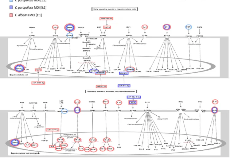
Hepaticfibrosis/stellate cell (HSC) activation pathway discriminates between the strong and attenuated inflammatory response toward the two species.The other signaling pathway that was simultaneously regulated by all three conditions af- ter 6 h was the hepatic fibrosis or hepatic stellate cell (HSC) pathway—a pathway involved in stellate cell activation during hepatic inflammation and injury. The early signaling events during the activation of HSCs and in activated HSCs are shown in Fig. 7. Similar to the HIF1-apathway, several genes showed either species-specific or treatment-influenced expression changes (Fig. 7). The regulation of the signal trans- duction pathway byC. parapsilosisat an MOI of 1:1 and 5:1 andC. albicansat an MOI of 1:1 was also statistically significant, although the direction of change could not be determined, due to the incoherent changes in gene expression (Fig. 8A). This pathway is associated with a number of biological functions related to inflammation, cellular activation, chemotaxis, apoptotic cell death, or tumor cell proliferation. In general,C.
albicansstimuli led to the overall activation of proinflammatory responses (e.g., upreg- ulation of chemotaxis, cellular activation), whileC. parapsilosistreatment resulted in ei- ther only a mild inflammatory response (e.g., immune cell activation with MOI of 5:1) or no significant effect (chemotaxis and cellular activation with MOI of 1:1; and inflam- mation and chemotaxis response for MOI of 5:1). Host cell apoptosis was inhibited by all three applied fungal conditions, althoughC. parapsilosisMOI 1:1 elicited the strong- est inhibitory effect (z-score =22.059). Interestingly, in contrast to the robust host tu- mor cell proliferation promoting effect of bothC. albicansandC. parapsilosisat an MOI of 5:1, the low-dose application of C. parapsilosis led to a mild inhibitory effect (z- score = 2.260, 2.248, and20.586, respectively) (Fig. 8B).
Similar to the HIF1-apathway, miRNA-mRNA target pairs could be identified afterC.
albicansandC. parapsilosisMOI 5:1 stimulus, but not after the lowerC. parapsilosisinfec- tion dose. Four miRNAs with altered expression were identified inC. albicans-treated cells, namely, miR-2110 (LFC = 5.00), miR-4485 (LFC =22.19), miR-34b (LFC =21.87), miR-4677 (LFC =21.73), and their potential counterparts included IGFBP5 (insulin-like growth fac- tor-binding protein 5; LFC =23.81), KLF6 (kruppel-like factor 6; LFC = 1.58), TGF-a(LFC = 1.76), and IL-8 (chemokine; LFC = 9.90), respectively (Fig. 8C). AfterC. parapsilosisMOI 5:1 treatment, miR-92a and its potential target (EDN1) as well as miR-543 (LFC =23.38) and its potential target IGFBP3 (insulin-like growth factor-binding protein 3; LFC = 4.20) were identified. Although no regulatory miRNAs were identified afterC. parapsilosisMOI 1:1 treatment, we found altered levels of expression of KLF6 and IGFBP5, suggesting that they were possibly regulated by other posttranscriptional regulatory processes.
In this pathway, each of the targeted genes primarily affect inflammatory functions.
Thus, the miRNA silencing observed here may contribute to the discrimination of the inflammatory response toC. albicansandC. parapsilosis.
DISCUSSION
In this study, we aimed to dissect and compare host responses triggered byC. albi- cansandC. parapsilosis—two common fungal residents of the oral microbial commu- nity—in oral ECs derived from a healthy individual (34). Ourfindings indicate that the EC immune response is more robust by 6 h of coincubation, by which time both spe- cies underwent morphology transition, rather than after 1 h, when bothC. albicansand C. parapsilosisare in a yeast form or only initiating their secondary morphology, sug- gesting that morphology transition could be a key trigger of the epithelial cell responses. ECs actively discriminate betweenC. albicansandC. parapsilosis, as shown FIG 6Results of the IPA analyses on the HIF1-asignaling-related molecular components and functions. (A) The significance of the pathway activation and direction were determined by thePvalue of overlap (,0.05) after performing an expression core analysis in IPA on the set of DEGs in each applied condition. (B) The direction of activation of the functions regulated by this signaling pathway was analyzed similarly. The blank (white) rectangles mean that we could not observe a significant regulation of that particular biological function under or after the corresponding treatment. (C) We selected the miRNA-mRNA target pairs involved in this signaling pathway after applying severalfiltering steps in IPA’s miRNA targetfilter tools. We only considered pairs that showed significant, opposite regulation in corresponding treatments, in which the targets were scientifically proven to be involved in HIF1-asignaling and the target site on the mRNA was either experimentally proven or strongly predicted by IPA based on base complementarity.
by the significant differences in host LDH release, chemokine, cytokine, and antimicro- bial peptide responses. In our model,C. parapsilosisfailed to evoke a robust, immediate proinflammatory response compared toC. albicans, which is similar to what has been observed in other experimental infection models (35–39). Thesefindings are also com- parable with earlier studies ofCandida-EC interactions, showing that onlyC. albicans triggers a strong inflammatory response during the colonization of the oral epithelial barrier (36). To aid the understanding of how oral ECs might discriminate between the two species and thus distinguish a species with a higher pathogenic potential from one that more commonly is a mucosal commensal, we examined host cell transcrip- tomic changes following yeast-EC interaction.
Ourfindings revealed significant differences in host cell transcriptomic responses that were species specific. WithC. albicanscoculture, the majority of signaling routes and pathways were specific to the inflammatory response and resulted in the activa- tion of, e.g., NF-κB and IL-17 signaling pathways (both are required for epithelium protection during oral candidiasis), which is in line with previous reports (40–42).
C. parapsilosischallenge, however, led to the activation of various, mainly inflamma- tion-independent pathways, such as carbohydrate metabolism-, hypoxia-, and cardio- vascular development-related responses, and interestingly, pathways frequently associ- ated with carcinogenesis, none of which have been previously associated with this species. The expression of genes related to carbohydrate metabolic processes was also upregulated in the case of bothC. parapsilosis doses. Glucose homeostasis mainte- nance has recently been suggested to be required for efficient anti-C. albicansimmune
FIG 7 Pathway explorer results on hepaticfibrosis/stellate cell activation signaling in IPA. Similar to the HIF1-a signal transduction network, regulated molecular components were visualized via pathway designer tools.
responses (43). According to Tucey et al (43),C. albicansdepletes glucose from human and murine macrophages during infection, thereby accelerating host cell death.
Although no studies are available comparing the carbon metabolism ofC. albicansand C. parapsilosis, high glucose tolerance and rapid proliferation ofC. parapsilosisin glu- cose-rich parenteral nutrition have previously been reported (44–46). This suggests enhanced glucose metabolic processes in this species, and also that duringC. parapsi- losisinfections, regulation of host glucose metabolism, as a virulence factor, might be even more momentous. Although further research is required to confirm this hypothe- sis, considering that the highest risk group ofC. parapsilosisinfections includes low- birth-weight neonates (47), the population primarily receiving parenteral nutrition, interfering with the pathogen’s carbon metabolic processes might reduce the risk of invasive candidiasis development in this patient group. Damaged tissues and inflam- mation are often coupled with local hypoxia (48). The lack of severe host cell damage and proinflammatory responses upon high-doseC. parapsilosischallenge suggests that the significantly altered hypoxic responses have other origins. Such responses could arise simply due to the elevated fungal burden rapidly depleting available oxygen lev- els through the rapid outgrowth of host cells. Host responses related to cardiovascular development and the activation of pathways frequently associated with carcinogenesis duringC. parapsilosistreatment are also unique, as no such phenomenon has been previously associated with this species. Changes of expression in tumorous pathways FIG 8 Results of the IPA analyses of hepaticfibrosis/stellate cell activation signaling. (A) Direction of activation of the hepaticfibrosis signaling-related functions were analyzed after an expression core analysis of the DEGs.
(B) Pathway activation was also examined. (C) miRNA-miRNA-targets were analyzed via miRNA targetfilter tools under similar conditions.
were dose dependent, as the low dose ofC. parapsilosistreatment (MOI of 1:1) had no effect on expression of the related pathways, while the high-dose treatment (MOI of 5:1) significantly enhanced singling routes related to carcinogenesis, similar to that observed with C. albicans treatment (MOI of 1:1). Such novel information sets the ground for a new aspect of future experimental investigations in thefield ofCandida research together with cancer biology.
Besides the species-specific activated pathways, signaling pathways with simultane- ous regulation by all three conditions were also found. Even among these, condition- specific transcriptional responses could be identified. One such pathway was hypoxia- inducible factor 1a(HIF1-a) signaling. HIFs, especially HIF1-a, have previously been demonstrated to regulate various innate immune processes (49). Although a study showed that HIF1-aactivation byb-glucan and commensal bacteria promotes protec- tion against subsequentC. albicansinfections (50, 51), suggesting the pathway’s inclu- sion in anti-Candidaresponses, little is known about its role in antifungal immunity regulation. Our results suggest that activation of the HIF1-a pathway is divergent.
WhileC. albicans stimulus promoted signaling processes related to cell survival and migration, or inhibition of ECM synthesis, glucose uptake and metabolism-related processes dominated afterC. parapsilosiscoculture. While regulation of EC protective cellular responses seems to be a priority in the case ofC. albicans, in line with previous reports (52), regulation of carbohydrate metabolism appears to be a unique character- istic ofC. parapsilosisstimulus. The HSC activation pathway (“hepatic stellate cell acti- vation pathway”), the other pathway simultaneously activated by all conditions, is not a singular signaling route in a strict sense, but rather a collection of several extracellu- lar signaling molecules and their related pathways whose activation altogether lead to the transformation of stellate cells into proliferative,fibrogenic myofibroblasts under suitable conditions. These pathways include, for example, TGF-b/SMAD and TGF- a/EGFR (epidermal growth factor receptor) pathways, which are important participants in these processes (53). These pathways are also known to influence epithelial cell functions, including their proliferation and chemotaxis (54, 55). In the HSC activation pathway, both species regulated signaling processes primarily involved in inflamma- tion. AlthoughC. albicanschallenge resulted in the overall activation of proinflamma- tory responses,C. parapsilosiscoculture led to only a mild effect. Species-specific regu- lation of both HIF1-a signaling and the HSC activation pathway might be what determines the outcome of the triggered innate immune responses of oral ECs.
Subsequent miRNA analyses revealed condition-specific posttranscriptional regula- tion of the transcriptomic responses. Among the identified dysregulated miRNA spe- cies, 4 were associated with coculturing ECs withC. parapsilosisat an MOI of 1:1, 10 with an MOI of 5:1, and 18 withC. albicans. Among the miRNAs identified duringC.
albicanstreatment, only miRNA-16-1p has been previously associated withC. albicans infections (28). Out of the remaining 17 differentially expressed miRNAs, miR-20a and hsa-let-7 have been reported to regulate antifungal responses, although only in Paracoccidioides brasiliensis(56). miR-16 and miR-4677 have been linked to antibacte- rial host responses (57, 58), while miR-3074, miR-335, miR-34b, miR-4485, and miR- 1246 have been associated with antiviral immune responses in variousin vitromodels (59–63). The remaining nine identified miRNA species have not yet been associated with microbe-induced inflammatory responses. In contrast, except for miR-3064 and miR-1294, all of the miRNAs differentially regulated in the presence ofC. parapsilosis have been suggested to regulate host responses during microbial stimuli. miR-210 was previously associated withC. albicans(26), miR-125b withP. brasiliensis(64), and miR- 92a with Paracoccidioides americana infections (65). Other than antifungal host responses, miR-4755 and miR-4677 deregulation was previously linked to bacterial stimuli (58, 66), miR-1305, miR-627, miR-543, and miR-581 to viral challenge, and miR- 12135 to parasitic infections (67). miR-1277 and miR-365 were associated with host responses upon both bacterial and viral infections (68–72). Thus, although fewer miRNA species could be coupled withC. parapsilosisinfections than withC. albicans,
the majority of these were confirmed regulators of antimicrobial responses. It is note- worthy that several of the miRNAs identified after bothC. albicansandC. parapsilosis stimuli have also been associated with various tumorigenic processes (73–78), further highlighting that fungal colonization might actively influence tumorigenic processes, as suggested previously (79, 80).
Subsequent analyses revealed that the yeast-specifically-identified miRNA species regulate the expression of genes involved in condition-specific activated pathways, including survival, proliferation, and inflammation inC. albicansand vascular develop- ment- and carbohydrate metabolism-related pathways inC. parapsilosiscocultures. For instance, miR-92a was identified in HIF1-asignaling, potentially regulating the expres- sion of GLUT1 (SLC2A1) and GLUT14 (SLC2A14), two glucose transporters required for carbohydrate metabolism maintenance (81) as well as the expression of EDN1 (endo- thelin-1) (82), a potent vasoconstrictor, duringC. parapsilosisinfection. Thefinding that both GLUT1 and GLUT14 are also upregulated uponC. albicansstimulus suggests that the significant activation of carbohydrate metabolic processes by C. parapsilosisis the result of an additive effect and that glucose metabolic regulators, other than the men- tioned glucose transporters, are also deregulated during the stimulus. In the same path- way, followingC. albicanschallenge, miR-34b was linked to TGF-aexpression, a known regulator of survival and cell proliferation after its activation by hypoxia-induced factors (such as HIF1-a) (83), and miR-2110 was found to repress PIK3R3 expression, a subunit of PI3K, thereby interfering with proinflammatory responses (84, 85).
In the HSC activation pathway, besides miR-92a and its target EDN-1, miR-543 was also identified, which targets IGFBP3, an IGF-binding protein previously linked to apo- ptosis regulatory processes (86). Although independent of IGFBP3 expression changes, apoptosis inhibition as a potential outcome of HSC activation was predicted to be the strongest following the low-doseC. parapsilosistreatment. WithC. albicansinfection, besides the above-mentioned miR-34b2TGF-a pair, miR-2110 was identified as a potential regulator of IGFBP5, miR-4485 was linked to KLF6 regulation, and miR-467 to regulating CXCL8 expression. KLF6 is a zinc finger transcription factor previously reported to promote inflammation in macrophages (87). IGFBP5, another IGF-binding protein, was reported to be a potent chemoattractant of immune cells (88). CXCL8 is a well-known chemokine secreted by oral ECs uponC. albicansstimuli (89), in line with our data. Thus, all three miRNA target genes are potential inflammation regulatory components of the anti-C. albicansoral EC response.
Taken together, the in-depth analyses of the two simultaneously, yet diversely regu- lated signaling pathways also support the major, species-specificfindings of the tran- scriptome functional analyses and suggest that the differentiating EC responses might indeed derive from altered posttranscriptional regulations. Although the obtained results shed some light on the potential underlying molecular mechanisms enabling species-specific host responses, further investigations and experimental studies are required to support thesefindings, such as by applying variousC. albicansandC. para- psilosisclinical isolates simultaneously, to confirm ourfindings or further reveal poten- tial strain-dependent differences, along with applyingin vivoexperimental setups to examine these interactions under complex oral environmental conditions.
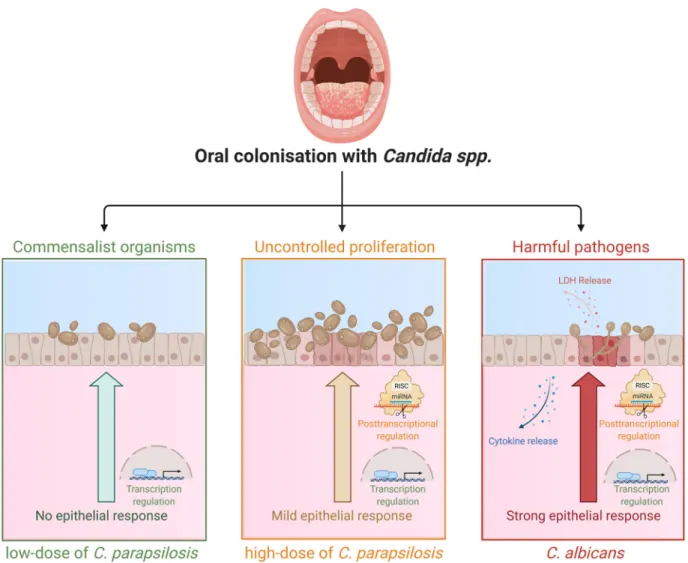
In summary, we can conclude that human oral ECs are able to actively differentiate between Candida species through altered posttranscriptional regulatory processes (Fig. 9). While the presence of C. parapsilosis stimulus does not generate a robust inflammatory response in ECs, an elevated fungal burden can initiate inflammatory responses, albeit in a much less rapid and robust manner compared toC. albicans.
Additionally, we found that different fungal burdens ofC. parapsilosisled to the vari- able induction of generic alterations with the higher MOI inducing a broader and more significant response. The species-specificfine-tuning of both HIF1-asignaling and HSC activation pathways via miRNA silencing could also be a key to the distinct epithelial responses. The in silico data acquired through this project aid our current
understanding of how healthy oral ECs might discriminate betweenCandidaspecies with high or low pathogenic potential in the human oral mucosa.
MATERIALS AND METHODS
Strains and growth conditions.In this study,Candida parapsilosisCLIB 214 andCandida albicans SC5314 laboratory strains were used.Candidastrains were maintained on solid penicillin-streptomycin- supplemented YPD medium at 4°C. Prior to host cell stimulation, yeast cells were grown overnight at 30°C in liquid yeast extract-peptone-dextrose (YPD) medium, washed three times with phosphate-buf- fered saline (PBS), and counted using a hemocytometer to adjust the desired cell concentration.
Stimulation of OKF6/TERT2 cells.The oral EC line OKF6/TERT2, a telomerase-deficient EC line derived from a healthy individual, was used for all experiments and maintained as described previously (34). Oral ECs were then plated in six-well plates in keratinocyte serum-free medium (K-SFM) supple- mented with 25mg/ml bovine pituitary extract (BPE), 2 ng/ml recombinant epidermal growth factor (rEGF), 2 mML-glutamine, and 0.5% penicillin-streptomycin, and cells were grown to 90% confluence.
OKF6/TERT2 cells were then stimulated withC. parapsilosisandC. albicansin serum-free K-SFM medium.
Depending on the experiment, either cell-free supernatants or host cells were collected following fungal exposure and stored at280°C or used immediately.
Lactate dehydrogenase assay.Host cell damage byC. albicansandC. parapsilosiswas determined by lactate dehydrogenase (LDH) cytotoxicity detection kit according to the manufacturer’s instructions.
OKF6/TERT2 cells were challenged with fungal cells at MOIs of 1:5, 1:2, 1:1, 2:1, and 5:1 or left untreated at various time points. During analysis, the values corresponding to the levels of LDH activity measured in untreated samples were subtracted from the values of stimulated samples. The percentage of cytotoxicity was determined as (optical density [OD] of the experimental value/OD of the positive control)100. 1%
Triton X-100-treated samples served as positive controls. Results are derived from three independent experiments.
FIG 9Altered innate immune response regulation in healthy oral ECs discriminate between low-doseC. parapsilosis, increased dose ofC.
parapsilosis, andC. albicansstimuli. (Created with BioRender.com.)
Total RNA and miRNA extraction.Total RNA and miRNA extraction from OKF6/TERT2 cells was car- ried out with miRNeasy minikits according to the manufacturer’s instructions with minor modifications, allowing for the simultaneous extraction of total RNA and miRNA. Cells were grown until 90% conflu- ence in tissue culturing six-well plates in supplemented K-SFM medium, washed once with PBS, and stimulated withC. albicans(MOI of 1:1) orC. parapsilosis(MOI of 1:1 and/or 5:1) in unsupplemented K- SFM medium. Following coincubation, host cells were washed two times with PBS and treated with the supplied QIAzol lysis reagent, avoiding the use of police rubber and extensive vertexing of the samples to prevent the lysis of fungal cells (hyphae). After host cell lysis, phase separation, and purification of RNA (both total and miRNA), we performed quantity and quality checks of the samples before proceed- ing to cDNA library preparation or cDNA synthesis and subsequent sequencing. Three independently treated biological parallels were used.
cDNA synthesis and real-time PCR analysis.For preliminary expression studies, 1,000 ng of RNA was utilized for cDNA synthesis using the RevertAidfirst strand cDNA synthesis kit. Primers for qPCR analyses are listed in Table S10 in the supplemental material. The amplification conditions were as fol- lows: one cycle of denaturation for 3 min at 95°C; denaturation at 95°C for 10 s; 49 cycles, with 1 cycle consisting of annealing at 60°C for 30 s and elongation at 65°C for 30 s; and afinal extension step at 72°C for 30 s.b2-Microglobulin was used as an internal control. Relative normalized expression values (unstimulated host cells served as controls) were calculated and presented.
Sequencing library preparation and RNA sequencing.miRNA sequencing libraries were prepared using NEBNext Multiplex Small RNA Library Prep Set for Illumina following the manufacturer’s protocol.
Libraries were size selected using AMPure XP beads and after validation with an Agilent 2100 Bioanalyzer instrument sequenced with an Illumina MiSeq DNA sequencer using Illumina MiSeq reagent kit V3-150.
Transcriptome analysis.We performed the preliminary quality analysis and trimming using FastQC and Cutadapt command line tools on the raw sequencefiles. Next, wefit the reads to the reference ge- nome index (GRCh38) using HISAT2 (90), with the parameters–dta–non-deterministic–rna-strandness.
Read numbers were calculated using the GenomicAlignments package, and differential gene expression in logarithmic fold change (LFC) was then performed using the DeSeq2 tool (91). Wefiltered out objects with read counts lower than 1 part per million (ppm). In the experimentally derived gene list, differen- tially expressed genes (DEGs) were counted above the absolute value of the logarithmic fold change of .1.5 and the adjustedPvalue of,0.05.
Short-read mapping and counting.The sequenced reads were mapped to known microRNA precur- sors, and novel sequences downloaded from miRBase (version 22) using miRDeep2.0 (92). Hits with a read count below 1 ppm werefiltered out from further analysis. The distorting effect caused by the hits that were expressed at an exceptionally high level (and have the largest variance) was corrected via DeSeq2, and theP value was corrected by null distribution using the fdrtool package. The cutoff values for the significant hits were set at aPvalue of,0.05 and the absolute value of the logarithmic fold change of.1.5.
Overrepresentation analyses.Upon completion of the genome-wide RNA and miRNA expression analyses, gene expression data were interpreted using overrepresentation analyses (ORA) and gene set enrich- ment analyses (GSEA) provided in the Bioconductor package, DOSE (93) and clusterProfiler (94) (Fig. 3A). The two ORAs—KEGG overrepresentation test and GO overrepresentation test—as well as the GO GSEA were car- ried out, against a constant background, for which purpose, the human genome wide annotation package (“org.Hs.eg.db”) was used (95). During both analyses, the most robust Benjamini and Hochberg (96) (“BH”) method was used for the multiple comparisonPvalue adjustment, and pathwaysP,0.05 were considered significantly overrepresented. The enrichment results were visualized as dotplots via the enrichplot package.
For further data mining, we calculated the semantic similarity (SS) of the found GO terms to establish connec- tions between genes targeted by a specific miRNA via the ViSEAGO package (97). These results were visualized on a multidimensional scaling plot (MDS) that represents the distance among the set of enriched GO terms on thefirst two dimensions, which highlight possible clustering patterns.
Causal analyses.We employed causal analysis methods included in the Qiagen licensed, leading-edge bioinformatical software Ingenuity Pathway Analysis (IPA), and we ran expression core analyses on our samples.
Among the included algorithms, we used downstream effect analysis (DEA) to observe each treatment’s effect on the biological functions of the host cells. Furthermore, we concluded miRNA-target analyses tofind possible miRNA-mRNA target pairs with significant, anti-correlated expression. We employed thePvalue of overlap and the activation Z-score to determine the significance of the prediction in IPA, which are the two most important parameters to achieve this (98). ThePvalue of overlap determines the statistical significance based on the overlap of the observed and predicted regulated gene sets, while the activation Z-score predicts the direction of regulation depending on the parallelism in the observed and predicted up/downregulatory patterns. In our experiments, only the predictions withPvalues of,0.05 were considered significant hits. We further specified that only experimentally proven or strongly predicted intermolecular relationships should be considered.
Statistical analysis.All statistical analyses were performed with GraphPad Prism v 6.0 software using parametricttests or nonparametric Mann-Whitney tests. The values for the groups examined were considered statistically significantly different atP,0.05.
Data availability.Sequencing data are accessible under the BioProject accession numberPRJNA715092.
SUPPLEMENTAL MATERIAL
Supplemental material is available online only.
TABLE S1, XLSXfile, 0.1 MB.
TABLE S2, XLSXfile, 0.01 MB.
TABLE S3, XLSXfile, 0.02 MB.
TABLE S4, XLSXfile, 0.03 MB.
TABLE S5, XLSXfile, 0.02 MB.
TABLE S6, XLSXfile, 0.01 MB.
TABLE S7, XLSXfile, 0.01 MB.
TABLE S8, XLSXfile, 0.1 MB.
TABLE S9, XLSXfile, 0.04 MB.
TABLE S10, XLSXfile, 0.01 MB.
ACKNOWLEDGMENTS
M.H. and this research work were supported by the Szeged Scientists Academy under the sponsorship of the Hungarian Ministry of Innovation and Technology (FEIF/
433-4/2020-ITM_SZERZ). This work was supported by grants 20391-3/2018/FEKUSTRAT, NKFIH K 123952, and GINOP-2.3.2.-15-2016-00015. A.G. was further funded by LP2018- 15/2018. László Bodai was supported by the ÚNKP-20-5-SZTE-642 New National Excellence Program of the Ministry for Innovation and Technology and by the János Bolyai Research Scholarship (BO/00522/19/8) of the Hungarian Academy of Sciences.
The human oral epithelial cell line, OKF6/TERT-2, was kindly provided by J.
Rheinwald (Harvard University, Cambridge, Massachusetts).
We declare that we have no conflicts of interest.
A.G. and R.T. contributed to the concept and design of this project. M.H. and R.T.
carried out the majority of experiments with the help of G.N., N.Z., C.V., J.D.N., and M.H., and G.N., L.B., and P.H. analyzed the acquired data. M.H. prepared the manuscript and thefigures, which were revised by R.T. with A.G. All authors reviewed the manuscript, contributed to the discussion, and approved thefinal version.
REFERENCES
1. Avila PC, Schleimer RP. 2009. Airway epithelium, p 366–397.InKay AB, Bousquet J, Holt PG, Kaplan AP (ed), Allergy and allergic diseases, 2nd ed.
Wiley-Blackwell, Hoboken, NJ.
2. Schleimer RP, Kato A, Kern R, Kuperman D, Avila PC. 2007. Epithelium: at the interface of innate and adaptive immune responses. J Allergy Clin Immunol 120:1279–1284.https://doi.org/10.1016/j.jaci.2007.08.046.
3. Calderone RA, Clancy CJ. 2011. Candida and candidiasis. American Society for Microbiology, Washington, DC.
4. Vázquez-González D, Perusquía-Ortiz AM, Hundeiker M, Bonifaz A. 2013.
Opportunistic yeast infections: candidiasis, cryptococcosis, trichosporo- nosis and geotrichosis. J Dtsch Dermatol Ges 11:381–393.https://doi.org/
10.1111/ddg.12097.
5. Pappas PG, Lionakis MS, Arendrup MC, Ostrosky-Zeichner L, Kullberg BJ.
2018. Invasive candidiasis. Nat Rev Dis Primers 4:18026.https://doi.org/10 .1038/nrdp.2018.26.
6. Sobel JD. 2006. The emergence of non-albicans Candida species as causes of invasive candidiasis and candidemia. Curr Infect Dis Rep 8:427–433.
https://doi.org/10.1007/s11908-006-0016-6.
7. Manolakaki D, Velmahos G, Kourkoumpetis T, Chang Y, Alam HB, De Moya MM, Mylonakis E. 2010. Candida infection and colonization among trauma patients. Virulence 1:367–375.https://doi.org/10.4161/viru.1.5.12796.
8. Ghannoum MA, Jurevic RJ, Mukherjee PK, Cui F, Sikaroodi M, Naqvi A, Gillevet PM. 2010. Characterization of the oral fungal microbiome (myco- biome) in healthy individuals. PLoS Pathog 6:e1000713.https://doi.org/
10.1371/journal.ppat.1000713.
9. Peters BA, Wu J, Hayes RB, Ahn J. 2017. The oral fungal mycobiome: char- acteristics and relation to periodontitis in a pilot study. BMC Microbiol 17:157.https://doi.org/10.1186/s12866-017-1064-9.
10. Berkovits C, Tóth A, Szenzenstein J, Deák T, Urbán E, Gácser A, Nagy K. 2016.
Analysis of oral yeast microflora in patients with oral squamous cell carci- noma. SpringerPlus 5:1257.https://doi.org/10.1186/s40064-016-2926-6.
11. Olivas-Escárcega V, Ruiz-Rodríguez MDS, Fonseca-Leal MDP, Santos-Díaz MÁ, Gordillo-Moscoso A, Hernández-Sierra JF, Pozos-Guillén ADJ. 2008.
Prevalence of oral candidiasis in chronic renal failure and renal transplant pediatric patients. J Clin Pediatr Dent 32:313–317.https://doi.org/10 .17796/jcpd.32.4.f53343t742150l73.
12. Abu-Elteen KH, Hamad MA, Salah SA. 2006. Prevalence of oral candida infections in diabetic patients. Bahrain Med Bull 28:1–8.
13. Singh GK, Capoor MR, Nair D, Bhowmik KT. 2017. Spectrum of fungal infection in head and neck cancer patients on chemoradiotherapy. J Egypt Natl Canc Inst 29:33–37.https://doi.org/10.1016/j.jnci.2017.01.006.
14. Gaitán-Cepeda LA, Sánchez-Vargas O, Castillo N. 2015. Prevalence of oral candidiasis in HIV/AIDS children in highly active antiretroviral therapy era.
A literature analysis. Int J STD AIDS 26:625–632.https://doi.org/10.1177/
0956462414548906.
15. Almeida RS, Brunke S, Albrecht A, Thewes S, Laue M, Edwards JE, Filler SG, Hube B. 2008. The hyphal-associated adhesin and invasin Als3 of Candida albicans mediates iron acquisition from host ferritin. PLoS Pathog 4:
e1000217.https://doi.org/10.1371/journal.ppat.1000217.
16. Noble SM, Gianetti BA, Witchley JN. 2017. Candida albicans cell-type switching and functional plasticity in the mammalian host. Nat Rev Micro- biol 15:96–108.https://doi.org/10.1038/nrmicro.2016.157.
17. Moyes DL, Wilson D, Richardson JP, Mogavero S, Tang SX, Wernecke J, Höfs S, Gratacap RL, Robbins J, Runglall M, Murciano C, Blagojevic M, Thavaraj S, Förster TM, Hebecker B, Kasper L, Vizcay G, Iancu SI, Kichik N, Häder A, Kurzai O, Luo T, Krüger T, Kniemeyer O, Cota E, Bader O, Wheeler RT, Gutsmann T, Hube B, Naglik JR. 2016. Candidalysin is a fungal peptide toxin critical for mu- cosal infection. Nature 532:64–68.https://doi.org/10.1038/nature17625.
18. Moyes DL, Runglall M, Murciano C, Shen C, Nayar D, Thavaraj S, Kohli A, Islam A, Mora-Montes H, Challacombe SJ, Naglik JR. 2010. A biphasic innate immune MAPK response discriminates between the yeast and hyphal forms of Candida albicans in epithelial cells. Cell Host Microbe 8:225–235.https://doi.org/10.1016/j.chom.2010.08.002.
19. Tóth R, Nosek J, Mora-Montes HM, Gabaldon T, Bliss JM, Nosanchuk JD, Turner SA, Butler G, Vágvölgyi C, Gácser A. 2019. Candida parapsilosis:
from genes to the bedside. Clin Microbiol Rev 32:e00111-18.https://doi .org/10.1128/CMR.00111-18.
20. Rippon JW. 1982. Medical mycology: the pathogenic fungi and the patho- genic actinomycetes. WB Saunders Company, Eastbourne, United Kingdom.
21. Dismukes WE, Pappas PG, Sobel JD (ed). 2003. Clinical mycology. Oxford University Press, New York, NY.
22. Tata W, Thepbundit V, Kuansuwan C, Preechasuth K. 2019. Distribution of Candida species in oral candidiasis patients: association between sites of