Role of pattern recognition receptors (PRR) in the pathogenesis of non-alcoholic steatohepatitis
(NASH)
Doctoral thesis
Tímea Csák M.D.
Semmelweis University
Clinical Medicine School of Doctoral Studies
Supervisor: Gyöngyi Szabó M.D., PhD
Reviewers: Gabriella Pár M.D., PhD.
Klára Werling M.D., PhD.
Comprehensive exam committee:
President: János Banai M.D., CSc
Members: László Herszényi M.D., PhD.
György Székely M.D., CSc
Budapest
2012
1
This work was performed at the Department of Medicine of the University of Massachusetts Medical School in
Worcester, USA between October 2008 and April 2011.
2 TABLE OF CONTENTS
Table of contents 2
List of abbreviations 6
1. Introduction 7
2. Review of the literature 8
2.1 Non-alcoholic fatty liver disease and non-alcoholic steatohepatitis 8
2.1.1 Definition 8 2.1.2 Epidemiology 8 2.1.3 Clinical aspects of NASH: symptoms, diagnosis and treatment 10
2.1.4 Fatty liver and viral infections 12
2.1.5 Pathogenesis 12
2.2 Pattern recognition receptors (PRRs) 16
2.2.1. Toll-like receptors (TLRs) 16
2.2.1.1 TLRs and their ligands 16
2.2.1.2 TLRs in liver diseases 19
2.2.2 RIG-I-like receptors (RLRs) 19
2.2.2.1 RLRs and their ligands 19
2.2.2.2 RLRs in liver diseases 21
2.2.3 Inflammasomes 22
2.2.3.1 Definition 22
2.2.3.2 Types of inflammasomes 22
2.2.3.3 Function of inflammasomes 23
2.2.3.4 Inflammasome activating ligands 24
2.2.3.5 Inflammasome expression in the liver 28
2.2.3.6 Role of inflammasomes in liver diseases 30
3. Aim of the thesis 31
3.1 To examine the role of toll-like receptor 4 signaling in the pathogenesis of NASH 31
3.2 To examine the role of IL-1β and inflammasomes in the pathogenesis of NASH 31
3
3.3 To examine the pathomechanism of decreased antiviral response in steatohepatitis
32
4. Materials and Methods 33
4.1 Animal studies 33
4.2 Biochemical analysis and cytokine measurements 33
4.3 Histopatological analysis 34
4.4 RNA analysis 34
4.5 Protein analysis 35
4.5.1 Preparation of cell lysates 35
4.5.2 SDS-PAGE electrophoresis 35
4.5.3 Native gel electrophoresis 35
4.5.4 Immunoprecipitation 35
4.6 Functional assays 36
4.6.1 Caspase-activity assays 36
4.6.2 NADPH activity assay 36
4.6.3 Cytotoxicity assay 36
4.7 In vitro experiments 36
4.7.1 Cell lines 37
4.7.2 Primary cells 37
4.8 Flow cytometry analysis 37
4.9 Human liver samples 37
4.10 Statistical analysis 38
5. Results 39
5.1 Deficiency in myeloid differentiation factor-2 (MD2) and toll-like receptor 4 (TLR4) expression attenuates non-alcoholic steatohepatitis and fibrosis in mice 39
5.1.1 MD-2 or TLR4 protects from MCD diet-induced liver fat deposition and inflammation 39
5.1.2 MD-2 and TLR4 deficiency attenuates oxidative stress 44
4
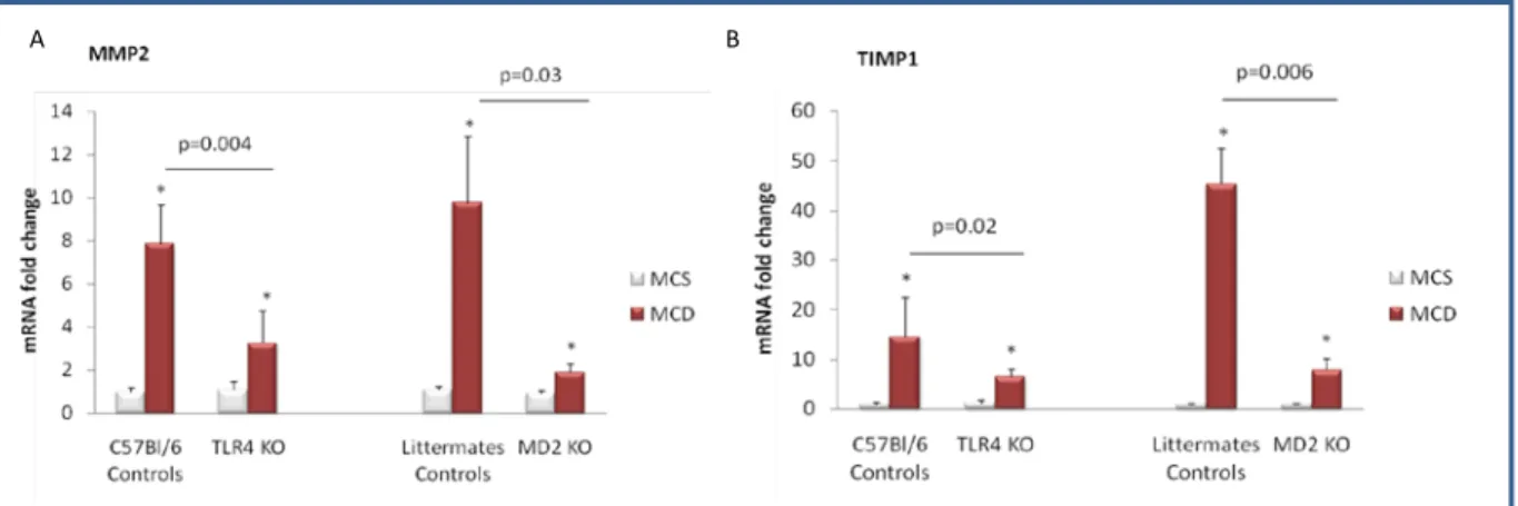
5.1.3 MD-2 and TLR4 deficiency protects from NASH-associated liver fibrosis
47
5.2 Fatty acids and endotoxin activates the inflammasome in non-alcoholic steatohepatitis 51
5.2.1 MCD diet-induced steatohepatitis is associated with increased IL-1β production and inflammasome activation in the liver 51
5.2.2 Long-term, but not short-term high fat diet feeding is associated with inflammasome activation in the liver 53
5.2.3 Increased inflammasome expression in human NASH 59
5.2.4 LPS induces upregulation of the inflammasome in the liver 60
5.2.5 Inflammasome is upregulated in hepatocytes in NASH 61
5.2.6 Fatty acids and LPS induce inflammasome activation in hepatocytes and mononuclear cells 63
5.2.7 IL-1β production in hepatocytes occurs in inflammasome-dependent (caspase-1) and inflammasome-independent (caspase-8-dependent) 67
5.2.8 Palmitic acid-treated hepatocytes transmit danger signals and induce inflammasome activation in liver mononuclear cells 68
5.2.9 ASC deficiency does not prevent liver injury and fat deposition in the MCD-diet model of NASH 72
5.2.10 Caspase-1 deficiency does not prevent liver injury and fat deposition in the MCD-diet model of NASH 75
5.2.11 Caspase-8 as an alternate to cleave IL-1β 77
5.2.12 Interleukin-1 receptor deficiency attenuates hepatic steatosis, but does not prevent MCD-diet-induced liver injury or fibrosis 78
5.3 Mitochondrial antiviral signaling protein defect links impaired antiviral response and liver injury in steatohepatitis in mice 82
5.3.1 Type-I IFN induction is decreased in steatohepatitis in response to poly I:C stimulation 82
5.3.2 Impaired Type-I IFN induction in steatohepatitis is restricted to the RIG- I/Mda5 pathway 83
5
5.3.3 Abnormal MAVS function in NASH involves decreased protein levels,
dissociation from the mitochondria and impaired oligomerization 85
5.3.4 Mitochondrial damage occurs in the fatty liver 90
5.3.5 Increased poly I:C-induced liver damage occurs without excessive pro- inflammatory cytokine induction in steatohepatitis 93
5.3.6 Poly I:C promotes a switch from apoptosis to necrosis and increases RIP3 expression in steatohepatitis 96
5.3.7 Altered MAVS and RIP3 mRNA expression in human NASH 100
6. Discussion 101
7. Conclusion 114
8. Summary 116
9. Összefoglalás 117
10. Reference list 118
11. List of own publications 146
12. Acknowledgement 150
6 LIST OF ABBREVIATIONS
AIM2, absent in melanoma 2; ALP, alkaline phosphatase; ALT, alanine aminotransferase;
ASC, apoptosis-associated speck-like CARD-domain containing protein; AST, aspartate aminotransferase; CLR, C-type lectin receptors; CpG, cytidine-phosphate-guanosine-rich DNA; DAMP, danger associated molecular pattern; dsRNA, double stranded RNA; FFA, free fatty acid; GGT, gamma-glutamyltransferase; HCV, hepatitis C virus; HFD, high fat diet; HIV, human immunodeficiency virus; HMGB1, high mobility group box protein-1;
HSC, hepatic stellate cell; IFN, interferon; IKK, IkB kinase; IL, interleukin; IPS-1, IFNβ promoter stimulator protein-1; IRAK, LI-1R-associated kinase; IRF, interferon regulatory factor; ISG, interferon-inducible gene; JNK, Jun N-terminal kinase; LMNC, liver mononuclear cell; LPS, lipopolysaccharide; LRR, leucin-rich repeats; LSEC, liver sinusoidal endothelial cells; MAVS, mitochondrial antiviral signaling protein; MCD, methionine-choline deficient; MCS, methionine-choline supplemented; MD2, myeloid differentiation factor 2; Mda5, melanoma differentiation-associated gene 5; Mult1, murine UL16-binding proteinlike transcript 1; MMP, matrix metalloproteinase; MyD88, Myeloid differentiation factor 88; NADPH, nicotinamide adenine dinucleotide phosphate; NAFLD, non-alcoholic fatty liver disease; NALP1, NACHT, LRR and PYD domains-containing protein 1; NALP3, NACHT, LRR and PYD domains-containing protein 3 / cryoporin;
NASH, non-alcoholic steatohepatitis; NFkB, Nuclear factor kB; NK, natural killer cell;
NLR, NOD-like receptors; NLRC4, NLR family CARD-domain containing -4; PA, palmitic acid; PAMP, pathogen associated molecular pattern; PKC, protein kinase C; Poly I:C, polyinosinic-polycytidylic acid; PSMA7, proteasome subunit alpha type 7; Rae-1α, retinoic acid early inducible-1α; RIG-I, retinoic acid-inducible gene-I; RIP3, receptor interacting protein-3; RLR, RIG-I-like receptor; ROS, reactive oxygen species; αSMA, α smooth muscle actin; TAK, TGF-β activated kinase; TBARs, thiobarbituric acid reactive substances; TBK1, TANK-binding kinase 1; TGFβ, transforming growth factor β; TIMP, tissue inhibitor metalloproteases; TLR, toll-like receptor; TNF, tumor necrosis factor;
TRAIL, TNF-related apoptosis inducing ligand; TRAF, TNFR-associated factor; TRIF, TIR domain-containing adaptor inducing IFN-beta; VISA, virus-induced signaling adaptor; zVAD, (carbobenzoxy-valyl-alanyl-aspartyl-[O-methyl]- fluoromethylketone)
7 1. INTRODUCTION
Non-alcoholic fatty liver disease (NAFLD) is one of the most common liver diseases affecting over 1/3 of the population in the Western world. The histopathology spectrum of NAFLD includes steatosis alone, steatosis with inflammation and steatohepatitis (non alcoholic steatohepatitis/NASH) that includes necroinflammation with or without fibrosis.
(1). The pathogenesis of NAFLD/NASH is not fully understood yet. Recently the role of innate immunity has been implicated in the pathogenesis of NASH (1). Pattern recognition Toll-like receptors (TLR) and NOD-like receptors (NLR) are key components of the innate immune system in the recognition of pathogens, but they also sense danger signals released from damaged cells (94). The inflammation induced via the TLRs or NLRs contribute to the pathogenesis of several autoinflammatory diseases.
Fatty liver is highly sensitive to the TLR4 ligand lipopolysaccharide (LPS or endotoxin), that is a bacterial wall component of Gram-negative bacteria. Furthermore, increased plasma endotoxin levels were detected in steatohepatitis both in mice and humans.
However, the role of the TLR4-MD2 receptor complex in NASH is yet to be evaluated.
Inflammasomes, large caspase-1-activating multiprotein complexes that sense both danger signals through the intracellular NLRs, are major contributors to inflammation. The NALP3 inflammasome is involved in sensing endogenous danger signals, and promotes the cleavage and maturation of the pro-inflammatory cytokine pro-IL-1β to promote/sustain inflammation. The inflammasome activation is a two step process, in which the first step usually by a TLR ligand such as LPS induce the up-regulation of the inflammasome and pro-IL-1β, and a second signal activate the inflammasome. The cell- specific expression and role of the inflammasome in the liver are yet to be evaluated in NASH.
While the factors determining progression of NASH are yet to be fully defined, the clinical importance of increased susceptibility of the fatty liver to viral infections is emerging. Co- morbidity of NASH with viral infections caused by RNA viruses, such as hepatitis C and HIV remains a clinical challenge. The pathomechanism behind the impaired antiviral immuntity is not fully clarified yet.
8 2. REVIEW OF THE LITERATURE
2.1. NON-ALCOHOLIC FATTY LIVER DISEASE AND NON-ALCOHOLIC STEATOHEPATITIS
2.1.1. Definition
Non alcoholic fatty liver disease (NAFLD) is a chronic liver disease with wide spectrum of hepatic abnormalities somewhat similar to alcoholic liver disease but without significant alcohol consumption. Along with obesity, hypertension, hypercholesterolemia, hyperuricaemia and insulin resistance, NAFLD is the part of the metabolic syndrome. The histopatological spectrum of NAFLD includes steatosis alone, steatosis with inflammation and steatohepatitis (NASH) with necroinflammation (with or without fibrosis). The last form, which is progressive, can lead to cirrhosis and even to hepatocellular carcinoma (1).
Steatosis Steatosis with inflammation Steatohepatitis with necroinflammation
Fibrosis
Cirrhosis
Hepatocellular carcinoma Figure 1. Natural history of NAFLD
2.1.2. Epidemiology
Since its first description in 1980 (2), non-alcoholic fatty liver disease (NAFLD) became one of the most common liver diseases in the Western world (3) due to the increasing prevalence of obesity. The prevalence of obesity is shown in Figure 2 and 3.
?
9
Figure 2. Prevalence of obesity in adult females in 2010 based on the WHO Global InfoBase.
Figure 3. Prevalence of obesity in adult males in 2010 based on the WHO Global InfoBase.
The estimated prevalence of NAFLD is around 20-30% in the general population in the Western world (4), while its prevalence is over 90% among obese subjects (5). Non- alcoholic steatohepatitis (NASH) occurs approximately in 2-3% of the general population (6
Notably, NAFLD affects not only the adults, but its prevalence is also increasing in the childhood: ~3% overall and ~50% in obese children (
), and its prevalence in the morbidly obese subjects is ~37% (5).
7).
10
2.1.3. Clinical aspects of NASH: symptoms, diagnosis and treatment
Most of the patients with NAFLD are asymptomatic and the diagnosis is incidental during either routine blood tests (elevated liver enzymes) or ultrasound examination (liver steatosis). Fatigue, partially due to the accompanying sleep apnea and right upper quadrant abdominal discomfort due to the hepatomegaly may occur. Later, with the progression of the disease, myriad of signs and symptoms of chronic liver disease, cirrhosis and liver failure develop. In addition, since NAFLD is the “hepatic manifestation” of metabolic syndrome, these patients often suffers from non-liver related symptoms of the co- morbidities, such as type II diabetes, hypertension etc.
Increased serum alanine aminotransferase (ALT), aspartate aminotransferase (AST), gamma-glutamyltransferase (GGT) and sometimes alkaline phosphatase (ALP) levels with exclusion of other etiologies of chronic liver diseases, such as alcohol, virus, metabolic diseases, raise the possibility of NASH (8). However, in some histologically proven NASH patients normal ALT values can be observed (9). Higher serum ferritin levels were also reported in 50% of NASH patients (10). In case of suspicion of NASH, ultrasound is used to detect hepatic fat deposition in form of hyperechogenic liver (11
The most emerging question is to differentiate the simple steatosis from the progressive steatohepatitis using non-invasive techniques. However, to date there are no available diagnostic laboratory tests that can reliably differentiate steatosis from steatohepatitis. The measurement of keratin-18 fragments as marker of apoptosis correlates with the severity of liver disease (
).
12) therefore may help in the staging. There are several scoring systems that might be useful in the diagnostic algorithm of NAFLD/NASH (13,14,15), including BARD score (16), NAFLD fibrosis score (17), FIB-4 score (13), APRI (AST to platelet ratio index) (18), ALT/AST ratio (13), FibroTest (19), ELF (European Liver Fibrosis) test (20), Fibroscan (21), SteatoTest (22), NASHTest (23), but they are not routinely used yet in the clinical practice (24
To date, liver biopsy remains the gold standard of the diagnosis of NASH (24, ).
25).
The morphological signs of NASH are the followings: predominantly macrovesicular steatosis (mild: <33%, moderate: 33-66%, severe: >66%); inflammatory cell infiltration in form of portal inflammation, interface hepatitis, lobular inflammation and/or confluent
11
necrosis involving monocytes-macrophages, neutrophils and lymphocytes; hepatocyte ballooning; and Mallory-Denk bodies. As the disease progresses fibrosis appears, first in the perivenular and perisinusoidal area, then portal fibrosis and bridging formation occurs that finally leads to cirrhosis (24). To evaluate the histological findings scoring systems (Brunt score, NAFLD activity score, Ishak[Knodell] score) has been developed that allows semiquantitative assessment (26, 24,27
Besides the lack of sensitive and specific diagnostic markers that makes the diagnosis and the accurate evaluation of prevalence difficult, the optimal treatment is also still awaited. However, the enormous effort in the NASH research field to better understand the pathogenesis of the disease brings closer to the development of efficient, specific and targeted therapy. The recent knowledge about the NASH pathogenesis will be discussed below in chapter 2.1.5.
).
Since, insulin resistance is a key feature of NASH, dietary and lifestyle modifications that improve insulin sensitivity are the first steps in the management of NASH (28), although the long-term efficacy is questionable mostly because of the lack of compliance (29). When the body mass index (BMI) of the patient reaches 35-40, bariatric surgery can be performed to help the patients in the weight-loss, but there are no randomized controlled trials to judge the benefit-risk ratio (29). As pharmacologic treatment the followings are used: insulin sensitizers (metformin, pioglitazone, rosiglitazone); antioxidants and hepatoprotectants (Vitamin E, Silymarin [30]);
ursodeoxycholic acid (UDCA [31]) and TNFα antagonists (Pentoxyfilline [32]) (29).
Among the insulin sensitizers, pioglitazone has been reported to be the most efficient (33,34), but none of them improved the fibrosis convincingly, and the discontinuation of the drug resulted in the re-elevation of the liver enzymes (35). The antioxidant Vitamin E alone or in combination with pioglitazone is efficient and recommended (36) but only in selected patient population. Several other drugs have been tried in the treatment of NASH in animal experiments and clinical trials including glucagon-like peptide 1 (GLP-1) (exenatide) (37), betaine (38), angiotensin receptor blockers (39), endocannabinoid antagonist rimonabant (40), lipid lowering fibrates and statins (41), but there is no final conclusion and consensus about their use in the NASH therapy.
12
2.1.4. Fatty liver and viral infections
While the factors determining progression of NASH are yet to be fully defined, the clinical importance of increased susceptibility of the fatty liver to ischemia (42,43), bacterial lipopolysaccharide (LPS) (44), and drug-induced (45,46
Co-morbidity of NASH with RNA viral infections, such as hepatitis C and HIV remains a clinical challenge (
) liver damage is emerging.
47). HCV-infected patients with significant steatosis or superimposed NASH have rapid progression of liver disease, increased rate of fibrosis, and a decreased likelihood of sustained virological response (SVR) to standard antiviral therapy (48). Patients with chronic HCV infection exhibit high prevalence of metabolic abnormalities (49,50), while HCV clearance by sustained virological response ameliorates insulin resistance (48,50). In human immunodeficiency virus (HIV) infection highly active anti-retroviral therapy (HAART) induces extensive alterations to liver lipid metabolism, including liver damage and sometimes even liver failure (51,52,53,54,55
The susceptibility of fatty liver to virus-induced liver damage urges the better understanding of changes of antiviral immune responses in steatotic livers.
). Fatty liver also may complicate viral infections, such as hepatitis A virus (HAV)-, cytomegalovirus (CMV)-, or Epstein-Barr virus (EBV) resulting in “acute on chronic” liver failure.
2.1.5. Pathogenesis
In 1998, the “two-hit hypothesis” of NASH pathogenesis was proposed in which the initial step (1st hit) involves fat accumulation in the liver as a result of excessive delivery of free fatty acids (FFA) from the adipose tissue, and imbalance of lipid synthesis and export in hepatocytes (56). The steatosis then increases the susceptibility of the liver to
“2nd hits”, such as oxidative stress, gut-derived bacterial endotoxin, inflammatory cytokines/adipokines etc. which in turn lead to the progression of the disease, steatohepatitis and fibrosis (57). However, challenging this dogma, there are increasing evidences that free fatty acids (FFA) can directly cause liver injury and inflammation without additional second hits (58,59).
13
Recently, a “multiple parallel hits” model was proposed by Tilg H et al. where they even question the order of steatosis-inflammation, and suggest that in some cases of NASH inflammation may precede steatosis and itself lead to fat accumulation (60
Insulin resistance, one of the key factors of NAFLD pathogenesis, lead to impairment of β-oxidation of FFAs (
).
61) and also impairment of suppression of adipose tissue lipolysis (62) thus promoting hepatic lipid accumulation. Vice versa, steatosis can cause insulin resistance by activation of serine kinases including Jun N-terminal kinase (JNK), inhibitor of nuclear factor κB (IKK) and protein kinase C (PKC) (63). Furthermore, insulin resistance may augment the inflammation in NASH (64
Beyond the insulin resistance, increasing evidence suggests the crucial role of innate immune system in the NASH pathogenesis (
).
65,66), and innate immunity is also important in the development of insulin resistance (67). The pro-inflammatory cytokines TNF-α and IL-6 are involved in the progression of NALFD (68). Increased serum and liver TNF-α levels were observed in NASH patients, that correlated with the severity of the disease (69,70,71). The increased serum IL-6 levels in obese patients reduced with weight loss (72) and its pathogenic role was reported in high fat diet induced hepatic steatosis and insulin resistance (73), although contradictory studies exist as well (74
The pro-inflammatory cytokines can be produced in the liver, but also can be secreted by the adipocytes and the liver might be the main target for adipose tissue derived TNF-α and IL-6 (60). Obesity affects the macrophage recruitment and also macrophage phenotype in the adipose tissue (65). Other adipocyte-derived cytokines (adipokines) that has been investigated in the NASH pathogenesis are the leptin and adiponectin. Higher leptin and lower adiponectin levels were reported in patients with NAFLD (
).
75,76).
Although there are high leptin levels, but due to receptor disorders, leptin resistance was observed (77). In contrary, adiponectin is an anti-inflammatory cytokine and high level of adiponectin diminishes hepatic steatosis (78
Not only the adipose tissue, but the gut-microbiota contributes too to the development of steatohepatitis (
).
79). Endotoxin or lipopolysaccharide (LPS), a bacterial wall component was suggested as a “2nd hit”, resulting in progressive liver injury (56).
Increased plasma endotoxin levels were detected in mice with steatohepatitis (80), and in
14
humans with NAFLD (81). Importantly, fatty liver is highly sensitive to LPS and the deficiency of its receptor attenuates hepatic steatosis and inflammation in an animal model of NASH (82). The fact that probiotics might be useful in the therapy of steatohepatitis (83,84) support that gut microbiota has significant influence on the systemic immune responses. Furthermore, deficiency of Toll-like receptor 5 (TLR5) affect the composition of gut microbiota and lead to the development of metabolic syndrome (85
As subcellular localizations of the pathologic events, the role of both the mitochondria and the endoplasmatic reticulum is well-established. Structural and functional abnormalities of the mitochondria were reported (
). The role of TLRs in liver disease, particulary in NASH, will be discussed in chapter 2.2.3.
86,87) in NASH patients as well as in animal models of steatohepatitis (88). Mitochondria is one of the main sources of reactive oxygen species (ROS) and therefore it is significant contributor the oxidative stress observed in NASH (89). Several factors, including saturated FFAs can induce endoplasmatic reticulum (ER) stress (90) that leads to inflammation. An ER-related process, autophagy has evolved recently as another pathway involved in steatohepatitis (91
Finally, genetic factors such as patatin-like phospholipase 3 (PNPLA3) ( ).
92
The two-hit hypothesis and the multiple hypothesis models are shown as Figure 4 and Figure 5.
) might also help to explain why some patients have progressive steatohepatitis while others not (60).
The knowledge about the pathogenesis of NASH is continuously broadening, but it is not fully clarified yet.
15
Figure 4. The “modified two hit hypothesis model” of NASH pathogenesis. The figure is based on the publication of Dowman JK et al., Q J Med 2010;103: 71-83.(93)
I. Liver loaded with lipids; II. Adipose tissue: Adipokines (leptin, adiponectin, TNF-α, IL-6); PPARγ; FAS;
III. Gut: 1. Absence of microbiota lead to increased activity of phosphorylated AMPK, 2. Complex carbohydrates metabolized to SCFAs (proprionate, acetate), Gpr41 and Gpr43 ligands, shortage of SCFAs
inflammation, 3. Decreased (by microbiote) fasting induced adipocyte factor (Fiaf), a circulating lipoprotein lipase (LPL) inhibitor, 4. TLR (toll-like receptor) ligands (TLR5, TLR9, TLR4), 5. Nutrients:
trans fatty acids (TFA); fructose; 2,3,7,8-tetrachlorodibenzodioxin (TCDD, AhR [aryl hydrocarbon receptor]
ligand)
Figure 5. The “multiple parallel hits model” of NASH pathogenesis. The figure is based on the publication of Tilg H & Moschen AR, Hepatology 2010;52: 1836-1846.
16
2.2. PATTERN RECOGNITION RECEPTORS (PRRs)
As mentioned above, both bacterial components and danger signals released from damaged cells plays crucial role in the pathogenesis of NASH. Pattern recognition receptors including toll-like receptors (TLRs), Retinoic acid-inducible gene (RIG)-I like receptors (RLRs), NOD-like receptors (NLRs) and C-type lectin receptors (CLRs) are the initial sensors of infections/PAMPs (pathogen-associated molecular patterns) and endogenous danger signals/DAMPs (danger associated molecular patterns). TLRs and CLRs are transmembrane proteins, while RLRs and NLRs are cytoplasmic sensors expressed by both immune and non-immune cells (94
This thesis focuses on the role of TLRs and inflammasomes in the pathogenesis of NASH, and discusses the role of RLRs in terms of impaired antiviral immunity in steatohepatitis.
). The activation of PRRs by their ligands induces the transcription of several genes involved in the innate immune responses, such as pro-inflammatory cytokines, chemokines, Type-I interferons, antimicrobial proteins and many others (94). In addition, some NLRs form multiprotein complexes called inflammasomes that are important for the maturation of some pro-inflammatory cytokines (94).
2.2.1 Toll-like receptors (TLRs) 2.2.1.1. TLRs and their ligands
The Toll pathway was initially identified in Drosophila melanogaster involved in the embryonic development (95,96). Later on it became evident that the Toll pathway involved not only during embryogenesis, but also important in the innate immunity (97).
Furthermore, they were identified in other species than Drosophila as an evolutionary conserved signaling cascade (98
They consist from an N-terminal leucine-rich repeats (LRRs), a transmembrane region and a cytoplasmic Toll/IL-1R homology (TIR) domain. To date twelve TLRs have been identified in mice and ten in humans (94).
).
17
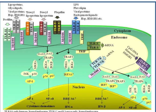
Figure 6 shows the TLRs with their corresponding ligands and the intracellular signaling cascades.
Figure 6. Toll-like receptors, their ligands, and the TLR signaling cascade
TLR signaling can be divided into myeloid differentiation factor 88 (MyD88)- and TIR domain-containing adaptor inducing IFN-β (TRIF)-dependent pathways that will be discussed below demonstrated on TLR4 since it triggers both MyD88- and TRIF- pathways.
TLR4 forms a receptor complex with myeloid differentiation factor 2 (MD2) on the cell surface and two TLR4-MD2 complexes create a homodimer (Figure 7.). TLR4 is the main sensor of endotoxin (LPS), but beyond LPS, it also recognizes bacterial flavolipin, mannan, viral proteins (99) and endogenous danger signals, including heat shock proteins, HMGB1, hyaluronan and fibronectin fragments (100
Ligand engagement recruits MyD88 to the receptor via TIRAP/Mal followed by an interaction between MyD88 and IL-1R-associated kinase (IRAK)-4. IRAK4 then activates
).
18
IRAK1 that associates with TNFR-associated factor 6 (TRAF6) and lead to the activation of TGF-β-activated kinase-1 (TAK1), and TAK-1 binding protein (TAB) 1,2 and 3. That later complex phosphorylates IκB kinase (IKK) and MAP kinase (MAPK) 6.
Phosphorilated IκB degrades and thus the free nucear factor-κB (NFκB) can translocate to the nucleus. MAP kinases activate another transcription factor, AP-1. NFκB and AP-1, finally, lead to the transcription of several pro-inflammatory genes (100). The MyD88 signaling induced by the endosomal TLR9 and TLR7 or 8 is somewhat different (without details depicted in Figure 6.) and lead to IFN-β production via IRF7, 5 or 1 (100).
TLR4-ligands may activate the TRIF pathway as well via TRIF-related adaptor molecule (TRAM). Active TRIF associates with TRAF6 and TRAF3. TRAF3 activates the kinases IKKε and TBK-1 (TANK-binding kinase 1) leading to the activation of interferon regulatory factor (IRF) 3 and 7 and Type-I interferon production. TRIF-induced TRAF6 pathway downstream is similar to MyD88-induced TRAF6 signaling resulting in pro- inflammatory cytokine and chemokine transcription. TLR3 uses only TRIF as adaptor (100).
Figure 7. Structure and downstream signaling of TLR4-MD2 complex
19 2.2.1.2. TLRs in liver diseases
Toll-like receptors are expressed in several cell types in the liver (101
The pathogenic role of TLRs has been demonstrated in infectious (HCV, HBV, endotoxin-induced liver damage, Listeria, Salmonella and malaria infections) and non- infectious (alcoholic liver disease, NASH, ischemia-reperfusion injury, primary biliary cirrhosis, autoimmune hepatitis) liver diseases and in their complications (hepatic fibrosis, hepatocellular carcinoma). The role of TLRs in liver diseases was reviewed by Seki et al.
(101)
). The hepatic resident macrophages (Kupffer cells) express TLR-2, -3, -4 and -9 and produce pro- and anti-inflammatory cytokines in response to TLR ligands. The plamocytoid dendritic cells (pCDc) produce IFN-α and also pro-inflammatory cytokines in response to TLR7/8 and TLR9 stimulation, while conventional-DCs respond to TLR-2,-3 and-4 ligands with cytokine production in the liver. The hepatic natural killer (NK) cells express TLR-1,-2,-3,- 4,-6, -7/8, -9 and those TLR-ligands together with Kupffer-cell derived IL-12 induce IFN-γ production. TLRs activate B-cell proliferation, while T-cells are rather indirectly activated by the TLRs. Beyond the immune cells, liver parenchymal cells also express TLRs.
Hepatocytes although do express all TLRs, but they can respond only to TLR2 and TRL4 ligands. Liver sinusoidal endothelial cells (LSEC) respond to TLR4 ligand LPS, and hepatic steallate cells (HSCs) can be stimulated by TLR4 and TLR9 ligands. (101)
2.2.2. RIG-I-like receptors (RLRs) 2.2.2.1. RLRs and their ligands
The cytoplasmic RIG-I-like helicase receptors include retinoic acid inducible protein I (RIG-I), melanoma differentiation associated gene 5 (MDA5), and laboratory of genetics and physiology 2 (LGP2). They contain a C-terminal regulatory domain, a central DEAD box helicase/ATPase domain and 2 N-terminal caspase recruitment domains (CARDs) except LGP2 that lacks the CARD domain. They have low expression, but viral infections and Type I IFNs highly enhance the RLR expression (94). Upon engagement by viral dsRNA (either genomic RNA of dsRNA viruses or dsRNA generated during the replication of ssRNA or dsRNA transcribed from dsDNA by polymerase III) RLR
20
signaling results in Type-I IFN and pro-inflammatory cytokine production (102,103).
Although, both RIG-I and MDA5 sense viral RNA, their ligand specificity somewhat differs. RIG-I recognizes short dsRNA and the presence of 5`triphosphate increases Type I IFN induction. In contrast, MDA5 senses long dsRNA such as the synthetic poly I:C (102).
RIG-I is important for the detection of Paramyxoviridae (eg. Sendai virus), Influenza A virus, Rhabdoviridae (eg. Vesicular Stomatitis Virus), Myxoma virus and Herpesviridae (eg. Ebstein Barr virus), while MDA5 is crucial for the recognition of Caliciviridae, Picornaviridae (eg. Encephalomyocarditis virus), Murine hepatitis virus and Vaccinia virus (104
RLR stimuli lead to the interaction between the CARD domain of the RLR receptor and the CARD domain of the adaptor protein called MAVS (mitochondrial antiviral signaling protein) or IPS-1 (IFN-β-promoter stimulator 1) or VISA (virus-induced signaling adaptor) (
). Both RIG-I and MDA5 contribute in the detection of Reoviridae and Flaviviridae such as Hepatitis C virus (103). LGP2 likely cooperate with RIG-I and MDA5, since LGP2 deficiency decreases certain viruses-induced IFN production (104).
105). The name MAVS will be used further on in that thesis. MAVS is localized in the outer membrane of the mitochondria that is crucial for the downstream signaling. Beyond the N-terminal CARD domain MAVS contains a C-terminal TM domain, that is responsible for the mitochondrial anchorage (106). In addition, Baril M et al. has recently shown that the TM domain is the main determinant of the self-interaction, and they demonstrated that MAVS oligomerization is essential for the downstream activation of IRF3 and NFκB (107). The TM-dependent dimerization of MAVS provides an interface for binding and activating TRAF3 (106). Similarly to the TRIF signaling pathway, TRAF3 activation results in the phosphorilation and oligomerization of IRF3 and IRF7 via TBK1 and IKKε, and the active IRF3 lead to the production of Type-I IFNs (94).
MAVS also interacts with Receptor interacting protein kinase 1 (RIP1) and Fas-associated death domain protein (FADD) that lead to the complex formation with caspase-8 and 10, and to the cleavage and activation of these caspases and apoptosis. The RIP1/TRADD/FADD complex also induces inflammatory cytokine production via NFκB activation (102). The RLR signaling is shown in Figure 8.
21
Figure 8. RLR signaling
2.2.2.2. RLRs in liver diseases
Helicase receptors are expressed in several cell types in the liver, including hepatocytes, conventional dendritic cells, Kupffer-cells, NK cells, endothelial cells and fibroblasts (108,109). Immunohistochemistry revealed that in naïve mice MDA5 is broadly expressed in hepatocytes and interstitial cells (Kupffer cells and endothelial cells) in the liver, but synthetic dsRNA (poly I:C) (110
The RLR-mediated antiviral responses are extensively studied in hepatitis C infection in the liver (
), as well as Type I IFNs further increase its expression.
111,112). RIG-I is essential for the innate immune signaling in hepatocytes triggered by HCV genome. However, HCV has evolved strategies to disrupt the host antiviral response eg. by cleaving MAVS from the mitochondria (113) by the viral NS3/4A protease. MAVS can be cleaved by other viruses too such as the 3ABC protease of hepatitis A virus (114). The target of the viral cleavage is at the C-terminal region (Q428, C508), such as in case of apoptotic cleavage (D429) (113). Beyond viral hepatitis, the role of RLRs in the pathogenesis of biliary diseases has been suggested recently (115).
22 2.2.3. INFLAMMASOMES
The third big group of pattern recognition receptors is the family of Nod-like receptors (NLRs) (94). NLRs are composed from a C-terminal leucin-rich-repeat (LRR) domain that plays role in the recognition of ligands, a central NACHT (NAIP, CIITA, HET-E and TP-1) domain that is responsible for the oligomerization and dNTPase activity, and an N-terminal CARD or pyrin (PYD) domain. Based on the NACHT domain three subfamilies have been distuinguished: a) NODs, [NOD1-5, CIITA] b) NLRPs or NALPs [NLRP / NALP 1-14] and c) IPAF [IPAF, NAIP] subfamily. Several NLRs plays role in the formation of a multiprotein complex called inflammasome.
2.2.3.1. Definition
Inflammasomes are intracellular multiprotein complexes that in response to pathogens or danger molecules activate the cysteine protesase caspase-1 that in turn results in the maturation of pro-inflammatory cytokines, including IL-1β and IL-18, the proteolytic inactivation of IL-33 and furthermore they contribute to the regulation of cell survival and cell death.
2.2.3.1. Types of inflammasomes
To date four main prototypes of inflammasomes are characterized: NLRP1 (NALP1); NLRP3 (NALP3, cryporin); NLRC4 (IPAF) and the recently described AIM2.
With the exception of AIM2, the nomenclature of inflammasomes is based on the NOD- like receptor (NLR) that form complex with the effector molecule pro-caspase-1 with or without the help of an adaptor molecule and lead the auto-activation of the caspase-1.
NLRP1 (NALP1), the first described inflammasome, is able to interact directly with caspase-1 due to its C-terminal CARD domain. However, the presence of ASC enhances the activity of the complex in humans. Murine NLRP1 is unable to bind to ASC because it does not contain functional PYD domain.
23
NLRP3 (NALP3), the most fully characterized member of the inflammasome family, consists of the PYD, NACHT and LRR domain containing Nod-like receptor, NLRP3, the adaptor molecule ASC and the effector molecule pro-caspase-1. Since NLRP3 does not contain CARD domain, the presence of the adaptor molecule is necessary for the complex formation.
IPAF (NLRC4) also contains a CARD domain resulting in direct interaction with caspase- 1, but some studies suggested the requirement of ASC for the maximal caspase-1 activation.
AIM2, is a PYD and HIN-200 domain containing protein that recruit caspase-1 via the ASC adaptor molecule, since itself is lacking the CARD domain.
NLRP3 inflammasome AIM2 inflammasome
NLRP1 inflammasome IPAF inflammasome
Figure 9. Structure of the NLRP3 (NALP3), NLRP1 (NALP1), IPAF and AIM2 inflammasomes
2.2.3.3. Function of inflammasomes
Inflammasome activation leads to auto-activation of the 45kDa inactive pro- caspase-1 precursor into p20 and p10 subunits that form the active caspase-1 (116). The cysteine protease caspase-1 belongs to the inflammatory caspases together with caspase--
24
11 and -12 in mice and caspase-4 and -5 in humans (117). Active caspase-1 cleaves the precursors of IL-1β and IL18 to their mature form or inactivates IL-33 (117,118
IL-1β is a pro-inflammatory cytokine, a central regulator of inflammation that binds to IL-1 receptor (IL-1R) to exert its broad biological effects. The IL-1R also recognizes IL-1α and binds IL-1R antagonist (IL-1Ra), the latter has an inhibitory effect on the IL-1R (
).
119). The transcription, translation and secretion of IL-1β are tightly regulated (119). IL-18 or IFN-γ inducing factor, activates Natural Killer (NK) cells to produce IFNγ (120). IL-18 precursor is constitutively expressed in human PBMCs and mouse spleen cells, but its maturation and secretion is controlled by the inflammasomes (121). IL-33 is a chromatin-associated cytokine of the IL-1 family that drives Th2 responses (122,123). The full-length active IL-33 is cleaved and inactivated by caspase-1 (118).
Beyond, the maturation of pro-inflammatory cytokines, inflammasomes activation regulates cell death. Pyroptosis, first described in Salmonella infected macrophages, is a caspase-1 dependent cell death showing similarities to apoptosis (DNA-damage), but it does not depend on apoptotic caspases and it is accompanied with loss of plasma membrane integrity and lack of chromatin condensation (124
Finally, we have to mention that caspase-1 can promote cell survival via SREBPs in HeLa and CHO cell lines (
). NLRP1 (NALP1), NLRC4 (IPAF) and NAIP activate pyroptosis (119) while NLRP3 (NALP3) contributes to another NLR-dependent cell death, pyronecrosis. Pyronecrosis shows similarities with necrosis, since it is not caspase-depedent and leads to breakdown of plasma membrane without chromatin condensation. Pyronecrosis utilizes the inflammasome adaptor molecule, ASC, and involves the lysosomal cathepsin B (119). Both pyroptosis and pyronecrosis elicit inflammation.
125).
2.2.3.4. Inflammasome activating ligands
Inflammasome activation is a 2-step process in which signal 1 results in up- regulation of inflammasome expression (mostly from TLR activation) and signal 2 triggers
25
functional inflammasome activation by an inflammasome activator (116). Inflammasome activators can be pathogen-associtaed (PAMPs) or endogenous danger molecules (DAMPs) summarized in Table 1.
Table 1. Known activators of inflammasome NLRs
Inflammasome Activator
NLRP3 (NALP3, cryoporin)
Large particles via phagocytosis Monosodium urate crystals (MSU) (126
CPPD (calcium pyrophosphate dehydrate) (126) ) Alum (127
Silica ( ) 128 Asbestos (
) 129 Cholesterol crystals (
)
130 Amyloid beta (
) 131 Hyaluronan (
) 132 Hemozoin (
) 133
Vaccine adjuvants (poly lactide-co-glycolide and polystyrene microparticles) (
) 134
Bacterial toxins (pore forming) )
Listeria monocytogenes Lysteriolysin O (135 Staphylococcus aureus alpha-toxin (135,125,
,125) 136 Aeromonas hydrophila aerolysin (135,125)
) Streptolysin (137
Nigericin (135) )
Maitoxin (Dinoflegellates) (125) Ion channels and activators ATP(P2X7) (135)
Influenza virus M2 channel protein (138
PAMPs (only if transferred to the cytoplasm by eg. Streptolysin O poreformin toxin)
)
LPS, lipid A, PGN, MDP, LTA, Pam3, ssRNA, dsRNA, CpG DNA (139,140 NLRP1
(NALP1)
) Bacillus anthracis lethal toxin (141
MDP (
) 142
NLRC4 (IPAF)
)
Gram negative bacteria (flagellin-dependent and independent) Salmonella typhymurium (143
Shigella flexneri (
) 144
Legionella pneumophila ( )
145 Pseudomonas aeruginosa (
) 146 AIM2
) dsDNA
bacterial (147,148 viral (148)
) mitochondrial (149 host (148)
)
26
To date only the bacterial wall component muramyl dipeptide (MDP) and the Bacillus anthracis lethal toxin has been shown to activate NRLP1 inflammasome (141,142). The exact pathomechanism has not clarified yet, but potassium efflux has been suggested to play role in the NLRP1 inflammasome activation (
NRLP1 (NALP1) inflammasome
150,151). We have to mention that NLRP1 localize mostly in the nucleus in contrary to other inflammasomes that are cytoplasmic (152).
NLRP3 is the most characterized inflammasome. The activation of NLRP3 is tightly regulated at transcriptional level via NFκB (
NLRP3 (NALP3) inflammasome
153
To date three major pathways have been implicated in NLRP3 inflammasome activation: 1. ROS production, 2. lysosomal disintegration and 3. potassium efflux. The recent knowledge about the NLRP3 activation is summarized in Figure 10. Since NLRP3 inflammasome can be activated by several, not even alike ligands, the theory that those stimuli can activate the NLRP3 inflammasome via ROS production as a common pathogenic pathway is attractive. Several groups have reported that ROS scavengers suppress inflammasome activation (149,
). Cell priming with an NFκB activator such as the TLR4-ligand LPS is the first and critical step of inflammasome activation (150). Although up-regulation of NLRP3 expression is required, but not sufficient for the inflammasome activition (150). Several stimuli have been shown to serve as second signal for the activation of NLRP3 inflammasome. The exact mechanism by that the huge varieties of activators lead to the auto-activation of caspase-1 is not fully clarified yet.
154,155,156). Mitochondria can serve as source of ROS (149), and NADPH oxidase may also contribute, since the blockage of NADPH oxidase inhibits inflammasome activation (150). Furthermore, both large particles (157) and ATP (152) induce ROS production. However, how can ROS induce inflammasome activation has not been clarified yet. Recently, the ROS-dependent release of thioredoxin- interacting protein (TXNIP) from thioredoxin and direct interaction between TXNIP and
27
NLRP3 has been described (153). Notably, some ligands that lead to ROS production, does not lead to inflammasome activation (158
The second pathway of inflammasome activation is induced by crystals or large particles such as silica, asbestos, alum, amyloid, monosodium urate, cholesterol (126-134).
The particles are phagocytosed and after the fusion of the phagosome and lysosome, the breakdown of the phagolysosomal membrane results in the release of lysosomal content into the cytoplasm inducing inflammasome activation. The role of cathepsin B, a lysosomal protease has been implicated in the NLRP3 activation (131). Notably, caspase-1 activation by large particles is not impaired in cathepsin B-deficient macrophages (
).
159 The third pathway is via potassium efflux. Extracellular ATP can stimulate the P2X7 purinergic receptor that in turn results in potassium efflux and the recruitment of pannexin. The later one is a membrane pore that allows the delivery of extracellular PAMPs and DAMPs into the cytosol (139).
).
NLRC4 (IPAF) inflammasome is activated by the flagellin of Gram-negative bacteria including Salmonella typhimurium, Pseudomonas aeruginosa and Legionella pneumophila and some Gram positive flagellated bacteria such as Listeria monocytogenes (143-146). On the other hand, NRLC4 can be activated by non-flagellated bacteria as well, including the Gram negative Shigella flexneri (144). The steps of NRLC4 inflammasome activation are not explored yet.
NLRC4 inflammasome
Muruve et al. described that bacterial, viral and mammalian host DNA can trigger caspase-1 activation. Later AIM2, a HIN200 protein has been identified as a cytosolic dsDNA sensing inflammasome (147,148). It was the first description of a non-NLR family member that forms inflammasome complex and lead to caspase-1 activation. Since then, it has been shown that RIG-I also can trigger caspase-1 activation via the adaptor molecule ASC (
AIM2 inflammasome
160).
28
AIM2 is also unique in terms of that direct ligand binding has been proven (147). It is important in the recognition of bacterial DNA, DNA viruses and may also contribute to the pathogenesis of autoimmune diseases by recognizing mammalian DNA (161).
The characterization of other NLRs and the exact pathomechanism leading to inflammasome activation is still awaited.
Figure 10. Signaling transduction pathways of inflammasome activation
2.2.3.5. Inflammasome expression in the liver
The expression of inflammasomes and subcellular localization of the different NLRs varies between tissues (159). Early studies showed highest expression of NLRP3 (CIAS1) and NLRP1 (NAC) in peripheral blood leukocytes, while the liver showed relatively low levels (162,163). The liver expresses NLRP1, 2, 3, 6, 10, 12 and NLRC4 at the mRNA levels (164). The expression of PRRs is lower in solid organs compared to
29
spleen likely due to the lack of higher number of splenic immune cells (164,165
The liver is comprised of both parenchymal (hepatocytes) and immune cells (macrophages, dendritic cells, T-cells, NK/NKT-cells), where hepatocytes represent the majority of the cell populations. The role of inflammasomes has been mostly studied in immune cells, but there is increasing evidence that NLRs exist in non-immune cells as well, including keratinocytes (159,
).
However, human livers express higher level of NLRP10 (164), while murine livers are high in NLRP6 expression (164) compared to the spleen. The significance of these NLRs is yet to be evaluated.
166), myoblasts (167), fibroblasts and endothelial cells (168), osteoblasts (169), spinal cord motoneurons (170
The liver resident macrophages, Kupffer cells produce significant amount of IL-1β that would suggest (
), pyramidal neurons and oligodendrocytes (159).
171) the presence of inflammasomes, however, surprisingly, Kummer et al. showed that certain macrophages, including Kupffer cells are negative for NLRP1 staining (159). The presence of NLRP3 inflammasome and/or inflammasome activation has been also shown in sinusoidal endothelial cells (172) and stellate cells (173
The NLRs are just one components of the inflammasome complex, most of the inflammasome require an adaptor protein ASC, and the expression of caspase-1 is also prerequisite of the inflammasome assembly. ASC is expressed in several tissues, including hepatocytes and interlobular bile ducts (
). However, to our best knowledge, there are no published data on hepatocytes.
174), stellate cells (173). A marked, constitutive expression of caspase-1 (ICE) has been reported in the liver (175
Of course, the presence of inflammasome components is required but does not necessarily mean the activation of the complex. We will discuss below the relevance of inflammasomes in the different liver diseases.
). The cell-specific expression of the inflammasome in the liver is shown as Figure 11.
30
Figure 11. Expression of inflammasome components in the liver
2.2.3.6. Role of inflammasomes in liver diseases
The role of inflammasome activation has been implicated in several liver diseases including acetaminophen-induced liver injury (176), ischaemia-reperfusion liver injury (177), P.acnes plus endotoxin-induced liver injury model (178
There is increasing evidence that gut microbiota, increased gut permeability and endotoxin play a crucial role in the pathogenesis of both alcoholic (ASH) and non- alcoholic steatohepatitis (NASH) (60). Therefore, our aim was to explore the role of inflammasomes in NASH.
) and liver fibrosis (173).
31 3. AIM OF THE THESIS
The aim of thesis was to explore the role of innate immunity in the pathogenesis of NASH. The first part of the work focus on the role of the Gram negative bacterial wall component endotoxin and its receptor, Toll-like receptor 4 in the development of diet- induced steatohepatitis and fibrosis. The second part of the work was designed to investigate the role of the pro-inflammatory cytokine IL-1β and the inflammasome complexes that are responsible for the IL-1β-maturation in the pathogenesis of NASH.
Finally, with the third part of the work we aimed to explore the pathogenesis behind the susceptibility of fatty liver to viral diseases.
3.1.
There is increasing evidence that non-alcoholic steatohepatitis is accompanied with increased gut permeability and increased serum endotoxin levels (80-85). Furthermore, our group previously showed increased susceptibility to gut-derived endotoxin, lipopolysaccharide (LPS) in steatohepatitis (44). Toll-like receptor 4 (TLR4) and MD2 are the major receptors for LPS (99). Therefore the aims of the present study were:
To examine the role of toll-like receptor 4 signaling in the pathogenesis of NASH
To investigate the role of TLR4 and its adaptor MD2 in the development of diet- induced hepatic fat accumulation, liver injury, inflammation and fibrosis. To perform the experiments we employed wild type, TLR4- and MD2-deficient mice fed with methinone-choline deficient (MCD) diet to induce steatohepatitis.
3.2. To examine the role of IL-1β and inflammasomes in the pathogenesis of NASH
The intracellular multiprotein complexes called inflammasomes are responsible for the maturation of the pro-inflammatory cytokine IL-1β that plays important role in numerous chronic and acute inflammatory diseases. Bacterial endotoxin, that has been implicated as 2nd hit in the NASH pathogenesis, is a key factor of the inflammasome activation.
Therefore we aimed to investigate the role of the inflammasomes and IL-1β in the pathogenesis of NASH. The aims in details were:
32
To test whether there is inflammasome activation and increased IL-1β production in animal models of liver steatosis (ob/ob mice and short term high fat diet feeding) and steatohepatitis (MCD diet-induced steatohepatitis and long term high fat diet feeding).
To test whether hepatocytes express the inflammasomes.
To explore potential inflammasome activators in steatohepatitis performing in vitro experiments on immune cells (RAW macrophages and isolated murine liver mononuclear cells) and hepatocytes (Hepa 1-6 cells and primary murine hepatocytes).
To investigate the clinical significance of the inflammasomes and IL-1 signaling in the development of MCD diet-induced steatohepatitis using mice deficient in the following genes: 1. ASC (inflammasome adaptor), 2. Caspase-1 (inflammasome effector), 3. IL-1 receptor.
To check the inflammasome expression in liver biopsy samples from NASH patients.
3.3. To examine the pathomechanism of decreased antiviral response in steatohepatitis As we mentioned above, the co-morbidity of NASH with RNA viral infections, such as hepatitis C and HIV virus remains a clinical challenge and the susceptibility of fatty liver to virus-induced liver damage urges the better understanding of changes of antiviral immune responses in steatotic livers. Therefore the aims of the present study were:
To test the hypothesis that mice with steatohepatitis are more susceptible to virus induced liver injury and if yes to explore the underlying pathomechanism. To perform the experiment we employed mice fed with MCD diet to induce steatohepatitis and challenged them with Poly I:C to mimic viral infection. We evaluated the liver injury using biochemical and histological methods.
To investigate whether mice with steatohepatitis have impaired antiviral immunity to viral challenge and explore the underlying pathomechanism. To perform the experiment we used the MCD-diet induced animal model of steatohepatitis, challenged the mice with Poly I:C as a mimic of viral infection and evaluated the mounted antiviral response including interferon and cytokine production. In addition we investigated the intracellular signaling cascade step by step induced by Poly I:C.
33 4. MATERIALS AND METHODS
4.1 Animal studies
This study was approved by Institutional Animal Use and Care Committee (IACUC) at University of Massachusetts (UMASS) Medical School.
Six-eight week-old C57Bl/6 wild type (wt) mice (n=6-16/group) were fed with either methionine-choline deficient (MCD) diet for 5 or 8 weeks; or high fat diet (HFD; Harlan Laboratories Inc., South Easton, MA, USA) for 4 weeks or 9 months. Control mice received either an MCD-identical, but DL-methionine (3 g/kg) and choline bitartrate (2 g/kg) supplemented (MCS) diet (Dyets Inc., Bethlehem, PA, USA), or regular rodent chow diet. We also used 9 weeks old, female leptin deficient (ob/ob; B6.V-Lep ob/J from Jackson Laboratories) mice with their own age and gender-matched control group (C57Bl/6J). All mice had unrestricted access to water. The presence of steatohepatitis was proven histologically in the MCD diet-fed mice, while fat deposition was proven by liver triglyceride assay in the HF diet fed mice and the ob/ob mice.
TLR4 ligand lipopolysaccharide (LPS) (Sigma, St. Louis, MO, USA; 0.5mg/bwkg to MCS/MCD mice; 12ug/mouse to ob/ob mice) or TLR3/RLR ligand polyinosinic:polycytidylic acid (Poly I:C) (InvivoGen, San Diego, CA, USA), a synthetic double stranded RNA (5mg/bwkg); or TLR9 ligand CpG-ODN (InvivoGen, San Diego, CA, USA), (5mg/bwkg) were injected intaperitoneally for 2 or 6 hours.
The following knock-out mice were used: MD-2, TLR4-, MyD88-, ASC-, caspase- 1or IL-1R-defcient mice with their appropriate controls. Furthermore wild type (WT) mice transplanted with MyD88-deficient bone marrow (WT/MyD88) and MyD88-deficient mice transplanted with WT bone marrow (MyD88/WT) were employed.
4.2 Biochemical analysis and cytokine measurements
Serum alanine aminotransferase (ALT) was determined using a kinetic method (D- TEK, Bensalem, PA, USA), liver triglyceride levels were assessed using L-Type Triglyceride H kit (Wako Chemicals USA Inc., VA, USA). Serum TNFα, IL-6 and IL-1β
34
levels were determined by BDTM Cytometric Bead Array (BD Biosciences, Sparks, MD, USA), serum IFNβ and HMGB1 protein levels were measured by ELISA (PBL Biomedical Laboratories, Piscataway, NJ, USa and IBL Transatlantic, Toronto, Canada;
respectively). Liver thiobarbituric acid reactive substances (TBARS) were assayed using whole liver homogenates and Oxi-TEK TBARS assay kit (ZeptoMetrix Corp., Buffalo, NY, USA).
4.3 Histopatological analysis
Sections of formalin-fixed, paraffin-embedded livers were stained with: 1) hematoxylin and eosin to assess histological features of steatohepatitis, 2) picro-sirius red stain to evaluate for hepatic collagen deposition. OilRed O tissue staining method on OCT- embedded frozen sections was used to quantify the steatosis. Liver sections were also subject to immunohistochemical staining for macrophages with monoclonal F4/80 antibody (Abcam, Cambridge, MA) and α–smooth muscle actin with a monoclonal antibody against α–smooth muscle actin (Lab Vision Corporation, Fremont, CA) using a labeled streptavidin-biotin immunoenzymatic antigen detection system (UltraVision Mouse Tissue Detection System Anti-Mouse-HRP/DAB, Lab Vision Corp). Image J and Microsuite software (Olympus Soft Imaging Solutions GmbH, Munster, Germany) was used for image analysis at indicated magnification on 20 high-power fields.
4.4 RNA analysis
RNA was purified using the RNeasy kit (Qiagen Sciences, Maryland, USA) and on- column DNA digestion. cDNA was transcribed with the Reverse Transcription System (Promega Corp., Madison, WI). Real-time quantitative polymerase chain reaction was performed using iCycler (Bio-Rad Laboratories Inc., Hercules, CA); primer sequences are shown in Table 1. All specific mRNA levels were normalized against the housekeeping gene, 18S, in the same sample.
35 4.5 Protein analysis
4.5.1 Preparation of cell lysates
Whole liver lysates were extracted from frozen liver using RIPA buffer (Boston Bioproducts, Ashland, MA, USA). Isolation of mitochondrial and cytosolic fraction from fresh liver tissue was based on the principle of differential centrifugation using Mitochondrial Extraction kit (Imgenex Co., San Diego, CA, USA).
4.5.2 SDS-PAGE electrophoresis
Whole liver, cytoplasmic or mitochondrial extracts were prepared. Samples with equal amounts of protein were separated in polyacrylamide gel, and proteins of interest were identified on the nitrocellulose membrane with specific primary antibodies followed by horseradish peroxidase–labeled secondary antibodies and chemiluminescence assay.
The following antibodies were employed: MAVS (Santa Cruz Biotechnology Inc.; Cell Signaling), cytochrome c (Imgenex), caspase-1 p10 (Santa Cruz Biotechnology Inc.), cleaved caspase-8 (Imgenex), RIP3 (Abcam), PSMA7 (Abcam), HMGB1 (Abcam), phoshoserine (Abcam), IRF3 (Cell Signaling), phosphoIRF3 (Cell Signaling), IL-1β (R&D), β-actin (Abcam), β-tubulin (Abcam), Tim23 (BD Biosciences).
4.5.3 Native gel electrophoresis
Native PAGE Novex Bis-Tris Gel System (Invitrogen Life Science, Carlsbad, CA, USA) was used. Liver samples were lysed using 5% Digitonin as mild detergent and separated on Native PAGE Novex 3-12% Bis-Tris Gels. Proteins were transferred to PVDF membrane, fixed with 8% acetic acid diluted in distilled water and identified with specific primary antibodies followed by HRP labeled secondary antibodies and chemiluminescence assay.
4.5.4 Immunoprecipitation
Whole liver lysates were precleared with anti-rabbit IgG beads followed by overnight incubation with 5ug of the primary antibody (PSMA7 or MAVS) and precipitated with IgG beads. The immunprecipitates were lysed and denatured using β-
36
mercaptoethanol containing buffer and heating. The proteins were separated on polyacrylamid gel, transferred to nitrocellulose membrane and detected by specific antibodies (MAVS, PSMA7).
4.6 Functional assays
4.6.1 Caspase-activity assays
Caspase-1 activity was determined in freshly prepared whole liver lysates using colorimetric assay. Caspase-1 activity analysis is based on the cleavage of substrate WEHD-pNA (R&DSystems, Minneapolis, MN, USA).
Caspase-3 activity was determined in freshly prepared whole liver lysates using colorimetric assay. Caspase-3 activity analysis is based on the cleavage of substrate DECD-pNA (GenScript, Piscataway, NJ, USA).
4.6.2 NADPH activity assay
NADP+/NADPH concentrations from whole liver extracts with comparable protein amounts were determined using EnzyChrom NADP+/NADPH assay kit (ECNP-100) (BioAssay Systems, Hayward, CA), as manufacturer recommended.
4.6.3 Cytotoxicity assay
The lactate dehydrogenase (LDH) assay (Sigma-Aldrich, St. Louis, MO, USA) was used to measure the amount of cytoplasmic LDH released into the medium as an indicator of membrane integrity and cell viability.
4.7 In vitro experiments
Cell lines or primary cells were stimulated with LPS (100 or 1000ng/ml for 2, 6, 18hours), fatty acids (palmitic acid with BSA 0.33mM, 0.165mM; oleic acid 0.66mM, 0.33mM; linoleic acid 0.66mM, 0.33mM for 2,6,18,24,36 hours) or their combinations with or without ZVAD (40μM). Poly I:C (10μg/ml) was used to stimulate hepatocytes with or without Lipofectamin 2000 (6 hours.)
37 4.7.1 Cell lines
Hepa1-6 mouse hepatoma cell-line and RAW 264.7 mouse leukemic monocyte- macrophage cell-line were employed.
4.7.2 Primary cells
Animals received anesthesia with ketamine (100 mg/kg) and xylazine (10 mg/kg);
the livers were perfused with Hank’s balanced saline solution (HBSS) followed by in vivo digestion with 0.33 mg/ml Liberase RI Enzyme (F. Hoffmann-La Roche Ltd; Basel Switzerland) in HBSS. The LMNCs and hepatocytes were purified from whole liver cell suspension obtained after tissue disruption using centrifugation at slow speed (500g).
Hepatocytes were washed twice with 2% fetal bovine serum (FBS) containing PBS and were plated on collagen-coated plates. The LMNCs were further purified by subsequent isolation in Percoll 40/70 gradient density at 800g and harvested from the gradient interface. Purity of cell population was assessed by qPCR.
4.8 Flow cytometry analysis
Liver mononuclear cells were washed in saline supplemented with 2% fetal bovine serum (FBS) and stained for surface NK cell marker NK1.1 (BD Bioscience, San Jose, CA). In some experiments LMNCs were stimulated with a cocktail of PMA (50 ng/ml), ionomycin (1µg/ml), and brefeldin A (10µg/ml) in RPMI1640+10% FBS for 4 hours and stained for CD68 and intracellular TNFα using specific fluorescent labeled antibodies and CytoFix/CytoPerm Kit (BD Bioscience, San Jose, CA). The cells were gated by size and granularity and their fluorescence was analyzed using the LSR flow cytometer.
4.9 Human liver samples
The study meets the ethical guidelines of the 1975 Declaration of Helsinki and was approved by the Committee for the Protection of Human Subjects in Research at the University of Massachusetts. All participants gave a written consented to participate in the study.
38
Human liver tissue was obtained from biopsies from six (2 males and 4 females; age: 45±8 years), clinically and biopsy-proven NASH patients. The histology showed steatosis (<1/3 hepatocytes: n=2, 1-2/3 hepatocytes: n=3, >2/3 hepatocytes: n=1) with rare hepatocyte ballooning (none: n=2, <1/3 hepatocytes: n=4) and inflammation with inflammatory score 1-4. Lobular inflammation was present in 5 patients. Fibrosis was not detected in any of the patients. Human liver tissue from chronic hepatitis C infected patients (n=5) were used as diseased controls. Human normal liver (n=4) total RNA was purchased from OriGene Technologies (Rockville, MD, USA)
4.10 Statistical analysis
Statistical significance was determined using the nonparametric Kruskal-Wallis test, Mann-Whitney tests, where appropriate. Data are shown as mean ± standard error and were considered statistically significant at p≤ 0.05.
39 5. RESULTS
MD-2 and TLR4 complex is the major receptor for endotoxin (
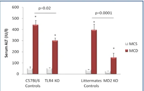
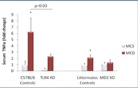
5.1.1 MD-2 or TLR4 protects from MCD diet-induced liver fat deposition and inflammation
179) that has been shown to contribute to activation of the inflammatory cascade in alcoholic steatohepatitis (ASH) leading to liver damage. Given the common pathophysiological features of ASH and NASH, we aimed to identify the role of MD-2/ TLR4 complex in an experimental model of NASH using mice deficient in MD-2 or TLR4 and their genotype control counterparts. Feeding a methionine-choline-sufficient (MCS) diet resulted in no signs of hepatic steatosis or inflammation in any of the mice (Figure 12-16). In contrast, mice of control genotypes fed a methionine-choline-deficient (MCD) diet for 8 weeks developed significant hepatic steatosis; MD-2- and TLR4-deficient mice on MCD diet showed lower liver fat accumulation, identified after OilRed O staining, compared to the mice of control genotypes (Figure 12). Consistent with the development of hepatic steatosis, liver triglyceride levels were significantly increased in MCD-diet-fed control genotype mice but to a significantly lower extent in MD-2- or TLR4-deficient mice (Figure 13). These findings suggested that TLR4/MD2 complex deficiency is partially protective against MCD-induced liver steatosis.
5.1 Deficiency in myeloid differentiation factor-2 (MD2) and toll-like receptor 4 (TLR4) expression attenuates non-alcoholic steatohepatitis and fibrosis in mice
40
Figure 12. Mice of control genotypes and those deficient (knock-out, KO) in TLR4 (TLR4 KO) and MD-2 (MD-2 KO) were fed methionine-choline-deficient (MCD) or methionine- choline-sufficient (MCS) diets for 8 weeks. Liver tissue was subjected to H&E (top panel) and OilRed O (bottom panel), one representative slide from n=6-16/group is shown.
C57Bl/6 controls TLR4 KO Littermates controls MD2 KO M
C S
M C D
M C S
M C D
Hematoxilin-eosin (200x)
OilRed O (400x)
C57Bl/6 controls TLR4 KO Littermates controls MD2 KO
41
Figure 13. Mice of control genotypes and those deficient (knock-out, KO) in TLR4 (TLR4 KO) and MD-2 (MD-2 KO) were fed methionine-choline-deficient (MCD) or methionine- choline-sufficient (MCS) diets for 8 weeks. Liver triglycerides were determined as described in the Methods. (*) represents p<0.05 compared to corresponding MCS group;
n=6-16/group
Feeding of MCD diet lead to accumulation of inflammatory cells into the liver in mice of control genotypes, and to a lesser extent in MD-2- or TLR4-KO mice, as indicated by the increase in content of F4/80+ cells in the livers of MCD-fed animals, compared to MCS diet-fed controls (Figure 14).