www.nature.com/scientificreports
Sequential activation of different pathway networks in ischemia- affected and non-affected
myocardium, inducing intrinsic
remote conditioning to prevent left ventricular remodeling
Noemi Pavo
1, Dominika Lukovic
1, Katrin Zlabinger
1, Abelina Zimba
1, David Lorant
2, Georg Goliasch
1, Johannes Winkler
1, Dietmar Pils
4,15, Katharina Auer
3, Hendrik Jan Ankersmit
4, Zoltán Giricz
5, Tamas Baranyai
5, Márta Sárközy
6, András Jakab
7,
Rita Garamvölgyi8, Maximilian Y. Emmert
9,10,11, Simon P. Hoerstrup
9,10,11,
Derek J. Hausenloy
12,13, Péter Ferdinandy
5,6,14, Gerald Maurer
1& Mariann Gyöngyösi
1 We have analyzed the pathway networks of ischemia-affected and remote myocardial areas after repetitive ischemia/reperfusion (r-I/R) injury without ensuing myocardial infarction (MI) to elaborate a spatial- and chronologic model of cardioprotective gene networks to prevent left ventricular (LV) adverse remodeling. Domestic pigs underwent three cycles of 10/10 min r-I/R by percutaneousintracoronary balloon inflation/deflation in the mid left anterior descending artery, without consecutive MI. Sham interventions (n = 8) served as controls. Hearts were explanted at 5 h (n = 6) and 24 h (n = 6), and transcriptomic profiling of the distal (ischemia-affected) and proximal (non-affected) anterior myocardial regions were analyzed by next generation sequencing (NGS) and post-processing with signaling pathway impact and pathway network analyses. In ischemic region, r-I/R induced early activation of Ca-, adipocytokine and insulin signaling pathways with key regulator STAT3, which was also upregulated in the remote areas together with clusterin (CLU) and TNF-alpha. During the late phase of cardioprotection, antigen immunomodulatory pathways were activated with upregulation of STAT1 and CASP3 and downregulation of neprilysin in both zones, suggesting r-I/R induced intrinsic remote conditioning. The temporo-spatially differently activated pathways revealed a global myocardial response, and neprilysin and the STAT family as key regulators of intrinsic remote conditioning for prevention of adverse remodeling.
1Department of Cardiology, Medical University of Vienna, Vienna, Austria. 2Department of Anaesthesiology, Medical University of Vienna, Vienna, Austria. 3Department of Obstretrics and Gynecology - Molecular Oncology Group, Medical University of Vienna, Vienna, Austria. 4Department of Surgery, Medical University of Vienna, Vienna, Austria.
5Department of Pharmacology and Pharmacotherapy, Semmelweis University, Budapest, Hungary. 6Department of Biochemistry, Faculty of Medicine, University of Szeged, Szeged, Hungary. 7Department of Biomedical Imaging and Image-guided Therapy, Medical University of Vienna, Vienna, Austria. 8Institute of Diagnostic Imaging and Radiation Oncology, University of Kaposvar, Kaposvar, Hungary. 9Swiss Centre for Regenerative Medicine, University of Zurich, Zurich, Switzerland. 10Division of Surgical Research, University Hospital of Zurich, Zurich, Switzerland. 11Clinic for Cardiovascular Surgery, University Hospital of Zurich, Zurich, Switzerland. 12The Hatter Cardiovascular Institute, University College London, London, UK. 13Cardiovascular and Metabolic Disorders Program, Duke-NUS Graduate Medical School, Singapore, Singapore. 14Pharmahungary Group, Szeged, Hungary. 15CeMSIIS - Center for Medical Statistics, Informatics, and Intelligent Systems, Medical University of Vienna, Vienna, Austria. Correspondence and requests for materials should be addressed to N.P. (email: noemi.pavo@meduniwien.ac.at)
Received: 04 November 2016 Accepted: 31 January 2017 Published: 07 March 2017
OPEN
Pre-infarction angina, termed “warm-up angina”, increases myocardial resistance to subsequent ischemia, thereby reducing infarct size and mortality1. Exposure of the myocardium to single or repetitive brief episodes of ischemia and reperfusion (I/R), before sustained ischemia, induces cardioprotection, which enhances the ability of the myocardium to withstand the next ischemic insult2,3. Most ischemic preconditioning (IPC) protocols incor- porate a sustained coronary occlusion following the IPC-stimulus, causing acute myocardial infarction (AMI).
Problematically, the extensive myocardial damage incurred can mask changes to the genomic, proteomic, or met- abolic profiles, which would be attributable solely to I/R-linked cardioprotection. Only few reports have employed repetitive I/R (r-I/R) without ensuing AMI to reveal the impact of r-I/R alone, using small animal models with open-chest procedures, or Langendorff perfused hearts in vitro4. Despite extensive research, translational break- throughs have not yet been reached5, mainly due to the recognition that the experimentally proven powerful IPC-related cardioprotective substances are effective before infarction, and logically should be applied prior to the onset of infarction in the clinical scenario. Therefore, to date, IPC could not achieve practical clinical relevance.
The mechanisms underlying the early (up to 3 hours) and late (from 24 hours to several days post i-I/R) phase of IPC seem diverse: rapidly released and activated transmitter molecules confer beneficial effects in the acute phase, whereas cardioprotection in second windows of protection (SWOP) is instigated by a more complex response that requires de novo synthesis of effector proteins6. In contrast to IPC, postconditioning (PostIC) and remote ischemic conditioning (RIPC) are already used in clinical settings, albeit recent meta-analyses revealed a lack of definitive success for both in human procedures. This is partially explained by the application of poten- tially confounding cardioprotective medications7.
Since the term IPC is closely associated with subsequent AMI, we use here the expression r-I/R injury for clear separation of the two distinct phenomena. While IPC (with AMI) represents the warm-up angina followed by ST-segment elevation myocardial infarction (STEMI), r-I/R corresponds to a stable angina in humans.
To date, high-throughput gene analyses have not yet been used for dissecting the effects of IPC or r-I/R in a closed-chest, catheter-based setting in a clinically relevant large animal model, without the masking effect of infarction7,8, even if this approach is useful for identifying single genes or pathways, or entire pathway networks, and is thus well suited for exploring the complexity of the cardioprotection mechanism9.
Here we analyzed the effects of the r-I/R stimulus without subsequent infarction-related injury in a porcine model using next generation sequencing (NGS) combined with pathway network analysis. Our aim was to reveal the complex transcriptomic response that is involved in cardioprotection in ischemic and ischemia-unaffected regions of the heart, and to identify novel genes and networks that are responsible for protection of the heart against I/R. We analyzed the relevant genes and pathways at an early time point after r-I/R injury (early win- dow, trigger of cardioprotection) and 24 h later (second window, performing cardioprotection), and elucidated the time-sequence of the activation of pathways and genes with conditioning function in the ischemic and the ischemia non-affected region, termed “intrinsic remote conditioning” responsible for prevention of LV adverse remodeling.
Results
A translational model of r-I/R and IPC.
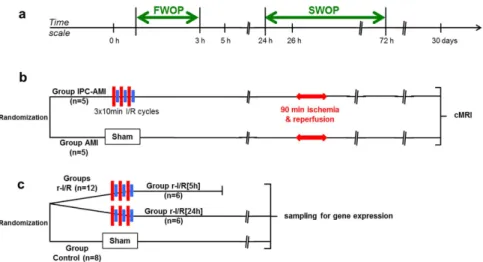
In contrast to our translational model, small animal open-chest models, with high heart rates differ substantially from the biological scenario presented by the human transient ischemic heart attack. Furthermore, in vitro models lack the pathophysiological complexity of the organism. Our assumption, that the typical 30 sec myocardial r-I/R cycles used in rodents for mimicking r-I/R or IPC, would be inadequate in provoking a sustained ischemic response in either pigs or humans, was evidence-based from our previous work showing that indications of adaptive mechanisms can be observed after a minimum of 5 min coro- nary occlusion in pigs10. We found that three 10 min cycles of r-I/R failed to provoke irreversible myocyte injury, as assessed by echocardiography, left ventricular (LV) hemodynamic measurements, necroenzyme release, or micro- scopic imaging, but induced substantial transcriptomic changes not only within the ischemic injury, but also in the remote myocardial area. Therefore, we induced r-I/R in pigs by using 3 × 10 min I/R by percutaneous balloon inflation/deflation of the mid left anterior descending coronary artery (LAD), followed by recovery of the animals.Sham-operated animals served as control (group control). At the pre-specified (5 h and 24 h) follow-ups (groups r-I/R [5 h] and r-I/R [24 h], respectively), the heart was explanted and the transcriptomics of the ischemia-affected distal anterior, border mid anterior and ischemia-non-affected proximal anterior parts of the heart were deter- mined (Fig. 1). In order to prove the cardioprotective effect of the 3 × 10 min r-I/R stimulus, we have additionally induced AMI 26 h post r-I/R (SWOP phase) (group IPC-AMI) or sham I/R (group AMI) (Fig. 1).
The r-I/R stimulus induces transient alterations in LV-hemodynamics without persistent myo- cardial dysfunction.
The results of hemodynamic measurements, both invasive and non-invasive, at base- line, directly after the r-I/R stimulus and at follow-up are shown in Supplementary Table S2. As a direct response to the r-I/R stimulus, there was a significant decrease in LV systolic pressures, and an increase in LV end-diastolic pressure and isovolumic relaxation time with normalization at 5 h or 24 h follow-ups.Biomarkers and histology.
The r-I/R stimulus failed to change plasma levels of troponin I, myoglobin or CK (Fig. 2a). Histologic images of the proximal, mid and distal anterior wall regions showed no signs of morpho- logical myocardial injury (Fig. 2b).r-I/R leads to time-dependent changes in gene expression compared to non-conditioned
hearts.
Summary of retrieved genes. The comparison of the groups r-I/R [5 h] and r-I/R [24 h] with the con- trol group showed that a total of 13758 genes were retrieved by NGS. PCA showed good discriminatory power between the three experimental groups with adequate similarity within the r-I/R groups even though originatingwww.nature.com/scientificreports/
group and controls were greater than between the r-I/R [24 h] group and controls, suggesting more pronounced alterations from normal cell function at 5 h than at 24 h after r-I/R.
Venn diagrams showed significantly altered gene expression between control and r-I/R animals (regardless of the time) and between r-I/R -induced early and late changes for proximal (ischemia-unaffected), mid (border) and distal (ischemia-affected) anterior regions (Fig. 3b). Remarkably, also the proximal region, which was not directly affected by ischemia, showed distinct changes in gene expressions already after 5 h. A total of 5789 genes were differentially expressed between the myocardial samples of the groups r-I/R [5 h] and r-I/R [24 h], equally affecting all three regions.
In the distal anterior wall region, a total of 1328 and 1762 genes were differentially expressed at 5 h and at 24 h, respectively. In the mid anterior region (border zone of ischemia/reperfusion), 1617 and 1087 genes were dereg- ulated 5 h and 24 h post r-I/R, respectively. Regarding the proximal anterior wall region, 1451 and 1276 genes showed differential expression at 5 h and 24 h post r-I/R.
Activated pathways and pathway networks of ischemia-affected and non-affected myocardial regions. Table 1 shows the results of the pathway analyses of the retrieved genes according to the different regions at 5 h and 24 h post r-I/R stimulus compared to controls. Results are presented as SPIA plots with false discovery rates of 5%
and 10% for the corresponding pathways (Supplementary Fig. S1). Pathway analysis revealed that there were multiple networks involved at 5 h in the ischemia-affected area, primarily including the calcium signaling, the adipocytokine and insulin signaling pathways. At 24 h (during the late phase of cardioprotection), the immuno- modulatory antigen processing and presentation, focal adhesion and extracellular matrix-receptor interaction pathways all were involved in the distal anterior area. The Ca-signaling pathway and the antigen processing and presentation pathways were also activated in the remote myocardial areas at 5 h and 24 h, respectively, indicating decisive roles in intrinsic remote conditioning (Table 1 and Supplementary Fig. S2). Table 2 and Fig. 4 display significantly deregulated genes, which are part of these relevant pathways.
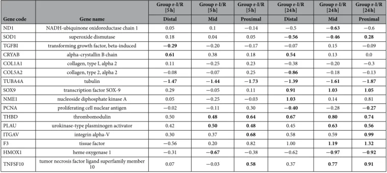
Table 3 and Supplementary Table S3 show a list of relevant selected genes of additional pathways that are significantly involved, showing expressional changes at 5 h and 24 h in the different regions. The genes are listed according to functional classes or established pathways, and changes in expression are indicated according to time points and regions.
Major pathways with known role in ischemia/reperfusion. The key regulator STAT3 is overexpressed in the ischemic, but also in the border and non-ischemic areas at 5 h, and mediates the Ca-signaling, adipocytokine and insulin signaling pathways. It is involved in both the PI3K/Akt and the JAK-STAT pathways. Moreover, SHISA2 showed a striking upregulation in both the early and late phases, suggesting the involvement of Wnt-signaling in ischemic preconditioning-induced cardioprotection11.
Activation of PI3K and its downstream targets Akt and glycogen synthase kinase 3β (GSK3B), has been demonstrated to be essential for ischemic preconditioning-induced cardioprotection involved in the RISK path- way12. Briefly, CREB-1 shows significant upregulation in the early phase, inducing transcriptional regulation of the cAMP pathway. Transcriptional changes in either GSK3A or GSK3B could not be detected in r-I/R-induced injury without subsequent AMI, neither at the early nor at the late phase, suggesting that the involvement of these proteins in cardioprotection does not depend on enhanced transcription. Indeed, most studies imply a post-translational regulation of GSK3B via phosphorylation at serine 913. The PI3K-Akt pathway with the genes Figure 1. Study design. (a) Time scale of the experiments. FWOP: first window of protection; SWOP: second window of protection. (b) Efficacy study randomizing domestic pigs into two groups without (Group AMI) or with ischemic preconditioning (IPC; Group IPC-AMI) 26 h before acute myocardial infarction (AMI). Cardiac magnetic resonance imaging (cMRI) at 30 days. (c) Gene expression analysis study randomizing pigs into groups with repetitive ischemia/reperfusion (r-I/R) and sacrificing at 5 h (upstream phase of SWOP) and 24 h (SWOP phase) or control.
found deregulated at 5 h and 24 h post r-I/R is displayed in Supplementary Fig. S3. Our data show that CREB, RTK and ITGA were up- and c-Myb and GF were downregulated. At 24 h, RTK and ITGA showed stable upreg- ulation, while c-Myb displayed baseline levels. We found no modulation of anti-apoptotic Bcl-2, and the tran- scription and cell survival factor NF-κB showed modest upregulation at 5 h and at 24 h (Supplementary Fig. S3).
The recruitment of STAT3 and the activation of the JAK-STAT pathway play a major role in upregulation of cardioprotective and anti-apoptotic proteins14. Other STAT proteins (STAT-1, 5 and 6) have been shown to be upregulated as well, but their precise functional role is not yet fully elucidated15. Expression of STAT3 is upregulated in the early phase of r-I/R-induced changes, along with altered expression of several associated proteins, such as TNFα , connective tissue growth factor, osteopontin, tenascin, and several paracrine factors and cytokines16. STAT3-KO mice developed cardiac fibrosis, LV remodeling and heart failure16. Activation of STAT3 with TNF and its receptors is referred to as the survivor activating factor enhancement (SAFE) pathway17. Supplementary Table S4 lists significantly up- or downregulated genes in the RISK and SAFE pathways that have been shown to be upregulated in different phases of IPC (I/R injury with subsequent AMI). Interestingly, r-I/R without subsequent infarction did not result in significant activation of these pathways as a whole, even though some components were deregulated.
Figure 2. Laboratory data, histology and fluorescence microscopy of the animals with repetitive ischemia/
reperfusion (r-I/R) [5 h] and r-I/R [24 h]) or without (group control) r-I/R. (a) Serum troponin I (TnI), plasma myoglobin, and serum creatine kinase (CK) at baseline, immediately after r-I/R (post-r-I/R) or sham- r-I/R, at 5 h and 24 h. Animals of groups r-I/R [5 h] and r-I/R [24 h] were pooled as group r-I/R at baseline (n = 12) and post r-I/R (n = 12). Group control: n = 8, 5 h post-r-I/R: n = 6 and 24 h post-r-I/R n = 6. No significant differences between the groups were observed at any time point. Each data point represents the result of one animal, and mean ± s.d. is indicated for each group. (b) Haematoxylin-eosin staining (20x magnification) and alpha-actin (green) with DAPI (blue) staining (scale bar 10 μ M) of myocardial samples of the proximal, mid and distal anterior regions. No structural changes of the myocardium after r-I/R without subsequent infarction were observed.
www.nature.com/scientificreports/
Regulation of further pathways and genes. In the late phase (24 h), expression of STAT1 was upregulated together with CASP3, both of which have a central role in the activated pathways and are promoting apoptosis.
Transcription of CLU (a secreted chaperone that is protective against apoptosis) is significantly elevated both at 5 h and 24 h post r-I/R in ischemic and non-ischemic regions, with an even more pronounced increase at the later time point. CLU has been found to induce ischemic tolerance in neurons18, but its role in cardiac r-I/R injury is currently obscure. CLU is secreted in response to cellular stress, promotes cell survival, modulates matrix metal- loproteinase expression, and stimulates angiogenesis. Together with leptin, it binds to the leptin receptor, which is subsequently internalized and activates transcriptional pathways including the JAK/STAT pathway19. These roles and the marked increase after r-I/R are indicative of an important role in restoring cell function post I/R, and/or indicate a surfeit of CLU to ensure future cardioprotection.
Similarly to a previous experiment by El-Adawi et al.20, the intrinsic angiotensin II autocrine loop was acti- vated early after r-I/R involving neprilysin (MME), which is recently acknowledged as playing a significant role in LV adverse remodeling. However, without subsequent deep and long-lasting ischemia leading to AMI, the expres- sion of this endopeptidase was almost reduced to baseline levels at 24 h, while even being significantly down reg- ulated in the distal area. MME and the peptidase DPP4 are both upregulated 5 h after r-I/R, but down regulated in the distal zone after 24 h. Among other effects, substance P, which is cleaved by both peptidases21, enhances angiogenesis and vasodilatation through release of nitric oxide22. The activation of MME and DPP4 at the 5 h time point possibly indicates repression of angiogenesis, followed by de-repression of substance P and angiogenesis in the distal area after 24 h. In addition, the inducible nitric oxide synthase (iNOS2) was reduced at all time points.
This indicates a potential cross talk between the MME/DPP4 system and the JAK/STAT pathway (Fig. 4).
Supplementary Table S5 demonstrates a selection of well-documented genes with currently unknown func- tion in the cardiovascular system. The upregulation of expression of some of these genes were stronger than the known cardioprotection-associated genes and further characterization may identify these gene as new potential biomarkers and/or targets.
Confirmation of gene expression changes of the anterior wall regions after the r-I/R-stimulus. Quantitative RT-PCR of selected targets confirmed the findings of the NGS analysis (Fig. 5). The r-I/R stimulus resulted in a significant increase of clusterin (CLU) at 5 h with further upregulation at 24 h, and similar, but less pronounced increase in CASP3. Both results are well in line with the sequencing data. We also examined the expression of HIF-1α , the master regulator of cellular oxygen homeostasis. Interestingly, both NGS and qPCR failed to identify a significantly altered transcriptional regulation of HIF-1α in response to the r-I/R stimulus, even though a trend towards increase in HIF-1α expression at 5 h was observed. This could be attributed to short I/R cycles and to the Figure 3. Principal component analysis (PCA) and Venn diagrams of the gene expression analysis.
(a) Isomaps of 13948 analyzed genes of the myocardial samples from the proximal, mid and distal anterior wall regions from control animals (black) and animals 5 h (r-I/R [5 h], green) or 24 h (r-I/R [24 h], red) after the repetitive ischemia/reperfusion (r-I/R) stimulus. The distal anterior wall was regarded as ischemia/reperfusion- affected area. (b) Overlap of the altered gene expression in the proximal, mid and distal anterior regions following the r-I/R stimulus. Venn diagrams show genes with significantly altered expression in the proximal (blue), mid (green) and distal anterior wall (red) regions in the groups r-I/R [5 h] and r-I/R [24 h] compared to controls, and r-I/R [5 h] compared to r-I/R [24 h].
fact that activity of HIF-1α is upregulated primarily by stabilization of the encoded protein, via reduced hydrox- ylation (mediated by HIF prolyl-hydroxylases), rather than de novo protein synthesis.
IPC reduces myocardial scar tissue area leading to significantly higher LV EF 30 days after MI during SWOP.
In order to prove the cardioprotective effect of the 3 × 10 min r-I/R stimulus, we have addi- tionally induced AMI 26 h post r-I/R (SWOP phase) (group IPC-AMI) or sham I/R (group AMI). The AMI was induced by 90 min catheter-based occlusion of the mid left anterior descending coronary artery followed by rep- erfusion. The results of cardiac MRI measurements at 30 days are summarized in Table 4. IPC treatment before MI in SWOP resulted in a significant reduction of myocardial scar tissue area, smaller LV ESV and higher LV EF, as well as better segmental wall motion of the ischemia-affected and remote areas, confirming the cardioprotec- tive and reverse remodeling effect of IPC in the late window in this pig model.Discussion
Time-dependent activation of adaptive genomic responses of the myocardium to ischemic stimulus.
This is the first study investigating an r-I/R stimulus without subsequent MI in a translational large animal model by the means of NGS, able to retrieve all mRNA transcripts, to identify cardioprotective signaling and the de novo synthesized proteins mediating cardioprotection of the late window. Pathway network and gene expression analyses confirmed time-dependent reprogramming of the ischemic and ischemia non-affected LV gene expression in response to the r-I/R stimulus without MI, with activation of the Ca-signaling, adipocytokine and insulin signaling pathway in the ischemic region early after I/R, and the immunomodulatory pathways at the time of the induced cardioprotection, during the late window of protection. The pathway network analysis pro- vides evidence of spatiotemporal changes in the activation of diverse pathways and complex gene expression pro- files of ischemic and non-ischemia-affected zones of the LV. Our data demonstrate that the r-I/R stimulus triggers a distinct adaptive genomic response of the myocardium in a time-dependent manner. Spatiotemporal analysis suggested protective responses in remote, non-ischemia-affected myocardial regions, which might be a target for prevention of ischemic LV remodeling. In contrast to previous experiments, which primarily examined selected genes or groups of related genes, the methods used here, i.e. NGS and pathway network modeling, allowed us to identify the complex responses to I/R in a more comprehensive manner. We analyzed the expressional changes of over 13000 genes by NGS mapping. The results confirmed the reprogramming of the LV in response to the r-I/R injury stimulus. Our data revealed that numerous cardioprotective proteins are necessary for the late windows of protection, which relies on de novo protein synthesis.Location Name of pathway ID of pathway p- value*
Group r-I/R [5 h]
Distal anterior Calcium signaling pathway 4020 0.000 Adipocytokine signaling pathway 4920 0.008
Insulin signaling pathway 4910 0.009
Mid anterior Calcium signaling pathway 4020 0.002
Adipocytokine signaling pathway 4920 0.022 Proximal anterior Calcium signaling pathway 4020 0.001 Group r-I/R [24 h]
Distal anterior Antigen processing and
presentation 4612 0.005
Focal adhesion 4510 0.044
Extracellular matrix-receptor
interaction 4512 0.044
Mid anterior Antigen processing and
presentation 4612 0.000
Complement and coagulation
cascades 4610 0.002
Graft-versus-host disease 5332 0.004
Allograft rejection 5330 0.004
Type I diabetes mellitus 4940 0.004
Viral myocarditis 5416 0.041
Cytosolic DNA-sensing pathway 4623 0.042 Proximal anterior Antigen processing and
presentation 4612 0.000
Complement and coagulation
cascades 4610 0.010
Table 1. Significantly activated pathways in distal (ischemia-affected), mid (border zone of ischemia) and proximal (non-ischemia affected) anterior wall regions of the heart at 5 h (group r-I/R [5 h]) or 24 h (group r-I/R [24 h]) after repetitive ischemia/reperfusion (r-I/R) without subsequent myocardial infarction.
*Multiplicity correction at false discovery rate 5%.
www.nature.com/scientificreports/
Gene networks and pathways altered by r-I/R.
We have identified different networks of pathways at the site of the ischemic injury that were activated sequentially, including Ca-signaling, adipocytokine and insulin signaling pathways at 5 h and several immunomodulatory pathways at 24 h. We found that altered gene expression and related pathway activation were more pronounced at 5 h than at 24 h after r-I/R, underlining the significance of early de-novo protein synthesis, reaching their full effectivity in the SWOP. STAT3 proved to be a key regulator of pathway networks in the early phase, while STAT1 and CASP3 are central nodes in the late phase of cardiopro- tection. We found an early upregulation of neprilysin both in the ischemic, but also in the non-ischemic region.This endopeptidase has recently been acknowledged as an important signaling molecule of heart insufficiency.
Our data suggests also its active role in LV remodeling after I/R injury. In the remote myocardium, Ca-signaling and immunomodulatory pathways were activated at 5 h and at 24 h, indicating intrinsic remote conditioning, in contrast with the neurohumoral mechanism of the classic remote conditioning applying repetitive ischemia in the Figure 4. Significantly deregulated gene- and pathway networks at 5 h (group r-I/R [5 h)] and 24 h (group r-I/R [24 h]) after repetitive ischemia/reperfusion (I/R) in the I/R-affected (distal anterior) and –non- affected (proximal anterior) myocardial areas. (a) 5 h after r-I/R in the myocardial area subjected to r-I/R.
A strong activation of cytokines, their receptors, and downstream signaling molecules of the JAK/STAT and other pathways were found. In particular, strong up regulation of hexokinase 2 (HK2), the hepatocyte growth factor nuclear receptor (MET), interferon regulatory factor 1 (IRF1), alpha-crystallin B chain (CRYAB), and the glucose transporter Glut4 (SLC2A4) resulted, all of which are enhancing either angiogenesis or energy consumption (glucose uptake and glycolysis). Upregulation of peptidases neprilysin (MME) and dipeptidyl peptidase 4 (DPP4), both mediating substance P hydrolysis and inactivation, also indirectly regulates angiogenesis through NO production. (b) 5 h after r-I/R in the non-ischemic area. The genes with differential expression in the non-ischemic area are somewhat similar to those found in the ischemic area. A stronger activation of chemokines (CXCL9) and members of the JAK/STAT pathway (JAK3, STAT1) was detected, but no significant regulation of downstream modulators of angiogenesis and glycolysis. This indicates a weaker effect in the remote area, which is not directly affected by ischemia. (c) 24 h after r-I/R in the myocardial area subjected to r-I/R. At this time point, a strong up regulation of chemokines was encountered, with a less pronounced differential expression of components of the JAK/STAT pathway (upregulation of STAT1, but not STAT3 or JAK3) or of the NF-κ B pathway. Downregulation of critical components of the regulation of collagen production was found, and reduced expression of peptidases MME and DPP4, which enhance angiogenesis through derepression of substance P. (d) 24 h after r-I/R in the non-ischemic area. In the non-ischemic area, lower regulatory effects were detected. The alterations in chemokines and the JAK/STAT pathway is similar to the ischemic area at this time point, but no effect on genes that are essential for collagen production or substance P inhibition was present.
Gene code Gene name
Group r-I/R
[5 h] Group r-I/R
[5 h] Group r-I/R
[5 h] Group r-I/R
[24 h] Group r-I/R
[24 h] Group r-I/R [24 h]
Distal Mid Proximal Distal Mid Proximal
NCOA1 nuclear receptor coactivator 1 0.13 0.28 0.35 − 0.11 − 0.01 0.12
CPT1A carnitine O-palmitoyltransferase 1 0.57 0.54 0.65 0.04 0.25 0.24
IKBKG nuclear factor kappa-B essential modulator 0.94 0.77 0.99 0.46 0.46 0.48
PPARA peroxisome proliferator-activated receptor alpha 1.24 1.19 0.89 − 0.33 − 0.45 − 0.09
SLC2A4 Solute carrier family 2 0.82 0.55 0.38 −0.92 −0.55 −1.04
STAT3 Signal transducer and activator of transcription 3 0.65 0.64 0.66 − 0.33 0.03 0.05
TNFRSF1A tumor necrosis factor receptor superfamily
member 1 A 0.50 1.30 0.23 0.21 1.33 0.04
HK2 hexokinase-2 3.36 1.64 1.09 −0.86 −1.43 −0.49
ALB albumin −1.96 −1.35 −1.59 −0.22 −0.7 −0.5
TRAF6 TNF receptor-associated factor 6 0.31 0.41 0.35 −0.46 −0.03 −0.11
CAMKK2 calcium/calmodulin dependent protein kinase
kinase 2 −0.58 −0.51 −0.45 −0.8 −0.66 −0.77
MET hepatocyte growth factor receptor precursor 0.74 0.58 0.33 0.26 0.16 0.01
TNFRSF1B tumor necrosis factor receptor superfamily
member 1B 0.48 0.33 0.35 0 0.21 0.22
IKBKB inhibitor of nuclear factor kappa-B kinase subunit
beta −0.83 −0.8 −0.89 −0.66 −0.33 −0.51
NEK6 serine/threonine-protein kinase Nek6 0.21 0.18 0.31 −0.47 −0.19 −0.35
BMP6 bone morphogenetic protein 6 0.53 0.27 0.57 0.10 0.40 0.33
MAPK14 mitogen-activated protein kinase 14 0.11 0.22 0.40 −0.23 −0.03 0.14
IL4R interleukin-4 receptor subunit alpha 0.54 0.50 0.61 0.41 0.41 0.33
CXCL10 C-X-C motif chemokine 10 0.99 1.13 1.23 1.90 2.21 2.37
HMGB1 high mobility group protein B1 0.14 0.17 0.28 −0.08 −0.02 0.15
CLU clusterin 2.48 2.3 2.14 3.67 3.71 3.69
FAS tumor necrosis factor receptor superfamily
member 6 precursor 0.54 0.43 0.66 0.05 0.17 0.37
CAT catalase −0.8 −0.62 −0.8 −0.5 −0.2 −0.2
ARNTL aryl hydrocarbon receptor nuclear translocator-
like protein 1.10 1.29 0.94 −0.10 −0.09 0.16
JAK3 tyrosine-protein kinase 0.35 0.51 0.72 0.33 0.64 0.51
IRF1 interferon regulatory factor 1 0.60 0.53 0.76 0.77 0.97 0.88
CD14 monocyte differentiation antigen CD14 1.11 1.21 1.14 0.41 0.82 0.53
STAT1 signal transducer and activator of transcription 1 0.29 0.5 0.57 0.68 0.83 0.98
CAV1 caveolin-1 0.40 0.38 0.48 −0.25 −0.05 0.17
F2R proteinase-activated receptor 1 precursor 0.51 0.40 0.56 −0.09 0.22 0.32
CXCL9 C-X-C motif chemokine 9 0.50 0.66 1.22 1.50 1.93 2.00
CCR2 chemokine (C-C motif) receptor 2 1.61 2.03 1.89 0.83 1.40 1.17
TGFBR2 TGF-beta receptor type-2 −0.21 −0.02 0.07 −0.73 −0.15 −0.27
DPP4 dipeptidyl peptidase 4 0.52 0.69 0.82 −0.47 0.01 0.22
COL5A1 collagen alpha-1 (V) chain 0.58 0.62 0.23 0.38 0.4 0.4
SOX9 transcription factor SOX-9 0.29 −0.05 0.11 0.91 1.03 −0.37
SPARC SPARC precursor 0.02 −0.15 0.12 −0.63 −0.25 −0.32
TIMP1 metalloproteinase inhibitor 1 precursor 0.32 0.05 0 1.08 0.55 0.24
CCL2 C-C motif chemokine 2 0.07 0.09 0.35 0.94 0.77 0.76
CXCL12 stromal cell-derived factor 1 0.01 −0.25 0.35 −0.68 −0.22 −0.05
NOS2 nitric oxide synthase, inducible −0.73 −0.86 −0.44 −0.83 −0.6 −0.68
CASP3 caspase-3 subunit 0.42 0.49 0.41 0.64 0.58 0.85
NFKB NF-kappa-B inhibitor 0.31 −0.32 0.8 0.3 0.33 0.34
TYK2 non-receptor tyrosine-protein kinase −0.74 −0.57 −0.48 −0.48 −0.22 −0.28
KIT mast/stem cell growth factor receptor precursor 1 1 0.89 −0.78 −0.6 −0.44
MMP2 72 kDa type IV collagenase 0.08 −0.11 0.09 −0.65 −0.12 −0.25
APOE apolipoprotein E precursor 0.06 0.31 0.35 1.45 −0.54 1.22
GPC3 glypican 3 −0.19 −0.13 −0.44 −0.74 −0.2 −0.52
MME neprilysin 1.15 0.92 1.07 −0.81 −0.09 −0.03
ACE angiotensin-converting enzyme 0.15 0.25 0.08 0.4 −0.57 0.01
Continued
www.nature.com/scientificreports/
peripheral muscles. The complexity of the gene regulation patterns revealed in this study suggests that profound biological reactions are triggered by r-I/R and shows that powerful bioinformatics analyses of pathways, gene and protein interactions have the potential to reveal novel targets for therapeutic intervention for clinical relevance.
During conditioning r-I/R cycles, the myocardium activates adaptive mechanisms in an attempt to maintain cell function during actual and delayed hypoxic conditions, and to counterbalance apoptotic signaling by limiting DNA-damage. Unaware of how long the ischemic event will last, the myocardium initially responds in an identi- cal manner to that seen during sustained ischemia. Once a conditioning response is confirmed, the question is to determine the threshold of ischemic burden that will trigger conditioning-induced signaling (instilling ischemic memory) towards irreversible damage.
With reference to previous studies, our NGS data confirms the involvement of pathways and networks already known to be implicated in I/R, such as Ca-signaling, energy metabolism including mitochondrial respiratory chain proteins, myocyte and matrix structure proteins, and stress response proteins including cell fate effec- tors12,17,23. The strength and novelty of our study lies in its comprehensive approach in identifying all intercon- nected transcripts and interacting pathways, and their regulation with respect to activation, in the ischemic and in the non-ischemic myocardium. The discovery of several activated pathways and upregulated genes in the ischemia-affected and non-affected regions with yet unknown cardiovascular function may serve as a starting point for further research on mitigation of I/R injury and post-ischemic left ventricular remodeling.
Implications for therapeutic development.
Taken together, our data indicates that complex cellular and molecular mechanisms are responsible for cardioprotection through r-I/R, and further regulatory pathways than just the SAFE and RISK pathways are triggered. A number of genes with yet unknown role in I/R have been iden- tified and their precise role and mechanism in cardioprotection may be characterized by further investigations.The profound alterations of several distinct pathways indicate that a multi-targeted therapeutic approach may be feasible. Regarding the concept of systems pharmacology, the precise role of all involved pathways should be taken into account, including the resilience of individual targets to interventional strategies, in order to develop effective therapies. Clearly, I/R is difficult to implement in the clinic. Instead, the elucidation of the functional out- comes may direct the development of novel pharmacologic or gene therapies based on the molecular changes that are responsible for cardioprotection. In this regard, the insight generated by this study facilitates further target characterization and selection to prevent ischemic injury and reverse remodeling of the human heart.
Study Limitation.
Indeed, r-I/R without subsequent infarction did not cause significant myocardial damage and did not elevate the cardiac markers. The 10 min duration of ischemia was selected based on the guidelines on the management of the stable coronary artery disease24 defining the stable angina as ischemic pain lasting up to 15 min, and also on our previous observations in pigs10 that at least 5 min coronary occlusion is needed to manifest ischemia in intracardiac electrocardiograms. The transition of reversible to non-reversible myocardial ischemia has no clear time-threshold, and depends on a number of different factors, including comorbidities24. Our earlier experiments10 showed that 10 min r-I/R causes neither permanent low electrical signals nor the seg- mental wall motion disturbances typical for infarction. However, it results in an incomplete recovery of the myo- cardial electrical activity even at 24 h post I/R injury, which might also explain the trigger of cardioprotective protein synthesis as a possible mechanism of the second window of cardioprotection in pigs. Although de-novoGene code Gene name
Group r-I/R
[5 h] Group r-I/R
[5 h] Group r-I/R
[5 h] Group r-I/R
[24 h] Group r-I/R
[24 h] Group r-I/R [24 h]
Distal Mid Proximal Distal Mid Proximal
ND1 NADH-ubiquinone oxidoreductase chain 1 0.05 0.1 −0.14 −0.5 −0.63 −0.6
SOD1 superoxide dismutase 0.18 0.04 0.05 −0.56 −0.46 0.28
TGFBI transforming growth factor, beta-induced −0.29 −0.20 −0.17 −0.07 0.15 −0.09
CRYAB alpha-crystallin B chain 0.61 0.38 0.18 0.54 0.13 0.0
COL1A1 collagen, type I, alpha 2 0.11 −0.25 0.23 −0.38 −0.20 −0.3
COL5A2 collagen, type 2, alpha 2 −0.08 −0.07 0.25 −0.86 −0.18 −0.13
TUBA4A tubulin −1.47 −1.44 −1.73 −1.39 −1.61 −1.87
SOX9 transcription factor SOX-9 0.29 −0.05 0.11 0.91 1.03 1.05
NME1 nucleoside diphosphate kinase A 0.05 −0.25 −0.03 1.03 0.14 0.81
PCNA proliferating cell nuclear antigen −0.02 −0.11 0.30 −0.40 −0.28 −0.27
THBD thrombomodulin 0.50 0.48 0.64 0.67 0.80 0.74
PLAU urokinase-type plasminogen activator 0.42 0.50 0.48 0.45 0.63 0.56
ITGAV integrin alpha-V 0.30 0.37 0.68 0.58 0.59 0.99
F3 tissue factor −0.56 0.20 0.82 1.00 1.19 1.32
HMOX1 heme oxygenase 1 −0.31 −0.67 −0.38 −0.62 −0.97 −0.92
TNFSF10 tumor necrosis factor ligand superfamily member
10 0.07 −0.03 0.58 0.37 0.77 0.91
Table 2. Deregulated genes and their expression (fold changes), which are involved in the activated pathway networks in the distal (ischemia-affected), mid (border zone of ischemia) and proximal (not ischemia-affected) anterior wall regions of the heart at 5 h group r-I/R [5 h] or 24 h group r-I/R [24 h] after repetitive ischemia/reperfusion (r-I/R) without subsequent myocardial infarction.
Functional group Gene code Gene name Function
Group
r-I/R[5 h] Group
r-I/R[5 h] Group
r-I/R[5 h] Group
r-I/R[24 h] Group
r-I/R[24 h] Group r-I/R[24 h]
Distal Mid Proximal Distal Mid Proximal
Apoptosis/survival FAIM Fas apoptotic inhibitory molecule
Protects against death receptor-triggered
apoptosis −1.7 −1.6 −1.7 −1.7 −1 −1.2
SHISA2 Shisa family member Attenuates both FGF
and WNT signaling 2.7 2.5 2.7 1.8 2.4 1.9
TNFRSF12A Tumor necrosis factor receptor superfamily,
member 12 A
Promotes angiogenesis and the proliferation of
endothelial cells
1.8 1.3 1.8 2 1.2 1.2
CLU/APOJ Clusterin Secreted chaperone.
suggested role in cell
death 2.5 2.3 2.5 3.6 3.7 3.7
CASP3 Caspase 3, apoptosis-related cysteine peptidase
Apoptosis. necrosis.
and inflammation
pathway 0.5 0.6 0.5 0.6 0.6 0.9
ADRA 1B Adrenoceptor alpha 1B Regulates growth and
proliferation 2.2 2.1 2.2 0.7 0.5 0.3
Oxidative stress NOS2 Nitric oxide synthase Generates nitric oxide. high affinity for
Ca2+ /calmodulin −0.7 −0.8 −0.7 −0.8 −0.6 −0.7
GSS Gluthathione synthethase Protects cells from oxidative damage by
free radicals −0.8 −0.6 −0.8 0 0.1 0.1
HSF4 Heat shock transcription
factor 4 Activates heat-shock
response genes −1 −0.9 −1 −0.2 0 −0.1
TRAP1/HSP75 TNF receptor-associated protein 1
Modulates the balance between oxidative phosphorylation and
aerobic glycolysis
−2.1 −2.1 −2.1 0 0.1 0
HIF1-α Hypoxia inducible factor 1-α subunit
Master transcriptional regulator of the adaptive response to
hypoxia
0.1 0.3 0.1 −0.5 −0.1 0.2
TXN Thioredoxin
Thioredoxin reductase.
glutaredoxin and glutathione reductase
activities. inhibits caspase-3
−0.3 0.3 −0.4 0.3 −0.4 0.2
DNA damage/repair ERCC4/XPF Excision repair cross- complementation group 4
Catalytic component of a structure- specific DNA repair
endonuclease
1.7 1.7 1.7 0.1 0 0.1
LIG1 DNA ligase I, ATP-
dependent Integral role in DNA
repair and replication −0.8 −0.8 −0.8 −0.5 −0.2 −0.4
Ca-signaling CNN1 Calponin 1, basic, smooth
muscle
Implicated in the regulation and modulation of smooth
muscle contractions
2 1.8 2 2 1.3 0.8
MYL1 Myosin, light chain 1, alkali,
skeletal, fast ATPase cellular motor
protein 1.5 1.6 1.5 0.4 0.5 −0.3
KCNT2 Potassium channel.
subfamily T, member 2 Ca2+ -activated
potassium channel 3 3.3 3 1.7 2.6 3
CAMTA2 Calmodulin binding
transcription activator 2 Calmodulin-binding
transcription activator 0 0 0 −0.5 −0.4 −0.4
ATP2B3 ATPase, Ca2+ transporting, plasma membrane 3
Critical role in intracellular calcium
homeostasis 0.8 0.5 0.8 1.3 0.3 0.5
CAMKK2 Calcium/calmodulin- dependent protein kinase
kinase 2, beta
Phosphorylates the downstream kinases in the calcium/calmodulin-
dependent (CaM) kinase cascade
−0.6 −0.5 −0.6 −0.8 −0.7 −0.8
AGT Angiotensinogen
Essential component of the renin- angiotensin system
(RAS)
1.3 1.3 1.3 0.3 1.1 1.2
Cell structure MYOC Myocilin Role in cytoskeletal
function −2.4 −2.5 −2.4 −0.5 0.4 −0.7
COL4A2 Collagen, type IV, alpha 2 Major structural component of
basement membranes 0.6 0.4 0.6 0.3 0.4 0.4
www.nature.com/scientificreports/
synthesis of protein is crucial for cardioprotection in SWOP, a complete interpretation of the IPC-induced mech- anisms necessitates integration of our NGS findings with proteomic, and other methods that can investigate and verify functional effects and post-translational modifications.
Here, IPC was performed on healthy animals, but atherosclerosis and other cardiovascular co-morbidities may attenuate cardioprotection by IPC25. Unfortunately, no animal model exists to simulate the complete palette of human atherosclerotic coronary artery disease. However, as the pig circulation and heart anatomy is very similar to humans, the porcine closed-chest catheter-based coronary intervention model is accepted as the most suitable translational model for human coronary ischemia and treatment.
Conclusion
We demonstrate for the first time that r-I/R stimuli provokes sequential changes in pathway networks and gene expression profiles not only in ischemic but also in the non-ischemia-affected regions of the myocardium; we introduce the term “intrinsic remote conditioning”, describing an intrinsic protective mechanism against adverse
Functional group Gene code Gene name Function
Group
r-I/R[5 h] Group
r-I/R[5 h] Group
r-I/R[5 h] Group
r-I/R[24 h] Group
r-I/R[24 h] Group r-I/R[24 h]
Distal Mid Proximal Distal Mid Proximal
GATA4 GATA binding protein Myocardial
differentation and
function 0 0 0 0.2 0.4 0.1
MEF2c Myocyte Enhancer Factor
2 C Role in myogenesis −0.2 −0.2 −0.2 −0.4 −0.2 0
PKC Protein kinase C Cell signaling.
cell adhesion. cell
transformation. 1 1.1 1 0.2 0.1 0.7
Immunomodulation SELL/LECAM 1 Selectin L Cell surface adhesion
protein 1.6 1.5 1.6 0.2 0.7 0.8
CXCL10 Chemokine (C-X-C Motif) ligand 10
Chemotactic for monocytes and
T-lymphocytes 0.9 1.1 0.9 1.9 2.2 2.4
Protein turnover CAPN2 Calpain 2, (M/II) large
subunit Calcium-activated
neutral protease −3.4 −3.9 −3.4 −1.6 −1.2 −1.6
Energy metabolism HK2 Hexokinase 2
Couples extramitochondrial
glycolysis to intramitochondrial
oxidative phosphorylation
3 2.6 3 −0.9 −1.4 −0.5
APOD Apolipoprotein D
Closely associated with the enzyme lecithin:cholesterol
acyltransferase
2.3 2.3 2.3 0.1 0.4 0.5
Cell signaling MSTN Myostatin Negative regulator
of skeletal muscle
growth −1.9 −1.1 −1.9 −2.3 −1 −1.7
FGF16 Fibroblast growth factor 16
Required for normal cardiomyocyte proliferation and heart development
−1.9 −1.8 −1.9 0.8 0.8 1.3
NFKBIB
Nuclear Factor Of Kappa Light Polypeptide Gene Enhancer In B Cells
Inhibitor, Beta
Inhibits NF-kappa-B by complexing with and trapping it in the
cytoplasm
0.4 −0.1 0.8 0.3 0.8 0.2
CTGF Connective tissue growth factor
Mitoattractant secreted by vascular
endothelial cells 1.9 1.5 1.9 0.8 1.1 0.9
TYK2 Tyrosine kinase 2 Involved in the
initiation of type I
IFN signaling −0.7 −0.6 −0.7 −0.5 −0.3 −0.3
CREB CAMP responsive element binding protein 1
Synchronization of circadian rhythmicity and the differentiation
of adipose cells
0.9 0.6 0.9 0.6 0.4 0.6
MAPK1/ERK2 Mitogen-activated protein kinase 1
Transduces signals from growth factors
and phorbol esters 0.3 0.3 0.3 −0.3 −0.1 0
STAT1 Signal transducer and
activator of transcription 1 Transcription of IFN-
stimulated genes 0.3 0.5 0.3 0.7 0.8 1
Table 3. Genes with significantly altered expression according to functional groups in distal (ischemia- affected), mid (border zone of ischemia) and proximal (not ischemia-affected) anterior wall regions of the heart at 5 h (group r-I/R [5 h]) or 24 h (group r-I/R [24 h]) after repetitive ischemia/reperfusion (r-I/R) without subsequent myocardial infarction.
LV remodeling. This experimental approach, using the current, clinically relevant animal model, provides a useful tool for the identification of early and late r-I/R-induced gene expression networks, and may reveal relevant path- ways for targeted drug intervention. Concerning the complexity of the response, it is likely that the simultane- ous regulation of multiple targets (e.g. mechanisms optimizing cellular metabolism, contractility, inflammation, DNA-repair, and cell survival) is a viable strategy for induction of robust cardioprotection.
Methods
Porcine Model of Ischemic Preconditioning.
Animal investigations were carried out in accordance with the “Position of the American Heart Association on Research Animal Use,” as adopted by the AHA on November 11, 1984. The study was approved by the Ethics Committee on Animal Experimentation at the University of Kaposvar, Hungary. The study design is displayed in Fig. 1.Domestic pigs underwent cardiac catheterization (Supplementary Methods). The r-I/R protocol consisted of three repetitive cycles of 10 min I/R via percutaneous balloon occlusion and deflation in the mid left anterior descending coronary artery (LAD) under general anesthesia. Details are described in the Supplementary Methods.
Figure 5. Real-time polymerase chain reaction (RT-PCR) of selected candidate genes in the control (C), and repetitive ischemia/reperfusion groups (r-I/R [5 h] and r-I/R [24 h]) in the different anterior wall regions. A significant increase in clusterin (CLU) was found 5 h after the r-I/R stimulus with a further increase at 24 h. Similar, but less pronounced changes of expression of CASP3 resulted. A trend towards increase in hypoxia-inducible factor-alpha (HIF-1α ) resulted. These results verify the NGS data. Each data point represents the result of one animal, and mean ± s.d. is indicated for each group. *p < 0.05 between Group r-I/R [5 h] vs group control in the corresponding myocardial regions.
cMRI parameter group IPC-AMI (n = 5) group AMI (n = 5) p-value*
LVEF, % (IQR) 44.02 (42.31–48.75) 38.60 (37.80–39.80) 0.016
CO, l/min (IQR) 3.31 (2.96–3.56) 3.00 (2.80–3.00) 0.346
LVEDV, ml (IQR) 62.29 (59.59–73.74) 75.60 (75.10–78.30) 0.117
LVESV, ml (IQR) 39.97 (30.43–42.75) 47.00 (46.10–47.10) 0.047
LV scar tissue, % (IQR) 5.15 (3.83–8.33) 16.20 (14.10–17.70) 0.028
LVM, mg (IQR) 69.59 (62.09–76.26) 73.80 (70.40–80.80) 0.251
Infarction transmurality, % (IQR) 55.35 (54.83–61.97) 69.80 (63.50–74.10) 0.175
MO, % (IQR) 0.14 (0.08–0.25) 0.90 (0.50–1.80) 0.053
RVEF, % (IQR) 44.35 (32.53–44.81) 40.70 (39.20–41.50) 0.917
Segmental contraction velocity of
ischemia–affected anterior area 14.45 (12.63–16.76) 12.24 (9.98–14.68) 0.044 Remote anterior area 22.54 (20.39–23.97) 20.12 (18.77–23.55) 0.086
Table 4. Cardiac magnetic resonance imaging (cMRI) data collected 30 days after reperfused acute myocardial infarction (AMI) in pigs with and without ischemic preconditioning (IPC), induced by 3 times 10 min ischemia/reperfusion. AMI was induced 26 h after IPC (group IPC-AMI) or sham-IPC procedure (group AMI). Data are given in median and interquartile ranges (IQR). Fonts in bold indicate statistical significance (p < 0.05). *p-values were calculated by the Mann-Whitney-U-test. LVEF indicates left ventricular ejection fraction; CO, cardiac output; LVEDV, left ventricular end-diastolic volume; LVESV, left ventricular end-systolic volume; LV, left ventricle; LVM, left ventricular mass; MO, microvascular obstruction; RVEF, right ventricular ejection fraction.

![Table 1. Significantly activated pathways in distal (ischemia-affected), mid (border zone of ischemia) and proximal (non-ischemia affected) anterior wall regions of the heart at 5 h (group r-I/R [5 h]) or 24 h (group r-I/R [24 h]) after repetitive isch](https://thumb-eu.123doks.com/thumbv2/9dokorg/1379779.113698/6.892.233.605.72.503/significantly-activated-pathways-ischemia-affected-ischemia-proximal-repetitive.webp)
![Table 3. Genes with significantly altered expression according to functional groups in distal (ischemia- (ischemia-affected), mid (border zone of ischemia) and proximal (not ischemia-affected) anterior wall regions of the heart at 5 h (group r-I/R [5 h]](https://thumb-eu.123doks.com/thumbv2/9dokorg/1379779.113698/11.892.61.832.73.797/significantly-expression-according-functional-ischemia-ischemia-affected-ischemia.webp)
![Figure 5. Real-time polymerase chain reaction (RT-PCR) of selected candidate genes in the control (C), and repetitive ischemia/reperfusion groups (r-I/R [5 h] and r-I/R [24 h]) in the different anterior wall regions](https://thumb-eu.123doks.com/thumbv2/9dokorg/1379779.113698/12.892.157.831.73.312/polymerase-reaction-selected-candidate-repetitive-ischemia-reperfusion-different.webp)
