Full author list is available on page 428 Key Words: coronary occlusion
◼ methods ◼ percutaneous coronary intervention ◼ treatment outcome
ABSTRACT: Outcomes of chronic total occlusion (CTO) percutaneous coronary intervention (PCI) have improved because of advancements in equipment and techniques. With global collaboration and knowledge sharing, we have identified 7 common principles that are widely accepted as best practices for CTO-PCI.
1. Ischemic symptom improvement is the primary indication for CTO-PCI.
2. Dual coronary angiography and in-depth and structured review of the angiogram (and, if available, coronary computed tomography angiog- raphy) are key for planning and safely performing CTO-PCI.
3. Use of a microcatheter is essential for optimal guidewire manipulation and exchanges.
4. Antegrade wiring, antegrade dissection and reentry, and the retro- grade approach are all complementary and necessary crossing strate- gies. Antegrade wiring is the most common initial technique, whereas retrograde and antegrade dissection and reentry are often required for more complex CTOs.
5. If the initially selected crossing strategy fails, efficient change to an alternative crossing technique increases the likelihood of eventual PCI success, shortens procedure time, and lowers radiation and con- trast use.
6. Specific CTO-PCI expertise and volume and the availability of special- ized equipment will increase the likelihood of crossing success and facilitate prevention and management of complications, such as perforation.
7. Meticulous attention to lesion preparation and stenting technique, often requiring intracoronary imaging, is required to ensure opti- mum stent expansion and minimize the risk of short- and long-term adverse events.
These principles have been widely adopted by experienced CTO-PCI operators and centers currently achieving high success and acceptable complication rates. Outcomes are less optimal at less experienced centers, highlighting the need for broader adoption of the aforementioned 7 guiding principles along with the development of additional simple and safe CTO crossing and revascularization strategies through ongoing research, education, and training.
Emmanouil S. Brilakis, MD, PhD
et al
WHITE PAPER
Guiding Principles for Chronic Total Occlusion Percutaneous Coronary Intervention
A Global Expert Consensus Document
© 2019 American Heart Association, Inc.
Circulation
https://www.ahajournals.org/journal/circ
Downloaded from http://ahajournals.org by on January 27, 2022
ST ATE OF THE AR T
C hronic total occlusions (CTOs) are completely occluded coronary arteries with Thrombolysis In Myocardial Infarction 0 flow with an estimated duration of at least 3 months. In recent years, the success rates of CTO percutaneous coronary inter- vention (PCI) have substantially improved, in concert with the maturation and refinement of the key indi- cations, equipment, and techniques for recanalization of occluded coronary arteries. Global collaboration and sharing of knowledge and techniques have led to the emergence of 7 key principles for the performance of CTO-PCI that can help training, clinical practice, and education in this field (Table 1).
This document was conceived during CTO-PCI meet- ings in 2018 (CTO Summit, Multi-Level CTO, and Euro- CTO). An initial document draft was created by a group of CTO-PCI experts from North America, Europe, and Japan. A total of 113 CTO-PCI experts from 56 coun- ties were invited to participate, of whom 101 from 50 countries provided comments and approved the final document.
SYMPTOM IMPROVEMENT IS THE PRIMARY INDICATION FOR CTO-PCI
Two published randomized, controlled clinical trials
1,2and several observational studies
3have reported symp- tom improvement after successful CTO-PCI. The Eu- roCTO multicenter trial (A Randomized Multicentre Trial to Evaluate the Utilization of Revascularization or Opti- mal Medical Therapy for the Treatment of Chronic Total Coronary Occlusions) randomly assigned 396 patients to CTO-PCI versus optimal medical therapy alone. At 12 months, in comparison with patients randomly assigned to medical therapy only, patients randomly assigned to CTO-PCI had greater improvement in angina frequency (subscale change difference, 5.23; 95% CI, 1.75–8.71;
P=0.003) and quality of life (subscale change difference, 6.62; 95% CI, 1.78–11.46; P=0.007), as assessed with the Seattle Angina Questionnaire.
1The single-center IMPACTOR-CTO trial (Impact on Inducible Myocardial Ischemia of Percutaneous Coronary Intervention versus Optimal Medical Therapy in Patients with Right Coro- nary Artery Chronic Total Occlusion) randomly assigned 94 patients with isolated right coronary artery CTO to CTO-PCI versus optimal medical therapy alone.
2At 12 months, in comparison with optimal medical therapy, patients undergoing CTO-PCI had a significant reduc- tion in ischemic burden and improvement in 6-minute walk distance and quality of life as assessed by the Short Form-36 Health Survey. Such symptomatic im- provement confirms results from multiple observational studies and meta-analyses.
4–6Interpretation of random- ized CTO-PCI trials should take into consideration selec- tion bias, because the most symptomatic patients were less likely to be enrolled, and crossover between arms.
For example, no symptomatic benefits were observed in a third randomized trial, DECISION CTO (Drug-Eluting Stent Implantation Versus Optimal Medical Treatment in Patients With Chronic Total Occlusion).
7However this study enrolled patients with minimal symptoms, and crossover rates were high in both treatment arms, moving the outcomes toward the null. In addition, no sham-controlled trial has yet been performed, leaving the possibility that some of the observed benefit of CTO-PCI is mediated by the placebo effect. One such trial is underway, the SHINE-CTO trial (Sham-controlled Intervention to Improve QOL in CTOs; NCT02784418).
In observational studies, CTO-PCI relieved regional ischemia and has been associated with improved ex- ercise capacity, increased anaerobic threshold,
8and improvement in depression.
9Viable myocardium sup- plied by a CTO is a persistently ischemic zone.
10,11It remains undetermined whether CTO-PCI improves other cardiovascular outcomes, such as left ventricu- lar ejection fraction, risk for arrhythmias, and mortal- ity. Both regional and global left ventricular function improved after successful CTO-PCI in several carefully performed observational studies
12using paired car- diac magnetic resonance imaging in patients with de- monstrable viability or baseline dysfunction,
13but not in 2 randomized, controlled trials.
14,15These random- ized studies, however, were performed in patients with normal mean left ventricular ejection fraction and did not examine the presence of viable dysfunc- tional myocardium at baseline, nor did they assess ex- ercise-induced changes in left ventricular function. In patients with ischemic cardiomyopathy with reduced ejection fraction, the presence of ischemia and vi- ability in the myocardium supplied by the CTO vessel should be confirmed before considering CTO revascu- larization. Patients with coronary CTOs who received an implantable cardioverter defibrillator for primary
Table 1. Key Principles on the Indications and Technique of Chronic Total Occlusion Percutaneous Coronary Intervention
1 The principal indication for CTO-PCI is to improve symptoms.
2 Dual coronary angiography and thorough, structured angiographic review should be performed in every case.
3 Use of a microcatheter is essential for guidewire support.
4 There are 4 CTO crossing strategies: antegrade wire escalation, antegrade dissection/reentry, retrograde wire escalation, and retrograde dissection/reentry.
5 Change of equipment and technique increases the likelihood of success and improves the efficiency of the procedure.
6 Centers and physicians performing CTO-PCI should have the necessary equipment, expertise, and experience to optimize success and minimize and manage complications.
7 Every effort should be made to optimize stent deployment in CTO PCI, including the frequent use of intravascular imaging.
CTO indicates chronic total occlusion; and PCI, percutaneous coronary intervention.
Downloaded from http://ahajournals.org by on January 27, 2022
or secondary prevention had a higher risk for ven- tricular arrhythmias than patients with nonocclusive coronary artery disease.
16,17and a higher frequency of recurrent ventricular tachycardia after ablation
18; there have been no randomized studies, however, ex- amining whether CTO-PCI reduces the risk for subse- quent arrhythmias. In observational studies, patients presenting with ST-segment–elevation acute myocar- dial infarction and a CTO in a nonculprit coronary ar- tery had higher risk for developing cardiogenic shock and higher mortality.
19In several observational studies of successful versus failed CTO-PCI, patients with successful procedures had lower mortality than those who had unsuccessful pro- cedures, but observational studies are subject to bias.
4,20Observational studies have also demonstrated a lower incidence of major adverse cardiac events with CTO- PCI
21,22in comparison with medical therapy alone, even among patients with well-developed collateral circula- tion.
23Although CTO-PCI may improve hard outcomes, especially in patients with large ischemic burden (eg, ischemia of >10% of the myocardium) in whom com- plete revascularization is achieved,
24,25this hypothesis will require confirmation in well-designed, prospective, randomized, controlled clinical trials, such as the ongo- ing ISCHEMIA-CTO trial (Nordic and Spanish Random- ized Trial on the Effect of Revascularization or Optimal Medical Therapy of Chronic Total Coronary Occlusions With Myocardial Ischemia; NCT03563417) and the NOBLE-CTO study (Nordic-Baltic Randomized Registry Study for Evaluation of PCI in Chronic Total Coronary Occlusion; NCT03392415).
In the 2011 American College of Cardiology/Ameri- can Heart Association PCI guidelines, CTO-PCI carries a class IIA/level of evidence B recommendation: “PCI of a CTO in patients with appropriate clinical indications and suitable anatomy is reasonable when performed by operators with appropriate expertise.”
26The 2018 European Society of Cardiology/European Associa- tion of Cardiothoracic Surgery guidelines on myocar- dial revascularization CTO-PCI carries a class IIA/level of evidence B recommendation: “Percutaneous recanali- zation of CTOs should be considered in patients with angina resistant to medical therapy or with large area of documented ischemia in the territory of the occlud- ed vessel.”
27In summary, improving patient symptoms caused by myocardial ischemia (angina, exertional dys- pnea, and sometimes fatigue) despite optimal medical therapy remains the only benefit of CTO-PCI that has been demonstrated in randomized, controlled trials and should therefore currently be the primary indication for offering this procedure to patients. An office-based risk/benefit conversation with prospective patients un- dergoing CTO-PCI is strongly encouraged to provide realistic expectations before the procedure.
DUAL ANGIOGRAPHY AND DETAILED, STRUCTURED ANGIOGRAPHIC REVIEW
The simplest, yet most powerful technique for improving technical success and reducing complications of CTO- PCI is the performance of high-quality, simultaneous dual coronary angiography. The use of 2 catheters and pressure-monitoring systems adds little time and cost to the procedure. Dual coronary angiography allows better visualization and understanding of CTO anatomy and is pivotal in estimating the complexity of the lesion and the likelihood of success. Moreover, it improves proce- dural safety by elucidating the guidewire location during crossing attempts and facilitating the management of periprocedural complications, such as perforation. CTO- PCI with a single guide can be performed in selected cases with collateral circulation exclusively coming from ipsilateral vessels, for example, in CTOs located in a left dominant system.
28In the latter scenario, selective con- trast injection in the collateral donor branch through a microcatheter can be performed to reduce contrast administration and to avoid propagation of antegrade dissection zones. The ping-pong technique, ie, the use of 2 catheters in the left main coronary artery, will allow for easier guidewire and microcatheter management, especially when using a retrograde approach.
Before the procedure, a detailed review and analysis of the angiogram and, if available, coronary computed tomography angiography (CCTA) is essential for creat- ing a primary and secondary procedural plan and as- sessing the risk/benefit ratio of the procedure. To allow adequate time for procedural planning and preparation and for proper counseling of patients, ad hoc CTO-PCI is discouraged in most cases. CTO-PCI preplanning can also help minimize contrast and radiation dose, reduce patient and operator fatigue, allow additional evalua- tion (such as myocardial viability) to be performed, and enable detailed discussion with the patient about all the aspects of the CTO-PCI procedure.
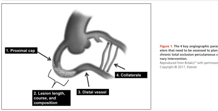
CTO anatomy will dictate the most likely success- ful and safest crossing strategies. Angiographic re- view of the CTO anatomy focuses on 4 characteristics (Figure 1): (1) proximal cap morphology; (2) occlusion length, course, and composition (eg, calcium); (3) qual- ity of the distal vessel; and (4) characteristics of the col- lateral circulation.
29,30Moreover, non-CTO lesions are reviewed, because assessing intermediate left main or other lesions may change clinical decision making lead- ing to alternative revascularization strategies, such as coronary artery bypass graft surgery or pre–CTO-PCI stenting of a donor artery.
Proximal Cap Morphology
Determining the location and morphology of the proxi-
mal cap is critical for selecting an optimal approach to
ST ATE OF THE AR T
Downloaded from http://ahajournals.org by on January 27, 2022ST ATE OF THE AR T
CTO-PCI. Attempts to cross ambiguous proximal caps may lead to perforation. Additional angiographic pro- jections using dual injection, selective contrast injection through a microcatheter located near the proximal cap, use of intravascular ultrasound,
31or preprocedural or real-time CCTA coregistration
32may help clarify the lo- cation of the proximal cap.
33If proximal cap ambiguity cannot be resolved, a retrograde approach is often rec- ommended as primary strategy.
Lesion Length, Course, and Composition
Lesion length is often overestimated with antegrade- only injections because of underfilling and poor opaci- fication of the distal vessel, from competing antegrade and retrograde coronary flow, leaving uncertainty about the location and morphology of the distal cap. Dual in- jection or preprocedural CCTA
34allows more accurate estimation of CTO length and the distal cap anatomy.
Severe calcification and tortuosity of the occluded seg- ment can hinder CTO crossing and increase the like- lihood of subadventitial guidewire entry. Advancing a knuckled (J-shaped) guidewire or changing to the ret- rograde approach is often preferred when the vessel course is unclear or highly tortuous,
35because such a J-shaped or knuckled wire allows advancement within the vessel architecture with a low risk of perforation.
36Distal Vessel
A distal vessel of large caliber (>2.0 mm) that fills well does not have significant disease and is free from ma- jor branches facilitates CTO recanalization. Conversely, small, diffusely diseased distal vessels are more chal- lenging to recanalize, especially after subintimal guide- wire entry. In some cases, however, distal vessels are
small because of hypoperfusion, leading to negative remodeling, and will increase in size after recanaliza- tion.
37Distal CTO caps in native coronary artery CTOs are more likely to be calcified and resistant to guide- wire penetration in patients with previous coronary artery bypass grafting. Moreover, distal vessel calcifi- cation may hinder wire reentry in case of subintimal guidewire entry.
Collateral Circulation
Evaluation of the collateral circulation is critical for de- termining the feasibility of the retrograde approach.
38High-quality angiography (ideally obtained during breath hold and without panning), allowing complete opacification of collateral vessels and obtained in opti- mal angiographic projections, should therefore be en- couraged as part of the routine diagnostic studies when a CTO is found. Retrograde access to the distal vessel can be obtained via septal collaterals, epicardial collaterals, or (patent or occluded) coronary bypass grafts. When assessing collateral channels, it is important to consider size, tortuosity, bifurcations, angle of entry to and exit from the collateral, and distance from the collateral exit to the distal cap. The most important predictor of suc- cessful guidewire and device crossing is lack of tortu- osity, followed by size.
39The size of the collaterals is often assessed by using the Werner classification (CC0, no continuous connection; CC1, threadlike connection;
and CC2, side branch-like connection).
10Crossing in- visible septal collateral channels is often possible with the surfing technique, letting the wire find the path of least resistance.
40It is helpful to carefully study previous angiograms for multiple potential collateral pathways, because the predominant collateral may change over the time before the procedure or during the course of
Figure 1. The 4 key angiographic param- eters that need to be assessed to plan chronic total occlusion percutaneous coro- nary intervention.
Reproduced from Brilakis30 with permission.
Copyright © 2017, Elsevier.
Downloaded from http://ahajournals.org by on January 27, 2022
ST ATE OF THE AR T PCI (shifting collaterals). Previously visualized collaterals that disappear at the time of the procedure may still be crossable. Whenever required, and after ensuring adequate backflow to prevent barotrauma, selective contrast tip injections through the microcatheter can be safely performed to outline collateral anatomy. Pat- ent bypass grafts represent an ideal retrograde conduit because of the absence of side branches, predictable course, and large caliber. Even occluded grafts can be used as retrograde pathways. However, in case of col- lateral circulation originating from the left anterior de- scending artery, that is supplied by a mammary artery, access via the internal mammary artery graft increas- es the risk of global ischemia and should be avoided whenever possible.
41Septal collaterals are typically safer and easier to navigate using very soft tip and polymer-jacketed guidewires in comparison with epicardial collater- als.
42,43In contrast to epicardial collaterals, septal col- laterals can be safely dilated with small balloons to facilitate microcatheter or device crossing if required.
The donor vessel proximal to the collateral origin, and collateral dominance (ie, presence of a single large vis- ible collateral), should also be assessed during retro- grade procedures to determine the risk for ischemia during retrograde crossing attempts. Careful review of collaterals before the procedure can reduce contrast and radiation dose, and the duration of the procedure.
In cases where the collateral anatomy is unclear or am- biguous, it can be helpful to perform selective injection of contrast into the collateral through the center lu- men of a microcatheter placed into the collateral by us- ing a 2- to 3-mL syringe. Furthermore, in cases where unfavorable noninterventional epicardial collaterals provide the dominant blood flow to the CTO, it can be useful to balloon occlude the epicardial collateral for 2 to 4 minutes to see if more favorable interventional collaterals can be recruited and identified for attempts at retrograde crossing.
CTO Scores
Angiographic and clinical characteristics, such as previ- ous CTO-PCI failure
44and previous coronary artery by- pass grafting,
45have been combined to create scores for estimating the difficulty and hazard of a specific CTO-PCI in various patient populations. The first and most commonly used CTO-PCI score is the J-CTO score (Multicenter CTO Registry of Japan), developed to esti- mate the likelihood of successful antegrade guidewire crossing within 30 minutes based on 5 criteria (at least 1 bend of >45° in the CTO entry or CTO body, occlusion length >20 mm, calcification, blunt proximal stump, and previously failed attempt).
46The J-CTO score has been validated in other CTO-PCI cohorts
44and is also associated with 1-year clinical outcomes.
47Other scores
include the PROGRESS-CTO score,
48the RECHARGE (Registry of Crossboss and Hybrid Procedures in France, the Netherlands, Belgium and United Kingdom) registry score,
49the CL-score (Clinical and Lesion related score),
50the ORA (ostial location, collateral filling of Rentrop <2, age >75) score,
51the Ellis et al
52score, the weighted angiographic scoring model (W-CTO score),
53and the CASTLE (coronary artery bypass grafting history, age [≥70 years], stump anatomy [blunt or invisible], tortuos- ity degree [severe or unseen], length of occlusion [≥20 mm], and extent of calcification [severe]) score.
54There are also CCTA-based scores, such as the CT-RECTOR multicenter registry (Computed Tomography Registry of Chronic Total Occlusion Revascularization) score
55and the Korean Multicenter CTO CT Registry Score.
56Various scores have similar predictive capacity for tech- nical success and are more accurate in antegrade-only cases.
57The risk of complications can be assessed by using the Progress-CTO complications score that uses 3 variables (age ≥65 years, lesion length >23 mm, and use of the retrograde approach) to stratify patients for the risk of periprocedural complications.
58In general, each score is only applicable to the pop- ulation from which it was derived and validated. Cal- culating ≥1 scores can promote detailed review of the angiogram and facilitate decision making. For example, medical therapy may be preferred over CTO-PCI in mildly symptomatic patients with highly complex occlu- sions. Complex CTOs (such as those with J-CTO score
≥2) are more likely to require dissection reentry and ret- rograde crossing techniques and should be performed by experienced operators.
USE OF A MICROCATHETER FOR GUIDEWIRE MANIPULATION
A microcatheter should be routinely used for supporting the coronary guidewire and allowing rapid guidewire switching during both antegrade and retrograde wire manipulation. Microcatheters improve the precision of both rotational and longitudinal guidewire movements both in fluid (blood-filled vessels) and in tissue (the oc- clusion itself) and allow the penetration force of the wire to be dynamically altered by changing the distance between the tip of the guidewire and the microcath- eter, with guidewires becoming stiffer when the micro- catheter is positioned close to the guidewire tip. Micro- catheters also allow rapid guidewire tip reshaping or exchange, while preserving previous guidewire crossing or advancement achieved. Microcatheters inherently dilate retrograde collateral channels and protect them from wire-induced trauma. Microcatheters can also be used to deliver contrast either for visualization or to ac- complish the Carlino technique (intralesional injection of 1–2 mL of contrast to elucidate microcatheter posi-
Downloaded from http://ahajournals.org by on January 27, 2022
ST ATE OF THE AR T tion and facilitate crossing), especially in wire-resistant
lesions.
59A microcatheter is preferred over an over-the- wire balloon because it has a marker at the distal tip, providing adequate fluoroscopic feedback of its actual position and also providing greater freedom of advance- ment with a lower profile and better wire-to-lumen in- ternal diameter ratio. Also, unlike over-the-wire plastic balloon catheter shafts that are prone to kinking, nearly all contemporary coronary microcatheters incorporate a kink-resistant metallic braid.
Similar to guidewires, microcatheter selection de- pends on the CTO angiographic characteristics, local availability, and expertise. In addition to using a micro- catheter, obtaining coaxial guide position and strong guide support can significantly facilitate CTO crossing.
CTO CROSSING STRATEGIES
There are 4 CTO crossing strategies, classified accord- ing to wiring direction (antegrade and retrograde) and whether or not the subintimal space is used (wiring ver- sus dissection and reentry; Figure 2).
29,30Antegrade Wiring
Antegrade wiring (also called antegrade wire esca- lation) is the most widely used CTO crossing tech- nique.
31,60–62Various guidewires are advanced in the antegrade direction (original direction of blood flow).
Guidewire choice depends on CTO characteristics. If there is a tapered proximal cap or a functional occlu- sion with a visible channel, a polymer-jacketed, low penetration force, tapered guidewire is used initially, with subsequent escalation to intermediate and high penetration force guidewires, as required. If there is a blunt proximal cap, antegrade wiring is usually started with an intermediate penetration force polymer-jacket- ed guidewire, or a composite core guidewire. Stiff, high penetration force guidewires may be required in highly resistant proximal caps or when areas of resistance are encountered within the body of the occlusion. After proximal cap crossing of 1 to 2 mm, however, deesca- lation to less penetrating guidewires should follow to navigate through the CTO segment.
Contralateral injection and orthogonal angiographic projections are critical for determining guidewire posi- tion during crossing attempts. If the guidewire enters into the distal true lumen, the microcatheter is then advanced into the distal true lumen, and the dedicat- ed CTO guidewire is then exchanged for a workhorse guidewire through the microcatheter to minimize the risk for distal vessel injury and perforation during bal- loon angioplasty and stenting (wire deescalation). If the guidewire exits the vessel structure, it should be withdrawn and redirected without advancing micro- catheters, balloons, or stents over it. If the guidewire
enters the subintimal space, it can be redirected, but if this maneuver fails, the wire can be left in place to aid directing a second guidewire into the distal true lu- men (parallel-wire technique), which can be assisted by a dual-lumen microcatheter or facilitated by the use of intravascular ultrasound.
31Alternatively, antegrade dis- section/reentry techniques can be used to reenter into the distal true lumen, as described below. Subintimal guidewire advancement distal to the distal cap should be avoided because it can lead to hematoma forma- tion, causing luminal compression and reducing the likelihood of success. Antegrade vessel reentry can be guided by intravascular ultrasound, although this ap- proach requires expertise and may be hindered by lim- ited wire maneuverability in the presence of the subin- timal intravascular ultrasound catheter.
Antegrade Dissection and Reentry
Antegrade dissection and reentry involves entering the subintimal space, followed by subintimal crossing of the CTO with subsequent reentry into the distal true lumen.
Antegrade dissection and reentry may be intentional or unintentional during antegrade wiring attempts. The first developed dissection reentry technique was named STAR (Subintimal Tracking And Re-Entry) and used inad- vertent, uncontrollable reentry into the distal lumen.
63This frequently necessitated stenting long coronary seg- ments with occlusion of numerous side branches, lead- ing to extensive vascular injury and high rates of in-stent restenosis and reocclusion.
63–65As such, the STAR tech- nique has evolved to a bailout strategy without stent implantation after ballooning, in preparation for a re- peat CTO-PCI attempt (subintimal plaque modification, also termed an “investment procedure”).
66–68The devel- opment of limited dissection/reentry techniques (using dedicated reentry systems
69,70or wire-based strategies
71) was an important advancement, because they minimize vascular injury, limit the length of dissection and subse- quent stent length, and increase the likelihood of side branch preservation.
36,69,72Such approaches have been associated with favorable clinical outcomes.
72–76Retrograde Approach
The retrograde technique differs from the antegrade approach in that the occlusion is approached from the distal vessel with guidewire advancement against the original direction of blood flow.
77A guidewire is ad- vanced into the artery distal to the occlusion through a collateral channel or through a bypass graft, followed by placement of a microcatheter at the distal CTO cap.
Retrograde CTO crossing is then attempted either with retrograde wiring (usually for short occlusions, especial- ly when the distal cap is tapered
78) or using retrograde dissection/reentry techniques.
Downloaded from http://ahajournals.org by on January 27, 2022
ST ATE OF THE AR T
The most commonly used retrograde crossing tech- nique is reverse controlled antegrade and retrograde tracking, in which a balloon is inflated over the ante- grade guidewire, followed by retrograde guidewire advancement into the space created by the antegrade balloon (Figure 2). In challenging reverse controlled antegrade and retrograde tracking cases, intravascular ultrasound can clarify the mechanism of failure and in- crease the likelihood of success.
79Guide catheter ex- tensions can also facilitate reverse controlled antegrade and retrograde tracking.
80Crossing Strategy Selection
Selecting the initial and subsequent crossing strategies depends on the CTO lesion characteristics and local equipment availability and expertise.
Several algorithms have been developed to facili- tate crossing strategy selection, such as the hybrid,
29Asia Pacific,
35and Euro-CTO
81algorithms. Antegrade crossing is generally preferred over retrograde crossing as the initial crossing strategy, given the higher risk of complications with the retrograde approach
60–62and the need for antegrade lesion preparation even when the retrograde approach is eventually required. Some retro- grade CTO-PCI complications, however, are caused by antegrade crossing attempts. The retrograde approach remains critical for achieving high success rates, espe- cially in more complex CTOs,
60,62and has been associ- ated with favorable long-term outcomes.
82CTOs with proximal cap ambiguity and flush aorto- ostial CTOs are often approached with a primary ret- rograde strategy. Alternatively, proximal cap ambiguity can be approached in the antegrade direction, espe- cially when no collateral or graft is available by using (1) intravascular ultrasound or preprocedural CCTA for determining the location of the proximal cap and ves- sel course,
32,35,83or (2) techniques to facilitate entry into
Figure 2. Illustration of the chronic total occlusion crossing techniques.
Reproduced from Brilakis30 with permission. Copyright © 2017, Elsevier.
Downloaded from http://ahajournals.org by on January 27, 2022
ST ATE OF THE AR T the subintimal space proximal to the occlusion, such as
the balloon-assisted subintimal entry (ie, inflation of a balloon proximal to the occlusion to cause a dissection, followed by subintimal guidewire entry and subintimal crossing of the occlusion) technique.
84CHANGE OF CROSSING STRATEGY
Flexibility is important for the success, safety, and ef- ficiency of CTO-PCI. If the initial or subsequent crossing strategy fails to achieve progress, small changes (such as modifying the guidewire tip angulation or changing guidewire) or more significant changes (such as con- verting from an antegrade to a retrograde approach) should be made, based on preprocedural planning.
29,35It is important to avoid getting stuck in a failure mode, in which excessive time, radiation, and contrast are ex- pended with little or no progress being made while re- peatedly attempting the same technique, because this will preclude the use of alternative strategies and in- crease the risk of complications.
Similar to selection of the initial crossing strategy, the timing and choice of subsequent crossing strate- gies depends on lesion characteristics, challenges en- countered with the original technique, and equipment availability and expertise, and can be guided by existing crossing algorithms.
29,35Only ≈50% to 60% of CTOs are successfully crossed with the initial strategy,
28,60,61highlighting the need for further refinements in the procedure-planning algorithms. Changing strategies can help maximize the likelihood of eventual success and limit contrast volume and radiation dose.
Reasons to stop a CTO-PCI attempt include occur- rence of a complication, high radiation dose (usually
>5 Gy air kerma dose in the absence of lesion cross- ing or substantial progress), large contrast volume ad-
ministration (>3.7× the estimated creatinine clearance), exhaustion of crossing options, or patient or physician fatigue. As with all interventions, careful assessment of individual risk versus benefit should guide decision making and choice of strategy during different stages of the procedure. On many occasions, it may be best to fail rather than to pursue highly aggressive strategies that may lead to serious complications.
60EQUIPMENT AND PHYSICIAN TEAM EXPERTISE
CTO-PCI should be performed within dedicated pro- grams that promote continual training and rigorous monitoring of outcomes.
85Higher CTO-PCI volume has consistently been associated with higher success rates.
52,86,87The performance of CTO-PCI by a skilled physi- cian and team is especially important to minimize and manage procedural complications. CTO-PCI car- ries increased risk of complications in comparison with non–CTO-PCI,
86especially perforation.
3Across multiple contemporary registries, tamponade oc- curred in 0.4% to 1.3% of cases (Table 2).
3,31,60–62,88,89Additional CTO-PCI adverse events include access site complications, donor vessel injury, arrhythmias, stroke, contrast-induced nephropathy, radiation der- matitis, emergency coronary bypass graft surgery, and death.
90The average complication risk is ≈3%, but varies widely between studies (Table 2) and increases with greater lesion complexity.
3,60–62,88,89Dual injection minimizes the risk for perforation by helping determine guidewire position. Placement of a safety guidewire in the CTO donor vessel can facilitate treatment if donor vessel injury occurs. Maintaining an activated clotting time of ≥300 to 350 seconds reduces
Table 2. Contemporary Series of Chronic Total Occlusion Percutaneous Coronary Intervention
Authors Acronym
Study
Period Centers Cases
Technical Success
Procedural Success
Overall MACE Death
Acute
MI Stroke TVR Tamponade Konstantinidis
et al89
EURO-CTO registry
2008–2015 53 17 626 85% – 0.6% 0.2% – – – 0.4%
Habara et al88 Japanese Retrograde Summit Registry
2012–2013 56 3229 – 88% 0.5% 0.2% 0.1% 0.1% – 0.3%
Tajti et al60 PROGRESS-CTO 2012–2017 20 3055 87% 85% 3.0% 0.3% 0.7% 0.1% 0.2% 0.5%
Suzuki et al31 Japanese CTO- PCI Expert
Registry
2014–2015 41 2846 90% 89% <2% 0.2% 1.2% 0.2% 0.2% 0.4%
Maeremans et al61
RECHARGE 2014–2015 17 1253 89% 86% 2.6% 0.2% 0.2% 2.2% 0.1% 1.3%
Wilson et al62 UK Hybrid 2012–2014 7 1156 90% — 1.6% 0.0% 0.8% 0.4% 0.0% 0.7%
Sapontis et al3 OPEN-CTO 2013–2017 12 1000 86% 85% 7.0% 0.9% 2.6% 0.0% 0.1% –
The studies are listed according to the number of patients included. EURO-CTO indicates European Registry of Chronic Total Occlusion; MACE indicates major adverse cardiac events; MI, myocardial infarction; OPEN CTO, Outcomes, Patient Health Status, and Efficiency in Chronic Total Occlusion Hybrid Procedures;
PROGRESS-CTO, Prospective Global Registry for the Study of Chronic Total Occlusion Intervention; RECHARGE, Registry of Crossboss and Hybrid procedures in France, The Netherlands, Belgium, and United Kingdom; UK hybrid, United Kingdom hybrid registry; and TVR, target vessel revascularization.
Downloaded from http://ahajournals.org by on January 27, 2022
ST ATE OF THE AR T the risk of donor vessel thrombosis; the activated clot- ting time should be checked at least every 30 minutes during the procedure. In case of perforation, covered stents and coils should be available to treat large ves- sel and distal vessel perforations, respectively. Prepro- cedural operator training in the proper use of these devices will ensure efficient use in the emergency set- ting. In case of epicardial collateral perforation,
43,91em- bolization from both directions (using coils, thrombin, fat, etc) is often needed to achieve sealing.
92Special attention should be given to patients with previous coronary bypass graft surgery, because perforation can result in life-threatening, difficult to access, loculated hematomas
93or bleeding in the mediastinum or pleu- ral cavities.
Meticulous attention should also be paid to minimiz- ing radiation dose and the risk for radiation skin injury.
This can be achieved by using low-frame rate fluorosco- py and the fluoroscopy-store function for documenting balloon and stent inflation instead of cine-angiography, using collimation, minimizing the distance of the image receptor from the patient, and intermittently changing the position of the image receptor during the proce- dure.
94,95Patients who receive high doses of radiation (eg, >5 Gray air kerma dose) require formal follow-up to evaluate for subacute skin injury. Similarly, contrast administration should be minimized through meticu- lous preprocedural planning and use of contrast-spar- ing devices to reduce the risk for contrast nephropathy.
OPTIMAL STENT DEPLOYMENT
CTO-PCI often involves placement of multiple stents in vessels that are calcified, diffusely diseased, and nega- tively remodeled. Given the often arduous and lengthy attempts required for CTO crossing, less attention may be given to stent optimization (maximal stent expan- sion and optimal inflow and outflow), potentially result- ing in higher rates of restenosis and stent thrombosis.
Full lesion expansion should be achieved before stent implantation by predilation with properly sized balloon or atherectomy. Intravascular imaging can facilitate the assessment of vessel size and calcification before stent- ing and the adequacy of stent expansion, apposition and lesion coverage, to reduce the risk for subsequent adverse events.
96–99Moderate diffuse disease distal to the CTO often does not require treatment, because the distal vessel often enlarges over time after restoring vessel patency.
100CONCLUSIONS
Extensive interactions and collaboration across the world have led to the advancements in CTO-PCI that are summarized in the 7 key principles outlined in this
global expert consensus document. These principles can guide training of new CTO-PCI operators and program development and facilitate further improvement in the success, safety, and clinical outcomes of CTO-PCI.
ARTICLE INFORMATION Authors
Emmanouil S. Brilakis, MD, PhD; Kambis Mashayekhi, MD; Etsuo Tsuchikane, MD, PhD; Nidal Abi Rafeh, MD; Khaldoon Alaswad, MD; Mario Araya, MD;
Alexandre Avran, MD; Lorenzo Azzalini, MD, PhD, MSc; Avtandil M. Babunashvili, MD; Baktash Bayani, MD; Ravinay Bhindi, MD; Nicolas Boudou, MD; Marouane Boukhris, MD; Nenad Ž. Božinović, MD; Leszek Bryniarski, MD, PhD; Alexander Bufe, MD; Christopher E. Buller, MD; M. Nicholas Burke, MD; Heinz Joachim Büttner, MD; Pedro Cardoso, MD; Mauro Carlino, MD; Evald H. Christiansen, MD; Antonio Colombo, MD; Kevin Croce, MD, PhD; Felix Damas de los Santos, MD; Tony De Martini, MD; Joseph Dens, MD, PhD; Carlo Di Mario, MD; Kefei Dou, MD; Mohaned Egred, MD; Ahmed M. ElGuindy, MD; Javier Escaned, MD, PhD; Sergey Furkalo, MD; Andrea Gagnor, MD; Alfredo R. Galassi, MD; Roberto Garbo, MD; Junbo Ge, MD; Pravin Kumar Goel, MD; Omer Goktekin, MD; Luca Grancini, MD; J. Aaron Grantham, MD; Colm Hanratty, MD; Stefan Harb, MD;
Scott A. Harding, MD; Jose P.S. Henriques, MD; Jonathan M. Hill, MD; Farouc A. Jaffer, MD, PhD; Yangsoo Jang, MD; Risto Jussila, MD; Artis Kalnins, MD;
Arun Kalyanasundaram, MD; David E. Kandzari, MD; Hsien-Li Kao, MD; Dimitri Karmpaliotis, MD, PhD; Hussien Heshmat Kassem, MD, PhD; Paul Knaapen, MD; Ran Kornowski, MD; Oleg Krestyaninov, MD; A. V. Ganesh Kumar, MD;
Peep Laanmets, MD; Pablo Lamelas, MD; Seung-Whan Lee, MD; Thierry Lefevre, MD; Yue Li, MD; Soo-Teik Lim, MD; Sidney Lo, MBBS; William Lombardi, MD;
Margaret McEntegart, MD, PhD; Muhammad Munawar, MD; José Andrés Na- varro Lecaro, MD; Hung M. Ngo, MD, PhD; William Nicholson, MD; Göran K.
Olivecrona, MD, PhD; Lucio Padilla, MD; Marin Postu, MD; Alexandre Quad- ros, MD; Franklin Hanna Quesada, MD; Vithala Surya Prakasa Rao, MD; Nico- laus Reifart, MD; Meruzhan Saghatelyan, MD; Ricardo Santiago, MD; George Sianos, MD, PhD; Elliot Smith, MD; James C. Spratt, MD; Gregg W. Stone, MD; Julian W. Strange, MD; Khalid Tammam, MD, PhD; Imre Ungi, MD, PhD;
Minh Vo, MD; Vu Hoang Vu, MD; Simon Walsh, MD; Gerald S. Werner, MD;
Jason R. Wollmuth, MD; Eugene B. Wu, MD; R. Michael Wyman, MD; Bo Xu, MD; Masahisa Yamane, MD; Luiz F. Ybarra, MD; Robert W. Yeh, MD; Qi Zhang, MD; Stephane Rinfret, MD, SM
Correspondence
Emmanouil S. Brilakis, MD, PhD, Minneapolis Heart Institute, 920 E 28th St
#300, Minneapolis, MN 55407. Email esbrilakis@gmail.com
Affiliations
Minneapolis Heart Institute and Minneapolis Heart Institute Foundation, Abbott Northwestern Hospital, MN (E.S.B., M.N.B.). Department of Cardiology and An- giology II University Heart Center Freiburg Bad Krozingen, Germany (K.M., H.J.B.). Toyohashi Heart Center, Aichi, Japan (E.T.). St. George Hospital Univer- sity Medical Center, Beirut, Lebanon (N.A.R.). Henry Ford Hospital, Detroit, MI (K.A.). Clínica Alemana and Instituto Nacional del Tórax, Santiago, Chile (M.A.).
Arnault Tzank Institut St. Laurent Du Var Nice, France (A.A.). Interventional Cardiology Division, Cardio-Thoracic-Vascular Department, San Raffaele Scien- tific Institute, Milan, Italy (L.A., M.C.). Department of Cardiovascular Surgery, Center for Endosurgery and Lithotripsy, Moscow, Russian Federation (A.M.B.).
Cardiology Department, Mehr Hospital, Mashhad, Iran (B.B.). Department of Cardiology, Royal North Shore Hospital and Kolling Institute, University of Syd- ney, Australia (R.B.). Rangueil University Hospital, Toulouse, France (N.B.). Car- diology department, Abderrahment Mami Hospital, Faculty of Medicine of Tu- nis, University of Tunis El Manar, Tunisia (M.B.). Department of Interventional Cardiology Clinic for Cardiovascular Diseases University Clinical Center Nis, Serbia (N.Z.B.). II Department of Cardiology and Cardiovascular Interventions Institute of Cardiology, Jagiellonian University Medical College, Cracow, Poland (L.B.). Department of Cardiology, Heartcentre Niederrhein, Helios Clinic Krefeld, Krefeld, Germany, Institute for Heart and Circulation Research, University of Cologne, Germany, and University of Witten/Herdecke, Witten, Germany (A.B.). St. Michael’s Hospital, Toronto, ON, Canada (C.E.B.). Cardiology Depart- ment, Santa Maria University Hospital (CHULN), Lisbon Academic Medical Cen- tre (CAML) and Centro Cardiovascular da Universidade de Lisboa (CCUL), Por-
Downloaded from http://ahajournals.org by on January 27, 2022
ST ATE OF THE AR T
tugal (P.C.). Department of Cardiology, Aarhus University Hospital, Denmark (E.H.C.). San Raffaele Hospital and Columbus Hospital, Milan, Italy (A.C.). Car- diovascular Division, Brigham and Women’s Hospital, Boston, MA (K.C.). Inter- ventional Cardiology Department, Instituto Nacional de Cardiología Ignacio Chávez Mexico City, Mexico (F.D.d.l.S.). SIU School of Medicine, Memorial Medical Center, Springfield, IL (T.D.M.). Department of Cardiology, Hospital Oost-Limburg, Genk, Belgium (J.D.). Structural Interventional Cardiology, Careggi University Hospital, Florence, Italy (C.D.M.). Center for Coronary Heart Disease, State Key Laboratory of Cardiovascular Disease, Fu Wai Hospital, and National Center for Cardiovascular Diseases, Chinese Academy of Medical Sci- ences and Peking Union Medical College,Beijing (K.D.). Freeman Hospital and Newcastle University, Newcastle upon Tyne, United Kingdom (M.E.). Depart- ment of Cardiology, Aswan Heart Center, Egypt (A.M.E.). National Heart and Lung Institute, Imperial College London, United Kingdom (A.M.E.). Hospital Clinico San Carlos IDISSC and Universidad Complutense de Madrid, Spain (J.E.).
Department of Endovascular Surgery and Angiography, National Institute of Surgery and Transplantology of AMS of Ukraine, Kiev (S.F.). Department of In- vasive Cardiology, Maria Vittoria Hospital, Turin, Italy (A.G.). Chair of Cardiolo- gy, Department of PROMISE, University of Palermo, Italy (A.R.G.). Director of Interventional Cardiology, San Giovanni Bosco Hospital, Turin, Italy (R.G.).
Zhongshan Hospital, Fudan University, Shanghai, China (J.G.). Sanjay Gandhi Post Graduate Institute of Medical Sciences Lucknow, India (P.K.G.). Memorial Hospital, Istanbul, Turkey (O.G.). Centro Cardiologico Monzino, IRCCS, Milan, Italy (L.G.). Saint Luke’s Mid America Heart Institute, Kansas City, MO (J.A.G.).
Belfast Health and Social Care Trust, United Kingdom (C.H., S.W.). LKH Graz II, Standort West, Kardiologie, Teaching Hospital of the University of Graz, Austria (S.H.). Wellington Hospital, Capital and Coast District Health Board, New Zea- land (S.A.H.). Academic Medical Centre of the University of Amsterdam, The Netherlands (J.P.S.H.). King’s College Hospital, London, United Kingdom (J.M.H.). Cardiology Division, Massachusetts General Hospital, Boston (F.A.J.).
Division of Cardiology, Severance Cardiovascular Hospital, Yonsei University Health System, Seoul, South Korea (Y.J.). Helsinki Heart Hospital, Finland (R.J.).
Department of Cardiology, Eastern Clinical University Hospital, Riga, Latvia (A.
Kalnins). Promed Hospital, Chennai, India (A. Kalyanasundaram). Piedmont Heart Institute, Atlanta, GA (D.E.K.). Department of Internal Medicine, National Taiwan University Hospital, Taipei (H.-L.K.). Columbia University, New York (D.K.). Cardiology Department, Kasr Al-Ainy Faculty of Medicine, Cairo Univer- sity, Egypt (H.H.K.). Fujairah Hospital, United Arab Emirates (H.H.K.). Depart- ment of Cardiology, VU University Medical Center, Amsterdam, The Nether- lands (P.K.). Department of Cardiology, Rabin Medical Center, Petach Tikva,
“Sackler” School of Medicine, Tel Aviv University, Petach Tikva, Israel (R.K.).
Meshalkin Novosibrisk Research Institute, Russia (O.K.). Department of Cardiol- ogy, Dr LH Hiranandani Hospital, Mumbai, India (A.V.G.K.). North Estonia Med- ical Center Foundation, Tallinn, Estonia (P. Laanmets). Department of Interven- tional Cardiology and Endovascular Therapeutics, Instituto Cardiovascular de Buenos Aires, Argentina (P. Lamelas). Department of Health Research Methods, Evidence and Impact, McMaster University, Hamilton, ON, Canada (P. Lamelas).
Department of Cardiology, Asan Medical Center, University of Ulsan College of Medicine, Seoul, South Korea (S.-W.L.). Institut Cardiovasculaire Paris Sud Hopi- tal prive Jacques Cartier, Massy, France (T.L.). Department of Cardiology, the First Affiliated Hospital of Harbin Medical University, China (Y.L.). Department of Cardiology, National Heart Centre Singapore (S.-T.L.). Department of Cardiol- ogy, Liverpool Hospital and The University of New South Wales, Sydney, Austra- lia (S.L.). University of Washington, Seattle (W.L.). Golden Jubilee National Hos- pital, Glasgow, United Kingdom (M. McEntegart). Binawaluya Cardiac Center, Jakarta, Indonesia (M. Munawar). Médico Cardiólogo Universitario - Hemodin- amista en Hospital de Especialidades Eugenio Espejo y Hospital de los Valles, Ecuador (J.A.N.L.). Choray Hospital, Vietnam (H.M.N.). WellSpan Health Sys- tem, York, PA (W.N.). Skane University Hospital, University of Lund, Sweden (G.K.O.). Department of Interventional Cardiology and Endovascular Therapeu- tics, ICBA, Instituto Cardiovascular, Buenos Aires, Argentina (L.P.). Cardiology Department, University of Medicine and Pharmacy “Carol Davila,” Institute of Cardiovascular Diseases “Prof. Dr. C.C. Iliescu,” Bucharest, Romania (M.P.). In- stituto de Cardiologia / Fundação Universitária de Cardiologia - IC/FUC, Porto Alegre, RS – Brazil (A.Q.). Interventional Cardiology Department, Clinica Com- familiar Pereira City, Colombia (F.H.Q.). Hyderguda Apollo, Hyderabad, India (V.S.P.R.). Department of Cardiology, Main Taunus Heart Institute, Bad Soden, Germany (N.R.). Nork-Marash Medical Center, Yerevan, Armenia (M.S.). Hospi- tal Pavia Santurce, PCI Cardiology Group, San Juan, Puerto Rico (R.S.T.). AHEPA University Hospital, Thessaloniki, Greece (G.S.). Department of Cardiology, Barts Heart Centre, St Bartholomew’s Hospital, London, United Kingdom (E.S.).
St George’s University Hospital NHS Trust, London, United Kingdom (J.S.). Cen- ter for Interventional Vascular Therapy, Division of Cardiology, New York-Pres- byterian Hospital/Columbia University Medical Center (G.W.S.). Department of
Cardiology, Bristol Royal Infirmary, United Kingdom (J.W.S.). Cardiac Center of Excellence, International Medical Center, Jeddah, Saudi Arabia (K.T.). 2nd De- partment of Internal Medicine and Cardiology Center, University of Szeged, Hungary (I.U.). Mazankowski Alberta Heart Institute, Edmonton, AB, Canada (M.V.). Interventional Cardiology Department, Heart Center, University Medical Center at Ho Chi Minh City, and University of Medicine and Pharmacy, Vietnam (H.V.). Medizinische Klinik I Klinikum Darmstadt GmbH, Germany (G.W.). Provi- dence Heart and Vascular Institute, Portland, OR (J.R.W.). Prince of Wales Hos- pital, Hong Kong (E.W.). Torrance Memorial Medical Center, CA (R.M.W.). Fu Wai Hospital, National Center for Cardiovascular Diseases, Chinese Academy of Medical Sciences, Beijing (B.X.). Saitima St. Luke’s International Hospital, Tokyo, Japan (M.Y.). London Health Sciences Centre, Schulich School of Medicine &
Dentistry, Western University, London, Ontario, Canada (L.F.Y.). Beth Israel Dea- coness Medical Center, Boston, MA (R.W.Y.). Shanghai East Hospital, Tongji University, China (Q.Z.). McGill University Health Centre, McGill University, Montreal, QC, Canada (S.R.).
Acknowledgment
We recognize the assistance of Dr Iosif Xenogiannis in the creation of this docu- ment.
Disclosures
Dr Brilakis: Consulting/speaker honoraria from Abbott Vascular, American Heart Association (associate editor Circulation), Boston Scientific, Cardiovascular Inno- vations Foundation (Board of Directors), CSI, Elsevier, GE Healthcare, InfraRedx, and Medtronic; research support from Regeneron and Siemens. Shareholder:
MHI Ventures. Board of Trustees: Society of Cardiovascular Angiography and Interventions. Dr Mashayekhi: Consulting/speaker/proctoring honoraria from Abbott Vascular, Ashai Intecc, AstraZeneca, Biotronik, Boston Scientific, Cardinal Health, Daiichi Sankyo, Medtronic, Teleflex, Terumo. Dr Tsuchikane: Consultant of Boston Scientific, Asahi Intecc, Nipro, and Kaneka. Dr Abi Rafeh: CTO Proctor and consultant for Boston Scientific and Abbott Vascular. Dr Alaswad: consul- tant and speaker for Boston Scientific, Abbott Cardiovascular, CSI, and LivaNova.
Dr Avran: Proctor for Boston Scientific, Biotronik, Abbott, Terumo, Biosensor, Medtronic. Dr Azzalini: Honoraria from Abbott Vascular, Guerbet, Terumo, and Sahajanand Medical Technologies; research support from ACIST Medical Sys- tems, Guerbet, Terumo. Dr Boudou: Proctorship fees from Boston Scientific, Terumo, Abbott Vascular, and Biotronik. Dr Buller: Intellectual property: Tele- flex; consultant: Abbott Vascular, Soundbite Medical, and Philips-Volcano. Dr Burke: Consulting and speaker honoraria from Abbott Vascular and Boston Scientific. Dr Croce: Proctor/speaking honoraria: Abbott, Boston Scientific, CSI, Philips; research grant: Teleflex, Takeda; advisory board: Abiomed, Cordis. Dr de los Santos: Speaker and proctor of Boston Scientific, Terumo, and Abbott.
Dr De Martini: Proctor and advisory board for Abbott and Boston Scientific. Dr Dens: Consulting/speaker honoraria from Abbott Vascular, Boston Scientific, IMDS, Orbus Neich, Terumo, and Topmedical (distributor for Asahi). Dr Di Mario:
research grant to institution from Amgen, Behring, Chiesi, Daiichi Sankyo, Ed- wards, Medtronic, Shockwave. Dr Egred: Honoraria, speaker and proctorship fees from Abbott Vascular, Boston Scientific, Vascular Perspectives, Philips/Vol- cano, Biosensors, and EPS. Dr ElGuindy: Proctorship fees from Boston Scientific.
Dr Gagnor: Consultant Boston Scientific, Terumo. Dr Garbo: Consultant Boston Scientific, Terumo, Philips Volcano, IMDS, and CID-Alvimedica. Dr Grantham:
Speaking fees, travel reimbursement, and honoraria from Boston Scientific, Ab- bott Vascular, and Asahi Intecc. Institutional research grants Boston Scientific.
Part-time employment and equity in Corindus Vascular Robotics. Dr Hanratty:
Proctoring for Abbott, Boston Scientific, Medtronic, and Teleflex. Dr Harb:
Consultant with Medtronic, speaker´s honoraria from Medtronic and Cardinal Health. Dr Harding: Proctor/speaker for Boston Scientific, Abbott Vascular, and Bio-Excel; consultant/speaker for Medtronic and Asahi. Dr Hill: Speaker, consul- tant, and proctor for Boston Scientific and Abbott Vascular. Dr Jaffer: Sponsored research from Canon, and Siemens; consultant for Boston Scientific, Abbott Vas- cular, Siemens, and Philips. Massachusetts General Hospital has a patent licens- ing arrangement with Canon, and Dr Jaffer has the right to receive royalties. Dr Jussila: Consulting agreement with EPS Vascular, Boston Scientific, and Terumo.
Dr Kalyanasundaram: Speaker, consultant, and proctor for Boston Scientific, Asahi, and Abbott Vascular. Dr Kandzari: Research/grant support: Medtronic, Boston Scientific, Biotronik; consulting honoraria: Medtronic, Boston Scien- tific, Biotronik, and CSI. Dr Kao: Speaker/proctor honoraria: Abbott Vascular, Asahi Intecc, Biotronik, Boston Scientific, Medtronic, Orbus Neich, and Terumo.
Dr Karmpaliotis: Honoraria Boston Scientific, Abiomed and Abbott Vascular.
Dr Kornowski: Co-founder of NitiLoop. Dr Krestyaninov: Speaker and proctor honoraria from Abbott Vascular. Dr Laanmets: Consultant for Terumo. Dr Lee:
Downloaded from http://ahajournals.org by on January 27, 2022
ST ATE OF THE AR T
Speaker and proctorship honoraria from Abbott Vascular, Boston Scientific, and Medtronic. Dr Lefevre: Proctoring for Terumo. Dr Lim: Travel support from Asahi Intecc, Terumo, Kaneka, Boston Scientific, and Abbott Vascular. Dr Lo: Travel support from Bioexcel and Abbott; speaker honoraria from Abbott, Boston Sci- entific, and Bioexcel; proctorshop fees from Bioexcel and Boston Scientific. Dr Lombardi: Speaking fees, honoraria, and travel expense reimbursement from Boston Scientific, Asahi-Intecc, Teleflex, Siemens, and Abbott Vascular; equity holder in Corindus Vascular Robotics; spouse employed by Phillips. Dr Nicholson:
Advisory boards and consulting: Abbott Vascular, Boston Scientific, Medtronic, and Corindus. Dr Olivecrona: Lecture/proctor honoraria: Biotronik, EPS vascu- lar, Biosensors, and Edwards Lifesciences. Dr Postu: Advisory board: Medtronic;
proctor: Boston Scientific; consultant: Terumo. Dr Quadros: Education support from Medtronic, Abbott Vascular, Boston Scientific, and Biotronic, and research grants from Medtronic. Dr Quesada: Proctor for Boston Scientific. Dr Saghat- elyan: Consulting/speaker honoraria from Asahi Intecc. Dr Trinidad: Proctor and Speaker for Boston Scientific and Abbott Vascular. Dr Smith: Speaker fees, honoraria, proctorship fees, Boston Scientific, Abbott Vascular, Vascular Perspec- tives, and Biosensors International. Dr Stone: Reports having served as a con- sultant to: Matrizyme, Miracor, Neovasc, V-wave, Shockwave, Valfix, TherOx, Reva, Vascular Dynamics, Robocath, HeartFlow, Gore, Ablative Solutions, and Ancora; having received speaker honoraria from Amaranth and Terumo; holding equity in Ancora, Cagent, Qool Therapeutics, Aria, Caliber, MedFocus family of funds, Biostar family of funds, Applied Therapeutics, and SpectraWAVE; serv- ing as a director in SpectraWAVE; and that his employer, Columbia University, receives royalties for sale of the MitraClip from Abbott. Dr Strange: Consulting fees from Abbott and Boston Scientific. Dr Tammam: Proctor for Boston Scien- tific, Terumo and Asahi. Dr Ungi: CTO Proctor and consultant for Boston Scien- tific. Dr Vo: Consultant for Abbott Vascular, Canadian Hospital Specialties, and Teleflex. Dr Wollmuth: Proctor/consultant for Abbott Vascular, Boston Scientific, and Asahi Intecc. Dr Wu: Consultant fees and speaker honorarium from Abbott and Boston Scientific, and research grant from Asahi. Dr Wyman: Consultant/
honoraria from Abbott, Abiomed, and Boston Scientific. Dr Yeh: Consulting/
advisory board: Abbott Vascular, Asahi Intecc, Boston Scientific, Medtronic, and Teleflex. Research grants: Abbott Vascular, Abiomed, and Boston Scientific. Dr Rinfret: Research support from SoundBite Medical; consultant, proctor and/or speaker for Abiomed, Boston Scientific, Abbott, and Teleflex. The other authors report no conflicts.
REFERENCES
1. Werner GS, Martin-Yuste V, Hildick-Smith D, Boudou N, Sianos G, Gelev V, Rumoroso JR, Erglis A, Christiansen EH, Escaned J, et al; EU- ROCTO trial investigators. A randomized multicentre trial to compare revascularization with optimal medical therapy for the treatment of chronic total coronary occlusions. Eur Heart J. 2018;39:2484–2493. doi:
10.1093/eurheartj/ehy220
2. Obedinskiy AA, Kretov EI, Boukhris M, Kurbatov VP, Osiev AG, Ibn Elhadj Z, Obedinskaya NR, Kasbaoui S, Grazhdankin IO, Prokhorikhin AA, et al. The IMPACTOR-CTO Trial. JACC Cardiovasc Interv. 2018;11:1309–1311. doi:
10.1016/j.jcin.2018.04.017
3. Sapontis J, Salisbury AC, Yeh RW, Cohen DJ, Hirai T, Lombardi W, McCabe JM, Karmpaliotis D, Moses J, Nicholson WJ, et al. Early procedur- al and health status outcomes after chronic total occlusion angioplasty: a report from the OPEN-CTO Registry (Outcomes, Patient Health Status, and Efficiency in Chronic Total Occlusion Hybrid Procedures). JACC Cardiovasc Interv. 2017;10:1523–1534. doi: 10.1016/j.jcin.2017.05.065
4. Christakopoulos GE, Christopoulos G, Carlino M, Jeroudi OM, Roesle M, Rangan BV, Abdullah S, Grodin J, Kumbhani DJ, Vo M, et al. Meta-analysis of clinical outcomes of patients who underwent percutaneous coronary interventions for chronic total occlusions. Am J Cardiol. 2015;115:1367–
1375. doi: 10.1016/j.amjcard.2015.02.038
5. Joyal D, Afilalo J, Rinfret S. Effectiveness of recanalization of chronic total occlusions: a systematic review and meta-analysis. Am Heart J.
2010;160:179–187. doi: 10.1016/j.ahj.2010.04.015
6. Ybarra LF, Dautov R, Gibrat C, Dandona S, Rinfret S. Midterm angina- related quality of life benefits after percutaneous coronary intervention of chronic total occlusions. Can J Cardiol. 2017;33:1668–1674. doi:
10.1016/j.cjca.2017.08.008
7. Lee SW, Lee PH, Ahn JM, Park DW, Yun SC, Han S, Kang H, Kang SJ, Kim YH, Lee CW, et al. Randomized trial evaluating percutaneous coronary intervention for the treatment of chronic total occlusion: the DECISION-CTO Trial. Circulation. 139:1674–1683. doi:
10.1161/CIRCULATIONAHA.118.031313.
8. Mashayekhi K, Neuser H, Kraus A, Zimmer M, Dalibor J, Akin I, Werner G, Aurel T, Neumann FJ, Behnes M. Successful percutaneous coronary in- tervention improves cardiopulmonary exercise capacity in patients with chronic total occlusions. J Am Coll Cardiol. 2017;69:1095–1096. doi:
10.1016/j.jacc.2016.12.017
9. Bruckel JT, Jaffer FA, O’Brien C, Stone L, Pomerantsev E, Yeh RW. Angina severity, depression, and response to percutaneous revascularization in patients with chronic total occlusion of coronary arteries. J Invasive Car- diol. 2016;28:44–51.
10. Werner GS, Ferrari M, Heinke S, Kuethe F, Surber R, Richartz BM, Figulla HR.
Angiographic assessment of collateral connections in comparison with inva- sively determined collateral function in chronic coronary occlusions. Circula- tion. 2003;107:1972–1977. doi: 10.1161/01.CIR.0000061953.72662.3A 11. Sachdeva R, Agrawal M, Flynn SE, Werner GS, Uretsky BF. The myocar- dium supplied by a chronic total occlusion is a persistently ischemic zone.
Catheter Cardiovasc Interv. 2014;83:9–16. doi: 10.1002/ccd.25001 12. Galassi AR, Boukhris M, Toma A, Elhadj Z, Laroussi L, Gaemperli O,
Behnes M, Akin I, Lüscher TF, Neumann FJ, et al. Percutaneous coronary intervention of chronic total occlusions in patients with low left ventricu- lar ejection fraction. JACC Cardiovasc Interv. 2017;10:2158–2170. doi:
10.1016/j.jcin.2017.06.058
13. Megaly M, Saad M, Tajti P, Burke MN, Chavez I, Gössl M, Lips D, Mooney M, Poulose A, Sorajja P, et al. Meta-analysis of the impact of successful chronic total occlusion percutaneous coronary intervention on left ventricular systolic function and reverse remodeling. J Interv Cardiol.
2018;31:562–571. doi: 10.1111/joic.12538
14. Henriques JP, Hoebers LP, Råmunddal T, Laanmets P, Eriksen E, Bax M, Ioanes D, Suttorp MJ, Strauss BH, Barbato E, et al; EXPLORE Trial In- vestigators. Percutaneous intervention for concurrent chronic total oc- clusions in patients with STEMI: the EXPLORE Trial. J Am Coll Cardiol.
2016;68:1622–1632. doi: 10.1016/j.jacc.2016.07.744
15. Mashayekhi K, Nührenberg TG, Toma A, Gick M, Ferenc M, Hochholzer W, Comberg T, Rothe J, Valina CM, Löffelhardt N, et al. A randomized trial to assess regional left ventricular function after stent implantation in chronic total occlusion: the REVASC Trial. JACC Cardiovasc Interv. 2018;11:1982–
1991. doi: 10.1016/j.jcin.2018.05.041
16. Nombela-Franco L, Mitroi CD, Fernández-Lozano I, García-Touchard A, Toquero J, Castro-Urda V, Fernández-Diaz JA, Perez-Pereira E, Beltrán- Correas P, Segovia J, et al. Ventricular arrhythmias among implantable cardioverter-defibrillator recipients for primary prevention: impact of chronic total coronary occlusion (VACTO Primary Study). Circ Arrhythm Electrophysiol. 2012;5:147–154. doi: 10.1161/CIRCEP.111.968008 17. Nombela-Franco L, Iannaccone M, Anguera I, Amat-Santos IJ, Sanchez-
Garcia M, Bautista D, Calvelo MN, Di Marco A, Moretti C, Pozzi R, et al. Impact of chronic total coronary occlusion on recurrence of ventricu- lar arrhythmias in ischemic secondary prevention implantable cardio- verter-defibrillator recipients (VACTO Secondary Study): insights from coronary angiogram and electrogram analysis. JACC Cardiovasc Interv.
2017;10:879–888. doi: 10.1016/j.jcin.2017.02.008
18. Di Marco A, Paglino G, Oloriz T, Maccabelli G, Baratto F, Vergara P, Bisceglia C, Anguera I, Sala S, Sora N, et al. Impact of a chronic total occlu- sion in an infarct-related artery on the long-term outcome of ventricular tachycardia ablation. J Cardiovasc Electrophysiol. 2015;26:532–539. doi:
10.1111/jce.12622
19. O’Connor SA, Garot P, Sanguineti F, Hoebers LP, Unterseeh T, Benamer H, Chevalier B, Hovasse T, Morice MC, Lefèvre T, et al. Meta-analysis of the impact on mortality of noninfarct-related artery coronary chronic total oc- clusion in patients presenting with ST-segment elevation myocardial infarc- tion. Am J Cardiol. 2015;116:8–14. doi: 10.1016/j.amjcard.2015.03.031 20. George S, Cockburn J, Clayton TC, Ludman P, Cotton J, Spratt J,
Redwood S, de Belder M, de Belder A, Hill J, et al; British Cardiovascular Intervention Society; National Institute for Cardiovascular Outcomes Re- search. Long-term follow-up of elective chronic total coronary occlusion angioplasty: analysis from the U.K. Central Cardiac Audit Database. J Am Coll Cardiol. 2014;64:235–243. doi: 10.1016/j.jacc.2014.04.040 21. Tomasello SD, Boukhris M, Giubilato S, Marzà F, Garbo R, Contegiacomo G,
Marzocchi A, Niccoli G, Gagnor A, Varbella F, et al. Management strate- gies in patients affected by chronic total occlusions: results from the Italian Registry of Chronic Total Occlusions. Eur Heart J. 2015;36:3189–3198.
doi: 10.1093/eurheartj/ehv450
22. Yang JH, Kim BS, Jang WJ, Ahn J, Park TK, Song YB, Hahn JY, Choi JH, Lee SH, Gwon HC, et al. Optimal medical therapy vs. percuta- neous coronary intervention for patients with coronary chronic total oc-
Downloaded from http://ahajournals.org by on January 27, 2022