Clinical Study
Analysis of Circulating MicroRNAs In Vivo following
Administration of Dexamethasone and Adrenocorticotropin
Ivan Igaz,
1Gábor Nyíry,
2Zoltán Nagy,
3Henriett Butz,
2Zsolt Nagy,
3,4Pál Perge,
3Peter Sahin,
1Miklós Tóth,
3Károly Rácz,
2,3Peter Igaz,
3and Attila Patócs
41Department of Gastroenterology, Szent Imre Teaching Hospital, T´et´enyi Street 12-16, Budapest 1115, Hungary
2Molecular Medicine Research Group, Hungarian Academy of Sciences and Semmelweis University, Szentkir´alyi Street 46, Budapest 1088, Hungary
32nd Department of Medicine, Faculty of Medicine, Semmelweis University, Szentkir´alyi Street 46, Budapest 1088, Hungary
4“Lend¨ulet-2013” Hereditary Endocrine Tumors Research Group, Hungarian Academy of Sciences and Semmelweis University, Szentkir´alyi Street 46, Budapest 1088, Hungary
Correspondence should be addressed to Attila Pat´ocs; patocs.attila@med.semmelweis-univ.hu Received 27 February 2015; Revised 2 June 2015; Accepted 10 June 2015
Academic Editor: Małgorzata Kotula-Balak
Copyright © 2015 Ivan Igaz et al. This is an open access article distributed under the Creative Commons Attribution License, which permits unrestricted use, distribution, and reproduction in any medium, provided the original work is properly cited.
Purpose. The interaction of hormones of the pituitary-adrenal axis and adrenal cortex-associated circulating microRNAs is mostly unknown. We have studied the effects of dexamethasone and adrenocorticotropin on the expression of five circulating microRNAs (hsa-miR-27a, hsa-miR-200b, hsa-miR-214, hsa-miR-483-5p, and hsa-miR-503) reported to be related to the adrenal cortex in plasma samples.Methods. Expression of microRNAs was studied in plasma samples of 10 individuals examined by 1 mg dexamethasone suppression test and another 10 individuals stimulated by 250𝜇g tetracosactide (adrenocorticotropin). Total RNA was isolated and microRNA expression was analyzed by real-time reverse transcription quantitative polymerase chain reaction normalized tocel- miR-39as reference.Results. Only circulatinghsa-miR-27aproved to be significantly modulatedin vivoby hormonal treatments:
its expression was upregulated by dexamethasone whereas it was suppressed by adrenocorticotropin. Secretedhsa-miR-27awas significantly induced by dexamethasonein vitro in NCI-H295R cells, as well. The expression ofhsa-miR-483-5pproposed as diagnostic marker for adrenocortical malignancy was not affected by dexamethasone or tetracosactide administration.Conclusions.
hsa-miR-27aexpression is modulated by hormones of the hypothalamic-pituitary-adrenal axis bothin vitro andin vivo. The biological relevance ofhsa-miR-27amodulation by hormones is unclear, but the responsiveness of circulating microRNAs to hormones of the pituitary-adrenal axis is noteworthy.
1. Introduction
MicroRNAs (miRNA, miR) are short nonprotein coding RNA molecules involved in the posttranscriptional regula- tion of gene expression as parts of the epigenetic machinery.
MicroRNAs were shown to be implicated in the regulation of several basic homeostatic processes like cell proliferation, apoptosis, development, immune regulation, hormone secre- tion, and so forth, [1]. Alterations of tissue microRNA profiles have been described in a wide array of diseases, for example, atherosclerosis, inflammatory diseases, and tumors [1–3].
Beside tissue microRNAs, novel data show that microRNAs are released in the circulation by three main mechanisms: (i)
passive release from damaged cells (inflammation, necrosis), or (ii) active release packed in membrane vesicles (microvesi- cles, exosomes, and apoptotic bodies), or (iii) active release in complex with macromolecules like high density lipoprotein or Argonaute proteins [4]. The physiological function of circulating microRNAs is mostly unknown, but it is hypoth- esized that they might act as hormones conveying epigenetic information to distant tissues [5].
There are some data that the expression of tissue microR- NAs is affected by hormones. Tissue microRNA profiles of steroid-producing organs have been shown to be modulated by treatment with hormones, for example, adrenocorti- cotropin (ACTH), dexamethasone, and estradiol [6,7]. There
Volume 2015, Article ID 589230, 6 pages http://dx.doi.org/10.1155/2015/589230
are also findings showing that circulating microRNA levels might also be influenced by hormone actions [8]. To the best of our knowledge, the circulating microRNA levels after administration of dexamethasone and ACTH affecting the hypothalamic-pituitary-adrenal axis have not been studied in humansin vivo, yet.
Adrenocortical cancer (ACC) is a rare tumor with an incidence of 0.5–2/million/year. The preoperative diagnosis of malignancy in adrenal tumors is very difficult. It is rather difficult to establish malignancy in small tumors and to exclude it in large adrenal tumors. Some circulating microRNA biomarkers, includinghsa-miR-483-5p,have been proposed as promising markers of malignancy in ACC [9–11].
Keeping in mind that during the evaluation of an adrenal mass dexamethasone suppression and ACTH stimulation tests are widely used diagnostic approaches, the question might be raised whether the expression level of circulat- ing microRNAs including proposed biomarkers for adreno- cortical malignancies is affected during these functional endocrine tests.
Our objective has been to study whether the expression of selected circulating microRNAs is affected by dexamethasone and ACTH administration in vivo in plasma samples of humans. We have included microRNAs used in the diagnosis of adrenocortical cancer to assess whether their plasma levels are affected by these treatments.
We have selected five microRNAs (hsa-miR-27a, hsa- miR-200b, hsa-miR-214, hsa-miR-483-5p, and hsa-miR-503) whose tissue counterparts were shown to be modulated by ACTH or dexamethasone in an animal model (hsa- miR-27a, hsa-miR-200b, hsa-miR-214,andhsa-miR-503) [7]
and/or involved in the pathogenesis of ACC (hsa-miR-214, hsa-miR-483-5p,andhsa-miR-503) [9–11].hsa-miR-483-5pis overexpressed not only in the tissue of adrenal cancer but also as a circulating microRNA in patient’s blood [9–11].
We have studied these selected microRNAs in altogether 20 individuals examined for hypercortisolism (Cushing’s syn- drome) by low-dose dexamethasone test [12] and for adrenal insufficiency or late onset congenital adrenal hyperplasia (21- hydroxylase deficiency) by ACTH (tetracosactide) test [13].
2. Subjects and Methods
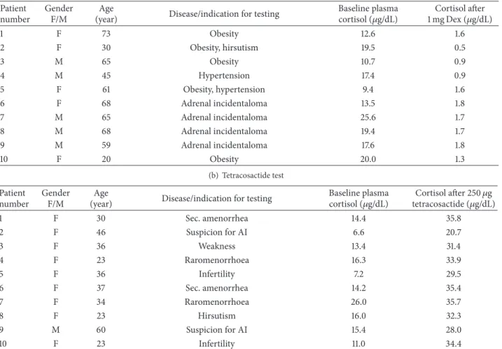
2.1. Patients. 10 patients were tested for suspected hypercor- tisolism by low-dose overnight (1 mg) dexamethasone sup- pression test suffering from obesity, hirsutism, hypertension, and adrenal incidentaloma. Another 10 patients have been examined by 250𝜇g tetracosactide (Cosyntropin, Sandoz Inc.) for suspected Addison’s disease or late onset congenital adrenal hyperplasia (deficiency of 21-hydroxylase) suffering from weakness, secondary oligomenorrhea, infertility, or hir- sutism. Baseline cortisol was taken between 7.00 and 9.00 a.m.
in fasting condition. Dexamethasone was taken at 11.00 pm, and blood was drawn the next morning between 7.00 and 9.00 a.m. Blood was taken one hour after tetracosactide adminis- tration. Patient data are included inTable 1. All tested indi- viduals have been eventually found to be free from any func- tional disturbance of the hypothalamic-pituitary-adrenal axis. The study was approved by the Ethical Committee
of the Hungarian Health Council and informed written consent was obtained from all patients involved.
2.2. RNA Isolation and Real-Time Reverse Transcription Quan- titative Polymerase Chain Reaction (RT-qPCR) from Plasma Samples. RNA isolation has been performed as described in our previous study [10]. Briefly, EDTA-anticoagulated blood was taken from patients and centrifuged at 3000 rpm for 20 minutes at 4∘C. All extracted plasma samples were stored at
−80∘C until further processing.
Total RNA was isolated from 200𝜇L plasma with Qiagen miRNeasy Mini Kit (Qiagen GmbH, Hilden, Germany) according to the manufacturer’s protocol with minor mod- ifications, as described earlier [10]. RNA concentration was measured by NanoDrop 1000 Spectrophotometer (Thermo Fisher Scientific Inc., Waltham, MA, USA), and the quality and quantity were determined by an Agilent 2100 Bioanalyzer (Agilent Tech. Inc., Santa Clara, CA, USA). RNA Integrity (RIN) numbers of RNA isolated from plasma samples were low (around 2.0), that is, similar to reported findings on RNA isolated from blood [14]. RNA was stored at−80∘C until use.
10 ng of total RNA was reverse transcribed with TaqMan MicroRNA Reverse Transcription Kit (Applied Biosystems, Foster City, CA, USA) and the specific looped RT primer. RT- qPCR was performed by TaqMan Fast Universal PCR Master Mix (2x) (Applied Biosystems) on a 7500 Fast Real-Time PCR System (Applied Biosystems) according to the manufacturer’s protocol. The following probes have been used: hsa-miR- 27a(000408),hsa-miR-200b(002251),hsa-miR-214(002306), hsa-miR-483-5p (002338), hsa-miR-503 (001048), and cel- miR-39(000200) as reference gene [15]. Samples were run in triplicate.
2.3. In Vitro Treatment of NCI-H295R Cells with Dexametha- sone. The NCI-H295R adrenocortical carcinoma cell line was purchased from the American Type Culture Collection and maintained in the recommended media. For treatments, hormone-free fetal bovine serum (FBS) was prepared as follows: 0.1 g dextran coated charcoal (C6241, Sigma-Aldrich, St. Louis, MO) was added to 6 mL FBS and incubated for 24 h at 4∘C. Then the mixture was centrifuged at 3000×g for 10 min and the supernatant was filtered through a 0.22𝜇m filter. Cells were seeded on 6-well plates as 106cells/well using media containing 2.5% hormone-free FBS. Next day, cells were synchronized by serum starvation for 24 h. On the following day, 2.5% hormone-free FBS was added in the presence of 100 nM dexamethasone or vehicle (DMSO). After 8 h incubation, cells and supernatans were harvested and total RNA was extracted. Dexamethasone treatments were repeated four times.
Total RNA was extracted using miRNeasy Mini Kit (Qiagen) both from cells and culture medium according to the manufacturer’s protocol with minor modifications, as described earlier [16]. RNA concentration was measured by NanoDrop 1000 Spectrophotometer (Thermo Fisher Sci- entific Inc.). RIN numbers determined by an Agilent 2100 Bioanalyzer (Agilent Tech. Inc., Santa Clara, CA, USA) varied between 9.0 and 10.0. RNA was stored at−80∘C until use. RT- qPCR reactions were performed by Taqman miRNA Assays
Table 1: Characteristics of patients.
(a) Dexamethasone test Patient
number
Gender F/M
Age
(year) Disease/indication for testing Baseline plasma
cortisol (𝜇g/dL) Cortisol after 1 mg Dex (𝜇g/dL)
1 F 73 Obesity 12.6 1.6
2 F 30 Obesity, hirsutism 19.5 0.5
3 M 65 Obesity 10.7 0.9
4 M 45 Hypertension 17.4 0.9
5 F 61 Obesity, hypertension 9.4 1.6
6 F 68 Adrenal incidentaloma 13.5 1.8
7 M 65 Adrenal incidentaloma 25.6 1.7
8 M 68 Adrenal incidentaloma 19.4 1.7
9 M 59 Adrenal incidentaloma 17.6 1.8
10 F 20 Obesity 20.0 1.3
(b) Tetracosactide test Patient
number
Gender F/M
Age
(year) Disease/indication for testing Baseline plasma
cortisol (𝜇g/dL) Cortisol after 250𝜇g tetracosactide (𝜇g/dL)
1 F 30 Sec. amenorrhea 14.4 35.8
2 F 46 Suspicion for AI 6.6 20.7
3 F 36 Weakness 13.4 31.4
4 F 23 Raromenorrhoea 16.3 33.9
5 F 36 Infertility 7.2 29.5
6 F 37 Sec. amenorrhea 14.2 35.4
7 F 34 Raromenorrhoea 26.0 35.7
8 F 23 Hirsutism 16.0 32.3
9 M 60 Suspicion for AI 15.4 28.0
10 F 23 Infertility 11.0 34.4
AI: adrenal insufficiency.
(Applied Biosystems) using specific primer/probe combi- nations:hsa-miR-27a(000408) andcel-miR-39 (000200) as reference gene [15].
2.4. Statistical Analysis. To identify microRNAs showing significant expression changes, Student’s 𝑡-test or Mann- Whitney𝑈test was used depending on the results of Shapiro- Wilks normality test [10]. Data were expressed asΔCt; thus higherΔCt indicates lower expression, whereas lowerΔCt shows higher expression. Statistical analysis of RT-qPCR data was done by Statistica 7.0 (StatSoft Inc., Tulsa, OK, USA) software.
3. Results
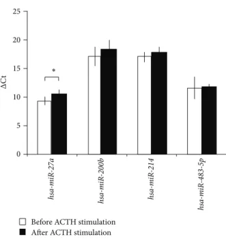
3.1. Expression of Circulating MicroRNAs in Dexamethasone and ACTH Stimulation Tests In Vivo. From the five microR- NAs selected, only one circulating microRNA,hsa-miR-27a, turned out to be significantly modulated by dexamethasone and tetracosactide treatment in our study. Most interestingly, dexamethasone and tetracosactide treatments resulted in opposite changes ofhsa-miR-27aexpression as dexametha- sone upregulated its plasma level, whereas tetracosactide suppressed its expression (Figures1and2).
The expression of hsa-miR-503 proved to be so low in the plasma samples that we have decided to exclude it from further analysis (data not shown). We have not found any cor- relation between the changes of cortisol levels and circulating microRNAs neither in the dexamethasone nor in the tetra- cosactide tests (data not shown). There has been no correla- tion between basalhsa-miR-27levels and body weight either.
To confirm the dexamethasone responsiveness of hsa- miR-27a, we have performed in vitro experiments on the adrenocortical NCI-H295R cell line. We have observed dex- amethasone responsiveness in vitro, as well. Dexametha- sone significantly induced secretedhsa-miR-27aexpression in NCI-H295R culture medium (Figure 3). Dexamethasone induced intracellularhsa-miR-27ain NCI-H295R cells too, but this has not reached statistical significance (data not shown). These results demonstrate that hsa-miR-27a is secreted by NCI-H295R cells and the level of secretedhsa- miR-27ais induced by dexamethasone, as well.
4. Discussion
We have found that the expression of circulatinghsa-miR-27a is modulated by hormonal treatmentsin vivoin humans, as its expression is induced by dexamethasone and suppressed
25 20
15
10
5
0
∗
Before dexamethasone After dexamethasone
ΔCt hsa-miR-27a hsa-miR-200b hsa-miR-214 hsa-miR-483-5p
Figure 1: Expression change of microRNAs in plasma after 1 mg overnight dexamethasone test normalized tocel-mir-39.ΔCt values are represented: increasedΔCt indicates lower expression, whereas decreasedΔCt indicates higher expression (mean±SD).∗𝑝 < 0.05, 𝑛 = 10.𝑡-test was performed following the Shapiro-Wilks normality test.
by ACTH. Dexamethasone induced secreted hsa-miR-27a in vitro, as well. The expression of the most promising circulating microRNA marker of adrenocortical malignancy, hsa-miR-483-5p,was not affected by these treatments also used as diagnostic tests supporting its applicability as a biomarker.
Circulating microRNAs are promising biomarkers in several diseases including tumors and atherosclerosis [17].
There are some data that their levels might be affected by hormonal changes, for example, in patients suffering from polycystic ovarian syndrome, the serum concentration of four microRNAs appeared to be in part correlated with serum free testosterone concentration [8].
To the best of our knowledge, the association of cir- culating microRNAs and the hormonal actions affecting the hypothalamic-pituitary-adrenal axis in vivo has not been explored in humans, yet. Some circulating microRNAs have been proposed as useful biomarkers for prediction of malignancy of adrenocortical tumors [9–11]. Since adreno- cortical cancer is frequently associated with adrenocortical hormone overproduction [18,19], the potential association of hypothalamic-pituitary-adrenal axis functioning and circu- lating microRNA levels might also be of interest. However, no data about the expression changes of circulating microRNAs during dexamethasone or ACTH-tests have been presented to date.
We have selected five circulating microRNAs for studying their responsiveness to dexamethasone and ACTH adminis- trationin vivo. Among these,hsa-miR-214, hsa-miR-503,and hsa-miR-483-5phave been proposed as tissue biomarkers for
25 20
15
10
5
0
∗
Before ACTH stimulation After ACTH stimulation
ΔCt hsa-miR-27a hsa-miR-200b hsa-miR-214 hsa-miR-483-5p
Figure 2: Expression change of microRNAs in plasma after 250𝜇g tetracosactide test, normalized tocel-mir-39.ΔCt values are repre- sented: increasedΔCt indicates lower expression, whereas decreased ΔCt indicates higher expression (mean±SD).∗𝑝 < 0.05,𝑛 = 10.
Results of Mann-Whitney𝑈test.
30 25 20 15 10 5
0 Control Dexamethasone treated
∗
ΔCt
Figure 3: Expression change of secretedhsa-miR-27aafter 100 nM dexamethasone treatment for 8 hours in the NCI-H295R adreno- cortical cell line mediums normalized to cel-mir-39. ΔCt values are represented: increasedΔCt indicates lower expression, whereas decreasedΔCt indicates higher expression (mean±SD).∗𝑝 < 0.05, 𝑛 = 4. Results of𝑡-test.
adrenocortical malignancy [20–22], andhsa-miR-483-5phas been found to be significantly overexpressed in blood samples of adrenocortical cancer patients, as well [9–11]. Moreover, the tissue expression ofmiR-214andmiR-503was downregu- lated by ACTH in a rat model [7]. Two further microRNAs reported to be responsive to hormonal treatments in a rat model were included in our study: the expression of bothhsa- miR-27aandhsa-miR-200bwas shown to be downregulated by dexamethasone, whereas ACTH also downregulatedhsa- miR-27ain rat adrenals [7].
The expression of the five selected circulating microRNAs has been studiedin vivousing plasma samples of ten patients
before and after low-dose dexamethasone testing and samples from ten patients before and after tetracosactide adminis- tration. All individuals included turned out to be eventually free from any functional abnormality of the hypothalamic- pituitary-adrenal axis.
From the microRNAs selected, onlyhsa-miR-27 turned out to be significantly modulated by hormonal treatmentsin vivo. The expression of the other four circulating microRNAs that were shown to be associated with the adrenal was not affected by dexamethasone or ACTH-administration. The stable expression of circulatinghsa-miR-483-5pnot affected by these hormones supports its applicability as a biomarker of adrenocortical cancer. Our study has certainly limitations, since healthy individuals were tested, and a different response in ACC patients cannot be excluded.
Dexamethasone and tetracosactide treatments resulted in opposite changes ofhsa-miR-27aexpression as dexametha- sone upregulated its plasma levels, whereas tetracosactide suppressed its expression (Figures 1 and 2). In addition, secretedhsa-miR-27awas significantly induced by dexam- ethasone treatmentin vitroin the NCI-H295R adrenocortical cell line as well. Our findings in culture medium underline that dexamethasone induces the secretion of hsa-miR-27a from adrenocortical cells (Figure 3). The molecular mech- anism of hsa-miR-27a secretion in NCI-H295R cells and its interaction with dexamethasone, however, awaits further studies. In a rat model, ACTH-treatment also suppressed tissuemiR-27aexpression, but dexamethasone did the same.
This discrepancy might be related to species differences;
moreover, the expression of tissue and circulating microR- NAs can be different [11].
Thein vitroandin vivoaction of dexamethasone onhsa- miR-27aexpression is similar, since it upregulatedhsa-miR- 27a expression in both NCI-H295R adrenocortical cellsin vitro (both cellular and secreted) and circulating hsa-miR- 27a in vivo. The cellular origin of circulating microRNAs is, however, unclear, but these parallel changes in expres- sion might raise the possibility of its partial adrenocortical origin. There are several data underlining the relevance ofhsa-miR-27ain muscles, angiogenesis, adipogenesis and obesity, inflammation, immune response, lipid metabolism, atherosclerosis, and metabolic syndrome [23]. Circulating hsa-miR-27ahas been raised as a biomarker for left ventric- ular contractility after acute myocardial infarction [24] and hypertrophic cardiomyopathy [25], and it was found to be underexpressed in early stage non-small cell lung cancer [26].
All these tissues are targets for glucocorticoid actions mediated via the glucocorticoid receptor. Since dexametha- sone treatment also altered hsa-miR-27aexpression in the NCI-H295R adrenocortical cell line, it might be hypothesized that this microRNA may be also regulated via the gluco- corticoid receptor. As the transcription of hsa-miR-27a is made by RNA Polymerase II [27] it would be interesting to test whether a functional glucocorticoid response element is present in thehsa-miR-27apromoter (byin silicoprediction, a glucocorticoid response element can be localized within the hsa-miR-27apromoter (data not shown)).
hsa-miR-27ahas been shown to downregulate myostatin expression that is a major growth factor implicated in muscle
development and muscle atrophy. Increased myostatin ex- pression was associated with muscle wasting [28].miR-27a and myostatin appear to be involved in an autoregulatory loop as myostatin increasesmiR-27aexpression viaSMAD3 and miR-27a in turn inhibits myostatin expression in a murine model [28]. As glucocorticoids inhibit the transcrip- tional activation of SMAD3 [29], administration of dexam- ethasone might interfere with the myostatin-SMAD3-miR- 27aloop at multiple points. The ACTH-induced downregula- tion of circulating hsa-miR-27a might also be relevant, for example, in ACTH-dependent Cushing’s syndrome. The overall effects of these actions on myostatin,miR-27a, and SMAD3 would be difficult to predict at present, but these findings might be implicated in the pathogenesis of glucocor- ticoid-induced muscle atrophy characteristic for hypercorti- solism.
Levels of circulating hsa-miR-27a have been found to be strongly associated with fasting glucose levels and type 2 diabetes mellitus [30]. Since glucocorticoids are involved in the pathogenesis of insulin resistance [31], these findings raise the possibility that ACTH- and glucocorticoid-induced changes inhsa-miR-27aexpression might be relevant in the pathogenesis of various diseases, and most of all in hyper- cortisolism, but further studies are needed to establish the pathological relevance of these alterations.
5. Conclusions
By analyzing the expression of selected microRNAs based on literature data, we have established that hsa-miR-27ais significantly downregulated by ACTH and induced by dexa- methasone-treatmentin vivo. We have also observed thein vitro induction of secreted hsa-miR-27a in adrenocortical NCI-H295R cells by dexamethasone. The expression ofhsa- miR-483-5pproposed as a biomarker of ACC was not affected by hormonal treatments that underlines its applicability as a potential diagnostic test in the preoperative diagnosis of ACC. These data together highlight again that microRNAs are present in the circulation, and some of these are targets for hormone actions, and similarly to the hormone con- centration measurement, strict preanalytical and analytical conditions should be followed before sampling.
Conflict of Interests
The authors have no conflict of interests to report.
Acknowledgments
This study has been funded by Hungarian Scientific Research Grant (OTKA K100295 to Peter Igaz; OTKA, PD100648 to Attila Pat´ocs) and Technology Innovation Fund, National Developmental Agency (KTIA-AIK-2012-12-1-0010).
References
[1] I. Alvarez-Garcia and E. A. Miska, “MicroRNA functions in animal development and human disease,”Development, vol. 132, no. 21, pp. 4653–4662, 2005.
[2] M. Malumbres, “MiRNAs and cancer: an epigenetics view,”
Molecular Aspects of Medicine, vol. 34, no. 4, pp. 863–874, 2013.
[3] G. Siasos, C. Kollia, V. Tsigkou et al., “MicroRNAs: novel diag- nostic and prognostic biomarkers in atherosclerosis,”Current Topics in Medicinal Chemistry, vol. 13, no. 13, pp. 1503–1517, 2013.
[4] R. S. Redis, S. Calin, Y. Yang, M. J. You, and G. A. Calin, “Cell- to-cell miRNA transfer: from body homeostasis to therapy,”
Pharmacology and Therapeutics, vol. 136, no. 2, pp. 169–174, 2012.
[5] M. A. Cortez, C. Bueso-Ramos, J. Ferdin, G. Lopez-Berestein, A.
K. Sood, and G. A. Calin, “MicroRNAs in body fluids-the mix of hormones and biomarkers,”Nature Reviews Clinical Oncology, vol. 8, no. 8, pp. 467–477, 2011.
[6] A. Riester, O. Issler, A. Spyroglou, S. H. Rodrig, A. Chen, and F. Beuschlein, “ACTH-dependent regulation of MicroRNA as endogenous modulators of glucocorticoid receptor expression in the adrenal gland,”Endocrinology, vol. 153, no. 1, pp. 212–222, 2012.
[7] Z. Hu, W.-J. Shen, Y. Cortez et al., “Hormonal regulation of microRNA expression in steroid producing cells of the ovary, testis and adrenal gland,”PLoS ONE, vol. 8, no. 10, Article ID e78040, 2013.
[8] M. Murri, M. Insenser, E. Fern´andez-Dur´an, J. L. San-Mill´an, and H. F. Escobar-Morreale, “Effects of polycystic ovary syn- drome (PCOS), sex hormones, and obesity on circulating miRNA-21, miRNA-27b, miRNA-103, and miRNA-155 expres- sion,”Journal of Clinical Endocrinology and Metabolism, vol. 98, no. 11, pp. E1835–E1844, 2013.
[9] O. Chabre, R. Lib´e, G. Assie et al., “Serum miR-483-5p and miR- 195 are predictive of recurrence risk in adrenocortical cancer patients,”Endocrine-Related Cancer, vol. 20, no. 4, pp. 579–594, 2013.
[10] D. R. Szab´o, M. Luconi, P. M. Szab´o et al., “Analysis of circu- lating microRNAs in adrenocortical tumors,”Laboratory Inves- tigation, vol. 94, no. 3, pp. 331–339, 2014.
[11] D. Patel, M. Boufraqech, M. Jain et al., “MiR-34a and miR-483- 5p are candidate serum biomarkers for adrenocortical tumors,”
Surgery, vol. 154, no. 6, pp. 1224–1229, 2013.
[12] K. I. Alexandraki and A. B. Grossman, “Novel insights in the diagnosis of Cushing’s syndrome,”Neuroendocrinology, vol. 92, no. 1, pp. 35–43, 2010.
[13] E. S. Husebye, B. Allolio, W. Arlt et al., “Consensus statement on the diagnosis, treatment and follow-up of patients with primary adrenal insufficiency,”Journal of Internal Medicine, vol. 275, no.
2, pp. 104–115, 2014.
[14] J. H. Kim, H. O. Jin, J. A. Park, Y. H. Chang, Y. J. Hong, and J. K. Lee, “Comparison of three different kits for extraction of high-quality RNA from frozen blood,”SpringerPlus, vol. 3, no.
1, article 76, 5 pages, 2014.
[15] P. S. Mitchell, R. K. Parkin, E. M. Kroh et al., “Circulating microRNAs as stable blood-based markers for cancer detec- tion,”Proceedings of the National Academy of Sciences of the United States of America, vol. 105, no. 30, pp. 10513–10518, 2008.
[16] H. Butz, I. Lik´o, S. Czirj´ak et al., “MicroRNA profile indicates downregulation of the TGF𝛽pathway in sporadic non-func- tioning pituitary adenomas,”Pituitary, vol. 14, no. 2, pp. 112–124, 2011.
[17] H. Schwarzenbach, N. Nishida, G. A. Calin, and K. Pantel,
“Clinical relevance of circulating cell-free microRNAs in can- cer,”Nature Reviews Clinical Oncology, vol. 11, no. 3, pp. 145–156, 2014.
[18] M. Fassnacht, M. Kroiss, and B. Allolio, “Update in adreno- cortical carcinoma,” Journal of Clinical Endocrinology and Metabolism, vol. 98, no. 12, pp. 4551–4564, 2013.
[19] M. Terzolo, F. Daffara, A. Ardito et al., “Management of adrenal cancer: a 2013 update,”Journal of Endocrinological Investigation, vol. 37, no. 3, pp. 207–217, 2014.
[20] Z. T¨omb¨ol, P. M. Szab´o, V. Moln´ar et al., “Integrative molec- ular bioinformatics study of human adrenocortical tumors:
microRNA, tissue-specific target prediction, and pathway anal- ysis,”Endocrine-Related Cancer, vol. 16, no. 3, pp. 895–906, 2009.
[21] E. E. Patterson, A. K. Holloway, J. Weng, T. Fojo, and E. Kebe- bew, “MicroRNA profiling of adrenocortical tumors reveals miR-483 as a marker of malignancy,”Cancer, vol. 117, no. 8, pp.
1630–1639, 2011.
[22] P. S. H. Soon, L. J. Tacon, A. J. Gill et al., “miR-195 and miR-483- 5p identified as predictors of poor prognosis in adrenocortical cancer,”Clinical Cancer Research, vol. 15, no. 24, pp. 7684–7692, 2009.
[23] W.-J. Chen, K. Yin, G.-J. Zhao, Y.-C. Fu, and C.-K. Tang, “The magic and mystery of microRNA-27 in atherosclerosis,”Athero- sclerosis, vol. 222, no. 2, pp. 314–323, 2012.
[24] Y. Devaux, M. Vausort, G. P. McCann et al., “A panel of 4 microRNAs facilitates the prediction of left ventricular contrac- tility after acute myocardial infarction,”PLoS ONE, vol. 8, no. 8, Article ID e70644, 2013.
[25] R. Roncarati, C. V. Anselmi, M. A. Losi et al., “Circulating miR-29a, among other up-regulated microRNAs, is the only biomarker for both hypertrophy and fibrosis in patients with hypertrophic cardiomyopathy,”Journal of the American College of Cardiology, vol. 63, no. 9, pp. 920–927, 2014.
[26] N. H. H. Heegaard, A. J. Schetter, J. A. Welsh, M. Yoneda, E. D.
Bowman, and C. C. Harris, “Circulating micro-RNA expression profiles in early stage nonsmall cell lung cancer,”International Journal of Cancer, vol. 130, no. 6, pp. 1378–1386, 2012.
[27] Y. Lee, M. Kim, J. Han et al., “MicroRNA genes are transcribed by RNA polymerase II,”The EMBO Journal, vol. 23, no. 20, pp.
4051–4060, 2004.
[28] C. McFarlane, A. Vajjala, H. Arigela et al., “Negative auto-regu- lation of myostatin expression is mediated by Smad3 and MicroRNA-27,”PLoS ONE, vol. 9, no. 1, Article ID e87687, 2014.
[29] C.-Z. Song, X. Tian, and T. D. Gelehrter, “Glucocorticoid receptor inhibits transforming growth factor-𝛽signaling by directly targeting the transcriptional activation function of Smad3,”Proceedings of the National Academy of Sciences of the United States of America, vol. 96, no. 21, pp. 11776–11781, 1999.
[30] D. S. Karolina, S. Tavintharan, A. Armugam et al., “Circulating miRNA profiles in patients with metabolic syndrome,” The Journal of Clinical Endocrinology & Metabolism, vol. 97, no. 12, pp. E2271–E2276, 2012.
[31] E. B. Geer, J. Islam, and C. Buettner, “Mechanisms of gluco- corticoid-induced insulin resistance: focus on adipose tissue function and lipid metabolism,”Endocrinology and Metabolism Clinics of North America, vol. 43, no. 1, pp. 75–102, 2014.
Submit your manuscripts at http://www.hindawi.com
Stem Cells International
Hindawi Publishing Corporation
http://www.hindawi.com Volume 2014
Hindawi Publishing Corporation
http://www.hindawi.com Volume 2014
INFLAMMATION
Hindawi Publishing Corporation
http://www.hindawi.com Volume 2014
Behavioural Neurology
Endocrinology
International Journal ofHindawi Publishing Corporation
http://www.hindawi.com Volume 2014
Hindawi Publishing Corporation
http://www.hindawi.com Volume 2014
Disease Markers
Hindawi Publishing Corporation
http://www.hindawi.com Volume 2014
BioMed
Research International
Oncology
Journal ofHindawi Publishing Corporation
http://www.hindawi.com Volume 2014
Hindawi Publishing Corporation
http://www.hindawi.com Volume 2014
Oxidative Medicine and Cellular Longevity
Hindawi Publishing Corporation
http://www.hindawi.com Volume 2014
PPAR Research The Scientific World Journal
Hindawi Publishing Corporation
http://www.hindawi.com Volume 2014
Immunology Research
Hindawi Publishing Corporation
http://www.hindawi.com Volume 2014
Journal of
Obesity
Journal ofHindawi Publishing Corporation
http://www.hindawi.com Volume 2014
Hindawi Publishing Corporation
http://www.hindawi.com Volume 2014
Computational and Mathematical Methods in Medicine
Ophthalmology
Journal ofHindawi Publishing Corporation
http://www.hindawi.com Volume 2014
Diabetes Research
Journal ofHindawi Publishing Corporation
http://www.hindawi.com Volume 2014
Hindawi Publishing Corporation
http://www.hindawi.com Volume 2014
Research and Treatment
AIDS
Hindawi Publishing Corporation
http://www.hindawi.com Volume 2014
Gastroenterology Research and Practice
Hindawi Publishing Corporation
http://www.hindawi.com Volume 2014
Parkinson’s Disease
Evidence-Based Complementary and Alternative Medicine
Volume 2014 Hindawi Publishing Corporation
http://www.hindawi.com