Mutational Control of Bioenergetics of Bacterial Reaction Center Probed by Delayed Fluorescence
Delphine Onidas1,a, Gábor Sipka2,a, Emese Asztalos2 and Péter Maróti2*
1 Laboratoire de Chimie Physique UMR 8000, Batiment 350, Orsay-Cedex, Université de Paris-Sud, France, 91405
2 Department of Medical Physics, University of Szeged, Rerrich Béla tér 1, Szeged, Hungary, H-6720
* Corresponding author Phone: 36-63-544-120
E mail: pmaroti@sol.cc.u-szeged.hu Homepage: www.szote.u-szeged.hu/dmi
a These authors contributed equally to the present work.
Key words: Bacterial photosynthesis, reaction center protein, H-bond network, light-induced free energy changes, electron transfer
Abbreviations: BChl, monomeric bacteriochlorophyll; DL, delayed light; LDAO, N,N’-
dimethyldodecylamine N-oxide; P, primary electron donor, a non-covalently linked bacteriochlorophyll dimer; QA and QB, primary and secondary quinones; Rba., Rhodobacter; RC, reaction center; Triton X- 100, octylphenol polyethylene glycol ether; UQ, ubiquinone; WT, wild type.
Abstract
The free energy gap between the stable charge separated state P+QA-
and the excited bacteriochlorophyll dimer P* was measured by delayed fluorescence of the dimer in mutant reaction center proteins of the photosynthetic bacterium Rhodobacter sphaeroides. The mutations were engineered both at the donor (L131L, M160L, M197F and M202H) and acceptor (M265I and M234E) sides. While the donor side mutations changed systematically the number of H-bonds around P, the acceptor side mutations modified the energetics of QA by altering the van-der-Waals and electronic interactions (M265IT) and H-bond network to the acidic cluster (M234EH). All mutants decreased the free energy gap of the wild type RC (~890 meV), i.e. destabilized the primary charge pair by 60-110 meV at pH 8. The
destabilization showed slight pH-dependence (~ 10-15 meV/pH unit) at neutral pH and increased at high pH (wild type, donor side mutations and M265IT) or was absent (M234EH). It demonstrated the importance of H-bond structure around P in destabilization of the dipole and is in accordance with simultaneous and substoichometric proton binding by the acidic cluster near QB and proton release by residues at the donor side. The free energy change consisted of mainly enthalpic term but the acceptor side mutants showed half-half entropic and enthalpic contributions. This could include softening the structure of the iron ligand (M234EH) and the QA binding pocket (M265IT) and/or increase of the multiplicity of the electron transfer pathways of charge separation in the acceptor side upon mutation.
Introduction
Some selected proteins (enzymes) of the nature ensure conditions 1) to undergo specific chemical reactions and 2) to enhance the rate up to 1017 compared to the equivalent reaction in solution [1,2].
About one third of the proteins are redox proteins where the enhancement of the rates of electron and proton transfer together with the very high specificity of ligand binding has special importance [3].
These processes occur under a wide range of redox states and are determined by the structure, dynamics and energetics of the redox protein [4]. The energetics of the protein is controlled by the interactions of the constituents of the macromolecule [5]. Although the interactions are very complex, some amino acids are in key positions to determine the energetics. Mutations directed to these sites can cause major changes in interactions and functions of the protein [6,7].
The general principle of mutation control of redox proteins can be tested in reaction center (RC) of photosynthetic bacteria that can serve as model system for these studies (Fig. 1). The RC is part of the few number of membrane proteins whose 3D structures have been obtained at high (~ 2.5 Å, [8]) or very high (1.87 Å, [9]) resolutions. The structure reveals the binding sites and precise orientation of cofactors and their interaction with proteins and provide a solid basis to interpret results of absorption and
fluorescence studies at an atomic level [10]. The biochemical modification and the genetic
transformation of the strain Rhodobacter (Rba.) sphaeroides are well elaborated [11,12]. Additionally, the kinetics and thermodynamics of the charge transfer processes are quite easy to study by detection of flash induced absorbance changes of the cofactors in their different redox states. All these advantages allow subtle analysis of structure-energetics-function relationships including protein and electrostatic controls of the electron and proton transfer processes [13-15]. The bacterial RC performs its function with a very high quantum efficiency of near 100% [16] and has therefore long been a model system for the study of the conversion of light into chemical energy, with a large relevance to the design of artificial systems [17]. Results obtained on RCs are frequently used as a basis to understand the function of
similar redox and/or proton transport proteins involved in photoactive or respiratory systems [18]. Here, we aim to obtain a better understanding of which facts make the bacterial RC robust and yet flexible enough to function efficiently under different conditions.
The RC protein of purple photosynthetic bacterium Rhodobacter sphaeroides performs unique way of conversion of light energy into chemical free energy. The protein binds ubiquinone (UQ) and reduced cytochrome (cyt c2+) at the cytoplasmic and periplasmic sites, respectively, and releases UQH2 and cyt
c3+ as redox products [14,15,19,20]. The absorption of photon results in intraprotein electron transfer from the primary electron donor (bacteriochlorophyll dimer, P, situated on the periplasmic side of the membrane) through a series of intermediates (bacteriochlorophyll monomer, BChl and
bacteriopheophytine, BPheo, in the membrane) to stable electron acceptors (ubiquinones, QA and QB on the cytoplasmic side). The cofactors are arranged along two branches (A and B) of nearly perfect mirror- symmetry, but the electron transfer in wild type RC occurs on the A branch only. A non-heme iron atom (Fe) with its ligand is situated symmetrically to both quinones and facilitates the interquinone electron transfer. After subsequent transfer of the second electron and uptake of two protons from the aqueous bulk phase, the secondary quinone is completely reduced to quinol (QBH2) and leaves the RC. Thus, the light energy is converted to the reducing power of the QB/QBH2 redox couple that can be directly used to cover the free energy need of essential biological processes (ion transport, ATP synthesis etc.).
The photochemical utilization of the absorbed light energy is controlled by the free energy state of the primary stable charge pair P+QA-
(Fig. 2). The higher is the gap between P* and P+QA-
, the smaller free energy is utilized from the absorbed photon although the stabilization of the charge pair is increased. A convenient way of the determination of the free energy gap is based on the measurement of the delayed fluorescence from the bacteriochlorophyll dimer [21-23]. By comparison of the prompt (PF) and delayed fluorescence (DL) intensities emitted by the excited dimer (P*), the free energy gap between the excited dimer and the charge separated state P+QA-
can be determined from Boltzmann distribution of the
population between the two states unless the DL does not influence the thermal equilibrium (the DL is of
“leakage” type). The use of DL has the unique advantage of absolute determination of the free energy gap which goal can be rarely set for other similar methods. It has been applied to several related problems of the energetics of bacterial RCs including substoichiometric amount of proton uptake by isolated RC upon QA reduction [24,25] or pH-independent midpoint potential of QA in chromatophores [26].
In this study, the question is set how the free energy level of the charge separation would be modified upon mutations of some key residues on both the donor and acceptor sides of the RC. The amino acids targeted to site-directed mutagenesis are demonstrated in Fig. 1. As the intensity of the observed DL depends on the energetics of both sides, the changes of the free energy levels upon mutations can be nicely followed and interpreted by measurement of the DL from the
bacteriochlorophyll dimer. As in LEGO, the selected mutants show overall energetic changes that can be summed up from individual free energy changes induced by the modification of the number of H-
bonds around the dimer and by steric restriction, electrostatics and H-bond pattern of QA. The thermodynamic parameters of the P* → P+QA-
process indicate enhanced entropic term in the free energy change of the mutants at the acceptor side that shed some light in possibility of increased multiplicity of electron transfer pathways of stable charge separation.
Materials and Methods
The wild type strain and all expression strains used in this study are derived from the pink and
carotenoid-containing strain 2.4.1 of Rba. sphaeroides of wild-type constitution [27]. The construction of mutant strains harboring the different mutations, the mutagenesis procedures, growth conditions for mutant cells, as well as the RC preparation, have been described previously [28]. The cells were grown in Erlenmeyer flasks filled to 50% of the total volume with malate yeast medium supplemented with kanamycin (20 μg/ml) and tetracycline (2 μg/ml). The cultures were grown in the darkness at 30 °C on a gyratory shaker (100 rpm). The RCs were solubilized from the membrane by ionic detergent LDAO that was removed and replaced by a nonionic detergent Triton X-100 after a long (48 h) dialysis at 4°C by a frequent change of the dialyzing medium. The pH was measured with a combined glass electrode (model 91-03; Orion) calibrated with standards that spanned the measured pH range. The assay solution
contained 2 μM RCs with 0.03% Triton X-100, 100 mM NaCl, and 5 mM buffer, depending on the pH.
The following buffers were used: 2-(N-morpholino)-ethanesulfonic acid (MES; Sigma) between pH 5.5 and pH 6.5; 1,3-bis[tris(hydroxymethyl) methylamino]propane] (Bis-Tris propane; Sigma) between pH 6.3 and pH 9.5; Tris–HCl (Sigma) between pH 7.5 and pH 9.0; 3-(cyclohexylamino) propanesulfonic acid (CAPS; Calbiochem) and 2-(cyclohexylamino) ethanesulfonic acid (Ches, Sigma) above pH 9.5.
The photochemical function of RC of each mutant was characterized by the near-infrared absorbance spectrum and by measurement of the flash-induced P/P+ signal amplitude and P+QA-
→ PQA charge- recombination kinetics at 860 nm. The absorbance spectroscopy was performed on a kinetic
spectrophotometer of local design [24]. The secondary quinone activity of the RC was inhibited by addition of 100 μM terbutryn.
Delayed fluorescence measurements were performed as described in [23,26]. The home-built kinetic fluorometer was equipped with a frequency-doubled and Q-switched Nd:YAG laser (Quantel YG 781- 10, wavelength 532 nm, energy 20 mJ, duration 5 ns) and the detector was protected by an electronically controlled mechanical shutter (Uniblitz VS25). The free energy drop from P* to P+QA-, ΔGP*A, was
calculated by comparison of the delayed and prompt fluorescence yields, according to Arata and Parson [21]:
ph fl fl d p d B
0 A
P d
d
ln
k k t F
t F T
k
G .
∫Fd(t)dt and ∫Fp(t)dt are the integrated intensities of delayed and prompt fluorescence, measured in the same sample but at very different excitation intensities (both in the linear region) to give similar emission intensities. ∫Fd(t)dt is determined by a one-exponential fit to the decay of the delayed
fluorescence signal; ∫Fp(t)dt is determined by electronic integration of the prompt fluorescence, using a time constant (0.1 s) similar to that of the delayed fluorescence decay time. The Boltzmann factor is kBT (25 meV at room temperature), kfl is the radiative rate constant of prompt fluorescence (8·107 s−1, [21,22]), kd is the rate of decay of the delayed fluorescence, ηph is the quantum yield of photochemical trapping (≈ 1.0, [16]), and ηfl is the quantum yield of the prompt fluorescence (4·10-4, [29,30]). As the P+QA-
→ PQA charge recombination kinetics are not strictly exponential [31], the determination of kd
based on single exponential decomposition of the delayed fluorescence decay may introduce some systematic error in the determination of the free energy gap ΔGP*A [26,32].
Results
The kinetics of delayed fluorescence of the dimer was measured in a series of mutants at the donor (Fig.
3) and acceptor (Fig. 4) sides of the RC. In all selected mutants, both the amplitude (area) and the rate of the decay increased compared to those of the wild type.
Addition and removal of a single H-bond in the network around the dimer. One of the most fundamental questions is how the addition and removal of the single H-bond in the close vicinity of the dimer will change the observed delayed fluorescence. If an H-bond is introduced by the L131LH mutation, then the intensity of the DL would increase significantly relative to that measured in wild type RC (Fig. 3 inset).
This change indicates the increase of the midpoint potential of P/P+ due to the H-bond to the dimer. If, however, this mutation is compensated by an additional mutation at position M202 by removal of the H- bond (M202HL), then the intensity of the DL would decrease significantly close to the level of the wild type RC. The increase of the redox midpoint potential of P/P+ attributed to the mutation L131LH is compensated by the second mutation at M202LH by lowering the midpoint potential of the dimer. This
experiment clearly demonstrates that the H-bond to the dimer can be made responsible for the change of the free energy level of the dimer wherever this H-bonding is occurring in the vicinity of P.
Multiple H-bond formation on the donor side. The build up of H-bond system around the dimer can be continued by creation of double (L131LH-M160LH) and triple (L131LH-M160LH-M197FH)
mutations. The increase of the number of H-bonds increases the intensity of the DL and the increase is stepwise: the triple mutant shows about twice as large increase as the double mutant relative to that of the wild type. The exact location of the new H-bond is not critical to the increase of the DL as the mutation of relatively remote site can cause similar changes of energetics as sites closer to the dimer.
The H-bond network is extended around P and its modification induces similar energetic effects wherever the perturbation occurs. Similar effect of hydrogen bonds to the conjugated carbonyl molecules on the electrochemical titrations of these mutants was earlier observed [12,33].
The rate constants of the decay of DL as a function of free energy changes. Figure 5 demonstrates the decay rates of DL for several mutants in representation of the driving force of the P+QA-
→ PQA charge recombination. The data obtained from very different mutations on the acceptor and donor sides line up to a single (Marcus) parabola. As the back reaction occurs through a direct (tunnelling) pathway in these RCs, the plot can be approximated by Marcus-type electron transfer [34]. The observed rates do not show large changes upon increase of the driving force that involves a reorganisation energy very close to the actual free energies: λ = 705 meV. This value is somewhat lower than the relevant reorganization energies (800-900 meV) published earlier [12]. The decay rates of the DL behave very similarly to those of the charge recombination measured by absorption changes. This means that the decay of the DL of bacterial RC is governed by the kinetics of the charge recombination, i.e. it shows “leakage” character.
For comparison, the DL or thermoluminescence from Photosystem II of green plants do not demonstrate this property, mainly due to the much smaller (~400-500 meV) free energy gap. The disadvantage of much smaller DL in bacterial RC is compensated by simpler (Boltzmann) evaluation of the data.
pH-dependence of the free energy gap between P* and P+QA-. The free energies of the different states can be modified not only by mutations of nearby residue(s) but by change of the pH of the medium, as well (Fig. 6). If the redox transition (oxidation/reduction of the species) is accompanied by
unbinding/binding of protons at nearby residues, then the free energy of the species will
increase/decrease. It can be considered that the proton release/binding (from energetic point of view) destabilizes/stabilizes the redox agent. The affect is small but can be measured. At lower pH, the free energy gap is higher than at higher pH. The average slope (~ 10-15 meV/pH unit) is far away from the
value of 60 meV/pH unit that directly characterizes the net uptake/release of 1 H+ ion upon reduction/oxidation of the redox center. Neither QA/QA-
nor P/P+ participate directly in
protonation/deprotonation processes. The increased instability above pH 8 in wild type is due to acidic cluster in the vicinity of QB [35]. This behaviour is observed in the M253IT mutant but is completely lacking in the M234EH mutant. The decreasing stability of the P+QA-
charge pair can be clearly recognized at high pH upon increase of the number of H-bonds in the donor side mutations. This tendency indicates the possible significance of the system of H-bonds and water molecules in
transmittance of interaction energy between P+ and the protonatable groups on the periplasmic site of the RC.
Thermodynamic parameters. The direct measurement of the intensity of the DL offers the free energy of the P+QA- → P* process (ΔG0P*A) that consists of enthalpic (ΔH0) and entropic (T·ΔS0) terms: ΔG0 = ΔH0 - T·ΔS0. As the DL originates from the leakage of the thermal equilibrium between P+QA-
and P*, the observation of the temperature change of the DL (van’t Hoff plot) serves as an independent method to find the enthalpy change (ΔH0) of the P+QA-
→ P* transition. ΔH0 is obtained as the slope of the straight line in the van’t Hoff representation (Fig. 7). From these two quantities (ΔG0 and ΔH0), the third
parameter, the entropy change (ΔS0) can be derived (Table 1).
In wild type RC, the P+QA-
→ P* transition is primarily driven by enthalpy change and the entropic change has practically no significance [21]. The tendency is basically preserved in the donor site mutants where the enthalpic term remains dominant with slightly larger contribution of the entropic term. The acceptor side mutation shows principally different shape: the entropy change becomes a significant driving force in the P+QA-
→ P* transition. This observation should involve significant molecular
rearrangements in the M265IT and M234EH mutants relative to that in wild type RC. These specific and transient molecular conformations are prerequisite of the transition (charge separation/recombination) in QA related mutations and seem to be absent in the donor side mutations.
Discussion
To understand the light-induced charge separation and subsequent electron transfer reactions in bacterial RC, both kinetics and thermodynamics should be measured [36]. In this study, mutants directed to specific sites on the donor and acceptor sides were constructed and the changes in energetics were followed by detection of the delayed fluorescence of the bacteriochlorophyll dimer which method has been proved to be an adequate and sensitive tool in bacterial photosynthesis to gain both kinetic and
thermodynamic data. The selected mutants caused large changes of the free energies of the first stable separated charges of P and QA accompanied with major modifications in their enthalpic and entropic contributions and smaller but not insignificant variations on their pH-dependence. The discussion will focus on molecular details and interpretation of these observations.
Changes of Interactions in the Mutants. All the mutants used in this work demonstrated large drop of the free energy gap between P* and P+QA-. As neither of the mutations are accompanied with major structural changes, the observed energetic changes are attributed to slight modifications of the interactions between the cofactors (P and QA) and the protein environment including the extended H- bond network on the cytoplasmic site and (possibly) also on the periplasmic site of the RC. Below, these interactions will be specified.
The effect of hydrogen bonds to the conjugated carbonyl molecules on the oxidation/reduction midpoint potential of the dimer is well documented [12]. In wild type, the redox midpoint potential of P/P+ is poised to ~ 500 mV [24] and an additional hydrogen bond present in the wild type raises the potential by 60 to 120 mV while loss of the hydrogen bond decreases the potential by approximately 80 mV. The DL measurements on H-bond mutants supported these results. The free energy change caused by introduction of a single H-bond was compensated by the removal of an H-bond at a different site near the dimer (Fig. 3) and both the rates and the amplitude (area) of the decay of DL increased with
increasing number of the H-bonds (Figs. 5 and 6). Although the decrease of the free energy gap
determined from DL (Table 1) was not as large and graduate as those obtained from redox titration data, the tendency could be well recognized. The discrepancy can be attributed to the different conditions of the two measurements. While the redox equilibrium is achieved by chemical oxidation of the dimer, the DL method includes the redox changes of QA, as well. The interaction of the protein environment with the dimer in wild type RC sets the free energy gap to a maximum value which assures small portion (rate) of wasteful charge recombination. From the point of view of the yield of charge separation, this structure can be considered as an “optimum” structure. The H-bonds in the mutations decrease the free energy gap and the yield of photochemical utilization of the absorbed photon by interruption of the optimum structure. Systematic modulation of the H-bond pattern by mutagenesis produces a wide range of midpoint potentials of the dimer that sets the stage for building new functionality into the bacterial RC including photo-oxidation of tyrosine [37] and tight binding and oxidation of manganese [38].
The energetics of QA is determined by complex interactions with surrounding amino acids [20] and the mutation of M265IT used here can have several consequences. His-M219 and Ala-M260 form H-
bonds with the O4 (2.87 Å) and O1 (2.82 Å) carbonyls of QA, respectively (structure 1AIG.pdb). Trp- M252 and Ile-M265 are on opposite sides of the quinone and in van der Waals contact. The mutations of the M265 bulky isoleucine site to smaller and polar amino acids threonine and serine led to significant redox tuning. Differences in the measured rate of charge recombination indicated that the polar
mutations lowered the midpoint potential of QA by ~100 and 85 mV respectively [39,40]. Similar results (~115 and 60 mV for threonine and serine) were obtained when the free energy of QA- was directly measured via the delayed fluorescence from P+QA- (see Fig. 4. and [32]). Several factors may result in these free energy changes but their contributions have not yet been clarified. The mutation can affect the protein-quinone interactions by changing binding pocket structure, specific steric or van der Waals interactions with QA, or electrostatically. A movement of the peptide backbone away from the quinone may increase the hydrogen bond distance between the O1 carbonyl and the Ala-M260 peptide NH by
~0.1 Å. Additionally, the difference in size (van der Waals contact) of the amino acids and the introduction of the hydroxyl group can contribute to generate the observed shift. The orientations of quinone methoxy groups that may be implicated in redox poising can play minor role only as
substituting the native ubiquinone with anthraquinone, which lacks any methoxy groups, showed similar energetic effects [40]. Recent pulsed EPR measurements provided further details but could not offer final conclusion [41]. While nitrogen hyperfine parameters were nearly unchanged between the histidine M219 and the QA semiquinone, changes in the quadrupole parameters indicated a significant change in the electric field gradient. Additionally, resolution of the hyperfine coupling between the peptide nitrogen and the semiquinone decreased in the mutant. Although there is only a small effect on the binding site structure, yet it might have sufficient magnitude to suggest that the electric potential and/or field gradient are important contributions to the electronic structure to observe changes of the free energy.
There are accumulating evidences for conformational gating of the QA-
to QB interquinone electron transfer (see for summary in [20]), although the nature of this conformational gate is not clear. The majority of studies argue for a QB-related conformational change including the displacement of QB between distal and proximal binding states [8,42]. However, there are works that question that, and propose a rotated orientation of QA as the conformational change [43]. If it works, then the rotation of QA must have a very large effect on QA energetics. Our studies on the M265I related mutations did not support the direct involvement of QA in the conformational gating. The gating process has not been reflected in a modification neither of the interquinone electron transfer [40] nor of the energetics [32,41]
and pH-dependence (Fig. 6.). The rotation of QA can play minor (if any) role in the mechanism of the conformational gating of the interquinone electron transfer.
The glutamic acid M234 develops two bonds with the Fe atom and, together with four histidines (L190His, L230His, M219His and M266His), constitues the iron ligand. L190His and M219His are symmetrically bound to the Fe atom and also to QA and QB, with which they develop H-bonds. The QA- M219His-Fe-L230His-QB “wire” has been proposed as a structural and energetic connection of the two quinone pockets [44]. Because of the central position of M234Glu, lying between the two quinones and binding the Fe atom, it may influence the energetics of the QA site. The site-specific mutant M234EH adds a fifth His to the Fe ligand and the M234EL mutant replaces a bulky non-polar amino acid that is incapable of developing interactions with the Fe atom and/or does not allow to form a cavity filled with water molecules. Indeed, both mutants modified the H-bond network to QA and caused severe changes in the energetics of the primary quinone detected by DL.
pH-dependence of the stabilisation/destabilisation of the P+QA-
dipole. The static pH-dependence of the free energy gap ΔGP*A indicates the role of protonation/deprotonation of the protein that accompanies the charge separation. The measured pH-dependence of the free energy change is related to the integral of the proton uptake/release induced by the P → P+QA-
transition [19]. The P+QA-
dipole has two ends and therefore the stabilisation can occur by simultaneous uptake and release of protons at the quinone and dimer sides, respectively. The flash-induced uptake of 1 proton/RC by the acidic cluster near QB
corresponds to stabilisation energy of 60 meV. The measured stabilisation energy of P+QA-
state in wild type RC (e.g. Δ ΔG = ΔG(pH 6) - ΔG(pH 11)) amounts a couple of 10 meV that corresponds to
substoichometric net proton binding in agreement with earlier results [24,25]. The use of acceptor and donor side mutations makes it possible to separate the contribution of proton binding and release in the free energy stabilisation process, respectively.
The quinone-side mutations that modify the flash-induced uptake of H+ ions operate in agreement with the expectations based on recent studies. It has been proposed that protons are taken up by an acidic cluster in anticooperative interaction near the QB. The cluster is connected to QA by a chain of H- bonds all over the cytoplasmic face of the protein [35]. The relaxation of the P+QA- charge separated state and the subsequent electron transfer (charge recombination) require the spreading of the protons and the establishment of a favorable configuration for their distribution over the hydrogen bond network. This is also supported by theoretical calculations which have shown that upon formation of QA−
, structural waters (and associated hydrogen bond networks) on the cytoplasmic side of the RC
change their orientation/occupation [45,46]. The M265IT mutant does not modify the extended H-bond network between QA and the acidic cluster and therefore the pH-dependence of the free energy gap remains the same as experienced in the wild type RC. The M234E mutants, however, have major impact on the iron ligand and the H-bond network and do not show any stabilisation (rather slight
destabilisation) upon lowering the pH.
The donor side mutations in our study change the H-bond system around P and modify the pH- dependence of the stabilisation. By increase of the number of H-bonds, the stabilisation of the P+QA- dipole will be more pronounced indicating the not negligible role of donor side interactions beside those of the well characterized acidic cluster near QB. The light-induced net deprotonation of the few
protonatable groups on the periplasmic site due to P+ formation could be shown experimentally after re- oxidation of QA- [19,25]. Similarly as on the cytoplasmic side, the H-bond network may influence the interactions of the protonatable residues on the periplasmic side, modify the pKa values, and alter the stoichiometry of proton release, therefore the stabilisation of the dipole. Although the few protonatable residues near P do not form a closely associated cluster as the acidic amino acids on the acceptor side near QB, their effect on proton unbinding is certainly amplified by the H-bond system and water molecules available on the periplasmic side of the RC.
Thermodynamic parameters of the mutants. While the kinetic properties of charge separation and recombination are relatively easy to determine, the thermodynamic information is far less accessible.
The thermodynamics of the charge separation reveals the energy levels of the initial and final states and the driving force (Gibbs free energy) of the reaction, which is composed of enthalpic and entropic components. The pulsed photoacoustics (PA) and DL methods offer different contributions of enthalpic and entropic terms in charge separation of the native bacterial RC. In vivo PA measurements of
formation of P+QA-
showed a large positive entropy both in bacterial RC and in wild type photosystem I from Synechocystis sp. PCC 6803 [47-49]. It was demonstrated that the A-FX to FA/B step in
Synechocystis 6803 Photosystem I was entropy driven [50]. In contrast, the electron transfer in photosystem II was accompanied by a small negative entropy change. The observed difference in entropy between photosystem I (and bacterial RC) and photosystem II indicates that the apparent entropy may play a vital role in photosynthetic electron transfers. It was demonstrated that entropy controlled the charge separation in artificial photosynthetic systems [51] and showed up temperature dependence of charge recombination in bacterial RC [52].
While the PA finds the bacterial charge separation to be half enthalpic (ΔH0~-440 meV) and half entropic (T·ΔS0~+420 meV) [47], the DL yields negligible entropic contribution (T·ΔS0~0) in the free energy change of ΔG0 ~-890 meV (see Table 1) [21]. The apparent discrepancy may arise from a) different time regimes and b) different assumptions of the two methods. The DL method assumes equilibrium between a single charge-separated state P+QA-
and the excited P state on the 100 ms time scale while the PA method measures the heat released on the <100 ns time scale. The enthalpy is derived from the variation of the equilibrium constants with temperature (van’t Hoff plot of the DL) or from calorimetric measurements (PA) with the assumption of temperature independent thermodynamic parameters (enthalpy, heat capacity and volume change). The latter assumption is not necessarily valid that can cause the observed discrepancy between the two methods [52].
To explore the specific role of the protein matrix in the electron transfer, the bacterial RC was genetically modified at some key amino acids and showed the altered thermodynamics of electron transfer. The mutations at the donor side affected the free energy change of the charge separation but did not have significant influence on the entropic contribution. It remained as negligible as it was in the wild type. The acceptor side mutations, however, increased the partition of the entropy in the free energy change of the reaction and became commeasurable with the enthalpic term or could be even the dominant term. The positive sign of the entropy change upon charge separation calls the attention to increased number of macrostates in the acceptor side. The small amino acids instead of the bulky isolucine at M265 could allow for a greater number of conformations than in wild type and polar side chains from threonine (or serine) could stabilize a specific and alternate methoxy conformation through H-bonding. Similarly, the modification of the iron ligand by M234 mutants changed the H-bond network to QA and increased the number of substates constituting the electron pathways at the QA site. The polar side chains at the cytoplasmic side of the protein may play some role in entropy increase of the charge separation in these mutants. Due to slight steric/electrostatic rearrangement caused by the mutation, some negatively charged surface groups can be reoriented (or even repelled from the RC) which is a disordering process that would enhance the entropy of the reaction.
In standard formulations of the Marcus theory, it is assumed that the vibrations coupled to electron transfer have the same frequency in reactant and product states, which implies that the entropy change is zero [34]. As negligible entropy change for charge separation process is measured in wild type RC, the standard formalism of the Marcus theory can be applied. For the acceptor side mutants, however,
significant entropic contributions is observed and therefore the standard Marcus theory of electron transfer should be extended [49].
Conclusions. The delayed light emission from the dimer is one of the very few methods to measure directly the free energy change of the charge separation in bacterial RC. The selected mutants on both sides of the electron transfer revealed how the molecular interaction between the cofactors and the protein environment determine the free energy gap and how the H-bond networks extended with water molecules contribute to pH-dependent stabilisation of the first non-transient dipole P+QA-. What we have learned here can have further perspectives in a better understanding of enzyme energetics and activity and may be relevant for designing artificial enzymes for specific activities and for drug design.
Acknowledgements. We are indebted to Dr. Pierre Sebban for discussions at the early phase of the work and to Mrs. Valérie Derrien for preparation and cultivation of the mutants. Thanks to CNRS-MTA bilateral agreement 2010-2011, TÁMOP 4.2.2.A-11/1KONV-2012-0060, TÁMOP 4.2.2.B and COST Action on “Molecular machineries for ion translocation across biomembranes” (CM0902) programs for financial support.
References
[1] S.J. Benkovic, S. Hammes-Schiffer, A perspective on enzyme catalysis, Science, 301 (2003), 1196- 1202.
[2] O.A. Sytina, D.J. Heyes, C.N. Hunter, M.T. Alexandre, I.H.M. van Stokkum, R. van Grondelle, M.L.
Groot, Conformational changes in an ultrafast light-driven enzyme determine catalytic activity, Nature, 456, 7224 (2008) 1001-1008.
[3] C.A. Wraight, Chance and design - proton transfer in water, channels and bioenergetic proteins.
Biochim. Biophys. Acta 1757 (2006) 886-912.
[4] H. Frauenfelder, G. Chen, J. Berendzen, P.W. Fenimore, H. Jansson, B.H. McMahon, I.R. Stroe, J.
Swenson, R.D. Young, A unified model of protein dynamics, P. Natl. Acad. Sci. USA, 106, 13 (2009) 5129-5134.
[5] S.J. Benkovic, G.G. Hammes, S. Hammes-Schiffer, Free-energy landscape of enzyme catalysis, Biochemistry, 47, 11 (2008) 3317-3321.
[6] H.E. Townley, R.B. Sessions, A.R. Clarke, T.R. Dafforn, W.T. Griffiths, Protochlorophyllide oxidoreductase: A homology model examined by site-directed mutagenesis, Proteins-Structure Function and Genetics, 44, 3, (2001) 329-335.
[7] L. Wang, N.M. Goodey, S.J. Benkovic, A. Kohen, Coordinated effects of distal mutations on environmentally coupled tunneling in dihydrofolate reductase, Proc. Natl. Acad. Sci. USA, 103, 43 (2006) 15753-15758.
[8] M.H.B. Stowell, T.M. McPhillips, D.C. Rees, S.M. Soltis, E. Abresch, G. Feher, Light-induced structural changes in photosynthetic reaction center: Implications for mechanism of electron-proton transfer, Science 276 (1997) 812-816.
[9] J. Koepke, E.M. Krammer, A.R. Klingen, P. Sebban, G.M. Ullmann, G. Fritzsch, pH modulates the quinone position in the photosynthetic reaction center from Rhodobacter sphaeroides in the neutral and charge separated states, J. Mol. Biol. 371 (2007) 396–409.
[10] Z. Guo, N.W. Woodbury, J. Pan, S. Lin, Protein Dielectric Environment Modulates the Electron Transfer Pathway in Photosynthetic Reaction Centers, Biophys. J. (in press)
[11] M.R. Jones, Structural Plasticity of Reaction Centers from Purple Bacteria, in Advances in Photosynthesis and Respiration: The Purple Phototrophic Bacteria (C.N. Hunter, F. Daldal, M.
Thurnauer, J.T. Beatty, Eds.), Springer, Dordrecht, The Netherlands, 2009, pp. 295-321.
[12] J.C. Williams, J.P. Allen, Directed Modification of Reaction Centers from Purple Bacteria, in Advances in Photosynthesis and Respiration: The Purple Phototrophic Bacteria (C.N. Hunter, F.
Daldal, M. Thurnauer, J.T. Beatty, Eds.), Springer, Dordrecht, The Netherlands, 2009, pp. 337-353.
[13] P. Maróti P, C.A. Wraight, Kinetics of H+ ion binding by the P+QA-
state of bacterial photosynthetic reaction centers: rate limitation within the protein, Biophys. J. 73 (1997) 367–381.
[14] M.Y. Okamura, M.L. Paddock, M.S. Graige, G. Feher, Proton and electron transfer in bacterial reaction centers, Biochim. Biophys. Acta 1458 (2000) 148-163.
[15] C.A. Wraight, Proton and electron transfer in the acceptor quinone complex of photosynthetic reaction centers from Rhodobacter sphaeroides, Frontiers Biosci. 9 (2004) 309-337.
[16] C.A. Wraight, R.K. Clayton, The absolute quantum efficiency of bacteriochlorophyll photooxidation in reaction centers, Biochim. Biophys. Acta 333 (1973) 246–260.
[17] P. Maróti, M. Trotta, (2012) Artifical Photosynthetic Systems, in: CRC Handbook of Organic Photochemistry and Photobiology, Third Edition, Vol.1. (A. Griesbeck, M. Oelgemöller, F. Ghetti Eds.), Chapter 55, CRC Press, 2012, 1,694 Pages.
[18] D. Zannoni, B. Schoepp-Cothenet, J. Hosler, Respiration and Respiratory Complexes, in Advances in Photosynthesis and Respiration: The Purple Phototrophic Bacteria (C.N. Hunter, F. Daldal, M.
Thurnauer, J.T. Beatty, Eds.), Springer, Dordrecht, The Netherlands, 2009, pp. 537-561.
[19] P. Maróti: Flash-induced proton transfer in photosynthetic bacteria (minireview). Photosynthesis Research 37, 1-17 (1993)
[20] C.A. Wraight, M.R. Gunner, The Acceptor Quinones of Purple Photosynthetic Bacteria- Structure and Spectroscopy, in Advances in Photosynthesis and Respiration: The Purple Phototrophic Bacteria (C.N. Hunter, F. Daldal, M. Thurnauer, J.T. Beatty, Eds.), Springer, Dordrecht, The Netherlands, 2009, pp. 379-405.
[21] H. Arata, W.W. Parson, Delayed fluorescence from Rhodopseudomonas sphaeroides reaction centers: enthalpy and free energy changes accompanying electron transfer from P870 to quinones, Biochim. Biophys. Acta 638 (1981) 201–209.
[22] P.H. McPherson, V. Nagarajan, W.W. Parson, M.Y. Okamura, G. Feher, pH-dependence of the free energy gap between DQA and D+QA− determined from delayed fluorescence in reaction centers from Rhodobacter sphaeroides R-26, Biochim. Biophys. Acta 1019 (1990) 91–94.
[23] K. Turzó, G. Laczkó, Z. Filus, P. Maróti, Quinone-dependent delayed fluorescence from reaction centers of photosynthetic bacteria, Biophys. J. 79 (2000) 14–25.
[24] P. Maróti, C.A. Wraight, Flash-induced H+ binding by bacterial photosynthetic reaction centers:
comparison of spectrophotometric and conductimetric methods, Biochim. Biophys. Acta 934 (1988) 314–328.
[25] P.H. McPherson, M.Y. Okamura, G. Feher, Light-induced proton uptake by photosynthetic reaction centers from Rhodobacter sphaeroides R-26. I. Protonation of the one-electron states D+QA-
, DQA-
, D+QAQB-, and DQAQB-, Biochim. Biophys. Acta 934 (1988) 348–368.
[26] P. Maróti, C.A. Wraight, The redox midpoint potential of the primary quinone of reaction centers in chromatophores of Rhodobacter sphaeroides is pH independent, Eur. Biophys. J. 37 (2008) 1207–
1217.
[27] P.R. Pokkuluri, P.D. Laible, Y.L. Deng, T.N. Wong, D.K. Hanson, M. Schiffer, The structure of a mutant photosynthetic reaction center shows unexpected changes in main chain orientations and quinone position, Biochemistry 41 (2002) 5998–6007.
[28] J.A. Spitz, V. Derrien, L. Baciou, P. Sebban, Specific triazine resistance in bacterial reaction centers induced by a single mutation in the QA protein pocket, Biochemistry 44 (2005) 1338–1343.
[29] K.L. Zankel, D.W. Reed, R.K. Clayton, Fluorescence and photochemical quenching in photosynthetic reaction centers, Proc. Natl. Acad. Sci. U.S.A. 61 (1968) 1243–1249.
[30] N.W. Woodbury, M. Becker, D. Middendorf, W.W. Parson, Picosecond kinetics of the initial photochemical electron-transfer reaction in bacterial photosynthetic reaction centers, Biochemistry 24 (1985) 7516–7521.
[31] B.H. McMahon, J.D. Müller, C.A. Wraight, G.U. Nienhaus, Electron transfer and protein dynamics in the photosynthetic reaction center, Biophys. J. 74 (1998) 2567–2587.
[32] L. Rinyu, E.W. Martin, E. Takahashi, P. Maróti, C.A. Wraight, Modulation of the free energy of the primary quinone acceptor (QA) in reaction centers from Rhodobacter sphaeroides: contributions from the protein and protein–lipid (cardiolipin) interactions, Biochim. Biophys. Acta 1655 (2004) 93–101.
[33] J.P. Allen, J.C. Williams, Relationship between the oxidation potential of the bacteriochlorophyll dimer and electron transfer in photosynthetic reaction centers, J. Bioenerg. Biomembr. 27 (1995) 275- 283.
[34] R.A. Marcus, N. Sutin, Electron transfers in chemistry and biology, Biochim. Biophys. Acta 811 (1985) 265-322.
[35] H. Cheap, J. Tandori, V. Derrien, M. Benoit, P. de Oliveira, J. Köpke, J. Lavergne, P. Maróti, P.
Sebban, Evidence for delocalized anticooperative flash induced proton bindings as revaled by mutants at M266His iron ligand in bacterial reaction centers, Biochemistry, 46 (2007) 4510-4521.
[36] D.N. LeBard, V. Kapko, D.V. Matyushov, Energetics and kinetics of primary charge separation in bacterial photosynthesis, J. Phys. Chem. B 112 (2008) 10322-10342.
[37] L. Kálmán, R. LoBrutto, J.P. Allen, J.C. Williams, Modified reaction centers oxidize tyrosine in reactions that mirror Photosystem II, Nature 402 (1999) 696-699.
[38] L. Kálmán, M.C. Thielges, J.C. Williams, J.P. Allen, Proton release due to manganese binding and oxidation in modified bacterial reaction centers, Biochemistry 44 (2005)13266-13273.
[39] E. Takahashi, T.A. Wells, C.A. Wraight, Protein control of the redox potential of the primary quinone acceptor in reaction centers from Rhodobacter sphaeroides, Biochemistry 40 (2001) 1020- 1028.
[40] T.A. Wells, E. Takahashi, C.A. Wraight, Primary quinone (QA) binding site of bacterial photosynthetic reaction centers: mutations at residue M265 probed by FTIR spectroscopy, Biochemistry 42 (2003) 4064-4074.
[41] E. Martin, The binding pockets of QA and QB in the photosynthetic reaction center of Rba.
sphaeroides probed by pulsed EPR, PhD Dissertation, University of Illinois at Urbana-Champaign USA (2011).
[42] N. Ginet, J. Lavergne, Conformational control of the Q(A) to Q(B) electron transfer in bacterial reaction centers: Evidence for a frozen conformational landscape below-25 degrees C, J. Am. Chem.
Soc. 130, 29, (2008) 9318-9331.
[43] U. Heinent, L.M. Utschig, O.G. Poluektov, G. Link, E. Ohmes, G. Kothe, Structure of the charge separated state P(865)(+)Q(A)(-) in the photosynthetic reaction centers of Rhodobacter sphaeroides by quantum beat oscillations and high-field electron paramagnetic resonance: Evidence for light- induced Q(A)(-) reorientation, J. Am. Chem. Soc., 129, 51, (2007) 15935-15946.
[44] H. Cheap, S. Bernad, V. Derrien, L. Gerencsér, J. Tandori, P. de Oliveira, D.K. Hanson, P. Maróti, P. Sebban, M234Glu is a component of the proton sponge in the reaction center from photosynthetic bacteria, Biochim. Biophys. Acta 1787 (2009) 1505–1515.
[45] E. Alexov, J. Miksovska, L. Baciou, M. Schiffer, D.K. Hanson, P. Sebban, M.R. Gunner, Modeling the effects of mutations on the free energy of the first electron transfer from QA− to QB in
photosynthetic reaction centers, Biochemistry 39 (2000) 5940–5952.
[46] B. Rabenstein, G.M. Ullmann, E.W. Knapp, Electron transfer between the quinones in the
photosynthetic reaction center and its coupling to conformational changes, Biochemistry 39 (2000) 10487–10496.
[47] G.J. Edens, M.R. Gunner, Q. Xu, D. Mauzerall, The Enthalpy and Entropy of Reaction for Formation of P+QA-
from Excited Reaction Centers of Rhodobacter sphaeroides, J. Am. Chem. Soc.
122 (2000) 1479-1485.
[48] H.J.M. Hou, V.A. Boichenko, Y.C. Wang, P.R. Chitnis, D. Mauzerall, Thermodynamics of electron transfer in oxygenic photosynthetic reaction centers: a pulsed photoacoustic study of electron transfer in photosystem I reveals a similarity to bacterial reaction centers in both volume change and entropy, Biochemistry 40 (2001) 7109-7116.
[49] H.J.M. Hou, Enthalpy, Entropy, and Volume Changes of Electron Transfer Reactions in
Photosynthetic Proteins, in: “Application of Thermodynamics to Biological and Materials Science”
(M. Tadashi ed.) ISBN: 978-953-307-980-6, InTech, Chapter 3, (2011) pp 93-110.
[50] H.J.M. Hou, D. Mauzerall, The A-FX to FA/B Step in Synechocystis 6803 Photosystem I I is Entropy Driven, J. Am. Chem. Soc. 128 (2006)1580-1586.
[51] A.C. Rizzi, M. van Gastel, P.A. Liddell, R.E. Palacios, G.F. Moore, G. Kodis, A.L. Moore, T.A.
Moore, D. Gust, S.E. Braslavsky, Entropic Changes Control the Charge Separation Process in Triads Mimicking Photosynthetic Charge Separation, J. Phys. Chem. A 112 (2008) 4215-4223.
[52] Q. Xu, M.R. Gunner, Temperature Dependence of the Free Energy, Enthalpy, and Entropy of P+QA-
Charge Recombination in Rhodobacter sphaeroides R-26 Reaction Centers, J. Phys. Chem. B 104 (2000) 8035-8043.
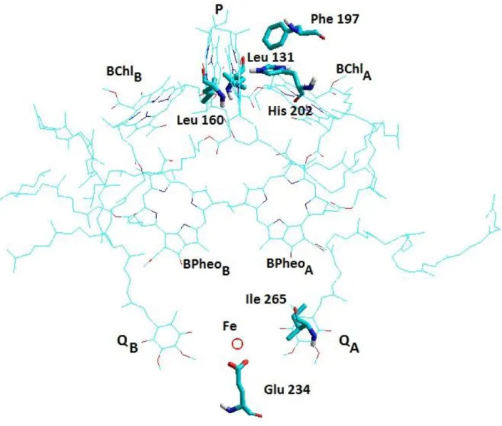
Figure 1. The twofold symmetric arrangement of the cofactors and location of the amino acid residues targeted for mutations in the atomic structure of the RC protein (Brookhaven Data Bank structure 3I4D.pdb). The periplasmic (electron donor) side is separated from the cytoplasmic (electron acceptor) side by a wide hydrophobic (membrane) band in the middle. The mutations directed to the indicated sites cause significant alteration of the energetics of the protein.
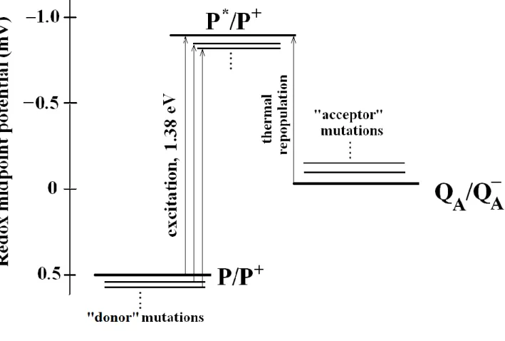
Figure 2. Redox midpoint potentials of the cofactors playing crucial role in generation of DL. Following the absorption of a photon of energy 1.38 eV (corresponding to 865 nm wavelength), the primary
electron donor, P, a dimer of bacteriochlorophylls becomes a strong reducer in its singlet excited state P*
(midpoint redox potential E'm (P*/P+) ~ −900 mV). The excitation energy of P → P* remains unchanged in the donor- and acceptor side mutations used in this study. The excitation of the dimer is followed by electron transfer from P* to the primary quinone yielding the charge separated P+QA-
state. The approximate redox midpoints of P/P+ and QA/QA-
of wild type RC at pH 8 are taken +500 mV and -50 mV, respectively [24]. The precursor of the DL is P* repopulated thermally from the P+QA-
state. Any energetic changes on the donor and acceptor sides due to mutations and/or other changes (e.g. pH) will affect the DL.
Figure 3. Kinetic traces of the DL of donor side mutants with increasing number of H-bonds in the vicinity of the dimer. The DL increases in single (L131LH), double (L131LH-M160LH) and triple (L131LH-M160LH-M197FH) mutants relative to that of wild type RC. The introduction of histidine residue at the donor side reflects addition of one H-bond to the network around the dimer. The addition (L131LH) and subsequent removal (L131LH-M202HL) of histidine residue recover the original level of DL of the wild type RC (inset).
Conditions: 2 μM RC, 100 mM NaCl, 0.03% Triton X-100, 100 μM terbutryn, pH 8 and wavelength of observed fluorescence 915 ± 10 nm. The mechanical shutter is closed during the excitation (a negligible but observable fraction of the very intense prompt fluorescence is creeping through the blades) and will open ~ 10 ms after the flash (see the large increase of the observed light intensity). The DL is the decaying light intensity detected in the dark. The rate constant of the decay is comparable to that of the charge recombination indicating the leakage character of the DL.
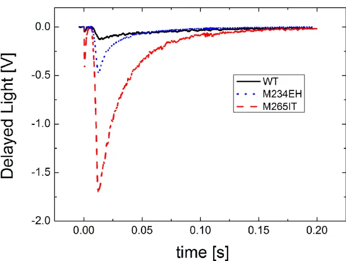
Figure 4. Kinetic traces of the DL of acceptor side mutants in the QA binding pocket (M265IT) and in the iron ligand (M234EH). The conditions are the same as in Fig. 3.
Figure 5. The free energy dependence of the observed rate constants of the decay of DL in different mutants. The fit to Marcus parabola (thin line) results kmax = 35 s-1 and λ = 705 meV for the maximum rate and reorganisation energy of the P+QA-
→ PQA process, respectively. Notations: Wild Type, L131LH-M202HL, ▲ L131LH, ► M234EH, ▼ L131LH-M160LH, ◄ M265IT, L131LH-
M160LH-M197FH.
Figure 6. The pH-dependence of the free energy drop from P* to P+QA-
in wild type RC and mutants at the dimer and at the QA binding site. ΔGP*A was determined from the intensity of delayed fluorescence, as described in Methods and Materials. Conditions: 2 μM RCs, 0.03% Triton X-100, 5 mM buffer (1 mM of each of Mes, Mops, Tricine, Ches, and Caps), 100 mM NaCl and 100 μM terbutryn.
Figure 7. van’t Hoff plots of the DL in wild type and different mutants. The area of the DL traces normalized to the concentration of the RC (ΔOD is flash-induced absorption change at 430 nm) show significant temperature-dependence: according to the Boltzmann’s relationship: larger intensity of DL can be obtained at elevated temperatures. The slope corresponds to the enthalpy change of the P+QA- → P* transition. The measured date of the different mutants are arranged (shifted) according to decreasing (increasing) slopes.
Mutants ΔG0 [meV]
ΔH0 [meV]
T·ΔS0 [meV]
@ T=295°K
Wild Type -897 -910 -13
Donor side
L131LH -833 -820 13
L131LH-M160LH -797 -735 62
L131LH-M160LH-M197LH -713 -710 3
Acceptor side M265IT -787 -360 427
M2234EH -820 -304 516
Table 1. Summary of the thermodynamic parameters (changes of the free energy (ΔG0), enthalpy (ΔH0) and entropy (T·ΔS0)) corresponding to the P* → P+QA-
charge separation determined for wild type and mutant RCs.