R E S E A R C H A R T I C L E Open Access
A pattern-triggered immunity-related phenolic, acetosyringone, boosts rapid inhibition of a diverse set of plant
pathogenic bacteria
Ágnes Szatmári1,2*, Ágnes M. Móricz1, Ildikó Schwarczinger1, Judit Kolozsváriné Nagy1, Ágnes Alberti3, Miklós Pogány1and Zoltán Bozsó1*
Abstract
Background:Acetosyringone (3,5-dimethoxy-4-hydroxyacetophenone, AS) is a syringyl-type phenolic compound rarely found in plants in free form. It has been shown earlier to inhibit the growth ofPseudomonasbacteria in the presence of hydrogen peroxide and peroxidase (AS mix).
Results:We detected elevated levels of free AS inNicotiana tabacumandN. benthamianaplants after inducing pattern-triggered immunity (PTI) by injecting bacterial elicitor flg22, or pathogenicity-mutantPseudomonas syringae pv.syringae61hrcC- bacteria; but not after inoculations with compatible or incompatible pathogens at the time of PTI onset. In this study, we demonstrate that the antibacterial effect of the AS mix is general, as growth of several Gram-negative and -positive phytopathogenic bacteria was characteristically inhibited. The inhibition of bacterial metabolism by the AS mix was rapid, shown by the immediate drop of luminescence intensity ofP. syringaepv.
tomatoDC3000 lx strain after addition of AS mix. The mechanism of the bacteriostatic effect was investigated using fluorescent reporter dye assays. SYTOX Green experiments supported others’ previous findings that the AS mix does not result in membrane permeabilization. Moreover, we observed that the mode of action could be depolarization of the bacterial cell membrane, as shown by assays carried out with the voltage sensitive dye DIBAC4(3).
Conclusions:Level of free acetosyringone is elevated during plant PTI responses in tobacco leaves (N. tabacum andN. benthamiana). When combined with hydrogen peroxide and peroxidase (AS mix), components of the mix act synergistically to inhibit bacterial metabolism and proliferation rapidly in a wide range of plant pathogens. This effect is related to depolarization rather than to permeabilization of the bacterial cell membrane. Similar AS mixture to the in vivo model might form locally at sites of invading bacterial attachment to the plant cells and the
presence of acetosyringone might have an important role in the inhibition of bacterial proliferation during PTI.
Keywords:Pattern-triggered immunity,Pseudomonas syringae, Acetosyringone, Elicitor, Oxidative burst, Antibacterial
© The Author(s). 2021Open AccessThis article is licensed under a Creative Commons Attribution 4.0 International License, which permits use, sharing, adaptation, distribution and reproduction in any medium or format, as long as you give appropriate credit to the original author(s) and the source, provide a link to the Creative Commons licence, and indicate if changes were made. The images or other third party material in this article are included in the article's Creative Commons licence, unless indicated otherwise in a credit line to the material. If material is not included in the article's Creative Commons licence and your intended use is not permitted by statutory regulation or exceeds the permitted use, you will need to obtain permission directly from the copyright holder. To view a copy of this licence, visithttp://creativecommons.org/licenses/by/4.0/.
The Creative Commons Public Domain Dedication waiver (http://creativecommons.org/publicdomain/zero/1.0/) applies to the data made available in this article, unless otherwise stated in a credit line to the data.
* Correspondence:szatmari.agnes@ttk.hu;bozso.zoltan@atk.hu
1Plant Protection Institute, ELKH Centre for Agricultural Research, Herman Ottó St. 15, Budapest 1022, Hungary
Full list of author information is available at the end of the article
Background
Similarly to the innate immune response of mammals, plants also have an inducible resistance system called pattern-triggered immunity (PTI). This form of plant resistance recruits pattern recognition receptors (PRRs) that have a structure like Toll-like receptors (TLRs) of mammals. PTI, similarly to innate immune responses, can be triggered by microbe associated molecular patterns (MAMPs). These include a conserved peptide from bacterial flagellin (flg22), elongation factor Tu (EF-Tu), peptidoglycan (PGN), lipopolysaccharides (LPSs), activator of XA21- mediated immunity in rice (Ax21) (reviewed in: [1,2]). PTI can also be induced in planta by non-pathogens:
saprophytes, commensals (e.g.Pseudomonas fluorescens) or pathogenicity mutants (e.g.Pseudomonas syringaepv.syrin- gae61hrcC-) [3–5]. PTI is effective against a broad range of microbes and is able to suppress opportunistic pathogens.
Its early signalling events include rapid calcium influx, activation of mitogen activated protein kinase (MAPK) phosphorylation cascades, which then lead to production of reactive oxygen species (ROS), callose deposition, and defence gene expression, presumably mediating inhibition of microbial growth [6]. Disease-inducing, virulentP. syringae pv. tabaci in tobacco plant is able to block the MAMP- induced expression level alterations of specific genes coding for signal transduction and transcription regulation-related proteins [7].
During our previous work, we identified over 400 expressed sequence tags (ESTs) representing 176 indi- vidual genes that are activated during PTI in Nicotiana tabacum. Phenylpropanoid synthesis-related genes were highly represented and covered a major part of the cur- rently known network of the phenylpropanoid pathway [8]. Our hypothesis was that elements of the phenylpro- panoid pathway might play an important role in the inhibitory effects of PTI on bacteria, either by leading to cell wall fortification or by some phenolic compounds having direct antimicrobial effect, or else, by acting as a signalling agent, affecting virulence factors of bacteria.
Lignification and induction of lignin monomers’ and related compounds’ synthesis has long been associated with plant defences [9,10], and lately with flg22-induced immunity inArabidopsis[11]. Recently, lignin deposition has been shown to spatially restrict bacterial pathogens and to limit their motility also inArabidopsis[12].
Baker and colleagues [13] have detected accumulation of acetosyringone (AS) and some other phenolics in N.
tabacumcell cultures, when treated with suspensions of P. syringae pv. syringae strain B7 (non-HR inducing).
Later they have also shown that P. fluorescens, a non- pathogenic species causes elevation of AS in the apoplast fluid of N. tabacum [14]. In this study we describe detection and identification of AS in N. benthamiana and N. tabacum leaves using high-performance liquid
chromatography-diode array detection-mass spectrom- etry (HPLC-DAD-MS), moreover, we show the MAMP (i.e. flg22) associated accumulation of this phenolic com- pound, and suggest that it is a marker of PTI in tobacco plants.
We sought for compound(s) that are accumulated during PTI in both Nicotiana model species, because while N. tabacum is a classical model plant to study bacterial interactions in the Solanaceae family, parallel data fromN. benthamianamight later prove to be useful as well, as transient silencing and overexpression studies are easier to carry out in the latter species. We investi- gated if appearance of AS is specific for PTI in N.
benthamiana and whether it correlates temporally with the onset of PTI, using HPLC-DAD-MS. Based on reports of AS as an enhancer of laccase activity [15] and having antibacterial activity when combined with horse- radish peroxidase and hydrogen peroxide [16], we tested a wide range of plant pathogenic bacteria to detect antibacterial activity. We also used luminescentP. syringae pv.tomatoDC3000 bacteria to monitor effects on bacterial metabolic activity. Finally, to gain information about the mode of action of AS we used fluorescent dyes to measure membrane permeabilization and membrane depolarization of affected bacteria.
Results
Identification of acetosyringone, a metabolite correlated with PTI onset
Our first aim was to detect and identify phenolic metabol- ite that are specifically more (or less) abundant in N.
benthamiana leaves in association with the onset of PTI.
N. benthamianaleaves were infiltrated with Pseudomonas syringaepv. syringae hrcC- (P. s. syringae hrcC-) suspension or flg22 peptide to induce PTI, and water as a control. Both PTI inducers were used at concentrations that caused reli- able HR-inhibition at 6 h post inoculation (flg22 at 50μM, P. s. syringae hrcC- at 109CFU/ml), based on works of Nguyen et al. and Chakravarthy et al. [17,18]. Inhibition of HR, sometimes referred to as an“HR test”is an indicator of the onset of effective PTI and correlates well with the onset of local resistance that is able to halt bacterial prolif- eration after a challenge inoculation [3,8,17]. Leaf samples were taken 6 h later and extracts were analyzed by HPLC- DAD-MS to find PTI-related phenolic compounds. Extrac- tion fromN. benthamianaleaves was performed with 90%
aqueous methanol according to recommendations in literature [19].
We detected several putative phenolic compounds (with characteristic UV spectra) in PTI-induced N.
benthamiana leaves (data not shown). Works of others [13,20] carried out onN. tabacumhelped identification of some of these compounds. In this manuscript, we focus on one specific compound (m/z197 [M + H]+) that
Szatmáriet al. BMC Plant Biology (2021) 21:153 Page 2 of 20
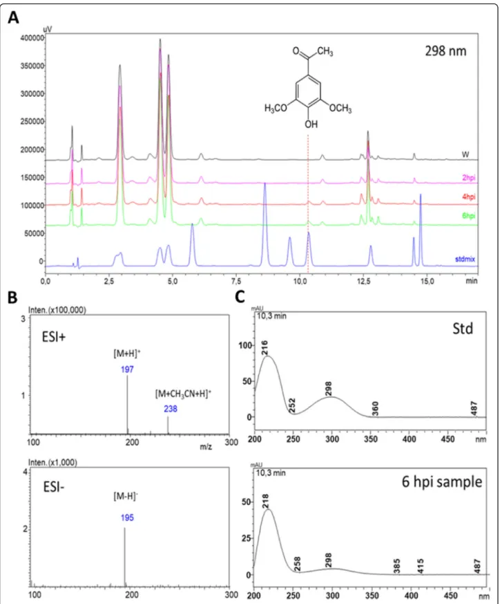
occurred selectively in PTI-induced (6 h post inoculation, hpi)N. benthamianaleaves (Fig.1a), and was identified as acetosyringone (3,5-dimethoxy-4-hydroxyacetophenon), by comparison of its MS (Fig.1b) and UV (Fig.1c) spectra and retention time (Fig. 1a) to those of an analytical standard.
Spiking the standard compound into the plant extracts resulted in clear elevation of the putative AS peak.
We found AS especially interesting, because besidesP.
s. syringae hrcC- treatment, it was also strongly induced in flg22 peptide-treatedN. benthamiana leaves, but was not detectable in water treated controls at 6 hpi (Fig.2a).
The abundance of AS correlated closely with PTI in the leaves of bothN. benthamiana(Fig.2a), and tobacco (N.
tabacum, Additional file 1). AS itself was first isolated fromN. tabacumleaf exudates and root culture medium, where it was established to be exudate specific, not passively leaking out from damaged plant cells [21]. Baker et al. have reported accumulation of AS in tobacco cell culture media [13] and in the apoplastic fluid [14] after treatment with different bacteria.
Changes in phenolic metabolite levels
To see if elevated level of AS occurs in different plant- bacterium interactions, we analyzed 6 hpi samples from N. benthamiana leaves injected with suspensions of a compatible bacterium: Pseudomonas syringaepv. tabaci, two incompatible bacteria:P. syringaepv. tomatoDC3000 andP. syringaepv. syringae61, PTI inducing bacterium:P.
syringaepv. syringae hrcC- (109CFU/ml each), and a PTI- inducing MAMP: flg22 peptide. Water-treated controls were also analyzed. Interestingly, AS displayed a unique pattern: it was only detected in flg22 andP. syringaepv.
syringae hrcC- treated samples – the two PTI- induced samples; but it was absent (below detection limit) in the rest of the samples at the used concentrations (Fig. 2b).
Time course experiments supported the gradual accumu- lation of AS during the course of the build-up of effective PTI both in N. benthamiana (Fig. 2a) and N. tabacum (Additional file1). It should be noted that there appears a co-timing with inhibition of HR and of proliferation of compatible bacteria upon challenge inoculation; which usually becomes effective also at 5–6 h after induction of PTI [22].
Antimicrobial effect of acetosyringone in combination with hydrogen peroxide and peroxidase against plant pathogenic bacteria
Several sources reported that AS or related acetophenones have antifungal or antibacterial effects. Lorimer and Perry [23] reported isolation of two major antifungal active compo- nents fromPlagiochila fasciculata (New Zealand liverwort).
These were identified as 2-hydroxy-4,6-dimethoxyacetophe- none and 2-hydroxy-3,4,6-trimethoxyacetophenone. The relatively high activity of the crude extract was due to the
high level of these compounds in the plant (6–7 mg/g dried plant for both). In vitro oxidation of AS with hydrogen peroxide by a peroxidase can create a prolonged oxidative environment similar to the oxidative burst in tobacco suspension cells inoculated with incompatible bacteria according to Mock et al. [16].
Based on the latter, we tested if in vitro oxidation of AS results in antimicrobial effects against bacterial strains corresponding to different plant (tobacco)-bacter- ium interactions (Fig. 3a-b). P. s. pv. tomato DC3000 and P. s. pv. syringae 61, and the PTI-inducing bacter- ium P. s.pv. syringae hrcC- and other plant pathogenic bacteria were tested, including wild type tumorigenic Agrobacterium tumefaciens strains (from cherry, sour cherry and dahlia) [24] and a disarmed laboratory strain (A. tumefaciens C58C1) [25]. Virulence genes of Agrobacteriaare activated by AS, and they are known to be poor inducers of PTI marker genes (Szatmári et al.
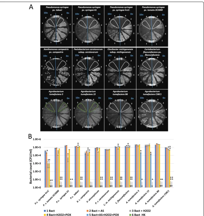
2006). We combined 50μM AS, 50μM H2O2and 0.72 U/ml peroxidase in phosphate buffer, containing 105 CFU/ml suspensions of different bacteria. Serial dilu- tions were plated after 3 h of co-incubation. No growth of the Pseudomonas, Xanthomonas, Pectobacterium, ClavibacterorCurtobacteriumstrains was detected after 3 h of incubation in the complete reaction mixture. On the contrary, Agrobacterium growth was retained in the case of two of the examined strains. When omitting dif- ferent components of the mixture, some Pseudomonas strains were still inhibited to various extent. For example CFU counts of P. s.pv. syringae hrcC- were lowered by one order of magnitude when treated with H2O2 and peroxidase. However, when the mixture was completed with AS, no growth was detected at all. AS alone was not effective against any of the strains, so the combin- ation of acetosyringone with peroxidase activity is essen- tial. Activation of tobacco peroxidase activity in planta at the time interval of AS production during PTI has been shown earlier [3].
When identical concentrations of AS, H2O2and peroxid- ase were combined with higher concentrations of bacteria, the mixture was not effective in CFU reduction. At 5 × 10⁷ CFU/ml Pseudomonas syringaepv. tabaciand Agrobacter- ium tumefaciensC58C1 were apparently unaffected, and at 5 × 10⁶CFU/ml they only showed a 5–6-fold reduction in CFU counts (Additional file2). There seems to be a strong factor depending on bacterial concentration that seems to counter the effect of oxidized AS.
We were curious about the changes occurring in the AS mix after combining the ingredients. We found that the amount of AS is diminishing over time only in the full AS mix, but not in the partial mixes containing AS+
POX or AS+H2O2(Additional file3A). New compounds appeared only in the latter throughout the 180 min period (Additional file3B).
Fig. 1Identification of PTI-related phenolic compound acetosyringone by HPLC-DAD-MS using analytical standard inNicotiana benthamiana.aHPLC-DAD analysis of phenolic compounds from PTI-induced and water-treated controlN. benthamianaleaves detected at 298 nm. PTI was induced byP. syringaepv.
syringae hrcC- bacteria, water was injected as control. Samples were taken at 2, 4, 6 hpi.bMS spectra of AS had the expectedm/zvalues in both positive and negative modes, identical to that of the standard.c) Identity of acetosyringone was supported by the similar UV spectra of the analytical standard and the unknown peak. Abbreviations: AS: acetosyringone, ESI+, ESI-: electrospray ionization positive and negative modes, Std: standard, W: water
Szatmáriet al. BMC Plant Biology (2021) 21:153 Page 4 of 20
The LC–mass spectrometry analysis of the full AS mix reaction yielded m/z peaks corresponding to AS itself (m/
z = 197, [M + H]+; (m/z = 195, [M-H]-); putative compounds syringaldehyde (m/z = 183, [M + H]+; (m/z = 181, [M-H]-) and 2,6-DMQB (2,6-dimethoxy-p-benzoquinone, m/z = 169, [M + H]+) and several other peaks corresponding to
unknown compounds (Additional file3B). Putative identifi- cation of the newly formed compounds was based on data from literature [26,27].
The catalytic cycle of oxidases like peroxidases and laccases in general involves an intermediate phenoxy radical after phenolic substrates release a proton and an electron when
Fig. 2Relative accumulation of acetosyringone inN. benthamianaleaves.aResponse to treatments withP. syringaepv.syringae hrcC- bacteria at 2, 6 and 24 hpi.bRelative accumulation of AS inN. benthamianaleaves in response to treatments with different bacteria at 6 hpi. Error bars indicate standard deviations. Asterisks indicate significant difference from corresponding water-treated controls according to student’s T-test (*p< 0.1; **p< 0.05). Red downward arrows indicate zero values. W: water-treated control; P. s.:Pseudomonas syringaepathovars; flg: flg22 peptide elicitor
they are oxidized in the enzymatic step to produce radicals.
The phenoxy radicals can be involved in the radical recombination, cross-coupling and self-coupling resulting in
disproportionation, dimerization, polymerization, or oxida- tion of other substrates [26,28]. The resulting compounds are expected to be the products of such reactions.
Fig. 3Antimicrobial effect of oxidized form of acetosyringone on different plant pathogenic bacteria. Bacteria (105CFU/ml) were added to reaction mixtures containing 50μM AS, 50μM H2O2, and 0.72 U/ml horseradish peroxidase, and various control mixtures from which one or two components were omitted. Serial dilutions were plated following 3 h of co-incubation for CFU determination.aTypical representative examples of serial dilutions plated on Kings’s B agar plates. Right half of each plate: no dilution (0×), left side of each plate: 10× dilution (indicated with blue lettering). Numbering of treatment combinations applied to the bacterial suspensions: 1. non-treated control; 2. AS; 3. H2O2; 4. H2O2+ POX;
5. AS + H2O2+ POX; 6. HK.bDiagram showing quantification of the results. Error bars indicate standard deviations. Asterisks indicate significant difference from corresponding water-treated controls according to student’s T-test (*p< 0.1; **p< 0.05). Downward arrows indicate zero values.
Abbreviations: Bact: bacterium, AS: acetosyringone, HK: heat-killed, POX: horseradish peroxidase
Szatmáriet al. BMC Plant Biology (2021) 21:153 Page 6 of 20
We used a luminescentP. syringaepv.tomatoDC3000 strain that is tagged chromosomally with theluxCDABE gene cluster [29] to monitor the changes in bacterial cell viability caused by oxidized AS. Viable P. s. pv. tomato DC3000 lux bacteria actively emit a constant level of lu- minescence. We planned to monitor the time dependent decrease of luminescence, when combined with oxidized AS. We found that the luminescence level dropped vir- tually immediately to the background level of heat killed bacteria already at the first measurement (5 min) after administration of the AS + H2O2+ POX cocktail (Fig.4).
This response was significantly faster than the effect caused by H2O2alone or H2O2combined with peroxid- ase. AS alone did not reduce bioluminescence. In fact, it seemed to significantly increase bioluminescence com- pared to untreated bacteria, a phenomenon that needs further investigations to find an explanation. Altogether, these results implied that metabolic activity of the bacteria was halted immediately after addition of the AS + H2O2+ POX cocktail. Therefore, not only is the mix- ture more effective in inhibiting bacterial proliferation
than H2O2+ peroxidase alone, but acts much quicker on the bacteria, at least as far as metabolic activity is concerned.
Testing viability and membrane depolarization of acetosyringone mix treated bacterial cells by enhanced fluorescence of selected dyes
We tested several fluorescent dyes to be able to detect viability of bacteria upon treatment with AS mixture (Fig. 5a-c). The SYTOX Green and TO-PRO-3 iodide assays are generally used to assess bacterial cell mem- brane integrity, while DIBAC4(3) is known to be able to report slower speed membrane depolarization. With DIBAC4(3) and TO-PRO-3 iodide we found that the H2O2 + POX and AS + H2O2 + POX mixtures caused bleaching of the added dye (Fig. 5b, c), testing by addition of heat killed bacteria (HK BACT), therefore no valid detection of staining was possible. Destaining en- hancing effect of acetosyringone (and other phenolic compounds) is known to facilitate destaining of even recalcitrant dyes by aggressive oxidation. AS seems to be
Fig. 4Bioluminescence changes of indicator bacteria after treatment with oxidized AS. 105CFU/ml ofP. syringaepv.tomatoDC3000 lux, a luminescentP. syringaestrain was added to reaction mixtures containing 50μM AS, 50μM H2O2, and 0.72 U/ml POX, and various control mixtures from which one or two components were omitted. Bioluminescence was measured at indicated time points. Different letters above the bars denote different levels of luminescence assigned by Tukey’s Test (P< 0.05). Error bars indicate standard deviations. AS: acetosyringone, HK: heat- killed,P.s. tomatoDC3000 lux:Pseudomonas syringaepv.tomatoDC3000 luminescent strain, POX: horseradish peroxidase
Fig. 5(See legend on next page.)
Szatmáriet al. BMC Plant Biology (2021) 21:153 Page 8 of 20
a superior mediator of bleaching as compared to other tested molecules [15]. Because the redox potential in the AS + H2O2+ POX mixture returns to nearly basal level after 2–3 h [16], we added the fluorescent dyes 3 h after adding the reaction mixtures to the wells. This allowed detection of SYTOX Green and DIBAC4(3) fluorescence in the HK BACT + H2O2+ POX and HK BACT + AS + H2O2 + POX mixtures as well (Fig. 5a, c), while in the case of TO-PRO-3 Iodide the bleaching still occurred and still no fluorescence was detectable. This phenomenon might mean that at 3 h there is still significant remaining oxidizing capacity in the H2O2+ POX and AS + H2O2+ POX mixtures that is able to bleach TO-PRO-3 (Fig. 5b), but not SYTOX Green and DIBAC4(3) (Fig. 5a, c). TO- PRO even seemed to go through significant fading in the control wells during the 3-h incubation. Therefore, we con- cluded that DIBAC4(3) is a suitable dye to detect bacterial cell membrane depolarization after treatment of cells by AS + H2O2+ POX mixture, given that the dye is added only 3 h after the addition of the original reaction mixture.
DIBAC4(3) is a voltage sensitive fluorescent dye, which can be used to monitor the dissipation of the membrane potential, not just increasing membrane permeability in general as in the case of propidium iodide (PI) or SYTOX Green dyes [30]. The latter dyes can only penetrate bacterial cells and dye the nucleoid when membrane integrity is compromised. However, the death of a cell can also result from dissipation of the trans- membrane potential, which can be detected by voltage sensitive fluorescent dyes such as DIBAC4(3). This dye reports longer term depolarization but does not measure instant changes [31].
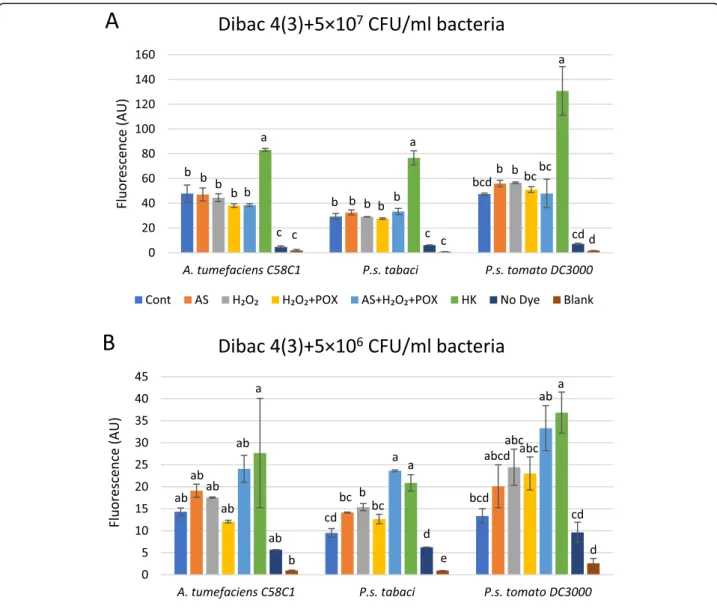
Depolarization of the bacterial membrane contributes to the inhibitory effect of oxidized acetosyringone on bacteria We compared elevations of DIBAC4(3) fluorescence values in control and AS mix-treated bacterial suspen- sions of three species at two cell concentrations. Bacteria were incubated in the mixtures for 3 h, then DIBAC4(3) was added, finally, fluorescence was recorded after 1 h of co-incubation (Fig.6a-b). The AS mixture affected fluor- escence differently at the two bacterial concentrations used. At 5 × 10⁷CFU/ml there was none or just a small elevation in fluorescence as compared to the untreated
control in any of the bacteria (Fig. 6a). In contrast, at 5 × 10⁶CFU/ml the fluorescence of the AS mix-treated bacteria was closer to the values of the heat killed con- trols (Fig.6b). This correlated with the previous finding, that higher concentrations of bacteria were less affected by the same AS mix as far as the drop in colony forming units is concerned (section 2.3). Reading of fluorescence below 5 × 10⁶CFU/ml bacterial density was not sensitive enough, therefore, we used microscopy to detect fluores- cent cells in more diluted suspensions.
Corresponding literature suggests that DIBAC4(3) is correlated to bacterial cell death in many, but by far not all cases, as membrane depolarisation does not necessar- ily mean full permeabilization of the membrane [30].
Therefore, to address this issue in the case of the AS mix, we found it necessary to carry out parallel experi- ments with a dye that measures cell viability by detecting membrane permeability to larger molecules, such as SYTOX Green (Fig.7).
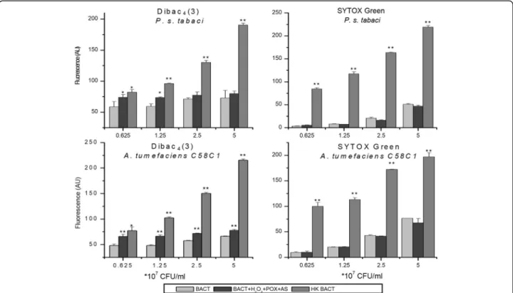
To test, if the level of cell membrane depolarisation correlates to or diverges from cell membrane permeabil- ity when AS mix is added to plant pathogenic bacteria, the same AS mix was applied to bacterial suspensions with increasing densities. A Pseudomonas and an Agro- bacterium strain were randomly selected as representa- tive strains. Heat killed bacteria were used as positive controls both for membrane depolarisation (DIBAC4(3)) and membrane permeability (SYTOX Green). At lower bacterial densities (0.625 × 10⁷ and 1.25 × 10⁷CFU/ml), significant difference was detected between DIBAC4(3) fluorescence of non-treated and AS mix-treated bacteria.
This difference diminished towards higher bacterial densities, in the case of both bacterial species, and became insignificant in the case ofPseudomonas syringaepv.tabaci.
These data indicated that membrane depolarisation is counteracted by bacteria, using an unknown mechanism. In contrast to DIBAC4(3), SYTOX Green fluorescence did not increase upon treatment with the AS mix. This was the case with both bacteria at all densities. Therefore we con- cluded that the AS mix might induce depolarization of the bacterial membrane, when bacterial density is relatively low, compared to the concentration of the components of the acetosyringone mix. On the other hand, no permeabilization of the bacterial membrane occurs.
(See figure on previous page.)
Fig. 5Evaluation of applicability of different fluorescent dyes for the detection of membrane permeability and membrane depolarisation in the AS mix system. Different fluorescent dyes were tested for usability to measure membrane permeability: SYTOX Green (a), TO-PRO (b); and membrane depolarisation: DIBAC4(3) (c). Fluorescence of indicator dyes in reaction mixtures containing 50μM AS, 50μM H2O2, and 0.72 U/ml POX, and various control mixtures from which one or two components were omitted was measured. Fluorescent dyes were added either immediately (0 h) or 3 h after preparation of the mixtures to see if bleaching occurs. HKP. syringaepv.tabacisuspension was also added 3 h after mixture preparation to 5 × 10⁶CFU/ml final density. Fluorescence was measured 4 h after preparation of the mixtures. Error bars indicate standard deviations. Asterisks indicate significant difference from corresponding controls according to student’s T-test (*p< 0.1; **p< 0.05). Abbreviations:
CONT: untreated bacteria, AS: acetosyringone, HK: heat-killed, POX: horseradish peroxidase, AU: arbitrary unit
Microscopic evaluation of cell viability
Fluorescent microscopy was used to augment fluores- cence measurements to visualize the depolarization of the bacterial cell membrane at low CFU and to compare this to the degree of membrane permeabilization, if detectable. The latter has been investigated by Postnikova et al. [32] on P. syringaepv. syringae using LIVE/DEAD® BacLight fluorescent stain, which utilizes propidium iodide (PI) to detect membrane permeabil- ity. The authors there found no permeabilization of the bacterial membrane by oxidized AS.
Here we used the same dyes as in section 2.5., as both dyes have been shown before to be suitable for micros- copy of bacteria. The voltage sensitive dye Dibac4(3) is known not to inhibit bacterial growth and proliferation, therefore it has even been used for time lapse micros- copy [30]. SYTOX Green has widely been applied for viability staining of bacteria. We used each dye with DAPI as a counterstain to envisage all bacteria in the microscope’s field of view. As shown earlier, the inhibi- tory effect of the AS mix is inversely proportional to bacterial counts, therefore we worked here with a low
Fig. 6Dependence of bacterial membrane depolarisation on bacterial density after treatment with oxidized AS. Suspensions of 5 × 10⁷(a) and (b) 5 × 10⁶CFU/ml ofP. syringaepv.tabaci, P. syringaepv.tomatoDC3000 orA. tumefaciensbacteria were added to reaction mixtures containing 50μM AS, 50μM H2O2, and 0.72 U/ml POX, and various control mixtures from which one or two components were omitted. Bacterial membrane polarity was tested with the voltage sensitive DIBAC4(3) stain added 3 h after treatments of bacteria. Error bars indicate standard deviations.
Different letters above the bars denote different levels of DIBAC4(3) fluorescence assigned by Tukey’s Test (p< 0.05). Abbreviations: Cont: non- treated control bacteria, AS: acetosyringone, HK: heat killed, POX: horseradish peroxidase, Blank: buffer only
Szatmáriet al. BMC Plant Biology (2021) 21:153 Page 10 of 20
(105CFU/ml) density of P. syringae pv. tabaci bacteria, similarly to the CFU reduction experiments (Additional file 2). Cells were incubated in the AS mix or buffer as control for 3 h, and dyes were added only then to pre- vent bleaching by oxidation (Fig.8). DIBAC4(3) fluores- cence was absent in untreated control samples, however it was remarkably strong in the AS mix-treated samples and in the heat-killed samples (Fig.8a). This means that bacterial membranes are depolarized when the cells are incubated in the AS mix. In contrast, SYTOX Green, the dye specific for permeabilized membranes left the AS mix-incubated bacteria unstained, meaning that the membranes did not become permeable to larger mole- cules (Fig. 8b). The results of microscopy experiments supported those obtained by fluorescence measurements.
Taken together, AS in combination with hydrogen perox- ide and horseradish peroxidase resulted in membrane depolarization, but negligible membrane permeability, based on staining with specific fluorescent dyes.
Investigation of thein plantaeffect of AS mix on compatibleP. syringaepv.tabaci
We carried out in vivo experiments to see if external administration of the AS mix enhances the ability of
tobacco plants to withstand the attack of compatible P.
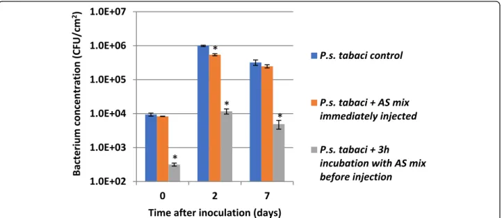
syringae pv. tabaci. Bacteria were suspended in 0.01 M potassium phosphate buffer (pH 6) to 106CFU/ml. AS mix components (AS, hydrogen peroxide, horseradish peroxidase) were added to final concentrations used in vitro (in section 2.4.). Then the prepared bacterial mixtures were injected into interveinal areas of tobacco leaves either immediately or after 3 h of incubation to see if the reaction mixture is able to inhibit bacterial cells in the intracellular space. Bacteria were re-isolated from the leaf tissue by grinding the leaves in a mortar and colony forming units were counted by dilution plating (Fig.9).
Our results indicated that while the applied AS mix effectively lowered colony forming unit numbers of P.
syringae pv. tabaci bacteria by cca. 2 orders of magnitude during the 3 h of incubation, no reduction in colony number was observed when the mixture was im- mediately injected into the leaf. P. syringae pv. tabaci bacteria proliferated at similar pace irrespective of the type of pre-treatment. In each case, bacterial counts increased about 1.5–2 orders of magnitude in the first two days, then declined slightly by the seventh day. The in vitro pre-treated bacteria could not reach a relatively
Fig. 7Membrane depolarization and permeability testing of bacteria after treatment with oxidized AS as a function of bacterial concentration. Dilution series of bacteria from 5 × 10⁷to 0.625 × 10⁷CFU/ml ofP. syringaepv.tabaciandA. tumefaciensC58C1 bacteria was added to reaction mixtures containing 50μM AS, 50μM H2O2, and 0.72 U/ml POX. Untreated bacteria were used as negative, and heat killed bacteria as positive controls. Bacterial membrane polarity was tested with the voltage sensitive DIBAC4(3) stain, and membrane permeability with SYTOX Green stain, both added 3 h after treatment of bacteria. Error bars indicate standard deviations. Asterisks indicate significant difference from corresponding controls according to student’s T-test (*p< 0.1; **p< 0.05). Abbreviations: BACT: bacterium, AS: acetosyringone, HK: heat-killed, POX: horseradish peroxidase, AU: arbitrary unit
Fig. 8(See legend on next page.)
Szatmáriet al. BMC Plant Biology (2021) 21:153 Page 12 of 20
higher apparent rate of proliferation as compared to the non-treated and the immediately injected bacteria. The ratio of colony forming units re-isolated from the plants inoculated with the non-treated control over that of the 3 h-preincubated bacteria increased from 30 to 65-fold by the end of the 7th day. This tells us, that if part of the bacteria were in VBNC state, they were apparently not able to return to normal state within the plant tissue, under the applied circumstances.
Discussion
Our results indicate that AS, a tobacco metabolite specifically appearing in N. tabacum and N. benthami- anaafter PTI induction either by HR-negative mutantP.
syringae pv. syringae hrcC- bacteria or the flg22 elicitor peptide at the time point (5–6 hpi) when PTI starts to be effective against bacterial multiplication, causes a rapid inactivation of certain plant pathogenic bacteria in vitro, when in an oxidative environment.
During our present study, AS was only detected in the PTI-induced (flg22 and P. syringae hrcC-) samples at 6 hpi. This was especially interesting, as it was found to rapidly inhibit several phytopathogenic Pseudomonas
bacterium species, when combined with hydrogen per- oxide and horseradish peroxidase in vitro. These results are even more interesting as over-expression of C4H (cinnamate 4-hydroxylase) has been shown to increase the accumulation of AS in elicited tobacco cell-suspension cultures [33]. Moreover, an OMT (O-methyltransferase) capable to synthesise AS was found in methyl jasmonate- treated tobacco cell-suspension cultures [34]. These changes in acetosyringone levels would be worth to be an- alyzed in the future in C4H and OMT overexpressing plants.
We demonstrated that the antibacterial effect of the AS mix is inversely proportional to bacterial density.
Moreover, someAgrobacterium isolates are more resist- ant to the AS mix than the remaining plant pathogenic bacteria tested. A straightforward explanation to these phenomena could be the antioxidant capacities pos- sessed by bacteria, e.g. the constitutive and inducible catalase enzyme activities. Xu and Pan [35] have shown that anAgrobacteriumstrain deficient in catalase activity was highly attenuated in the ability to cause tumours on plants compared with the wild type. Thus, catalase was considered a virulence factor of Agrobacterium, and our
(See figure on previous page.)
Fig. 8Fluorescent microscopic images ofP. syringaepv.tabacibacteria treated with an AS mix, and stained to evaluate changes in membrane permeability and polarity.P. syringaepv.tabaci(105CFU/ml) was treated with a mixture of AS + H2O2+ POX. After 3 h of shaking, (a) DIBAC4(3), an indicator dye of membrane depolarization or (b) SYTOX Green, an indicator of membrane permeability, and DAPI as counterstain were added and co-incubated for 1 h. After concentration of the bacteria, microscopic slides were prepared and photographed within 1–2 h. Bar = 100μm.
Abbreviations: Control: untreated bacteria AS: acetosyringone, HK: heat-killed, POX: horseradish peroxidase
Fig. 9Proliferation of compatibleP. syringaepv.tabaci(106CFU/ml inoculum) bacteria in tobacco (Nicotiana tabacum). Bacteria were suspended in 0.01 M potassium phosphate buffer pH 6. Mixture of AS+H2O2+ POX was added to the bacteria.N. tabacumleaves were infiltrated either immediately or after 3 h of incubation. Samples for bacterial counts were taken at 0, 2, 7 dpi. Error bars indicate standard deviations. Significant difference from untreated bacterial control according to student’s T-test is marked by a * (p< 0.1). Abbreviations: AS mix: acetosyringone combined with hydrogen peroxide and horseradish peroxidase, HK: heat killed, POX: horseradish peroxidase
results might provide a possible confirmation for an antioxidant enzyme being a virulence factor, however further investigations are needed. Agrobacteria are known to have a peroxide and phosphate limitation-inducible catalase gene (KatA), which was also shown to be regulated by a negative feedback loop [36, 37]. Complex regulatory mechanisms may eventually lead to higher resistance of Agrobacteriato oxidative stress. Pseudomonasspecies and other plant pathogens are also known to have functioning catalase and OxyR (a hydrogen peroxide-sensing transcrip- tional activator) [38], moreover, Pseudomonas syringae catalases were shown to be necessary for plant patho- genesis [39].
We investigated two possible antibacterial mechanisms of the AS mix using fluorescent dyes as reporters of membrane permeabilization and membrane depolarization.
We found that AS in combination with hydrogen peroxide and horseradish peroxidase resulted in membrane depolarization, but negligible membrane permeability. Our results were in concordance with those of Baker and colleagues [14] who have shown in flow cytometry experi- ments that PI was not able to penetratePseudomonas syrin- gae pv.syringaebacterial cells after treatment with the AS mix. They also have shown that these bacterial cells reached a viable but non-culturable (VBNC) state, supported by sus- tained cellular respiration. Our present data imply that this perceived VBNC state is reached following significant depolarization of the bacterial cell membrane. Mariano et al. [40] made similar observations, when investigating a toxin (Ssp6) secreted bySerratia marcescens, that was able to cause depolarization of target cells as detected by DIBAC4(3), without increasing membrane permeability for larger dye molecules such as PI, similarly to SYTOX Green in our present study. In another study, Spindler et al. [41]
reported an antimicrobial mechanism where membrane depolarization and permeabilization were not correlated.
The peptide antibiotic Bac8c caused almost immediate but reversible membrane depolarization of the Gram-negative bacteriumEscherichia coliat the IC50 concentration while no increase in membrane permeability was detected. At a higher concentration, depolarisation of the bacterial mem- brane still occurred within 5 min, and in that case, mem- brane permeabilization also followed, but only within 30 min. Our results underline that membrane depolarization and membrane permeabilization might be present simul- taneously in the activity mechanism of antimicrobial agents or these two effects might be decoupled as in the present case of oxidized AS.
Non-impaired proliferation of the AS mix treated, and immediately injected compatible bacteriain planta indi- cates that there might be one or more factors in the plant intercellular space that either scavenges hydrogen peroxide fast, and/or metabolizes or binds free AS. The provided 50μM concentration of H2O2 in the AS mix
might prove insufficient in vivo. In resistant interactions, plants might produce significantly higher amounts at the location of bacterial invasion. ROS production has especially been widely investigated during defence reac- tions (Reviewed in [42]). Respiratory burst associated with pathogen attack is thought to have a signalling role, moreover, ROS might also contribute to direct antimicro- bial effects in different pathosystems [43, 44]. Oxidative burst in Arabidopsis produced by apoplastic peroxidases has also been shown as an essential part of resistance [45].
Reactive oxygen species (ROS) producing cell wall peroxi- dases and a plasma membrane-localized NADPH oxidase, which are active during fungal infection, were re- cently identified by Kámán-Tóth and colleagues [46]
in Arabidopsis.
Our group reported induction of peroxidase genes and peroxidase activity during PTI in tobacco leaves earlier [3,8]; as well as localised production of hydrogen perox- ide in the cell wall adjacent to the attachment site of PTI-inducinghrpLmutant ofPseudomonas phaseolicola in N. tabacum using electron microscopy [22]. Co- localized accumulation of H2O2and peroxidases at the site of bacterial attachment to the plant cell wall and around bacteria themselves were also reported in leaves of various plants after infection with different bacteria including hrp mutant pathogens [47, 48]. These micro- scopic phenomena with the simultaneous accumulation of AS suggest that the conditions might be given locally for an antibacterial mix that, as we have shown, can be effective against wide range of bacterial pathogens. The in planta role of AS is however not clear at the moment, therefore further investigations would be necessary.
Based on the above cited literature one possibility is the formation of local points of high antimicrobial activity near the cell wall peroxidases or NADPH oxidases, when AS concentration is also elevated in the intercellular space is most probable.
Conclusion
During this work, we managed to reveal an indicator metabolite of pattern triggered immunity of Nicotiana plants. Modeling the oxidizing milieau of the intercellu- lar space of plants invaded by PTI-inducing bacteria in vitro, we demonstrated the ability of acetosyringone combined with hydrogen peroxide and peroxidase to in- hibit the growth of several different plant pathogenic bacteria. We have also shown that metabolic inhibition of the bacteria is almost immediate, moreover, that the antimicrobial effect is not based on membrane permeabilization, but on membrane depolarization of bacteria.
Based on our results we propose that besides some phenolic acids directly inhibiting bacterial growth, it seems that the interplay of a phenolic compound (e.g.
Szatmáriet al. BMC Plant Biology (2021) 21:153 Page 14 of 20
acetosyringone) and the oxidative burst (generated by hydrogen peroxide and peroxidase) might enhance antibacterial activity. Investigation of the in vivo role of AS, and its interrelation with other components of the complex ROS production system of plants however will require more complex methods in the future.
Our results add a new aspect to the types of of anti- microbial activities found in plants. Direct antimicrobial activity of metabolites derived from plants have been widely investigated (reviewed in [49]). Antimicrobial activity of ROS in plants has long been studied as well [50, 51]. Enhanced antimicrobial activity resulting from the reaction of secondary metabolites and hydrogen peroxide, leading to strong antimicrobial activity is a relatively novel area that is worth extensive investigation.
Plant metabolites that are produced after specific induc- tion, like PTI elicitors in this case, might be a rich source of compounds with indirect antimicrobial activity.
Methods Plant material
Tobacco plants (Nicotiana tabacum cv. Samsun and Nicotiana benthamiana) were grown in the greenhouse in soil (General potting mix from peat, clay and cow manure (Florimo® általános virágföld) pH 6.4+/−0.5, Matécsa Ltd., Kecel, Hungary, completed with 5 V/V%
perlite (Florimo® Kertészeti Perlit, 2-6 mm, Matécsa Ltd., Kecel, Hungary). Two days before and during the experi- ments, the 2–2.5-month-old tobacco plants were kept in a growth chamber with 16/8 h light/dark period at 23 °C.
Hypodermic syringes fitted with a 25 gauge needle were used for the infiltration of the 2–3 middle leaves with bacterial suspensions or solution of flg22 peptide. At the appropriate time points, leaf samples were frozen imme- diately in liquid nitrogen and stored at −70 °C until processing. Water-infiltrated adjacent leaves next to the treated ones (in the case of N. benthamiana) or fully separated interveinal areas on the same leaves (in the case ofN. tabacum) were used as control. Every experi- ment was carried out on at least two plants in parallel as biological replications. The experiments were also repeated at least twice using different plant generations to confirm the detected trends.
Bacterial and chemical treatments
All bacterial strains (Table 1) were cultured at 28 °C on King’s medium B [55]. Sources and reference publication of each bacterial strain is included in Table1. Overnight cultures of bacteria were suspended in distilled water and adjusted to OD 1.0 or OD 0.21 at 560 nm (109 or 108CFU/ml). Bacteria were diluted to the required density from this stock. Bacterial density was routinely verified by the plate dilution method before carrying out the experiments. Briefly, 10μl from the 10 times dilution
of the 105CFU/ml suspension was plated before starting the experiments, and colonies were counted 48 h later.
The flg22 peptide (Genescript, USA) was dissolved in sterile double-distilled water to a stock solution of 1 mM. This was further diluted in sterile double-distilled water to 50μM to infiltrateN. benthamianaplants.
High-performance liquid chromatography-diode array detection-mass spectrometry (HPLC-DAD-MS)
For sample preparation, 100 mg leaf samples were ground with a mortar and pestle under liquid nitrogen.
After addition of 500μl 90% methanol they were heated to 70 °C for 15 min. The samples were then sonicated two times with an ultrasonic homogenizer (series 4710, Cole Parmer Instrument Co., IL, USA) at 55% for 45 s.
Then the samples were centrifuged for 10 min at 13000 rpm. The supernatant was filtered through 0.22μm pore size hydrophilic PTFE syringe filters (Gen-lab Ltd., Hungary), and an aliquot was used for subsequent analysis.
Standards containing 0.02 mg/ml neocholorogenic acid, cryptochlorogenic acid, chlorogenic acid, acetosyr- ingone, salicylic acid, cinnamic acid, coumaric acid and caffeic acid (each from Sigma St Louis, MO, USA) were run parallelly. Gradient grade acetonitrile (Fisher Scientific, Pittsburg, PA, USA), formic acid (98–100%, Reanal, Budapest, Hungary) and pure water (purification equipment:
Merck Millipore Direct-Q 3 UV system) were used in HPLC mobile phase.
HPLC-DAD-MS was used to identify and quantify phenolic compounds. The analysis was performed on an LC-MS-2020 system (Shimadzu, Kyoto, Japan) equipped with a binary gradient solvent pump, a vacuum degasser, a thermostated autosampler, a column oven, a photo- diode detector and a single-quadrupole mass analyzer with electrospray ionization (ESI) interface. Chromato- graphic separations were carried out at 35 °C on a Reprospher 100 C18-DE column (150 mm × 3 mm ID, 5μm particle size, Dr. Maisch, Ammerbuch, Germany) using 0.8 ml/min mobile phase flow rate and 10μl injec- tion volume. The gradient of 5% aqueous acetonitrile with 0.1% formic acid (A) and acetonitrile with 0.1% for- mic acid (B) was as follows: 0–10 min, 5–15% B; 10–17 min, 15–65% B; 17.1–19.5 min, 100% B and 19.51–22 min, 5% B. ESI worked under the following conditions:
desolvation line (DL) temperature, 250 °C; heat block temperature, 400 °C; drying N2 gas flow, 15 l/min;
nebulizer N2gas flow, 1.5 l/min. Full mass scan spectra were recorded in the positive and negative ionization mode in the range of m/z 130–700 and selected-ion monitoring (SIM) acquisition was also performed detect- ing the base peak of phenolics found in the samples dur- ing pre-investigations. Data were acquired and processed using the program LabSolutions 5.42v (Shimadzu).
In vitro susceptibility testing
Susceptibility of the phytopathogenic bacteria (Table 1) to oxidized AS was tested by serial dilutions and plating based on the method published by Mock et al. (2015).
Briefly, acetosyringone (AS; Sigma St Louis, MO, USA) was dissolved in ethanol to gain a 50 mM stock solution, which was then diluted to 50μM final concentration in 10 mM potassium phosphate buffer (PPB), pH 6.
AS was used either alone, or in combination with 50μM H2O2 (from 30% H2O2, Sigma, St Louis, MO, USA) and horseradish peroxidase (POX). This mixture of acetosyringone combined with hydrogen peroxide and horseradish peroxidase is referred to as acetosyringone mix (AS mix). Type VI horseradish peroxidase (P8375-2KU, Sigma) stock (720 U/ml) was
prepared in 100 mM PPB, pH 6. Its final concentra- tion was 0.72 U/ml in 10 mM PPB, pH 6.
Bacteria were grown as indicated in section 5.2., then were diluted to 105CFU/ml in 10 mM PPB, pH 6. The following treatment combinations were applied to the bacterial suspensions: 1. non-treated control; 2. AS; 3.
H2O2; 4. H2O2 + POX; 5. AS + H2O2 + POX; 6. Heat- Killed (HK) at 70 °C for 10 min. Bacteria were incubated in the above treatment combinations in a thermoshaker (Stat Fax® 2200, Awareness Technology, Florida, USA) for 180 min at 27 +−0.1 °C with continuous shaking (200 rpm), then were diluted 10 times. 10μl of original and diluted mixtures were spread onto King’s B agar plates, and colonies were counted 2 days later. Treat- ments on each bacterial strain were carried out in Table 1Bacterium strains used in this study
Strain Pathogenicity / interaction type Notes Source and reference
Pseudomonas syringae pv. syringae61
HR causing in tobacco, Incompatible
Gram-negative Prof. Dr. A. Collmer, Cornell University, Ithaca, USA [52];
Pseudomonas syringae pv. syringae61-1530B
PTI causing in tobacco, No symptoms
Gram-negative hrcC-mutant strain
Prof. Dr. A. Collmer, Cornell University, Ithaca, USA [5];
Pseudomonas syringae pv. tabaciH10
Compatible pathogenic on tobacco
Gram-negative NCAIM1B.01601
Pseudomonas syringae pv.tomatoDC3000
Compatible pathogenic onArabidopsisand tomato
Gram-negative Prof. Dr. A. Collmer, Cornell University, Ithaca, USA [53];
Pseudomonas syringae pv.tomatoDC3000 Lux
Compatible pathogenic onArabidopsisand tomato
Gram-negative, with insertion of theluxCDABEoperon from Photorhabdus luminescens
Prof. Dr. Julia Vorholt, ETH Zurich, Switzerland [29,54];
Clavibacter michiganensis subsp.michiganensis
Tomato pathogen Gram-positive NCAIM1B.01276
Curtobacterium flaccumfacienspv.
flaccumfaciens
Bean pathogen Gram-positive NCAIM1B.01609
Pectobacterium atrosepticum Potato pathogen Gram-negative
(previouslyErwinia carotovora pv.atroseptica)
NCAIM1B.01611
Pectobacterium carotovorumsubsp.
carotovorum
Diverse host range (beet, potato etc.)
Gram-negative
(previouslyErwinia carotovora)
NCAIM1B.01109T
Xanthomonas
campestrispv.campestris
Pathogen of cabbage and other crucifers
Gram-negative NCAIM1B.01224
Agrobacterium tumefaciens C58C1
Disarmed laboratory strain Gram-negative
(updated nameRhizobium radiobacter)
Dr. Sándor Süle, Plant Protection Institute, ELKH Centre for Agricultural Research, Budapest, Hungary [25];
Agrobacterium tumefaciens15 Wild type isolate from cherry Gram-negative
(updated nameRhizobium radiobacter)
Dr. Sándor Süle, Plant Protection Institute, ELKH Centre for Agricultural Research, Budapest, Hungary [24];
Agrobacterium tumefaciens3 Wild type isolate from sour cherry
Gram-negative
(updated nameRhizobium radiobacter)
Agrobacterium tumefaciensB6 Wild type isolate from dahlia Gram-negative
(updated nameRhizobium radiobacter)
1National Collection of Agricultural and Industrial Microorganisms, Budapest, Hungary
Szatmáriet al. BMC Plant Biology (2021) 21:153 Page 16 of 20
duplicates at a time and were repeated at least twice on different days.
Monitoring changes in the acetosyringone reaction mixture by mass-spectrometry
Testing solutions were prepared as described in section 5.4 but in 0.01 M ammonium acetate buffer pH 6. AS in combination with H2O2or POX and AS mix were ana- lysed by flow injection analysis using an LC-MS 2020 system (without column). Water (purified with Merck Millipore Direct-Q 3 UV system) with 2% methanol (gradient grade, Molar Chemicals, Budapest, Hungary) was used as mobile phase with 0.3 mL/min flow rate and 10μL was injected immediately and at 5, 30, 60, 120 and 180 min after the preparation of the mixtures. The mix- tures were kept at 25 °C during this time. The ESI-MS settings was the same as given in section 5.3, but full mass scan spectra were recorded in the positive and negative ionization mode in the range of m/z 150–800.
Membrane depolarization and permeability assays For membrane depolarization and permeability assays, bacterial density was adjusted to given values between 5 × 10⁶and 5 × 10⁷CFU/ml. Testing solutions and mix- tures were prepared as described in section 5.4. Volumes of treatment combinations were 200μl, experiments were carried out in 96 well microtiter plates in dupli- cates. Mixtures were incubated in a thermoshaker (Stat Fax® 2200, Awareness Technology, Florida, USA) for 180 min at 27 +−0.1 °C with continuous shaking at 200 rpm. Fluorescent indicator dyes were then added, and co-incubated for 60 min.
The fluorescent dye DIBAC4(3) (Biotium Inc., CA, USA) stock solution was 1 mg/ml dissolved in DMSO.
This was further diluted 1:1000 in the experimental mixture.
SYTOX® Green and TO-PRO®-3 fluorescent dyes were from the SelectFX Nuclear Labeling Kit (Invitrogen, CA, USA). SYTOX® Green (60μg/ml in DMSO) and TO- PRO®-3 (210μg/ml in DMSO) stock solutions were both diluted 1:300 as recommended by the manufacturer.
Fluorescence was then recorded by an iBright™FL1000 Imaging System (Thermo Fisher Scientific Inc. MA, USA) in the GFP channel. Quantification was performed using the NIH ImageJ [56] software ReadPlate2.1 plugin.
Fluorescent microscopy
For fluorescent microscopy, bacterial density was ad- justed to 105CFU/ml. Testing solutions and mixtures were prepared as described in section 5.4. Volumes of treatment combinations were 1 ml. Mixtures were incu- bated in a thermoshaker (Stat Fax® 2200, Awareness Technology, Florida, USA) for 180 min at 27 +−0.1 °C with continuous shaking. Fluorescent indicator dyes
were then added, and co-incubated for 60 min. Bacterial cells were then centrifuged at 8000 rpm for 1 min. Then 990μl of the supernatant was discarded, the bacterial cells were re-suspended in the remaining 10μl, and taken for fluorescent microscopy immediately.
The fluorescent dyes DIBAC4(3) and SYTOX Green were used at the same concentrations as described in section 5.6. DAPI originated from the SelectFX Nuclear Labeling Kit (60μg/ml in DMSO; Invitrogen, CA, USA), and was used at 1:300 dilution, as recommended by the manufacturer.
Fluorescent microscopic images were collected using a Zeiss Axioskop 2 Plus fluorescent microscope (Carl Zeiss Microscopy GmbH). SYTOX Green and DIBAC4(3) (green fluorescence) were detected in epifluorescent mode using a filter set composed of a 450–490 nm excitation fil- ter, a 495 nm dichroic mirror, and a 500–550 nm barrier filter. DAPI (blue) fluorescence was detected using a fluor- escent filter set composed of a 365 nm excitation filter, a 395 nm dichroic mirror, and a 420–470 nm barrier filter.
Overlay images were generated using NIH ImageJ soft- ware [56]. Brightness and contrast adjustments were ap- plied across entire images and controls using NIH ImageJ.
Brightness and contrast were maintained so that every captured feature remains clearly visible in each image.
In vivo (in planta) bacterial proliferation testing
Tobacco plants were grown as described in 2.1. Density of Pseudomonas syringae pv. tabaci bacteria was ad- justed to 106CFU/ml. Testing solutions and mixtures were prepared in 0.01 M PPB pH 6 as described in sec- tion 5.4. Treatments were the following:1.bacteria (con- trol);2. bacteria + 50μm AS + 50μm H2O2+ 0.72 U/ml POX; mixture injected immediately;3. bacteria + 50μm AS + 50μm H2O2 + 0.72 U/ml POX; mixture injected following 3 h of shaking at 27 °C. Hypodermic syringes fitted with a 25 gauge needle were used for the infiltra- tion of the 3 middle leaves of tobacco with bacterial suspensions as described by [57]. Eight pieces of 11 mm diameter discs from three leaves per time point were homogenised in 800μl 10 mM MgCl2 in a mortar. The number of viable cells was calculated by the plate-count technique on King’s B agar plates. The experiment was carried out twice with similar results.
Abbreviations
PTI:pattern-triggered immunity; AS: acetosyringone; AS mix: mixture of acetosyringone, hydrogen peroxide and horseradish peroxidase;
POX: horseradish peroxidase; PRRs: plant resistance recruits pattern recognition receptors; TLRs: Toll-like receptors; MAMPs: microbe associated molecular patterns; EF-Tu: elongation factor Tu; PGN: peptidoglycan;
LPSs: lipopolysaccharides; MAPK: mitogen activated protein kinase;
ROS: reactive oxygen species; ESTs: expressed sequence tags; HPLC-DAD- MS: high-performance liquid chromatography-diode array detection-mass spectrometry; PPB: potassium phosphate buffer; HK: heat-killed; HK BACT: heat killed bacteria; P. s. syringae:Pseudomonas syringaepv.syringae; P.
s. tabaci:Pseudomonas syringaepv.tabaci; P. s. tomato:Pseudomonas syringae