Review Article
Multiple Myeloma of the Central Nervous System:
13 Cases and Review of the Literature
Gergely Varga ,
1Gábor Mikala,
2László Gopcsa,
2Zoltán Csukly,
2Sarolta Kollai,
3György Balázs,
4Tímár Botond,
5Nikolett Wohner,
1Laura Horváth,
1Gergely Szombath,
1Péter Farkas,
1and Tamás Masszi
113rd Department of Internal Medicine, Semmelweis University, Budapest, Hungary
2Department of Haematology and Stem Cell Transplantation, St. Istv´an and St. L´aszl´o Hospital, Budapest, Hungary
3Department of Neurology, Semmelweis University, Budapest, Hungary
4Heart and Vascular Center, Division of Diagnostic Imaging, Semmelweis University, Budapest, Hungary
51st Department of Pathology and Experimental Cancer Research, Semmelweis University, Budapest, Hungary
Correspondence should be addressed to Gergely Varga; vargager@gmail.com Received 27 November 2017; Accepted 18 March 2018; Published 23 April 2018
Academic Editor: Minesh P. Mehta
Copyright © 2018 Gergely Varga et al. This is an open access article distributed under the Creative Commons Attribution License, which permits unrestricted use, distribution, and reproduction in any medium, provided the original work is properly cited.
Central nervous system involvement is a rare complication of multiple myeloma with extremely poor prognosis as it usually fails to respond to therapy. We present 13 cases diagnosed at two centers in Budapest and review the current literature. The majority of our cases presented with high-risk features initially; two had plasma cell leukemia. Repeated genetic tests showed clonal evolution in 3 cases. Treatments varied according to the era, and efficacy was poor as generally reported in the literature. Only one patient is currently alive, with 3-month follow-up, and the patient responded to daratumumab-based treatment. Recent case reports show promising effectivity of pomalidomide and marizomib.
1. Introduction
Multiple myeloma (MM) typically presents with skeletal symptoms (back pain, vertebral fractures, and lytic lesions), cytopenias (anemia, thrombocytopenia, and neutropenia), renal failure, and infections related to immunoparesis [1].
Extramedullary propagation is a rare event with poor prog- nosis due to frequent treatment failure. Within this group, central nervous system (CNS) involvement is a particularly problematic complication with an even worse prognosis.
The treatment of MM developed a lot in recent years.
From the early 2000s, first thalidomide and its analogues and then bortezomib and newer proteasome inhibitors were introduced, followed by anti-CD38 and anti-CS1 antibodies getting FDA and EMA approval most recently. As a result, the expected survival of MM increased to 5–10 years in the major- ity of patients [2]. The longer survival corresponds to more extensive disease evolution that sometimes results in previ- ously unseen, more resistant, and more aggressive clones with
features uncommon in historical cohorts. Normally myeloma cells need constant support from the bone marrow stromal cells; however, evolution of the disease sometimes leads to clones that can survive independently in extramedullary tissues. This is rare at presentation (7%) but becomes increas- ingly common at subsequent relapses in the form of organ involvement, soft tissue tumors, plasma cell leukemia, and rarely (less than 1%) CNS involvement, which is the focus of this paper [3]. Due to its rarity, clinical trials are missing in this field; therefore, most of our knowledge is derived from case reports and retrospective reviews. As a consequence, we do not have evidence-based treatment guidelines.
Based on a review byDispenzieriandKyle, intracranial plasmacytomas or myelomas can be classified into four groups: (1) those extending from the skull pressing inward, (2) those growing from the dura mater or the leptomeninges, (3) those arising from the mucous membranes of a nasopha- ryngeal plasmacytoma, and (4) intraparenchymal lesions without evidence of extension from any of these other three
Journal of Oncology
Volume 2018, Article ID 3970169, 7 pages https://doi.org/10.1155/2018/3970169
(a) (b)
Figure 1: CT and MRI images of meningeal myeloma. (a) Contrast-enhanced CT; arrows: abnormally enhancing, leptomeningeal nodular lesions in the right frontal sulci (patient 4); (b) T1-weighted brain MRI after Gadolinium administration; arrows: abnormally enhancing dural lesion to be differentiated from subdural hemorrhage (patient 11).
(a) (b)
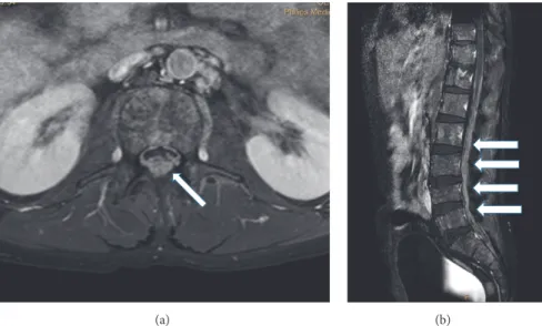
Figure 2: Gadolinium enhanced T1-weighted MRI images of leptomeningeal myeloma of the lumbosacral spine. (a) Axial and (b) sagittal image; arrows: contrast-enhanced myelomatous deposits (patient 11).
sites [4]. Leptomeningeal involvement is the most common form, often resulting in nerve root infiltration and cerebral nerve palsies.
2. Symptoms
Specific symptoms depend on the underlying abnormality.
Headache, confusion, cerebral nerve palsy, and radiculopathy are the most common general symptoms at presentation. In case of intraparenchymal lesions, focal neurologic symptoms can sometimes occur together with somnolence or seizures.
The diagnosis of CNS MM is based on imaging and/or cerebrospinal fluid (CSF) analysis, with stereotaxic brain biopsy, if applicable [5].
3. Imaging
In suspected CNS MM, the first diagnostic step is usu- ally contrast-enhanced MRI (head and/or whole spine).
The sensitivity of MRI is over 90%: it can demonstrate bone-originated plasmacytomas, intraparenchymal tumor- ous lesions, or leptomeningeal contrast-enhanced deposits (Figures 1 and 2). In a review, the latter was the most common form, present in 70% of the cases [6]. CT can be diagnostic as well but with lower sensitivity (80%), and iodinated contrast material is often contraindicated in myeloma [3]. However, the abnormalities are not necessarily specific; for example, they can mimic subdural hemorrhage as in patient 11 in our series [6] (Figure 1(b)).
(a) (b) [Ungated]
CD56APC
CD38ECD 103
102
101
100
103 102
101 100
(c)
[Ungated]
CD19PC5
CD138PE 103
102
101
100
103 102
101 100
(d)
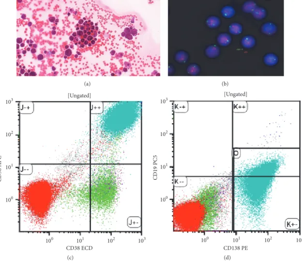
Figure 3: CSF analysis. (a) MGG stained cytospin preparation; (b) fluorescence in situ hybridization (FISH) with probes for 17p (normal);
((c) and (d)) flow cytometry: the plasma cells are CD38, 56, and 138 positive.
4. CSF Analysis
To cytologically confirm leptomeningeal disease, lumbar puncture is necessary: in the previously cited review, it was diagnostic in 90% of the cases [6]. Liquor opening pressure is typically raised, and the liquor cell count is elevated. Plasma cells can be cytologically identified (Figure 3(a)). Flow cytom- etry can confirm monoclonality and aberrant protein expres- sion (Figures 3(c) and 3(d)). CSF protein electrophoresis and free light chain analysis can indirectly confirm the CNS pres- ence of the aberrant plasma cell clone. Fluorescence in situ hybridization (FISH) on the cytospin preparation often shows high-risk features, most commonly deletion 17p (TP53) (Figure 3(b)) [12]. These genetic aberrations contribute to the resistance to traditional chemotherapy and radiotherapy [13].
5. Differential Diagnosis
Naturally, there can be many other causes for nervous system symptoms in myeloma patients, making the diagnosis of CNS propagation difficult. Many of the established MM drugs cause peripheral neuropathy. In case of bortezomib,
this is usually dominantly sensory, while in case of thalido- mide, it is typically a sensomotor neuropathy. Both cause axonal degeneration, and the symptoms are only reversible in case of quick drug interruption/dose reduction; however, if treatment continues, they can become permanent [14].
MM (and other M-protein secretory conditions from MGUS to Waldenstr¨om’s macroglobulinemia) can cause polyneu- ropathy on their own right. M-protein is present in 10%
of cryptogenic neuropathies, mostly IgM, and in 50% of the IgM cases the M-protein has an anti-myelin associated glycoprotein (anti-MAG) activity [15]. Another complication of the above-mentioned M-protein producing diseases is AL amyloidosis. In case of clinical suspicion, tissue biopsy is needed to confirm the diagnosis of amyloid deposition. The most common biopsy targets are abdominal subcutaneous fat and either rectal mucosal or gingival biopsy.
Other complications, common in MM, such as hyper- viscosity, renal failure, and hypercalcemia can also cause CNS symptoms, most commonly disorientation and som- nolence. Given the average age of the MM population plus the prothrombotic properties of the IMiDs, vascular events (both ischemia and hemorrhage) can occur. Apart from
Table1:Patientcharacteristics. Patient
AtdiagnosisPriortreatmentCNSpresentationSurvival(da AgeSexISS/PCLFISH/karyotypeM-proteinLinesThalBorLenASCTAgeSymptomsDiagnosisSystemic relapseISSNew FISHTreatmentResponseOS from CNS
OS from DG
PFS from CNS 169M31qampLC lambda1110069DoublevisionCT,CSFflowyes3NDEDAP,IT, Mel-ASCT,Len maintPR144335130 265M11qampLCkappa1010065ParaplegiaCSFflowyes1NDVTD,Mel-ASCT, LenmaintPR427897124 369F1NDIgG lambda2110074DoublevisionCSFIFXno1NDLenDexNR5620250 441F31qampLC lambda1110041
Headache, hypoglossus, andabducent paresis Clinical(unspecific MRI abnormalities)yes317pVRD-PACE,IT, EDAP,IRD, Mel-Benda-ASCTPR18029063 552M3del13qIgAkappa3110Allo55Abducent paresis,unable toswallowCSFcytospin,MRIno1ND
Thal+IT+ craniocaudalirrad +discontinue GVHDprophylaxis
CR1341103126 656F3hyperdIgAkappa3110160HeadacheMRIno1NDThal+ craniocaudalirrad, benda-VTDPR7762191104 754M3del13qIgAkappa2110156Oculomotor nerveparesisClinical(MRIorbit neg,CSFND)yes31qampVTDNR93103332 853M3complex, hypodiplIgG lambda3110257Abducent paresisClinical(MRIneg, CSFND)yes3samePAD-ThalMR2131563210 943M3,PCLcomplex,del 13qLC lambda1010044Paresis,cannot swallowCSFcytospin,MRI: tumorsyes3sameHDMTX,AraC, ITPD441400 1066M31qampIgG lambda2110067Abducent paresisMRIyes3sameIRDPD143860 1134M2t(4;14)IgG lambda3111136LeftlegparesisMRI,CSFflowyes1sameThio,Car,Thal, DexPR3855738 1260F3,PCLt(11;14),del17pIgGkappa2111160SeizuresCSFcytospinyes3sameVTD-PACE+ith triplet4x,Mel-TBI alloPD1103250 1372M2normalLCkappa4111073BackpainCSFflow,CTyesND1qampDVd+ITPR6069560 Mean56.58/12HR7/13 lambda258.212589763 Allo:allogeneicstemcelltransplantation;amp:amplification;ASCT:autologousstemcelltransplantation;Bor:bortezomib;Car:carfilzomib;CI:cranialirradiation;CSF:cerebrospinalfluid;CTD:cyclophosp thalidomide,anddexamethasone;CyBorDex:cyclophosphamide,bortezomib,anddexamethasone;Dara:daratumumab;del:deletion;DVd:daratumumab,bortezomib,anddexamethasone;flow:flowcyt hyperd:hyperdiploid;IFX:immunofixation;IRD:ixazomib,revlimid,anddexamethasone;IT:intrathecalchemotherapy;KTD:carfilzomib,thalidomide,anddexamethasone;LC:lightchain;Len:lena maint:maintenance;MPV:melphalan,prednisolone,andbortezomib;ND:notdone;PACE:cisplatin,doxorubicin,cyclophosphamide,andetoposide;PAD:bortezomib,doxorubicin,anddexamethas plasmacellleukemia;PomD:pomalidomideanddexamethasone;RT:radiotherapy;T-CED:thalidomide,cyclophosphamide,etoposide,anddexamethasone;Thal:thalidomide;Thio:thiotepa;VAD:vincr doxorubicin,anddexamethasone;VTD:bortezomib,thalidomide,anddexamethasone.
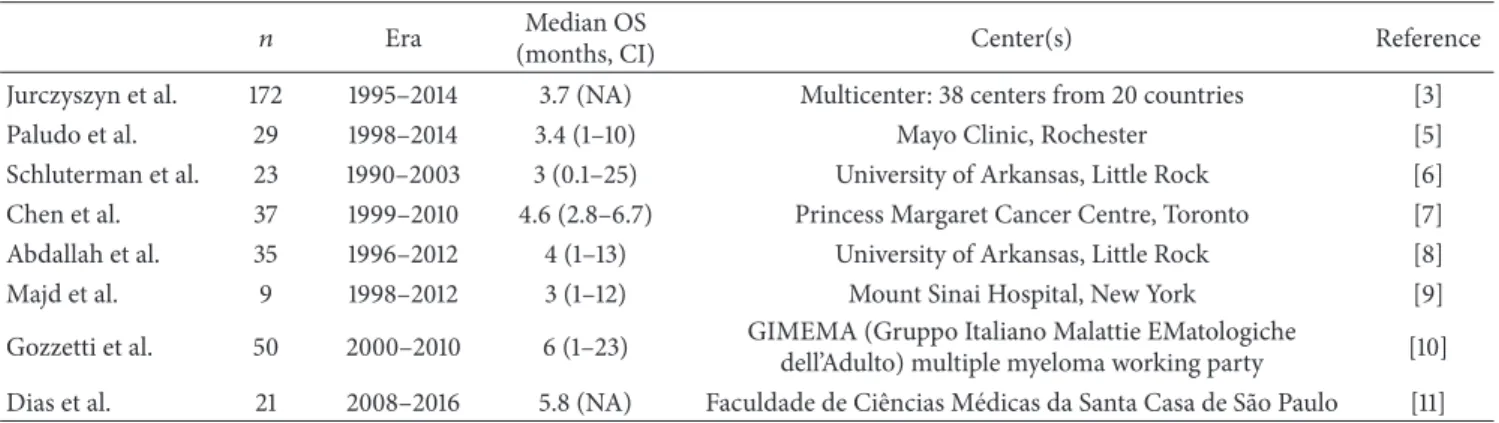
Table 2: Largest recently published case series.
𝑛 Era Median OS
(months, CI) Center(s) Reference
Jurczyszyn et al. 172 1995–2014 3.7 (NA) Multicenter: 38 centers from 20 countries [3]
Paludo et al. 29 1998–2014 3.4 (1–10) Mayo Clinic, Rochester [5]
Schluterman et al. 23 1990–2003 3 (0.1–25) University of Arkansas, Little Rock [6]
Chen et al. 37 1999–2010 4.6 (2.8–6.7) Princess Margaret Cancer Centre, Toronto [7]
Abdallah et al. 35 1996–2012 4 (1–13) University of Arkansas, Little Rock [8]
Majd et al. 9 1998–2012 3 (1–12) Mount Sinai Hospital, New York [9]
Gozzetti et al. 50 2000–2010 6 (1–23) GIMEMA (Gruppo Italiano Malattie EMatologiche
dell’Adulto) multiple myeloma working party [10]
Dias et al. 21 2008–2016 5.8 (NA) Faculdade de Ciˆencias M´edicas da Santa Casa de S˜ao Paulo [11]
CI: confidence interval; NA: not available.
these, autoimmunity, paraneoplasia, and infections have to be considered (herpes and JC viruses) in these patients with compromised immune system.
Most myeloma patients are taking multiple medications.
Among these, thalidomide has a sedative effect, but patients taking thalidomide often report vertigo, tremor, imbalance, and difficulty to walk too. These tend to improve quickly upon stopping thalidomide. Major analgesics (morphine and fentanyl) and adjuvant pain medications (gabapentin, pre- gabalin, tegretol, and tricyclic antidepressants) can blur the clinical picture further, especially if drug levels are unreliable due to liver and kidney failure.
6. Treatment and Survival
The treatment of CNS MM is highly problematic. On one hand, this complication is more common in otherwise high- risk MM (both clinically aggressive and genetically high risk), when chemotherapy is usually less effective. Another problem is that drugs tested and proven in relapsed MM, such as the proteasome inhibitors (bortezomib, carfilzomib, and ixazomib), cannot cross the blood-brain barrier, and standard chemotherapy drugs used to treat CNS lymphoma and brain cancer are not particularly effective in MM. The IMiDs (thalidomide, lenalidomide, and pomalidomide) do cross the blood-brain barrier; however, IMiD resistance is not uncommon in this group of patients. The anti-CD38 antibody daratumumab and anti-CS1 elotuzumab recently approved in relapsed MM were not prospectively tested in this particular group of patients; CNS involvement was an exclusion criterion in the registration trials. It is unknown whether these antibodies can get through the blood-brain barrier, and there is no published report about their intrathe- cal use, whatever tempting it would be theoretically. In one case report of a CNS MM case, it was found that MM cells in the CSF shred their surface CD38 after starting systemic daratumumab, which was interpreted as an indirect sign of CNS penetration of the drug [16]. It has to be mentioned here that intrathecal injection of bortezomib can be fatal and is therefore absolutely contraindicated!
As a salvage, in analogy to acute lymphoid leukemia, intrathecal combinational chemotherapy with methotrexate,
cytosine arabinoside, and dexamethasone (15 mg, 40 mg, and 4 mg, resp.) is often applied continuously until liquor clear- ance, usually in combination with high-dose chemotherapy, using drugs with known CNS penetration (methotrexate, cytosine arabinoside, idarubicin, and thiotepa), together with high-dose dexamethasone, similar to what we normally do in CNS lymphoma. In case of a response to this strategy, ret- rospective data support the use of craniocaudal radiotherapy and possibly ASCT as consolidation [7].
Recently, a case review reported the effectivity of poma- lidomide in CNS MM [17]. Pomalidomide is a drug of the IMiD class approved for lenalidomide refractory patients, which according to a subgroup analysis is more effective in high-risk cytogenetics, especially deletion 17p, than other IMiDs and has good CNS penetration [18]. Another drug that could have a role in this setting is marizomib, a potent natural second-generation proteasome inhibitor produced by a marine bacterium, which was used successfully in two cases of CNS MM [16].
Based on these data, it is not surprising that the overall survival in this condition is extremely poor. The survival data varies widely between studies but usually does not exceed a couple of months from diagnosis (median survival:
2–8 months, Table 2) [3, 5–11, 19]. It is significantly better, however, in those patients who had craniocaudal irradiation or ASCT. Although this data is biased as these are selected patients who responded to their initial treatment [3, 5], in more recent publications, nevertheless, there is a promising increase in survival [7].
7. 13 Cases of CNS MM over 10 Years in Our 2 Centers
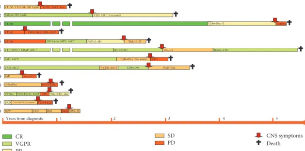
Out of 548 MM cases treated over 10 years at the 3rd Depart- ment of Internal Medicine, Semmelweis University, and the Department of Haematology and the Stem Cell Trans- plantation, St. Istv´an and St. L´aszl´o Hospital, we identified retrospectively 13 cases of CNS MM. Patients’ characteristics including symptoms, treatments, and survival are shown in Table 1, treatment courses and responses to various treatment lines are depicted graphically in Figure 4, and progression- free survival and overall survival given in Figure 5.
3 4 5 6 7 8 9 10 11 12 13
VTDx6 CyBorDex ×2 LenDex
VTDx3 VRD-PACE, IRD, ASCT
HD-Cyclo, MPV, ASCT Thal, CI, IT
VTD, MPVT, EDAP, ASCT RT, CTDx3
PAD, ASCT CyBorDex, Thal maint
VAD, ASCT T-CED, ASCT CyBorDex
PAD AraC, MTX
CyBorDex
VTDx4 VRD-PACE, ASCT IRD PAD
PR VGPR
CR SD
PD
VADx4 PADx2, allo
Thal, CI Benda-VTD
VTD PAD-Thal
Thio-KTD, allo
CNS symptoms Death
1 2 3 4 5
Years from diagnosis
MPV CTD IRD PomD DVd, IT
VTD-PACE, Len maint VTD-PACE MPVT IRD
Figure 4: Treatments and responses. Allo: allogeneic stem cell transplantation; amp: amplification; ASCT: autologous stem cell trans- plantation; Bor: bortezomib; Car: carfilzomib; CI: cranial irradiation; CSF: cerebrospinal fluid; CTD: cyclophosphamide, thalidomide, and dexamethasone; CyBorDex: cyclophosphamide, bortezomib, and dexamethasone; Dara: daratumumab; del: deletion; DVd: daratumumab, bortezomib, and dexamethasone; flow: flow cytometry; hyperd: hyperdiploid; IFX: immunofixation; IRD: ixazomib, revlimid, and dexamethasone; IT: intrathecal chemotherapy; KTD: carfilzomib, thalidomide, and dexamethasone; LC: light chain; Len: lenalidomide;
maint: maintenance; MPV: melphalan, prednisolone, and bortezomib; ND: not done; PACE: cisplatin, doxorubicin, cyclophosphamide, and etoposide; PAD: bortezomib, doxorubicin, and dexamethasone; PCL: plasma cell leukemia; PomD: pomalidomide and dexamethasone;
RT: radiotherapy; T-CED: thalidomide, cyclophosphamide, etoposide, and dexamethasone; Thal: thalidomide; Thio: thiotepa; VAD:
vincristine, doxorubicin, and dexamethasone; VRD: bortezomib, lenalidomide, and dexamethasone; VTD: bortezomib, thalidomide, and dexamethasone.
(days) PFS 63 (0–185) days OS 125 (84–165) days 0,0
0,2 0,4 0,6 0,8 1,0
800,00 600,00
400,00 200,00
,00
Figure 5: Progression-free survival (PFS) and overall survival (OS).
CNS symptoms started in each case at the time of relapse, on average 2 years from the initial diagnosis of MM (range:
3 months–5.3 years). These patients were younger than the usual MM patients, with more IgA and LC secretory cases than usually seen in MM. In 8/13 cases, high-risk cytogenetics were present at diagnosis, and we observed clonal evolution
in terms of new FISH findings in 3 patients. Two patients presented originally as primary plasma cell leukemia; in these cases, survival from the original diagnoses was only 5 and 12 months.
The dominant symptoms were cranial nerve palsies (dou- ble vision and dysphagia) but patients also presented with headache, seizures, paraparesis or paraplegia, and radicu- lopathy. The diagnosis was supported by imaging in 6 and by CSF analysis in 8 cases.
Treatment strategies varied according to the patients’ pre- senting symptoms, performance stage, and drug availability at the time. Ten patients had treatment containing an IMiD (5 lenalidomide and 5 thalidomide), five had high-dose salvage chemotherapy, four ASCT, two craniocaudal irradiation, and six intrathecal chemotherapy. One very recent case (#13) presenting with CNS relapse in September 2017 showed a remarkable response to systemic daratumumab. Dara- tumumab was combined with bortezomib and high-dose corticosteroids and the patient had 3 times intrathecal triplet chemotherapy treatment as well. With 3-month follow-up, he is alive and well, and his CSF that was previously full of plasma cells (Figure 3(a)) is now clear.
In one case, CNS MM presented as an isolated relapse fol- lowing allogeneic SCT, indicating that graft versus myeloma effect is less prominent in the CNS. In another case, after combination chemotherapy for CNS MM, we attempted allogeneic SCT with total body irradiation (TBI) based conditioning; this patient died in sepsis but was apparently free of disease.
Survival, in keeping with the literature, was very poor in our series; with the median being 3 months, the longest survival so far was 14 months.
8. Concluding Remarks
As in the 13 cases presented in our series, CNS MM is a real challenge to the treating physician. The diagnosis is complex;
probably many cases remain undetected. Although there are no clear guidelines regarding best treatment, aggressive management is necessary. Most recently, pomalidomide and marizomib showed promising results, and our cases sug- gest that monoclonal antibodies, particularly daratumumab, might have a role in this setting, too.
Conflicts of Interest
The authors have no conflicts of interest.
References
[1] S. V. Rajkumar, M. A. Dimopoulos, and A. Palumbo, “Inter- national Myeloma Working Group updated criteria for the diagnosis of multiple myeloma,”The Lancet Oncology, vol. 15, no. 12, pp. e538–e548, 2014.
[2] S. K. Kumar, A. Dispenzieri, M. Q. Lacy et al., “Continued improvement in survival in multiple myeloma: Changes in early mortality and outcomes in older patients,”Leukemia, vol. 28, no.
5, pp. 1122–1128, 2014.
[3] A. Jurczyszyn, N. Grzasko, A. Gozzetti et al., “Central nervous system involvement by multiple myeloma: A multi-institutional retrospective study of 172 patients in daily clinical practice,”
American Journal of Hematology, vol. 91, no. 6, pp. 575–580, 2016.
[4] A. Dispenzieri and R. A. Kyle, “Neurological aspects of multiple myeloma and related disorders,” Best Practice & Research Clinical Haematology, vol. 18, no. 4, pp. 673–688, 2005.
[5] J. Paludo, U. Painuly, and S. Kumar S, “Myelomatous involve- ment of the central nervous system , Clinical Lymphoma,”
Myeloma & Leukemia, vol. 16, no. 11, pp. 644–654, 2016.
[6] K. O. Schluterman, A. B.-T. Fassas, R. L. Van Hemert, and S.
I. Harik, “Multiple myeloma invasion of the central nervous system,”JAMA Neurology, vol. 61, no. 9, pp. 1423–1429, 2004.
[7] C. I. Chen, E. Masih-Khan, H. Jiang et al., “Central nervous system involvement with multiple myeloma: long term survival can be achieved with radiation, intrathecal chemotherapy, and immunomodulatory agents,” British Journal of Haematology, vol. 162, no. 4, pp. 483–488, 2013.
[8] A. O. Abdallah, S. Atrash, Z. Shahid, and etal., “Patterns of central nervous system involvement in relapsed and refractory multiple myeloma,”Clinical Lymphoma, Myeloma & Leukemia, vol. 14, no. 3, pp. 211–214, 2014.
[9] N. Majd, A. Demopoulos, and A. Chari, “Central nervous system involvement in multiple myeloma patients in the era of novel therapies, neurology,”Neurology, vol. 80, no. 7, 2013.
[10] A. Gozzetti, A. Cerase, F. Lotti et al., “Extramedullary intracra- nial localization of multiple myeloma and treatment with novel agents: a retrospective survey of 50 patients,”Cancer, vol. 118, no. 6, pp. 1574–1584, 2012.
[11] A. L. Dias, F. Higashi, A. L. Peres, P. Cury, E. d. Cruso´e, and V. T. Hungria, “Multiple myeloma and central nervous system involvement: experience of a Brazilian center,” Hematology, Transfusion and Cell Therapy, vol. 40, no. 1, pp. 30–36, 2018.
[12] H. Chang, S. Sloan, D. Li, and A. K. Stewart, “Multiple myeloma involving central nervous system: high frequency of chromo- some 17p13.1 (p53) deletions,”British Journal of Haematology, vol. 127, no. 3, pp. 280–284, 2004.
[13] A. B.-T. Fassas, F. Muwalla, T. Berryman et al., “Myeloma of the central nervous system: association with high-risk chromosomal abnormalities, plasmablastic morphology and extramedullary manifestations,”British Journal of Haematology, vol. 117, no. 1, pp. 103–108, 2002.
[14] I. Istenes, Zs. Nagy, and J. Demeter, “Chemotherapy-induced peripheral neuropathy: characteristics, diagnosis and treat- ment,”Hungarian Oncology, vol. 60, pp. 165–175, 2016.
[15] R. A. Rison and S. R. Beydoun, “Paraproteinemic neuropathy:
A practical review,”BMC Neurology, vol. 16, no. 1, article no. 13, 2016.
[16] A. Badros, Z. Singh, B. Dhakal et al., “Marizomib for central nervous system-multiple myeloma,”British Journal of Haema- tology, vol. 177, no. 2, pp. 221–225, 2017.
[17] A. Mussetti, S. Dalto, and V. Montefusco, “Effective treatment of pomalidomide in central nervous system myelomatosis,”
Leukemia & Lymphoma, vol. 54, no. 4, pp. 864–866, 2013.
[18] X. Leleu, L. Karlin, M. Macro et al., “Pomalidomide plus low- dose dexamethasone in multiple myeloma with deletion 17p and/or translocation (4;14): IFM 2010-02 trial results,”Blood, vol. 125, no. 9, pp. 1411–1417, 2015.
[19] L. Nieuwenhuizen and D. H. Biesma, “Central nervous system myelomatosis: review of the literature,” European Journal of Haematology, vol. 80, no. 1, pp. 1–9, 2008.
Stem Cells International
Hindawi
www.hindawi.com Volume 2018
Hindawi
www.hindawi.com Volume 2018
Endocrinology
International Journal ofHindawi
www.hindawi.com Volume 2018
Hindawi
www.hindawi.com Volume 2018
Disease Markers
Hindawi
www.hindawi.com Volume 2018
BioMed
Research International
Oncology
Journal ofHindawi
www.hindawi.com Volume 2013
Hindawi
www.hindawi.com Volume 2018
Oxidative Medicine and Cellular Longevity
Hindawi
www.hindawi.com Volume 2018
PPAR Research
Hindawi Publishing Corporation
http://www.hindawi.com Volume 2013
Hindawi www.hindawi.com
The Scientific World Journal
Volume 2018
Immunology Research
Hindawi
www.hindawi.com Volume 2018
Journal of
Obesity
Journal ofHindawi
www.hindawi.com Volume 2018
Hindawi
www.hindawi.com Volume 2018
Computational and Mathematical Methods in Medicine
Hindawi
www.hindawi.com Volume 2018
Behavioural Neurology Ophthalmology
Journal ofHindawi
www.hindawi.com Volume 2018
Diabetes Research
Journal ofHindawi
www.hindawi.com Volume 2018
Hindawi
www.hindawi.com Volume 2018
Research and Treatment
AIDS
Hindawi
www.hindawi.com Volume 2018
Gastroenterology Research and Practice
Hindawi
www.hindawi.com Volume 2018
Parkinson’s Disease
Evidence-Based Complementary and Alternative Medicine
Volume 2018 Hindawi
www.hindawi.com