The authors’ full names, academic de- grees, and affiliations are listed in the Ap- pendix. Address reprint requests to Dr.
Minard-Colin at Gustave Roussy, 114 Rue Edouard Vaillant, Villejuif 94805, France, or at veronique . minard@ gustaveroussy . fr.
*A complete list of the investigators in the European Intergroup for Childhood Non-Hodgkin Lymphoma and the Chil- dren’s Oncology Group is provided in the Supplementary Appendix, available at NEJM.org.
Drs. Minard-Colin and Aupérin and Drs.
Patte and Gross contributed equally to this article.
N Engl J Med 2020;382:2207-19.
DOI: 10.1056/NEJMoa1915315 Copyright © 2020 Massachusetts Medical Society.
BACKGROUND
Rituximab added to chemotherapy prolongs survival among adults with B-cell can- cer. Data on its efficacy and safety in children with high-grade, mature B-cell non-Hodgkin’s lymphoma are limited.
METHODS
We conducted an open-label, international, randomized, phase 3 trial involving patients younger than 18 years of age with high-risk, mature B-cell non-Hodgkin’s lymphoma (stage III with an elevated lactate dehydrogenase level or stage IV) or acute leukemia to compare the addition of six doses of rituximab to standard lymphomes malins B (LMB) chemotherapy with standard LMB chemotherapy alone. The primary end point was event-free survival. Overall survival and toxic effects were also assessed.
RESULTS
Analyses were based on 328 patients who underwent randomization (164 patients per group); 85.7% of the patients had Burkitt’s lymphoma. The median follow-up was 39.9 months. Events were observed in 10 patients in the rituximab–chemo- therapy group and in 28 in the chemotherapy group. Event-free survival at 3 years was 93.9% (95% confidence interval [CI], 89.1 to 96.7) in the rituximab–chemo- therapy group and 82.3% (95% CI, 75.7 to 87.5) in the chemotherapy group (hazard ratio for primary refractory disease or first occurrence of progression, relapse after response, death from any cause, or second cancer, 0.32; 95% CI, 0.15 to 0.66; one- sided P = 0.00096, which reached the significance level required for this analysis).
Eight patients in the rituximab–chemotherapy group died (4 deaths were disease- related, 3 were treatment-related, and 1 was from a second cancer), as did 20 in the chemotherapy group (17 deaths were disease-related, and 3 were treatment- related) (hazard ratio, 0.36; 95% CI, 0.16 to 0.82). The incidence of acute adverse events of grade 4 or higher after prephase treatment was 33.3% in the rituximab–
chemotherapy group and 24.2% in the chemotherapy group (P = 0.07); events were related mainly to febrile neutropenia and infection. Approximately twice as many patients in the rituximab–chemotherapy group as in the chemotherapy group had a low IgG level 1 year after trial inclusion.
CONCLUSIONS
Rituximab added to standard LMB chemotherapy markedly prolonged event-free survival and overall survival among children and adolescents with high-grade, high-risk, mature B-cell non-Hodgkin’s lymphoma and was associated with a higher incidence of hypogammaglobulinemia and, potentially, more episodes of infection. (Funded by the Clinical Research Hospital Program of the French Min-
ABS TR ACT
Rituximab for High-Risk, Mature B-Cell Non-Hodgkin’s Lymphoma in Children
V. Minard-Colin, A. Aupérin, M. Pillon, G.A.A. Burke, D.A. Barkauskas, K. Wheatley, R.F. Delgado, S. Alexander, A. Uyttebroeck, C.M. Bollard, J. Zsiros,
M. Csoka, B. Kazanowska, A.K. Chiang, R.R. Miles, A. Wotherspoon, P.C. Adamson, G. Vassal, C. Patte, and T.G. Gross, for the European Intergroup
for Childhood Non-Hodgkin Lymphoma and the Children’s Oncology Group*
Original Article
C
ure rates among children and ad- olescents with high-grade, mature B-cell non-Hodgkin’s lymphoma (mainly Burkitt’s lymphoma but also diffuse large B-cell lympho- ma) have dramatically improved over the past 30 years, with trials showing survival of approxi- mately 90%.1-4 However, well-known prognostic factors such as higher stage, elevated lactate dehydrogenase (LDH) level, leukemic bone mar- row, and central nervous system (CNS) involve- ment and treatment-related factors such as lack of early or complete response can identify pa- tients at high risk for treatment failure.Rituximab has shown efficacy in adults with B-cell cancers, including diffuse large B-cell lymphoma and Burkitt’s lymphoma, and is con- sidered to be the standard of care in addition to chemotherapy in most patients with high-grade B-cell non-Hodgkin’s lymphoma. The outcome in children and adolescents with B-cell non- Hodgkin’s lymphoma receiving chemotherapy alone is superior to that in adults; therefore, the potential benefits of rituximab must be balanced against potential unexpected and severe toxic effects. Furthermore, subtypes of mature B-cell non-Hodgkin’s lymphoma differ between adults and children in terms of molecular anomalies that may confer different prognoses and sensi- tivities to treatments.5,6
A phase 2 trial involving children showed that rituximab was active as a single-agent therapy for high-grade, high-risk, mature B-cell non- Hodgkin’s lymphoma and could be safely added to the lymphomes malins B (LMB) chemotherapy regimen.7 Therefore, we conducted an interna- tional, randomized, phase 3 trial (Inter-B-NHL ritux 2010) to establish whether the addition of rituximab to LMB chemotherapy could improve event-free survival among children and adoles- cents with high-grade, high-risk, mature B-cell non-Hodgkin’s lymphoma or leukemia.
Methods Trial Oversight and Design
In this trial, we investigated the efficacy and safety of adding rituximab to a modified LMB chemotherapy regimen. This academic trial in- volved two international cooperative groups — the European Intergroup for Childhood Non- Hodgkin Lymphoma (EICNHL) and the Children’s Oncology Group (COG) — spanning 12 coun-
tries (see the Supplementary Appendix, available with the full text of this article at NEJM.org).
The trial sponsors were Gustave Roussy (for countries in the EICNHL) and COG (for Austra- lia, Canada, and the United States) and included a partnership with F. Hoffmann–La Roche–Genen- tech, which provided partial funding and pro- vided rituximab at no cost but had no role in the design or conduct of the trial nor in the prepara- tion of the manuscript. The manuscript was written by the authors, who vouch for the accu- racy and completeness of the data and for the fidelity of the trial to the protocol, available at NEJM.org.
Parents and patients (if age appropriate) signed informed consent and assent forms before enroll- ment. The research protocol was approved in each country by the necessary ethics and regula- tory committees. An international, independent data and safety monitoring committee, which included two pediatric oncologists, one adult on- cologist, and one statistician, monitored progress and interim analysis reports.
Randomization was performed separately for patients at the EICNHL and COG sites. For pa- tients at the EICHNL sites, randomization was performed centrally at Gustave Roussy with the use of a minimization algorithm that accounted for histologic features (large-cell or non–large- cell), therapeutic group (B, C1, C3; defined in the next subsection), and the national group. To avoid predictability, a probability of 0.80 to as- sign the treatment that minimized imbalance was used. For patients at the COG sites, random- ization was performed centrally at the COG data center, with the use of block randomization stratified according to histologic features and therapeutic group. Physicians and patients were aware of the treatment-group assignments.
Patients
Eligible patients were 6 months to 18 years of age and had newly diagnosed high-grade, mature B-cell neoplasms (Burkitt’s lymphoma; diffuse large B-cell lymphoma; or high-grade, mature B-cell non-Hodgkin’s lymphoma not otherwise specified) and advanced St. Jude stage disease (stage III with an LDH level that was more than twice the institutional upper limit of the normal range or any stage IV or leukemia presentation).
Patients with primary mediastinal (thymic) large B-cell lymphoma were not eligible. Pathological
slides were centrally reviewed at the national level, but centralized review was not mandatory before enrollment. Detailed inclusion and exclu- sion criteria and initial workup data are provided in the Supplementary Appendix.
Therapeutic groups were defined as in the French–American–British (FAB)/LMB96 interna- tional study for all patients except those with blasts in cerebrospinal fluid (CSF), who were treated with high-dose methotrexate that was administered as a 24-hour infusion (group C3).3,4 The rationale for this modification was a retro- spective review of previous LMB studies that showed poorer survival among these patients than among other patients in group C.8 In the FAB/LMB96 study, outcomes in patients in group B who had stage I, II, or III disease with an LDH level that was equal to or less than twice the up- per limit of the normal range were excellent (event-free survival, 95%). Therefore, with regard to group B, only patients who had stage III dis- ease and an LDH level that was more than twice the upper limit of the normal range or non-CNS stage IV disease with bone marrow involvement of less than 25% were eligible for this trial.
Patients in group C1 had CSF-negative stage IV disease with bone marrow involvement of at least 25% (leukemia presentation), CNS-positive disease, or both. Group C3 included patients with CSF-positive stage IV disease.
Treatment
Chemotherapy was administered according to the FAB/LMB96-based protocol, with some minor modifications (Fig. S1 in the Supplementary Ap- pendix).3,4 All the patients received prephase treat- ment with low-dose cyclophosphamide, vincris- tine, and prednisone (COP regimen). Patients in group B received four cycles of chemotherapy, which was similar to the therapy used in group B4 in the FAB/LMB96 study. For all the patients in group C, the maintenance chemotherapy was reduced to two courses, in contrast to the four maintenance cycles that were used in the FAB/
LMB96 study. Information about monitoring the risk of jeopardizing efficacy with this reduction in therapy is presented in the Supplementary Ap- pendix. Patients in group C1 received high-dose methotrexate (at a dose of 8 g per square meter of body-surface area) over a period of 4 hours, as in the FAB/LMB96 study, whereas patients in group C3 (whose disease was CSF-positive) re-
ceived high-dose methotrexate (at a dose of 8 g per square meter) over a period of 24 hours.
Consecutive courses were administered as soon as blood counts recovered and the patient’s con- dition allowed, except for the maintenance cours- es, which were administered at 28-day intervals.
Rituximab was administered as an intravenous infusion (at a dose of 375 mg per square meter) on day 2 before (i.e., day −2) and day 1 of each of the two induction chemotherapy courses and on day 1 of each of the two consolidation courses, for a total of six doses.7 When a course had to be postponed, rituximab was administered at the planned time in order to ensure the intensity of the therapy.
An initial response evaluation was performed on day 7 after the receipt of the COP regimen.
Patients in groups B and C1 who had a reduction in the tumor size of less than 20% were switched to a more intensive therapeutic group (i.e., from group B to C1 or from group C1 to C3). Remis- sion assessment was performed after receipt of the first consolidation course in group B and after receipt of the second consolidation course in group C. In patients with a residual mass as assessed by radiographic evaluation, an excision or biopsy for pathological review was recom- mended. If a biopsy was not performed, the pa- tients were to continue receiving the assigned treatment. For patients in group B, if viable tu- mor cells were identified, the therapy was switched to the more intensive regimen that was given to patients in group C1. Patients in group C1 or C3 with biopsy-proven viable tumor cells after the second consolidation course (continu- ous infusion and high-dose cytarabine and eto- poside, with or without rituximab [depending on the randomly assigned treatment group]) were considered to have primary refractory disease and were considered to have had an event; such patients discontinued the protocol therapy. No treatment decisions were to be based on the re- sults of 18F-fluorodeoxyglucose positron-emission tomography–computed tomography.
End Points
The primary end point was event-free survival, which was defined as the minimum time be- tween randomization and the detection of residu- al viable tumor cells after receipt of the second consolidation course of therapy (i.e., primary re- fractory disease), relapse, progressive disease,
second cancer, or death from any cause, or the date of the last follow-up for patients who did not have any event. All events were validated by the steering committee. Details of the secondary end points (overall survival, complete remission at assessment time, toxic effects, and immune reconstitution as assessed by IgG levels) are pre- sented in the Supplementary Appendix.
Statistical Analysis
We expected the 3-year event-free survival to be 84% on the basis of data from the FAB/LMB96 study.3,4 Because this trial was designed to deter- mine the efficacy of adding rituximab to stan- dard treatment, a one-sided test was used. At a one-sided 5% level of statistical significance and assuming randomization in a 1:1 ratio, we cal- culated that 72 events would need to be observed in order for the trial to have 90% power to detect a hazard ratio of 0.50 (on the basis of an ex- pected 3-year event-free survival of 84% in the chemotherapy group and 92% in the rituximab–
chemotherapy group). It was estimated that 600 patients would need to undergo randomization in order for 72 events to be observed.
Interim analyses for efficacy with the use of the Lan–DeMets alpha-spending function ap- proach applied to an O’Brien–Fleming boundary, truncated at 3 SD, were planned.9 The first in- terim analysis was planned to take place when one third of the total expected events had oc- curred; the subsequent interim analyses were expected to occur yearly after that.
Event-free survival and overall survival were estimated by means of the Kaplan–Meier method, and 95% confidence intervals of yearly rates were estimated by the Rothman method. Hazard ratios with adjustment for therapeutic group, histologic type, and national group were estimated by the Cox model. Subgroup analyses according to three baseline characteristics (age, histologic features, and therapeutic group) were prespecified.
R esults Interim Analyses
The first interim analysis was based on 27 events in 310 patients, which corresponded to 37.5% of the expected events and a nominal alpha error of 0.00137. The median follow-up was 11.5 months.
Event-free survival at 1 year was 94.2% (95% con- fidence interval [CI], 88.5 to 97.2) in the ritux-
imab–chemotherapy group and 81.5% (95% CI, 73.0 to 87.8) in the chemotherapy group (hazard ratio for event [defined as primary refractory disease or first occurrence of progression, re- lapse after response, death from any cause, or second cancer], 0.33; 95% CI, 0.14 to 0.79).10 The full results of this interim analysis are presented in the Supplementary Appendix.
After the first interim analysis, randomization was stopped for efficacy in November 2015 on the recommendation of the independent data and safety monitoring committee. An additional 120 patients were subsequently enrolled and treated with rituximab and chemotherapy (single-group cohort) for prespecified secondary aims; these patients are not included in the analyses pre- sented here. The current analyses are based on 38 events, corresponding to 52.8% of the expect- ed events and a nominal alpha error of 0.00562.
The cumulative alpha error is 0.00699.
Analysis Sets
From December 2011 through November 2015, a total of 362 patients were enrolled at 176 cen- ters. Data from 1 patient were removed owing to withdrawal of consent. The 33 patients who were still receiving induction or consolidation chemotherapy after the closure of randomiza- tion in November 2015 were recommended to receive rituximab and chemotherapy regardless of the randomly assigned group. These patients were not included in the primary analysis but were included in the sensitivity analysis. Thus, the main analyses included 328 patients who had undergone randomization until August 26, 2015 (Fig. 1), including 4 patients who were de- clared to be ineligible: 3 patients after central pathological review (1 patient had precursor B-cell acute lymphoblastic leukemia and 2 had primary mediastinal B-cell lymphoma) and 1 patient who had stage III disease and an LDH level that was less than two times the upper limit of the nor- mal range.
Patients and Treatment
The characteristics of the patients at baseline were well balanced between the two treatment groups (Table 1). A total of 85.7% of the patients had Burkitt’s lymphoma. Three patients in the chemotherapy group received rituximab (2 owing to physicians’ decisions and 1 because of a diag- nosis of primary mediastinal B-cell lymphoma).
All the patients in the rituximab–chemotherapy group received rituximab, with 11 patients (7%) receiving fewer than six doses (Table S1).
Efficacy
The median follow-up was 39.9 months overall, with follow-up of 40.9 months (interquartile range, 35.1 to 48.9) in the rituximab–chemo-
therapy group and 39.1 months (interquartile range, 34.3 to 49.0) in the chemotherapy group.
Data regarding the response after the adminis- tration of the COP regimen and remission at the time of assessment are shown in Table S2. In the primary analysis, there were 38 events: 10 in the rituximab–chemotherapy group and 28 in the chemotherapy group (Table 2). Six patients
Figure 1. Randomization, Treatment, and Follow-up of the Patients.
The 33 patients who were still receiving induction or consolidation chemotherapy after the closure of randomization in November 2015 were recommended to receive rituximab and chemotherapy regardless of the randomly assigned group; these patients were not included in the main analyses. ALL denotes acute lymphoblastic leukemia, LDH lac- tate dehydrogenase, PMBL primary mediastinal B-cell lymphoma, and ULN upper limit of the normal range.
362 Patients underwent randomization from December 2011 through November 2015
181 Were assigned to chemotherapy group 181 Were assigned to rituximab–
chemotherapy group
16 Underwent randomization after Aug. 26, 2015, and
received induction or consolidation chemotherapy
after November 2015 (not included in main analyses)
17 Underwent randomization after Aug. 26, 2015, and
received induction or consolidation chemotherapy
after November 2015 (not included in main analyses)
165 Were assigned to chemotherapy group 164 Were assigned to rituximab–
chemotherapy group
1 Was excluded owing to withdrawal of informed consent
164 Were included in chemotherapy group
4 Were ineligible
1 Had stage III cancer with LDH <2×ULN 2 Had PMBL
1 Had precursor B-cell ALL
3 Received rituximab 1 Had PMBL
2 Had the assigned regimen changed per physician’s decision
11 Received <6 injections of rituximab
156 Were included in follow-up >1 yr
(median follow-up, 39.1 mo) 164 Were included in follow-up >1 yr (median follow-up, 40.9 mo)
164 Were included in efficacy analysis 164 Were included in efficacy analysis
Table 1. Characteristics of the Patients at Baseline.*
Characteristic
Chemotherapy Group (N = 164)
Rituximab–Chemotherapy Group (N = 164)
Male sex — no. (%) 137 (83.5) 135 (82.3)
Age
Mean — yr 8.6±4.4 9.2±4.0
Range — yr 2–17 2–17
Distribution — no. (%)
<6 yr 56 (34.1) 44 (26.8)
6 to <12 yr 67 (40.9) 73 (44.5)
12 to <15 yr 22 (13.4) 30 (18.3)
≥15 yr 19 (11.6) 17 (10.4)
Pathological diagnosis†
Burkitt’s lymphoma 142 (86.6) 139 (84.8)
Diffuse large B-cell lymphoma 12 (7.3) 19 (11.6)
High-grade B-cell lymphoma, not otherwise
specified 7 (4.3) 6 (3.7)
Primary mediastinal B-cell lymphoma‡ 2 (1.2) 0
Precursor B-cell acute lymphoblastic leukemia‡ 1 (0.6) 0
Prognosis group
Group B, low risk‡ 1 (0.6) 0
Group B, high risk 82 (50.0) 81 (49.4)
Group C1, without CSF blasts 63 (38.4) 65 (39.6)
Group C3, with CSF blasts 16 (9.8) 18 (11.0)
Primary mediastinal B-cell lymphoma‡ 2 (1.2) 0
Murphy stage
Stage III 73 (44.5) 70 (42.7)
Stage IV 32 (19.5) 32 (19.5)
Leukemic disease, mature B-cell acute leukemia 57 (34.8) 62 (37.8)
Primary mediastinal B-cell lymphoma‡ 2 (1.2) 0
Central nervous system involvement
No 120 (73.2) 119 (72.6)
Yes 44 (26.8) 45 (27.4)
Lactate dehydrogenase
≤2× ULN 18 (11.0) 17 (10.4)
>2× ULN 146 (89.0) 147 (89.6)
Primary site or type
Thorax 6 (3.7) 6 (3.7)
Abdomen or retroperitoneum 95 (57.9) 96 (58.5)
Head and neck, except skin and nodes 9 (5.5) 20 (12.2)
Peripheral lymph node 6 (3.7) 2 (1.2)
Cerebral lymphoma 3 (1.8) 2 (1.2)
Other tumor site 11 (6.7) 8 (4.9)
Clinical presentation of leukemia disease 34 (20.7) 30 (18.3)
* Percentages may not total 100 because of rounding. CSF denotes cerebrospinal fluid, and ULN upper limit of the nor- mal range (according to institutional values).
† These data were obtained from national pathological review in 235 patients (72%) or from local pathological diagnosis.
‡ Patients with these conditions were not eligible for the trial. On central review, these patients were found to be ineli- gible, but they were included in the intention-to-treat analyses.
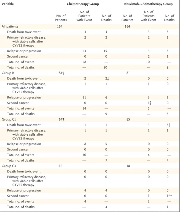
(three in each group) died from toxic events.
Four patients (two in each group) had primary refractory disease. The major between-group dif- ference in events was observed in relapse or pro- gression, which occurred in 3 patients in the rituximab–chemotherapy group and in 23 in the chemotherapy group; all events of relapse or progression occurred within 9 months after ran- domization. A total of 18 of these patients subse- quently died: all 3 of the patients in the ritux- imab–chemotherapy group and 15 of the 23 patients in the chemotherapy group. A second cancer developed in 2 patients in the rituximab–
chemotherapy group: 1 patient had melanoma, and 1 had fatal histiocytic sarcoma with a t(8;14) IGH–MYC anomaly identical to the primary lym- phoma. In total, 8 patients in the rituximab–
chemotherapy group died (4 deaths were dis- ease-related, 3 were treatment-related, and 1 was from a second cancer), as did 20 in the chemo- therapy group (17 deaths were disease-related, and 3 were treatment-related).
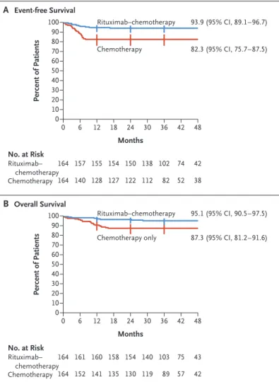
Event-free survival at 3 years was 93.9% (95%
CI, 89.1 to 96.7) in the rituximab–chemotherapy group and 82.3% (95% CI, 75.7 to 87.5) in the chemotherapy group (Fig. 2A). In the analysis of event-free survival, the hazard ratio for an event was 0.32 (95% CI, 0.15 to 0.66; one-sided P = 0.00096, which reached the significance level required for this second analysis). Overall sur- vival at 3 years was 95.1% (95% CI, 90.5 to 97.5) in the rituximab–chemotherapy group and 87.3%
(95% CI, 81.2 to 91.6) in the chemotherapy group (hazard ratio for death, 0.36; 95% CI, 0.16 to 0.82) (Fig. 2B).
The sensitivity analysis, which involved 361 patients (including those in the period between August 26, 2015, and November 2015, who were recommended to receive rituximab and chemo- therapy regardless of the randomly assigned group), showed similar results. Event-free survival at 3 years was 92.8% in the rituximab–chemo- therapy group and 83.4% in the chemotherapy group (hazard ratio for an event, 0.40; 95% CI, 0.21 to 0.78). Overall survival at 3 years was 94.3% in the rituximab–chemotherapy group and 88.5% in the chemotherapy group (hazard ratio for death, 0.45; 95% CI, 0.21 to 0.97). The bene- fit of rituximab was similar across subgroups defined according to age, histologic features, and therapeutic group (Fig. S2).
Adverse Events
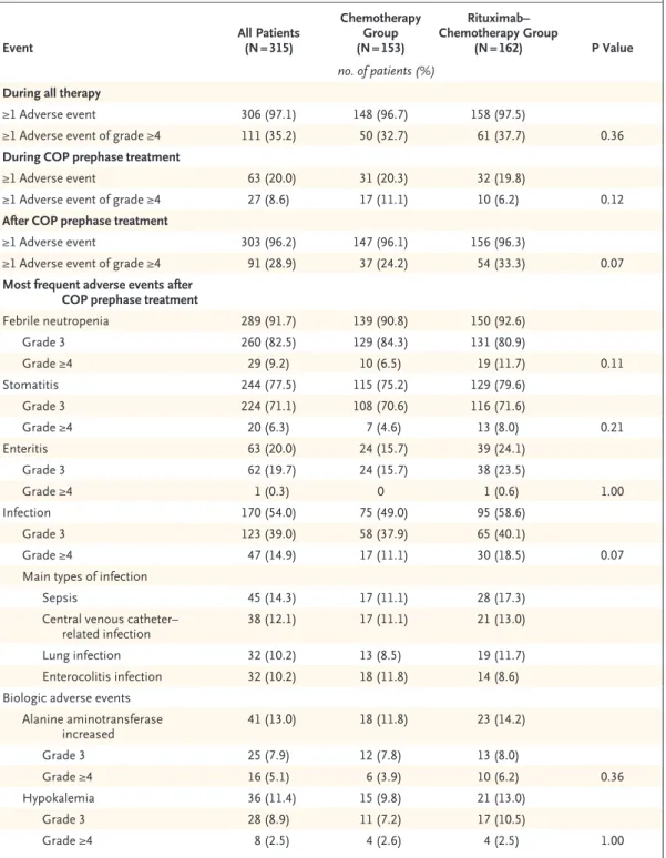
The incidence of rituximab infusion–related reac- tions was 33.3% during the first infusion (with 4.3% of the patients having a grade 3 event); the incidence decreased to 3.7 to 9.6% during subse- quent infusions (with 1.0 to 2.1% of the patients having a grade 3 event). No immediate infusion- related grade 4 toxic effect occurred. Treatment- related toxic effects of grade 4 or higher were observed in 35.2% of the patients overall, includ- ing in 28.9% of the patients after prephase treat- ment with the COP regimen (Table 3). The most frequent adverse events after prephase treatment were febrile neutropenia (in 91.7% of the patients), infection (in 54.0%), and stomatitis (in 77.5%).
Six patients (1.8% of the patients overall; three patients in each group) died from toxic effects.
Five of the deaths, four of which were from in- fection, occurred in patients receiving group C chemotherapy after the receipt of the first course of therapy with cyclophosphamide, vincristine, prednisone, doxorubicin, and methotrexate, with or without rituximab depending on treatment group (in three patients); after the receipt of the second course of therapy with cyclophospha- mide, vincristine, prednisone, doxorubicin, and methotrexate, with rituximab (in one patient); or after the receipt of a continuous infusion and high-dose cytarabine and etoposide, with ritux- imab (in one patient). One death from toxic effects occurred in a patient receiving group B chemotherapy who had undergone hemicolecto- my at diagnosis and died from small-bowel ob- struction and septic shock after the receipt of COPADM2.
The incidence of adverse events of grade 4 or higher was 37.7% in the rituximab–chemothera- py group and 32.7% in the chemotherapy group (P = 0.36). In an analysis that was focused on the period after the receipt of COP prephase treat- ment, the incidence of adverse events of grade 4 or higher was 33.3% in the rituximab–chemo- therapy group and 24.2% in the chemotherapy group (P = 0.07). After prephase treatment, the incidence of febrile neutropenia of grade 4 or higher was 11.7% in the rituximab–chemother- apy group and 6.5% in the chemotherapy group (P = 0.11), and the incidence of infection of grade 4 or higher was 18.5% and 11.1%, respectively (P = 0.07). No other acute unexpected toxic effects were reported in either group. The most frequent
Table 2. Number and Type of Events and Number of Deaths.*
Variable Chemotherapy Group Rituximab–Chemotherapy Group
No. of Patients
No. of Patients
with Event No. of
Deaths No. of Patients
No. of Patients
with Event No. of Deaths
All patients 164 164
Death from toxic event 3 3 3 3
Primary refractory disease, with viable cells after CYVE2 therapy
2 2 2 1
Relapse or progression 23 15 3 3
Second cancer 0 0 2 1
Total no. of events 28 — 10 —
Total no. of deaths — 20 — 8
Group B 84† 81
Death from toxic event 2 2‡ 0 0
Primary refractory disease, with viable cells after CYVE2 therapy
1 1 1 0
Relapse or progression 11 6 3 3
Second cancer 0 0 1§ 0
Total no. of events 14 — 5 —
Total no. of deaths — 9 — 3
Group C1 64¶ 65
Death from toxic event 1 1 3 3‖
Primary refractory disease, with viable cells after CYVE2 therapy
1 1 1 1
Relapse or progression 8 5 0 0
Second cancer 0 0 0 0
Total no. of events 10 — 4 —
Total no. of deaths — 7 — 4
Group C3 16 18
Death from toxic event 0 0 0 0
Primary refractory disease, with viable cells after CYVE2 therapy
0 0 0 0
Relapse or progression 4 4 0 0
Second cancer 0 0 1 1**
Total no. of events 4 — 1 —
Total no. of deaths — 4 — 1
* CYVE2 denotes the second course of continuous infusion and high-dose cytarabine and etoposide.
† One patient in the low-risk group B (who did not have an event) was counted in group B.
‡ One patient who was receiving group C1 chemotherapy died after receipt of the first course of therapy with cyclo- phosphamide, vincristine, prednisone, doxorubicin, and methotrexate.
§ One patient, a 14-year-old girl, received a diagnosis of melanoma that had developed on a congenital skin nevus 5 months after the diagnosis of Burkitt’s lymphoma.
¶ One patient with primary mediastinal B-cell lymphoma (who did not have an event) was counted in group B, and one (who had disease progression) was counted in group C1.
‖ One patient switched to group C3 after receipt of the COP regimen; the death occurred after receipt of the second course of group C3 chemotherapy (cyclophosphamide, vincristine, prednisone, doxorubicin, and methotrexate).
** One patient, a 6-year-old boy, had fatal histiocytic sarcoma with an identical t(8;14) IGH–MYC anomaly, which had been diagnosed when he was 16.6 months of age.
adverse events according to treatment course and all adverse events of grade 4 or higher are pre- sented in Tables S3 and S4, respectively.
Immunoglobulin Status
The percentage of patients with a low IgG level (less than the lower limit of the normal range) was significantly higher in the rituximab–chemo- therapy group than in the chemotherapy group at the end of therapy (70.3% vs. 46.8%, P = 0.002) and at 1 year after inclusion (55.9% vs. 25.4%, P<0.001), respectively (Table S5). Approximately twice as many patients in the rituximab–chemo- therapy group as in the chemotherapy group re- ceived intravenous immune globulin (15.8% vs.
7.0%). The primary reason for immune globulin replacement was a low immunoglobulin level without infection (Table S6). At 1 year after en- rollment, seven patients were still receiving intra- venous immune globulin in the rituximab–chemo- therapy group and three in the chemotherapy group. Complete and longer follow-up with re- gard to immune status and late infections have not been evaluated.
Discussion
In this randomized, international, phase 3 trial, we found that among children and adolescents with high-grade, high-risk, mature B-cell lym- phoma, the addition of six doses of rituximab to LMB therapy led to significantly better event-free survival outcomes (hazard ratio for an event, 0.32;
95% CI, 0.15 to 0.66; 3-year event-free survival, 93.9% in the rituximab–chemotherapy group vs.
82.3% in the chemotherapy group). The benefit was similar across the various therapeutic groups, including the group of patients with CNS dis- ease. Higher 3-year overall survival was also observed (95.1% in the rituximab–chemotherapy group vs. 87.3% in the chemotherapy group;
hazard ratio for death, 0.36; 95% CI, 0.16 to 0.82). Overall, the acute toxic effects that were associated with the addition of rituximab main- ly involved a higher incidence of myelotoxic ef- fects, but further follow-up is needed to provide information on long-term safety because ritux- imab therapy induced more hypogammaglobu- linemia than chemotherapy alone.
The benefit of rituximab therapy in this trial appeared to be similar to that observed in trials
involving adult patients. In a randomized French trial that compared the addition of four doses of rituximab to adult-adapted LMB chemotherapy with chemotherapy alone in 257 patients with Burkitt’s lymphoma (61% of the patients were
≥40 years of age), superior 3-year event-free sur- vival (75% in the rituximab–chemotherapy group vs. 62% in the chemotherapy group; hazard ratio for event, 0.59; P = 0.02) and overall survival (83% vs. 70%; hazard ratio for death, 0.51;
P = 0.01) were observed.11 A direct comparison of
Figure 2. Event-free Survival and Overall Survival, According to Treatment Group.
Shown are Kaplan–Meier estimates of event-free survival (i.e., freedom from primary refractory disease or first occurrence of progression, relapse after response, death from any cause, or second cancer) and of overall sur- vival. Vertical bars represent the Rothman 95% confidence intervals; point estimates of 36-month event-free survival and overall survival with 95%
confidence intervals are shown.
Percent of Patients
100 80 90 70 60 40 30 10 50
20
00 6 12 18 24 42 48
Months
B Overall Survival A Event-free Survival
93.9 (95% CI, 89.1–96.7)
82.3 (95% CI, 75.7–87.5)
No. at Risk Rituximab–
chemotherapy Chemotherapy
164 164
157 140
155 128
154 127
150 122
74 52 30
138 112
36
102 82
42 38
Percent of Patients
100 80 90 70 60 40 30 10 50
20 0
0 6 12 18 24 42 48
Months
95.1 (95% CI, 90.5–97.5)
87.3 (95% CI, 81.2–91.6)
No. at Risk Rituximab–
chemotherapy Chemotherapy
164 164
161 152
160 141
158 135
154 130
75 57 30
140 119
36
103 89
43 42 Rituximab–chemotherapy
Chemotherapy only Rituximab–chemotherapy
Chemotherapy
Table 3. Acute Adverse Events.*
Event All Patients
(N = 315)
Chemotherapy Group (N = 153)
Rituximab–
Chemotherapy Group
(N = 162) P Value
no. of patients (%) During all therapy
≥1 Adverse event 306 (97.1) 148 (96.7) 158 (97.5)
≥1 Adverse event of grade ≥4 111 (35.2) 50 (32.7) 61 (37.7) 0.36
During COP prephase treatment
≥1 Adverse event 63 (20.0) 31 (20.3) 32 (19.8)
≥1 Adverse event of grade ≥4 27 (8.6) 17 (11.1) 10 (6.2) 0.12
After COP prephase treatment
≥1 Adverse event 303 (96.2) 147 (96.1) 156 (96.3)
≥1 Adverse event of grade ≥4 91 (28.9) 37 (24.2) 54 (33.3) 0.07
Most frequent adverse events after COP prephase treatment
Febrile neutropenia 289 (91.7) 139 (90.8) 150 (92.6)
Grade 3 260 (82.5) 129 (84.3) 131 (80.9)
Grade ≥4 29 (9.2) 10 (6.5) 19 (11.7) 0.11
Stomatitis 244 (77.5) 115 (75.2) 129 (79.6)
Grade 3 224 (71.1) 108 (70.6) 116 (71.6)
Grade ≥4 20 (6.3) 7 (4.6) 13 (8.0) 0.21
Enteritis 63 (20.0) 24 (15.7) 39 (24.1)
Grade 3 62 (19.7) 24 (15.7) 38 (23.5)
Grade ≥4 1 (0.3) 0 1 (0.6) 1.00
Infection 170 (54.0) 75 (49.0) 95 (58.6)
Grade 3 123 (39.0) 58 (37.9) 65 (40.1)
Grade ≥4 47 (14.9) 17 (11.1) 30 (18.5) 0.07
Main types of infection
Sepsis 45 (14.3) 17 (11.1) 28 (17.3)
Central venous catheter–
related infection 38 (12.1) 17 (11.1) 21 (13.0)
Lung infection 32 (10.2) 13 (8.5) 19 (11.7)
Enterocolitis infection 32 (10.2) 18 (11.8) 14 (8.6)
Biologic adverse events Alanine aminotransferase
increased 41 (13.0) 18 (11.8) 23 (14.2)
Grade 3 25 (7.9) 12 (7.8) 13 (8.0)
Grade ≥4 16 (5.1) 6 (3.9) 10 (6.2) 0.36
Hypokalemia 36 (11.4) 15 (9.8) 21 (13.0)
Grade 3 28 (8.9) 11 (7.2) 17 (10.5)
Grade ≥4 8 (2.5) 4 (2.6) 4 (2.5) 1.00
* Shown are the numbers and percentages of patients who had at least one adverse event, those who had at least one adverse event of grade 4 or higher, and those who had the most frequent acute adverse events (i.e., events occurring in >10% of the patients overall). A total of 13 patients who did not have data on treatment or adverse events or who received only prephase treatment (a regimen of cyclophosphamide, vincristine, and prednisone [COP]) were not in- cluded in this analysis. Only nonhematologic adverse events of grade 3 or higher or cardiac adverse events of grade 2 or higher were recorded. P values are shown for events of grade 4 or higher.
outcomes in adults and children is difficult, how- ever, because the outcomes in children who are treated with chemotherapy are superior to those in adults, and molecular analyses suggest that there is age-related biologic heterogeneity in Burkitt’s lymphomagenesis.5,6
The data from this trial suggest that the ad- dition of rituximab may result in a higher inci- dence of severe (grade ≥4) acute adverse events (primarily febrile neutropenia and infection) af- ter prephase treatment. Although specific viral infections have been reported in approximately 2% of patients treated with rituximab, we did not observe such infections in the rituximab–
chemotherapy group (which included 164 pa- tients), but longer follow-up is needed.12,13 Two patients (1%) in the rituximab–chemotherapy group had second cancers: one patient had melanoma within a congenital skin nevus, and one patient had a clonally related histiocytic sarcoma suggesting transdifferentiation of the Burkitt’s lymphoma clone. Some reports have suggested that among adult patients with lym- phoma, the introduction of rituximab may in- crease the risk of a second cancer (especially acute myeloid leukemia, thyroid cancer, and melanoma),14 but a meta-analysis did not sup- port this finding.15 Finally, approximately twice as many patients in the rituximab–chemotherapy group as in the chemotherapy group had a low IgG level 1 year after trial inclusion, although very few patients received immune globulin re- placement for infections. An assessment of the long-term effects of combining rituximab with this chemotherapy regimen in children with non- Hodgkin’s lymphoma, including data on immune status, will be useful.
A previous phase 2 window study involving children with newly diagnosed high-grade, ma- ture B-cell non-Hodgkin’s lymphoma showed that 21% of the patients had a complete or par- tial response during a 5-day window after receiv- ing a single dose of rituximab16; another study is currently comparing one dose of rituximab with six doses in children with advanced-stage B-cell non-Hodgkin’s lymphoma (ClinicalTrials.gov num- ber, NCT03206671). With respect to pharmaco- kinetics, rituximab clearance appears to be relat- ed to total tumor burden; exposure to rituximab decreases as tumor metabolic volume increases.
Higher rituximab exposure is associated with a higher percentage of patients with a response and longer progression-free survival among patients with diffuse large B-cell lymphoma.17 A small cohort study involving children and adolescents with high-grade, mature B-cell non-Hodgkin’s lymphoma showed some age dependency with regard to rituximab pharmacokinetics.18 Thus, we are conducting additional pharmacokinetic analyses, including examination of the effects of age and tumor burden on rituximab disposition.
Although the results of the Inter-B-NHL ritux 2010 trial showed a benefit with the addition of rituximab in the treatment of children with high-grade, high-risk, mature B-cell non-Hodg- kin’s lymphoma, the use of rituximab in chil- dren with standard-risk B-cell non-Hodgkin’s lymphoma (approximately 40% of patients) is still questionable, because their survival after treatment with chemotherapy alone is very high (97 to 98%).19 It is conceivable that the addition of rituximab could allow for a reduction of cyto- toxic chemotherapy; however, given the very poor outcome in patients with refractory or relapsed disease,20 it would be ethically challenging to conduct such a study. Furthermore, although cur- rent chemotherapy is associated with substantial acute toxic effects, deaths from toxic effects are rare and the risks of clinically significant long- term sequelae are relatively modest with the use of this chemotherapy backbone.21 To help inform such a study, more data are necessary in order to evaluate the long-term safety of rituximab ad- ministered with chemotherapy in children with non-Hodgkin’s lymphoma.
This trial showed that the addition of ritux- imab to chemotherapy was effective therapy in children and adolescents with high-risk, high- grade, mature B-cell non-Hodgkin’s lymphoma and resulted in long-term complete remission in 95% of the patients. In addition, we found an effective global framework for academic-based, collaborative pediatric groups that leveraged both public and private sector support to conduct clinical trials involving children with a rare cancer.
Supported by a grant (PHRC2010) from the Clinical Research Hospital Program of the French Ministry of Health, by Cancer Research UK, the National Institute for Health Research Clini- cal Research Network, and Children’s Cancer Foundation Hong Kong, by grants (U10CA180886 and U10CA180899) from the
U.S. National Cancer Institute, and by F. Hoffmann–La Roche–
Genentech.
Dr. Minard-Colin reports receiving grant support, paid to her institution, from F. Hoffmann–La Roche; Dr. Aupérin, receiving grant support, paid to her institution, from F. Hoff- mann–La Roche; Dr. Burke, receiving consulting fees from F.
Hoffmann–La Roche, Janssen Pharmaceuticals, Merck, and Takeda Pharmaceutical; Dr. Adamson, receiving grant support, paid to Children’s Hospital of Philadelphia, from Roche Genen- tech, owning stock in Merck, and being employed by Sanofi and Genzyme US; Dr. Vassal, receiving grant support, paid to his institution, from F. Hoffmann–La Roche; and Dr. Patte, receiv- ing grant support, paid to her institution, from F. Hoffmann–La Roche. No other potential conflict of interest relevant to this article was reported.
Disclosure forms provided by the authors are available with the full text of this article at NEJM.org.
A data sharing statement provided by the authors is available with the full text of this article at NEJM.org.
We thank the members of the national and international review panels (P. Dartigues, T. Molina, J. Bosq, S. Perkins, K. McCarthy,
A. Ferrández, O. Balagué, F. Loong, J.C. So, and E.S.G. d’Amore);
all the European Intergroup for Childhood Non-Hodgkin Lym- phoma (EICNHL) and Children’s Oncology Group (COG) com- mittee members and investigators who participated in the trial;
F. Rotolo, of the Gustave Roussy Unit of Biostatistics and Epi- demiology; the national data managers (R. Banusz, E. Carraro, A. Raxworthy Cooper, V. Femke, O. Hung, A. Michiels, M. Peiró, J. Vreijling, O. Wajsen, and T. Yu); G. Goma and A. Mangin for data management for the patients at the EICNHL sites; J. Ander- son, A. Buxton, D. Hall, L. Saguilig, A. Miranda, T. Hlaing, and G. Galit for data management for the patients at the COG sites;
A. Tulard, D. Vuillier, and J. Rubino, of the Gustave Roussy clin- ical research team; S. Laghouati for pharmacovigilance; the members of the independent data and safety monitoring board (R. Sposto, F. Pein, R. Pinkerton, and M. Robert); and the F.
Hoffmann–La Roche–Genentech team. This study was initial- ly proposed to the Paediatric Committee by the Marketing Au- thorisation Holder on May 13, 2009, as the sole measure in the MabThera PIP (EMEA-000308-PIP01-08). The initial MabThera PIP was submitted on June 28, 2008, and was agreed on in the European Medicines Agency decision adopted on July 14, 2009.
Appendix
The authors’ full names and academic degrees are as follows: Véronique Minard-Colin, M.D., Ph.D., Anne Aupérin, M.D., Ph.D., Marta Pillon, M.D., Ph.D., G.A. Amos Burke, M.B., Ch.B., Ph.D., Donald A. Barkauskas, Ph.D., Keith Wheatley, Ph.D., Rafael F. Del- gado, M.D., Ph.D., Sarah Alexander, M.D., Anne Uyttebroeck, M.D., Ph.D., Catherine M. Bollard, M.D., Ph.D., József Zsiros, M.D., Ph.D., Monika Csoka, M.D., Ph.D., Bernarda Kazanowska, M.D., Ph.D., Alan K. Chiang, M.D., Ph.D., Rodney R. Miles, M.D., Ph.D., Andrew Wotherspoon, M.D., Ph.D., Peter C. Adamson, M.D., Ph.D., Gilles Vassal, M.D., Ph.D., Catherine Patte, M.D., and Thomas G.
Gross, M.D., Ph.D.
The authors’ affiliations are as follows: the Departments of Pediatric and Adolescent Oncology (V.M.-C., C.P.) and Clinical Research (G.V.), INSERM Unité 1015 (V.M.-C.), and the Unit of Biostatistics and Epidemiology and INSERM Unité 1018 (A.A.), Gustave Roussy, Université Paris-Saclay, Villejuif, France; the Department of Pediatric Hematology and Oncology, University of Padua, Padua, Italy (M.P.); the Department of Paediatric Haematology, Oncology, and Palliative Care, Cambridge University Hospitals NHS Foundation Trust, Addenbrooke’s Hospital, Cambridge (G.A.A.B.), Cancer Research UK Clinical Trials Unit, Institute of Cancer and Genomic Sci- ences, College of Medical and Dental Sciences, University of Birmingham, Birmingham (K.W.), and the Department of Histopathology, Royal Marsden NHS Foundation Trust, London (A.W.) — all in the United Kingdom; the Department of Preventive Medicine, Keck School of Medicine, University of Southern California, Los Angeles (D.A.B.); the Department of Pediatric Hematology and Oncology, University of Valencia, Valencia, Spain (R.F.D.); the Division of Haematology–Oncology, Hospital for Sick Children, Toronto (S.A.); the Department of Pediatric Hematology and Oncology, University Hospitals Leuven, Leuven, Belgium (A.U.); the Center for Cancer and Immunology Research, Children’s National Health System and George Washington University, Washington, DC (C.M.B.); Princess Máxima Center for Pediatric Oncology, Utrecht, the Netherlands (J.Z.); the Department of Pediatric Hematology and Oncology, Sem- melweis University, Budapest, Hungary (M.C.); the Department of Pediatric Bone Marrow Transplantation, Oncology, and Hematology, Wroclaw Medical University, Wroclaw, Poland (B.K.); the Department of Pediatrics and Adolescent Medicine, Li Ka Shing Faculty of Medicine, Queen Mary Hospital, University of Hong Kong, Hong Kong (A.K.C.); the Department of Pathology, University of Utah, Salt Lake City (R.R.M.); Children’s Hospital of Philadelphia, Philadelphia (P.C.A.); and the National Cancer Institute, Center for Global Health, Rockville, MD (T.G.G.).
References
1. Minard-Colin V, Brugières L, Reiter A, et al. Non-Hodgkin lymphoma in children and adolescents: progress through effec- tive collaboration, current knowledge, and challenges ahead. J Clin Oncol 2015;
33: 2963-74.
2. Woessmann W, Seidemann K, Mann G, et al. The impact of the methotrexate ad- ministration schedule and dose in the treatment of children and adolescents with B-cell neoplasms: a report of the BFM Group Study NHL-BFM95. Blood 2005;
105: 948-58.
3. Patte C, Auperin A, Gerrard M, et al.
Results of the randomized international FAB/LMB96 trial for intermediate risk B- cell non-Hodgkin lymphoma in children and adolescents: it is possible to reduce
treatment for the early responding pa- tients. Blood 2007; 109: 2773-80.
4. Cairo MS, Gerrard M, Sposto R, et al.
Results of a randomized international study of high-risk central nervous system B non-Hodgkin lymphoma and B acute lymphoblastic leukemia in children and adolescents. Blood 2007; 109: 2736-43.
5. Havelange V, Pepermans X, Ameye G, et al. Genetic differences between paedi- atric and adult Burkitt lymphomas. Br J Haematol 2016; 173: 137-44.
6. López C, Kleinheinz K, Aukema SM, et al. Genomic and transcriptomic chang- es complement each other in the patho- genesis of sporadic Burkitt lymphoma. Nat Commun 2019; 10: 1459.
7. Goldman S, Smith L, Anderson JR,
et al. Rituximab and FAB/LMB 96 chemo- therapy in children with stage III/IV B-cell non-Hodgkin lymphoma: a Children’s Oncology Group report. Leukemia 2013;
27: 1174-7.
8. Minard-Colin V, Auperin A, Leverger G. In childhood B-cell non Hodgkin’s lymphoma (B-NHL) and mature B-cell acute leukemia (B-AL) with CNS disease at diagnosis, patients with blasts in CSF are at higher risk of event. Ann Oncol 2011; 22: Suppl 4: iv112-iv114.
9. DeMets DL, Lan KK. Interim analysis:
the alpha spending function approach.
Stat Med 1994; 13: 1341-52.
10. Minard-Colin V, Auperin A, Pillon M, et al. Results of the randomized Inter- group trial Inter-B-NHL ritux 2010 for
children and adolescents with high-risk B-cell non-Hodgkin lymphoma (B-NHL) and mature acute leukemia (B-AL): evalu- ation of rituximab (R) efficacy in addition to standard LMB chemotherapy (CT) regi- men. J Clin Oncol 2016; 34: Suppl: 10507.
abstract.
11. Ribrag V, Koscielny S, Bosq J, et al.
Rituximab and dose-dense chemotherapy for adults with Burkitt’s lymphoma: a ran- domised, controlled, open-label, phase 3 trial. Lancet 2016; 387: 2402-11.
12. Focosi D, Tuccori M, Maggi F. Pro- gressive multifocal leukoencephalopathy and anti-CD20 monoclonal antibodies:
what do we know after 20 years of ritux- imab. Rev Med Virol 2019; 29(6): e2077.
13. Kusumoto S, Arcaini L, Hong X, et al.
Risk of HBV reactivation in patients with B-cell lymphomas receiving obinutuzu- mab or rituximab immunochemotherapy.
Blood 2019; 133: 137-46.
14. Tao L, Clarke CA, Rosenberg AS, et al.
Subsequent primary malignancies after
diffuse large B-cell lymphoma in the mod- ern treatment era. Br J Haematol 2017;
178: 72-80.
15. Fleury I, Chevret S, Pfreundschuh M, et al. Rituximab and risk of second pri- mary malignancies in patients with non- Hodgkin lymphoma: a systematic review and meta-analysis. Ann Oncol 2016; 27:
390-7.
16. Meinhardt A, Burkhardt B, Zimmer- mann M, et al. Phase II window study on rituximab in newly diagnosed pediatric mature B-cell non-Hodgkin’s lymphoma and Burkitt leukemia. J Clin Oncol 2010;
28: 3115-21.
17. Tout M, Casasnovas O, Meignan M, et al. Rituximab exposure is influenced by baseline metabolic tumor volume and pre- dicts outcome of DLBCL patients: a Lym- phoma Study Association report. Blood 2017; 129: 2616-23.
18. Barth MJ, Goldman S, Smith L, et al.
Rituximab pharmacokinetics in children and adolescents with de novo intermedi-
ate and advanced mature B-cell lymphoma/
leukaemia: a Children’s Oncology Group report. Br J Haematol 2013; 162: 678- 83.
19. Patte C, Zimmerman M, Auperin A, Reiter A. Similar results are currently ob- served in the LMB and BFM studies for B-cell Non-Hodgkin’s lymphoma and B-AL allowing future common studies. Pediatr Blood Cancer 2010; 55: 795. abstract.
20. Pearson ADJ, Scobie N, Norga K, et al.
ACCELERATE and European Medicine Agency Paediatric Strategy Forum for me- dicinal product development for mature B-cell malignancies in children. Eur J Cancer 2019; 110: 74-85.
21. Ehrhardt MJ, Chen Y, Sandlund JT, et al. Late health outcomes after contem- porary Lymphome Malin de Burkitt ther- apy for mature B-cell non-Hodgkin lym- phoma: a report from the Childhood Cancer Survivor Study. J Clin Oncol 2019;
37: 2556-70.
Copyright © 2020 Massachusetts Medical Society.
journalarchiveatnejm.org
Every article published by the Journal is now available at NEJM.org, beginning with the first article published in January 1812. The entire archive is fully searchable,
and browsing of titles and tables of contents is easy and available to all.
Individual subscribers are entitled to free 24-hour access to 50 archive articles per year.
Access to content in the archive is available on a per-article basis and is also being provided through many institutional subscriptions.