Haploidy versus Diploidy
in the Reproduction of Cell Type
WALTER TULECKE
Boy ce Thompson Institute for Plant Research, Yonkers, New York
Introduction
Haploid cells and organisms will be the main subject of this paper, and they will be compared to diploids in terms of their capacity for growth and differentiation. Examples of both plant and animal species will be discussed. Some emphasis will be placed on work performed in our labora- tory on haploid tissue cultures from plants, particularly those derived from Ginkgo biloba L.
If we consider the dominant forms of plant and animal life which sur- vive today, we observe that they are primarily diploid in chromosome complement. That is, they develop and function in a diploid (2n) state of complementary hereditary information, and transmit their characters through single genomes which are haploid (n). This simple well-known generalization is based on observations of the many biological forms which confront us in the field and in the laboratory, and this generaliza- tion holds, even though there are many interesting and informative exceptions.
Concerning the general framework of biological forms and functions, we may ask: What is special about the diploid state in comparison with the haploid? Are the differences between diploid and haploid cells re- flected in their functions? We recognize, for example, that the haploid state represents a type of specialization especially effective for sexual re- production. Similarly, we know that the diploid state not only assures survival and dominance of the species, but that it is also essential for the organization of complex organisms. In other words, complementary in- formation systems are usually needed for completely normal ontogeny of the organism.
T h e fusion of haploid nuclei to form the diploid organism requires a periodic reduction division. In a sense, this reduction division may be regarded as a hereditary cloning process because the gametes derived
217
218 WALTER TULECKE
from reduction division, as well as the zygotes derived from their fusion, are unique creations. Within this context of continuous variation, where sex and modulation of characters are involved, many questions can be asked. For example, were primitive cells haploid? Did the first fusion of haploid nuclei confer a greater capacity for competition and survival?
Was sex evolved from some of these early advantageous events? These are all interesting and pertinent queries. Our main concern, however, is with the following questions: What are the relative capabilities of haploid cells and organisms in comparison with those of their diploid counterparts?
What is known in this area and what are the basic problems?
For coherence, and in order to limit the scope of reference, it will be useful to define a few terms. A haploid cell or organism, for example, is understood to have a single basic set of chromosomes of a given species.
This set may be male (androgenetic) or female (gynogenetic); one genome, one game tic chromosome constitution, and haploid all are synonymous terms conventionally designated as (n). Euploids are multiples of the basic number; hence, diploids are the first of a series that includes triploids and tetraploids; higher genome sets are designated polyploids (e.g., autopolyploids which possess similar genomes, and allopolyploids which have dissimilar genomes).
Characteristics of Haploids
The most abundant haploid organisms are the lower forms, i.e., the algae, bacteria, yeasts, fungi, mosses, and liverworts. Haploid animals include the male bees and wasps, other Hymenoptera, and some members of the Rotifera, Homoptera, and Thysanoptera; haploid males usually develop from the eggs of diploid females, but the production of partheno- genetic haploid females is much less common (Suomalainen, 1950).
Among the higher plants, haploidy is known to occur in over 71 species of 39 genera in 16 families (Kimber and Riley, 1963). Haploids are reported in such well-known genera as Oenothera, Epilobium, Zea, Gossypium, Nicotiana, Datura, and Triticum. Some of them are spontaneous haploids of uncertain origin, and others are the result of hybridization or experi- mentation. Haploids derived experimentally, of course, provide more in- formation, since their origin can be determined. Most haploids are derived from the cells of the embryo sac, and hence are maternal in origin;
androgenetic haploids are relatively rare. T h e first examples of andro- genesis in maize (Zea mays) were reported by Goodsell (1961) and Chase (1963). Male sterile monoploid paternal plants were obtained in very low
HAPLOIDY VS. DIPLOIDY IN REPRODUCTION O F CELL T Y P E 219
frequencies (1/80,000) from special crosses. T h e sperm nucleus from the pollen apparently united with the cytoplasm of the egg which contained the plasmogenes for male sterility. The low frequency of progeny with this character indicates that the method is of little use in breeding pro- grams unless special lines or artificial induction can be used to increase its frequency. T h e significant fact, however, is that the expression of male sterility is governed by cytoplasmic factors from the maternal side. It will be very interesting to discover the nature of this type of cytoplasmic in- heritance, especially to determine whether some particular satellite DNA is involved.
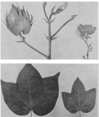
Haploid higher plants which have been found or induced experi- mentally, generally exhibit a weakened structural constitution. Haploid cotton plants, for example, possess zigzag stems, short internodes, usually produce no pollen, seeds, or bolls, and develop leaves and flowers reduced in size (Meyer and Justus, 1961). Doubling the chromosome number by colchicine treatment, however, may restore vigor to the progeny as shown in Fig. 1; leaves, flowers, and fruit are then restored to normal propor- tions. The origin of these haploids is not known, i.e., whether they develop parthenogenetically from the unfertilized egg, or androgenetically from the generative nucleus, although the former possibility seems more likely. These plants are propagated vegetatively by cuttings and usually do not produce either functional pollen or viable seeds. Haploid tomato plants of known genotype, however, were propagated vegetatively by Lindstrom (1941) for over 14 years and were found to be quite stable;
homozygous diploids derived from these plants were highly fertile. This stability in cotton is an exception, however, because plants with a single genome usually are quite variable in character; genetic balance and phenotypic uniformity are generally not maintained. Induced haploidy often leads to a serious disruption of development, especially in normally outbreeding heterozygous plants. Thus, the structural adaptations to out- breeding which are seen in some higher plants, serve to maintain heterozygosity; circumvention of this by the production of haploids usually leads to genetic imbalance. In contrast, highly inbred species have a minimum of allele differences because the introduction of new alleles is restricted. Haploids derived from inbred species might be expected to develop with greater frequency but with reduced vigor in comparison with haploids from heterozygous species which are fewer and more robust.
Animals of gametic chromosome constitution generally exhibit what is known as a haploid syndrome (Moore, 1955; Briggs and King, 1959;
Subtelny, 1958). Enucleated eggs of Rana pipiens implanted with nuclei
220 W A L T E R TULECKE
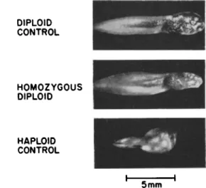
from haploid androgenetic blastulae develop as far as the beginning of gastrulation, but then become abnormal. Deficiencies in the gut, central nervous system, sense organs, and pronephros and cardiovascular systems, are collectively known as the haploid syndrome. In some of the recipient eggs development is delayed and the chromosome number is doubled.
These androgenetic autodiploids develop normally up to the early post
FIG. 1. Leaf and flowering branch from doubled haploid cotton plant (left) derived from haploid plant (right) by colchicine treatment. From Meyer and Justus (1961).
neurula stage and thereafter show abnormalities. T h e larvae feed and survive, but usually die before metamorphosis. Examples of these homo- zygous diploids and their haploid and diploid controls are shown in Fig. 2. Gynogenetic diploids are also retarded, but some reached the frog stage (Moore, 1955). Apparently, the maternal genome carries suffi- cient information and suitable cytoplasm for development to continue through metamorphosis, whereas the growth of androgenetic homozygous diploids is arrested earlier.
More than forty years ago Bridges (1925) described mosaic female
HAPLOIDY VS. DIPLOIDY IN REPRODUCTION OF CELL TYPE 221
individuals of Drosophila melanogaster which contained patches of haploid male tissue. These mosaic fruit flies were unusual because most natural haploids in animals are males. Bridges confirmed the presence of several sex-linked patroclinous characters and the single X chromosome in the haploid male tissue. In contrast, the development of fertilized sub- haploid eggs of Drosophila (Briggs and King, 1959) was arrested during cleavage; hyperhaploids of frogs terminated their development during gastrulation, and hyperdiploids of axolotls matured only to the feeding larval stage. Aneuploids in all these cases were sufficiently imbalanced to
DIPLOID CONTROL
HOMOZYGOUS DIPLOID
HAPLOID CONTROL
5mm
FIG. 2. Androgenetic homozygous larvae of Rana pipiens compared to a diploid control and an androgenetic haploid control. From Fig. 6, Subtelny (1958, p . 282).
impair development. We can assume, therefore, that a correct balance of genes in a complementary diploid condition is required for normal ontogeny; more evidence in support of this claim will be presented later.
I n d u c t i o n of H a p l o i d y
The artificial production of haploids may be brought about by a variety of techniques. Ultraviolet treatment of newt sperm, for example, is effec- tive in inducing haploid parthenogenesis from eggs of three species of Triturus (Selman, 1958); similar treatment of pollen induces haploid parthenogenesis in tobacco (Ivanov, 1938). Radiation treatment of pollen in general, however, does not enhance haploid frequencies (Brewbaker and Emery, 1962), and the results vary from species to species. In some
* #4
222 W A L T E R TULECKE
Zea mays hybrids there may be an increase in haploids (22/20,869 for irradiated and 1/11,707 for control), but the number of spontaneous haploids in some inbred lines may exceed 1%; in Antirrhinum majus maternal haploids may be about 0.05% (Knapp, 1939).
Induction of haploids from higher plants usually occurs through parthenogenetic development of the egg. In special cases, such as in Melilotus hybrids (Jaranowski, 1961), the synergids of the embryo sac may be stimulated to develop with the result that one or two synergid- derived haploid embryos and the normal diploid embryo may be found in a single seed. In this instance the haploid embryos compete satisfactorily with the diploid embryo, develop to maturity, and survive.
Other crossing techniques make use of alien cytoplasm to enhance the frequency of haploid development (Kihara and Tsunewaki, 1962). When Aegilops caudata cytoplasm was present in a cross with Triticum vulgäre var. erythrospermum (a variety of wheat), 1.7% haploids were obtained;
in a cross with Taylor's Triticale, 53% haploids were obtained. Without Aegilops cytoplasm, no haploids were formed in either cross. These ex- periments are interesting because they suggest that a suitable composition of cytoplasm in the mature egg favors the function of a single genome. In the absence of these favorable factors, the diploid state is required;
perhaps the diploid condition provides a similarly favorable cytoplasm.
A marked difference in the frequency with which haploids occur in the Mexican axolotl (Siredon mexicanum) was reported by Humphrey and Fankhauser (1957). Using dark (wild-type) and white (mutant, recessive) breeding stocks, they were able to detect androgenetic and gynogenetic haploids in various crosses by using color, number of chromophores, and the haploid syndrome as distinguishing characters; chromosome counts of tail clippings were also made. In crosses of white females with dark males, 213 offspring were spontaneous gynogenetic white and 11 were androge- netic dark haploids out of 31,273 examined; in the reciprocal cross, 4 gyno- genetic and 17 androgenetic haploids were obtained from 23,154 offspring.
Judging from the preponderance of female haploids we might conclude that this is probably a genetic character associated with the mutant strain of axolotl. When dark females were mated with white males, however, and the eggs placed in a refrigerator for 9-18 hours, a large number of white male haploids resulted. There was a low survival rate in these experi- ments; only 1/194 haploids were obtained with 1-9 hours cold treatment;
however, 63/110 survivors of an extended cold period were haploid. It was suggested that prolonged cold treatment essentially eliminated the egg nucleus of the dark females from participation in development and
H A P L O I D Y VS. DIPLOIDY IN REPRODUCTION O F CELL T Y P E 223
hence increased the incidence of white haploid male axolotls in these crosses.
This temperature-induced haploidy in the axolotl recalls some observa- tions made on pollen development in Hyacinthus orientalis by several workers, and reviewed and extended by Naithani (1937) and Stow (1933).
Stow found that he could induce embryo sac development in pollen from a particular variety of hyacinth by elevating the temperature in the premeiotic phase. Some of the pollen-derived embryo sacs were penetrated by germinating pollen tubes and the sperm nuclei released into the embryo sacs. This case of temperature-induced sex inversion in pollen was authenticated by Naithani who observed it as a normal occurrence in another variety of the same species.
Radiation Effects
The aging process in living organisms involves many factors, one of which is radiation damage. It might be expected that diploidy would provide an advantage over the haploid state, since each character would have an allele. Evidence to support this view has been obtained from ex- periments on Habrobracon (Clark et ah, 1963). Haploid and diploid males having the same life span were X-irradiated at dosages known to decrease the life span. The fact that irradiated diploid males lived longer than haploids, suggests that X-radiation damage (and subsequent aging) is retarded when alleles are present.
Other evidence comes from investigations on higher plants by Sparrow et al. (1961) who demonstrated a correlation between nuclear volume and tolerance to chronic radiation: the larger the nuclear volume, the greater the sensitivity to radiation; similarly, the higher the DNA content (at a given genome level), the greater the sensitivity. When replicate genomes were present, however, as in the case of two polyploid series with Chrysanthemum and Sedum, there was an increased resistance to radia- tion damage.
Quantitation and Biochemistry
T h e quantitative relationship between haploid and diploid cells has been determined in a variety of ways and, in general, is of the order of 1:2. An excellent demonstration was presented by Rudkin et al. (1955) who determined the DNA content of haploid and diploid salivary gland chromosomes of Drosophila melanogaster. They measured the ultraviolet absorption of paired chromosomes and of a single homologous chromo-
224 WALTER TULECKE
some in the same cell of a triploid fruit fly. The extinction values and the chromosome numbers fulfilled the criteria of Lambert's law of propor- tionality, indicating that the DNA content of paired chromosomes was approximately twice that of the single chromosome.
Other work by Mangelsdorf and Fraps (1931) established a quantitative relationship between the genes for yellow pigmentation (Y) and the amount of vitamin A in corn endosperm. This relationship is shown in Table 1: The presence of the y allele produced small amounts of vitamin A, whereas one, two, and three doses of the Y gene produced correspond- ing increases in vitamin A content.
TABLE I
QUANTITATIVE R E L A T I O N S H I P BETWEEN G E N E S FOR Y E L L O W P I G M E N T (Y) AND V I T A M I N A C O N T E N T O F CORN ENDOSPERM«
Number of genes for
yellow 0 1 2 3
Factorial composition of endosperm
y y y y y Y y Y Y Y Y Y
1928 0.05 2.50 5.00 7.00
Units of vitamin A 1929 0.05 2.00 5.00 8.00
per gram Average
0.05 2.25 5.00 7.50 β After Mangelsdorf and Fraps (1931}.
Another long recognized factor in haploid and diploid development is the nuclear-cytoplasmic ratio. Briggs (1949) demonstrated clearly and effectively that androgenetic haploids of Rana pipiens induced from small ova were smaller, more normal, and better able to feed, than haploids from large ova; the latter lasted only 18 days whereas those from small ova lasted 9 months. The functions of the single genome seem to be more dependent on the cytoplasm than those of the diploid genome. The sur- face-volume relationships of haploid cells appear to be nearer the critical level than those of diploid cells.
In the wasp Mormoniella, Mortimer and von Borstel (1963) were able to show that the sensitivity of sperm to radiation (in strains with domi- nant radiation-induced lethality) was doubled in diploid sperm in com- parison with the normal haploids. This observation suggests a strict cor- respondence between the amount of genetic material and the radiation effect. Other evidence of this relationship was reported by Olsen and Ogg (1963) for haploid and diploid strains of E. coli: Diploids were found to be more resistant to X rays than haploids. These findings, however, were
HAPLOIDY VS. DIPLOIDY IN REPRODUCTION OF CELL TYPE 225
incidental to their investigations. They used formic hydrogenlyase activ- ity to show that haploid and diploid cells respond differently when placed under anaerobic conditions in a growth medium without formate: When subsequently exposed to formate, the haploids produced hydrogen, while the diploids did not. Under inducing conditions (i.e., in a medium con- taining formate) the induction lag was longer in diploids and the maxi- mum inducible rates were lower than for haploid cells. Thus, on a quantitative basis the cells of E. coli and other organisms reflect their haploid-diploid constitution, but on a qualitative basis, the effect is more subtle as shown by the formic hydrogenlyase experiments. The addition of a second genome actually repressed enzyme activity, rate of induci- bility, and the rate of hydrogen production from formate. Other kinds of measurements on polyploid series in yeasts (Burns, 1956) and ferns (Partanen, 1965) tend to support this idea.
Growth and Differentiation Animals
Nuclear transplantation studies have helped to define the problems of genomes related to growth and development, especially in anuran embryo- genesis. For example, diploid nuclei from late gastrula cells of Rana pipiens were transplanted to enucleated eggs by King and Briggs (1955);
the larvae produced were found to be limited in their potential for differentiation. It was also found that for donor nuclei the later the stage of development, the more limited the differentiation. In other words, the events of ontogeny imposed a rather severe (and apparently permanent) restriction on the capacity for development.
The complementariness of maternal and paternal genomes was demon- strated in recent experiments by Subtelny (1965), who demonstrated that nuclei from androgenetic homozygous diploid blastulae of Rana pipiens developed into triploid froglets when transferred into nucleate ova;
androgenetic diploid blastulae developed only as far as the abnormal post neural stage; hence, it appears that the haploid chromosome set of the ova contributed to normal development in the triploid heterozygous condi- tion. This does not exclude the influence of maternal cytoplasm, however, since its integrity and composition depend in part on the resident ma- ternal genome. A suggestion of this type of influence is seen in the work of Hadorn (Briggs and King, 1959) who showed that androgenetic haploid hybrids of Triton palmatus (enucleate egg) χ Triton cristatus (sperm) normally die before showing specific characters; grafts of ectodermal por-
226 WALTER TULECKE
tions of gastrulae transplanted to T. alpestris survived, however, and showed skin protuberances typical of T. palmatus. This example of cyto- plasmic maternal inheritance emphasizes the importance of female cyto- plasm in influencing if not completely determining various characters.
Cell lines derived from human tissues show striking differences in growth, depending on whether they are diploid or heteroploid. The diploid cell lines undergo contact inhibition, show longer generation time, lack of growth in suspension culture, and lower cell densities, in
Time in days
0 10 30 50 70 90 J I 0 130 I50_ 170 190 210 230 250 270 8
6
4 xlO6
2
1 i s ° ·9
= 0.7
<u O
0.5 xlO5
0.3
4 10 16 22 28 34 4 0 46 52 58 63 63 63 Serial passages (2 1 split ratio)
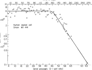
FIG. 3. T h e number of doublings of a diploid cell strain (WI-44) from human adult lung tissue from time of primary expiant to phase III decline. After Hayflick (1965).
comparison with heteroploid cells which show the opposite characters (Eagle, 1965). In other words, certain restrictions on growth are still evident in the diploid cell lines. In heteroploid cell lines, precise control of all replication is lost, as well as control over special cell functions. The importance of these generalizations is emphasized by the work of Hayflick (1965). He showed that human diploid cell strains cease to divide not as a function of the number of subcultures, but according to the number of cell doublings (i.e., about 40-60 generations), as shown in Fig. 3. More- over, it appears that the number of doublings occurring in adult lung tissue is lower than the number obtained from fetal lung tissue, which again emphasizes the fact that ontogenetic development does place in- creasingly greater restrictions on cellular capabilities, at least for growth.
HAPLOIDY VS. MPLOIDY IN REPRODUCTION OF CELL TYPE 227
Somatic cell genetics (Krooth, 1964) is rapidly becoming an important area of research, partly because of the implications for human genetics, for studies on differentiation, and for the recent advances in experimental somatic cell hybridization (Barski et al., 1960; Ephrussi and Weiss, 1965).
We can expect all the techniques of high resolution cell biochemistry to be utilized in the study of hybrid cells. Fusion of HeLa cells with rat lymphocytes, rabbit macrophages, and nucleated red blood cells of hens indicates a wide compatibility among cells of different species (Harris, 1965). The nuclei of these heterokaryons eventually fuse, and there is some evidence for stimulated RNA and DNA synthesis in differentiated cells following hybridization. These results are related to the earlier work of Moscona (1957) who obtained the reaggregation of dissociated em- bryonic cells of the chick and mouse. The application of these and other hybridization techniques to haploid cells of animals, and perhaps plants, can be expected in the future.
Plants
Totipotency is a character of diploid plant cells not generally shared by animal cells. Braun's (1959) demonstration of the recovery of normal plants from teratomatous crown gall tumors of tobacco was accomplished by isolating single cell clones of tissue, grafting into stems, and obtaining normal shoots from the implanted tissue. Induction of whole plants from diploid cells of carrots was achieved by Steward and co-workers (1964a,b) who suggested that free cells derived from young carrot embryos were virtually all totipotent if supplied with the appropriate nutrient. The free cells formed embryoids in a manner similar to that described for the zygote in the plant. Halperin and Wetherell (1964) have extended these studies to the wild carrot, presumably a more heterozygous plant than the inbred domestic carrot, and have found that callus tissues derived from various plant parts can also form adventive embryos. Regeneration from single cells of mosses (Ward, 1964) and ferns (Ito, 1960; Kato, 1964) as well as from tissue cultures of other plants such as Ranunculus sceleratus (Konar and Nataraja, 1965), Chicorium endiva, Lactuca sativa, and Petroselinum hortense (I. K. Vasil et al., 1964) and from the female game- tophytes of gymnosperms such as Cycas revoluta and Zamia floridana (La Rue 1948, 1954) and the derivation of tissues and plants from single isolated cells of tobacco by V. Vasil and Hildebrandt (1965) support the contention that plant cells are totipotent.
As far as differentiation is concerned, the gametophytes (n) of lower plants have been characterized by their lack of vascular tissue and this
228 W A L T E R TULECKE
has been considered typical of their haploid state. More recent work, how- ever, has shown that fern prothallia of Todaea barbara, which usually possess no vascular tissue, will develop such tissue when grown on the proper concentrations of sucrose and indole-3-acetic acid (Wetmore et al., 1964). Mosses lack vascular tissue, and there is no report of haploid sporophyte formation (Lai, 1963).
Apogamy, the formation of a sporophytic plant without gametic fu- sion, is a common occurrence in ferns and can be induced experimentally.
One of the more revealing methods of induction is that utilized by Whittier and Steeves (1960) and Whittier (1964). Prothallia of Pteridium acquilinum were supplied with carbon in the form of sugar, and induced to form both haploid and diploid sporophytes; prothallia that obtained carbon from photosynthesis produced no apogamous structures. Bristow (1962) was further able to show that cultures of Pteris cretica could be maintained as gametophytic in red light and be induced to form haploid apogamous leaves in white light; the leaves were then induced to form callus or sporophytes according to the amount of sugar provided. Induced organization of a callus culture of Lycopodium obscurum also produced, presumably, haploid sporophytes (De Maggio, 1964).
It is obvious that some of our conventional ideas about the potential of haploid cells for growth and differentiation will have to be corrected.
If a simple sugar can induce development of vascular tissue or complex formation of sporophytes, we are dealing with fundamental control mechanisms for form and function without the necessity of gametic fusion. This means that we know very little either about the mechanisms or about the limits of control of differentiation in haploids or diploids.
We have already noted that diploid animal cells appear to be limited in the number of duplications they undergo in vitro (Hayflick, 1965); plant cells do not appear to have this limitation. Relatively stable plant tissue cultures of diploid pea root callus were maintained by the use of a defined medium (Torrey, 1959); similar callus cultures on media containing 2,4-dichlorophenoxyacetic acid and yeast extract, changed from diploid
to tetraploid within one week. Other work on tissue cultures of carrot (Mitra et al, 1960) and Haplopappus gracilis (Mitra and Steward, 1961) grown on media supplemented with coconut water showed wide varia- tions in chromosome number. The Haplopappus cultures showed some instances of somatic pairing and several haploid cells, and it was assumed that somatic reduction had occurred. Even single cell clones of tobacco tissue (Cooper et al., 1964) were highly variable when grown on supple- mented media. Spruce tumor tissue (Risser, 1964) grown on defined media
HAPLOIDY VS. DIPLOIDY IN REPRODUCTION OF CELL TYPE 229
maintained a stable chromosome complement. Finally, the finding of Fox (1963) is important, since he observed that auxin- and cytokinin- independent strains of tobacco tissue possessed more stable chromosome numbers than strains requiring these compounds. A tentative generaliza- tion from these investigations seems to be that normal plant cells, like animal cells, can be grown indefinitely if grown as heteroploid popula- tions on supplemented media; diploid lines of plant cells are maintained with this complement if grown on defined media, especially if the tissues are autotrophic for growth hormones.
Haploid Tissue Cultures of Higher Plants
Male Gametophyte (Pollen)
Tissue cultures derived from the pollens of seed bearing plants are of interest for several reasons: They facilitate studies on pollen physiology;
they represent strains with genetically distinct characters when obtained from single pollen grains; they are useful for studies on differentiation and regeneration as extensions of the haploid male gametophyte.
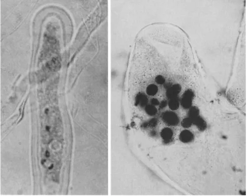
Ideally, the best material for pollen tissue cultures would be genetically well-known plants such as corn, tomato, or Oenothera. Despite attempts by several investigators, however (see La Rue, 1954; Tulecke, 1959, 1963) the pollens of angiosperms have not yet been induced to proliferate as haploid tissue cultures. Excellent pollen tube development was obtained from pollen of more than 30 species of angiosperms grown on various media, but the only indication of unusual nuclear division was observed in the pollen tubes of Antirrhinum ma jus (snapdragon). Extra nuclei in addition to the expected tube nucleus and two sperm nuclei were observed (Fig. 4). This development occurred on a defined medium containing 0.1 mg/ml of kinetin and 2,4-dichlorophenoxyacetic acid, and no cell divi- sion was obtained. These results are in contrast to the vigorous develop- ment of pollen tubes of gymnosperms such as Cupressus sempervirens (Fig. 4). This pollen germinates and grows on defined media for weeks or months whereas angiosperm pollens usually grow only for hours or days. Cupressus forms clusters of sperm nuclei within the pollen tube and represents a more primitive, less specialized condition of the male game- tophyte. Therefore, it is not surprising that gymnosperm pollens have been used successfully for the induction of androgenetic tissues.
The first tissue culture derived from pollen was obtained from Ginkgo biloba L. (Tulecke, 1953, 1957, 1960). Pollen grains distributed over the surface of nutrient media germinate and undergo either normal or ab-
230 WALTER TULECKE
normal development. Normal development proceeds from the four-celled microgametophyte (mature pollen grain) to the formation of two sperm cells, each about 100 μ in diameter and provided with cilia for motility.
Mature spermatozoids have been obtained in cultures, but no motile forms have been observed. Abnormal development involves extra divi- sions of the tube nucleus or the multiplication of cells from the original
FIG. 4. Observations on pollen development in vitro. Left, pollen tube of Antir- rhinum majus with three nuclei and dividing generative nucleus; right, cluster of male nuclei in pollen tube of Cupressus sempervirens.
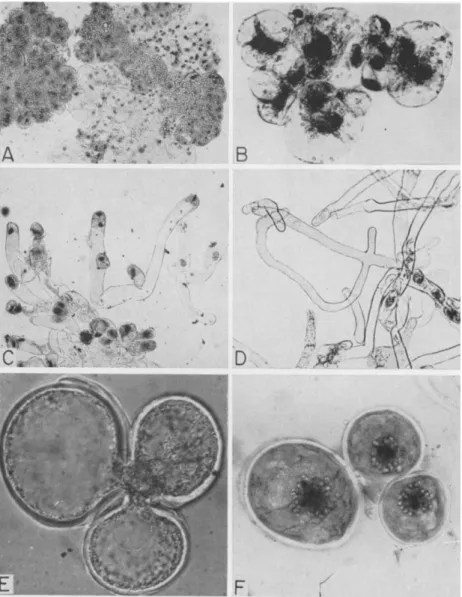
generative cell. Eventually, after 2-6 months growth, visible tissues may be seen in the pollen cultures. Some phases of this development are shown in Fig. 5. Other haploid tissue cultures of Taxus sp. (Tulecke, 1959), Torreya nucifera Sieb, and Zucc. (Tulecke and Sehgal, 1963), and Ephedra foliata Boiss. (Konar, 1963) have been derived from gymnosperm pollen. As shown in Fig. 6, the tissues have distinctive growth patterns.
Cells from the pollens of Ginkgo and Ephedra divide mitotically to form daughter cells in a manner typical of plants. The cells of the Taxus tissue
HAPLOIDY VS. DIPLOIDY IN REPRODUCTION OF CELL TYPE 231
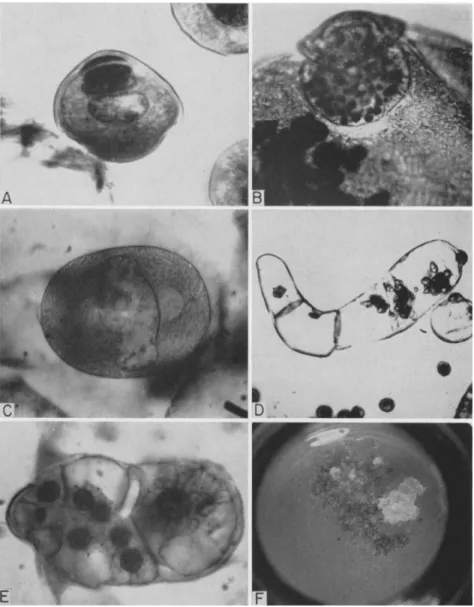
FIG. 5. Development of Ginkgo biloba pollen in vitro (normal): A, mature pollen grain; B, starch grains in plastids of generative cell; C, sperm mother cell prior to division to form two motile sperm; (abnormal): D, septate pollen tube; E, cluster of cells derived from the generative cell; F, tissue forming from pollen. The original pollen grain is about 25 μ in diameter and the sperm mother cell approximately 150 μ.
232 WALTER TULECKE
FIG. 6. Characteristics of haploid tissues derived from pollen: A, squash prepara- tion of Ginkgo tissue; B, cells of tissue from Ephedra pollen from Konar (1963); C-D, Taxus tissue showing pollen-tube type cells with nuclei often located terminally; E-F, yeast-like budding of Torreya tissue with derived uninucleate cells shown on the right.
The average cell size is about 50-200 μ.
HAPLOIDY VS. DIPLOIDY IN REPRODUCTION O F CELL T Y P E 233
grow in an elongated manner typical of growing pollen tubes; the nucleus and cytoplasm are localized at one end, and when division occurs unequal cells are formed, some of which are branched. T h e most unusual form of growth is observed in the cells of the tissue from Torreya pollen. Although they are very slow in growing, the cells divide by yeastlike budding. All of these tissues are white in appearance and show amyloplast development;
chlorophyll synthesis is apparently blocked. No differentiation beyond meristematic and parenchymatous cells is observed.
Observations on the chromosome complements of the tissues derived from the pollen of Taxus, Ginkgo, and Torreya indicate that haploid cells are still present in the tissue after several years in culture; polyploid
cells, including diploids and tetraploids, are also found in all tissues. T h e polyploid cells and nuclei are larger, but show no capacity for specialized differentiation.
Female Gametophyte
T h e female gametophyte of angiosperms is a much reduced structure, the embryo sac; in contrast, the same tissue in gymnosperms is large enough to bear two or more archegonia and to serve as a food reservoir for the developing embryo. Because of the generous size of the gametophy- tic tissue in Ginkgo biloba and because a haploid male gametophyte tissue was already available, a tissue culture of female origin was sought and obtained (Tulecke, 1964). T h e male and female gametophyte tissues are therefore available for comparative studies on maternal inheritance, plastid function, and differentiation.
The tissue culture of the female gametophyte proliferates well on a defined medium, contains chloroplasts, and is made up of several types of cells; chromosome counts indicate that the tissue is basically haploid.
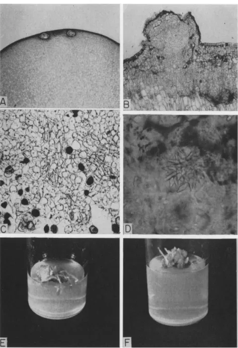
Some cells resemble those in the intact plant in their ability to form chloroplasts, store a resinous material, deposit a crystalline substance, differentiate tracheids, or continue growth as meristematic cells (Fig. 7).
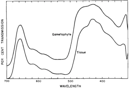
Absorption spectra of extracts from the female gametophyte and the tissue culture indicated that the pigments formed in vitro are similar to those found in the plant (Fig. 8). Quite often, female gametophyte tissues or cotyledons of gymnosperms are green while still within the seed. Appar- ently, all the precursors of pigment synthesis are formed and transported to the tissues and chlorophyll is formed in the dark before the seeds germinate. T h e female gametophyte tissue culture is normally green, but loses its chlorophyll when placed in the dark; growth proceeds equally well in the light and in the dark. On exposure to light the dark-grown
234 WALTER TULECKE
FIG. 7. Characteristics of a tissue culture derived from the haploid female gameto- phyte of Ginkgo biloba L.: A, section through the apical end of the female gametophyte
HAPLOIDY VS. DIPLOIDY IN REPRODUCTION OF CELL TYPE 235
tissue slowly regains its color. It is not yet known what precursors of chlorophyll are needed for greening in the dark, but the tissue should be useful for studying this phenomenon.
The differentiation of roots and shoots has been observed in some ex- periments with the female gametophyte tissue; examples are shown in Fig. 7. A total of 15 roots was observed originating from callus masses cultured on several different media. Abortive shoots were seen in only one
i 1 1 1 1 1 1 1 1 ι 1 1 1 1 1 i r
700 600 500 400 WAVELENGTH
FIG. 8. Absorption spectra of acetone extracts of Ginkgo biloba. Upper curve, the female gametophyte from the tree; lower curve, the tissue derived from the gametophyte.
culture in which five such organs were formed. Indirect evidence from cell and nuclear size indicated that the structures were haploid, but con- clusive chromosome counts still need to be obtained. Microscopic exami- nation of the roots and shoots revealed vascular strands; the ageotropic shoots possessed scalelike leaves and an abortive apical meristem. Several basal media and various supplements were used, but the most important component appeared to be purines such as the cytokinin, kinetin. Further
showing immature archegonia in the primary explant; explants lacking archegonia develop tissues equally well (the gametophyte is about 1 cm in diameter); B, growth of tissue from the primary explant; C, parenchyma, resin, and tracheidlike cells of the established tissue culture; D, crystal inclusion of cell from gametophyte tissue culture similar to inclusions in mature leaves; E, root formation; F, abortive shoot development.
236 WALTER TULECKE
work is needed to establish the quantitative and qualitative requirements for root and shoot formation.
In contrast to the specialized cell types found in the female gameto- phyte, no differentiation has ever been observed in the male gametophyte tissue of Ginkgo grown on a wide variety of media.
Conclusion
The evidence thus far available indicates that haploids of higher or- ganisms are limited in their ability to control full ontogeny, maturation, and reproduction. These deficiencies are probably related to the lack of complementary alleles normally present in the second genome. As a result, an autodiploid from a homozygous inbred species may be stunted, abnormal, or deficient; and one derived from a heterozygous species may be even more so. Furthermore, the maternal genome usually possesses a greater potential for controlling growth and development than the paternal; this is true of both plants and animals and is supported by evidence from gynogenetic and androgenetic haploids.
Our discussion has been limited, as far as possible, to the question of how haploidy and diploidy affect the development of cells and organisms.
We recognize, however, that the number of chromosomes is not highly important as long as the necessary characters are represented, but that gene sequence, the number of alleles, and factors affecting gene expression are important. T h e information obtained from haploids is useful because the state of complementary alleles has been largely eliminated. We have attempted to reduce the complexity of cells and organisms to a simpler, readable state; in this sense, haploids are useful and are likely to be increasingly helpful as more become available for experimental use.
Homozygosity is clearly the advantage offered by the haploids as experi- mental material. As potential breeding stock (i.e., as autodiploids), they are valuable gene pools of a certain uniformity. Hybrids of autodiploids of related species may show some pairing of chromosomes and these affini- ties are indicative of homologous chromosomes or segments. Hu (1960), for example, performed a karyotype analysis on two haploid species of rice and found similar bivalents and trivalents; he concluded that the five known discontinuous characters which separated the two species were not distinguishable at the chromosome level; hence, the species were rela- tively closely related. Studies of this type can be helpful in taxonomic investigations and in problems of phylogeny.
The usefulness of haploids is restricted by their relatively infrequent occurrence. Their use in breeding forest trees (Nei, 1963) or corn (Chase,
HAPLOID Y VS. DIPLOIDY IN REPRODUCTION OF CELL TYPE 237
1963), for example, will depend on improved techniques for inducing haploid plants. Two possible approaches for obtaining haploids would be the regeneration of plants from haploid tissue cultures and the induc- tion of somatic reduction followed by regeneration. These or other methods for obtaining haploid offspring would save time in achieving homozygosity and would aid genetic studies and breeding programs. The advantages, however, need to be weighed against the hazards of the generally poor development of haploids.
Androgenetic haploids of plants are of infrequent occurrence even among species where they are reported (Nicotiana, Crépis, Antirrhinum, and Zed). This fact alone emphasizes the need for obtaining tissue cul- tures from the pollens of angiosperms. When successful, this development should provide excellent material for the induction of androgenetic haploids. Such tissues could be used in investigations of the basic mecha- nisms which control male and female expressions and functions in plants.
From an evolutionary viewpoint, the diploid-haploid cycle of plants and animals has been selected as a mechanism for engendering variabil- ity. In the same way, many flowering plants appear to be evolving from diploid to polyploid lines; the original diploids may become extinct and eventually new diploids may arise from the existing polyploids (Raven and Thompson, 1964). These pseudohaploids actually provide a means for genetic variation, selection, and subsequent adaptation in recurrent cycles, in a way not greatly different from the normal haplontic-diplontic life cycles described in mathematical terms by Lindenmayer (1964). T h e polyploids may be better balanced genetically, more heterozygous, and isolated from diploids; their rate of evolution, however, is slower and their mutants are concealed.
Little reference has been made here to cytoplasmic inheritance, since this subject has been discussed by other participants in this symposium (see chapters by Schiff and by Srb). However, the evidence that plastids and mitochondria contain DNA (Gibor and Granick, 1964) suggests that haploid plants and tissues will be especially useful in studying cytoplasmic inheritance. In particular, haploid tissues should be useful in investigat- ing the functions of organelles in cells with a single genome. Some of the questions of maternal and paternal inheritance may be answered by this approach.
ACKNOWLEDGMENTS
This work was supported in part by a grant from the National Science Foundation (GB 3618). The recent work on pollen cultures was performed with the excellent tech- nical assistance of Mrs. Nanda Sehgal whose help is acknowledged.
238 WALTER TULECKE
REFERENCES
BARSKI, G. S., SORIEUL, S., AND CORNEFERT, F. (1960). Production dans des cultures in vitro de deux souches cellulaires en association, de cellules de caractère "hybride."
Compt. Rend. 251, 1825-1827.
BRAUN, A. C. (1959). A demonstration of the recovery of the crown-gall tumor cell with the use of complex tumors of single cell origin. Proc. Natl. Acad. Sei. U.S. 45, 932- 938.
BREWBAKER, J. L., AND EMERY, G. C. (1962). Pollen radiobotany. Radiation Botany 1, 101-154.
BRIDGES, C. B. (1925). Haploidy in Drosophila melanogaster. Proc. Natl. Acad. Sei. U.S.
11, 706-710.
BRIGGS, R. (1949). T h e influence of egg volume on the development of haploid and diploid embryos of the frog, Rana pipiens. J. Exptl. Zool. I l l , 255-294.
BRIGGS, R., AND KING, T . J. (1959). Nucleocytoplasmic interactions in eggs and embryos.
In " T h e Cell" (J. Brächet and A. E. Mirsky, eds.), Vol. 1, p p . 537-617. Academic Press, New York.
BRISTOW, J. M. (1962). T h e controlled in vitro differentiation of callus derived from a fern, Pteris cretica L., into gametophytic or sporophytic tissues. Develop. Biol. 4, 361-375.
BURNS, V. W. (1956). Temporal studies of cell division. I. T h e influence of ploidy and temperature on cell division in S. cerevisiae. J. Cellular Comp. Physiol. 47, 357-375.
CHASE, S. S. (1963). Androgenesis—its use for transfer of maize cytoplasm. / . Heredity 54, 152-158.
CLARK, A. M., BERTRAND, H . A., AND SMITH, R. E. (1963). Life span differences between haploid and diploid males of Habrobracon serinopae after exposure as adults to X rays. Am. Naturalist 97, 203-208.
COOPER, L. S., COOPER, D. C , HILDEBRANDT, A. C , AND RIKER, A. J. (1964). Chromosome
numbers in single cell clones of tobacco tissue. Am. J. Botany 51, 284-290.
D E MAGGIO, A. E. (1964). Organization in a gametophyte callus of Lycopodium and its morphogenetic implications. Proc. Natl. Acad. Set. U.S. 52, 854-859.
EAGLE, H. (1965). Metabolic controls in cultured mammalian cells. Science 148, 42-56.
EPHRUSSI, B., AND WEISS, M. C. (1965). Interspecific hybridization of somatic cells. Proc.
Natl. Acad. Set. U.S. 53, 1040-1042.
Fox, J. E. (1963). Growth factor requirements and chromosome number in tobacco tissue cultures. Physiol. Plantarum 16, 793-803.
GIBOR, A., AND GRANICK, S. (1964). Plastids and mitochondria: inheritable systems.
Science 145, 890-897.
GOODSELL, S. F. (1961). Male sterility in corn by androgenesis. Crop Sei. 1, 227-228.
HALPERIN, W., AND WETHERELL, D. F. (1964). Adventive embryony in tissue cultures of the wild carrot, Daucus carota. Am. J. Botany 51, 274-283.
HARRIS, H . (1965). Behaviour of differentiated nuclei in heterokaryons of animal cells from different species. Nature 206, 583-588.
HAYFLICK, L. (1965). T h e limited in vitro lifetime of human diploid cell strains. Exptl.
Cell Res. 37, 614-636.
Hu, C.-H. (1960). Karyological studies in haploid rice plants. IV. Chromosome mor- phology and intragenome pairing in haploid plants of Oryza glaberrima Steud., as compared with those in O. sativa L. Cytologia (Tokyo) 25, 437-449.
HAPLOIDY VS. DIPLOIDY IN REPRODUCTION OF CELL TYPE 239
HUMPHREY, R. R., AND FANKHAUSER, G. (1957). T h e origin of spontaneous and experi- mental haploids in the Mexican axolotl (Siredon or Ambystoma mexicanum). J.
Exptl. Zool. 134, 427-447.
ITO, M. (1960). Complete regeneration from single isolated cells of fern gametophyte.
Botan. Mag. (Tokyo) 73, 267.
IVANOV, M. A. (1938). Experimental production of haploids in Nicotiana rustica L.
Genetica 20, 295-386.
JARANOWSKI, J. (1961). Haploid-diploid twin embryos in Melilotus. Genet. Polon. 2, 129- 137.
KATO, Y. (1964). Physiological and morphogenetic studies of fern gametophyte by aseptic culture. III. Growth and differentiation of single cells isolated from callus tissues of Pteris vittata. Cytologia (Tokyo) 29, 79-85.
KIHARA, H., AND TsuNEWAKi, K. (1962). Use of an alien cytoplasm as a method of pro- ducing haploids. Japan. J. Genetics 37, 310-313.
KIMBER, G., AND RiLEY, R. (1963). Haploid angiosperms. Botan. Rev. 29, 480-531.
KING, T . J., AND BRIGGS, R. (1955). Changes in the nuclei of differentiating gastrula cells, as demonstrated by nuclear transplantation. Proc. Natl. Acad. Sei. U.S. 41, 321-325.
KNAPP, E. (1939). Haploïde Pflanzen von Antirrhinum majus. Ber. Deut. Botan. Ges.
57, 371-379.
KONAR, R. N. (1963). A haploid tissue from the pollen of Ephedra foliata Boiss. Phyto- morphology 13, 170-174.
KONAR, R. N., AND NATARAJA, K. (1965). Differentiation of embryoids in tissue cultures of floral buds of Ranunculus sceleratus L. Naturwissenschaften 52, 140-141.
KROOTH, R. S. (1964). "Somatic Cell Genetics." Univ. of Michigan Press, Ann Arbor, Michigan.
LAL, M. (1963). Experimental induction of apogamy and the control of differentiation in gametophytic callus of the moss Physcomitrium coorgense Broth. Plant Tissue Organ Cult., Symp., Delhi, 1961, pp. 363-381. Intern. Soc. Plant Morphologists, Univ.
of Delhi, Delhi, India.
LA RUE, C. D. (1948). Regeneration in the megagametophyte of Zamia floridana. Bull.
Torrey Botan. Club 75, 597-603.
LA RUE, C. D. (1954). Studies on growth and regeneration in gametophytes and sporo- phytes of gymnosperms. Brookhaven Symp. Biol. 6, 187-208.
LINDENMAYER, A. (1964). Life cycles as hierarchical relations. In "Form and Strategy in Science" (J. R. Gregg and F. T . C. Harris, eds.), p p . 416-470. Reidel Publ. Co., Dordrecht, Netherlands.
LINDSTROM, E. W. (1941). Genetic stability of haploid, diploid, and tetraploid genotypes in the tomato. Genetics 26, 387-397.
MANGELSDORF, P. C , AND FRAPS, G. S. (1931). A direct quantitative relationship between vitamin A in corn and the number of genes for yellow pigmentation. Science 73, 241-242.
MEYER, J. R., AND JUSTUS, N. (1961). Properties of doubled haploids of cotton. Crop. Sei.
1, 462-464.
MITRA, J., AND STEWARD, F. C. (1961). Growth induction in cultures of Haplopappus gracilis. II. T h e behavior of the nucleus. Am. J. Botany 48, 358-368.
MITRA, J., MAPES, M. O., AND STEWARD, F. C. (1960). Growth and organized development of cultured cells. IV. T h e behavior of the nucleus. Am. J. Botany 47, 357-368.
240 WALTER TULECKE
MOORE, J. A. (1955). Abnormal combinations of nuclear and cytoplasmic systems in frogs and toads. Advan. Genet. 7, 139-182.
MORTIMER, R. K., AND VON BORSTEL, R. C. (1963). Radiation-induced dominant lethality in haploid and diploid sperm of the wasp Mormoniella. Genetics 48, 1545-1549.
MOSCONA, A. (1957). T h e development in vitro of chimeric aggregates of dissociated embryonic chick and mouse cells. Proc. Natl. Acad. Sei. U.S. 43, 184-194.
NAITHANI, S. P. (1937). Chromosome studies in Hyacinthus orientalis L. III. Reversal of sexual state in the anthers of Hyacinthus orientalis L„ var. Yellow Hammer. Ann.
Botany (London) [N.S.] 1, 369-377.
NEI, M. (1963). T h e efficiency of haploid method of plant breeding. Heredity 18, 95-100.
OLSEN, R. H., AND OGG, J. E. (1963). Effect of haploid or diploid constitution of Escherichia coli on formic hydrogenlyase activity. / . Bacteriol. 86, 494-498.
PARTANEN, C. R. (1965). On the chromosomal basis for cellular differentiation. Am. J.
Botany 52, 204-209.
RAVEN, P. H., AND THOMPSON, H. J. (1964). Haploidy and angiosperm evolution. Am.
Naturalist 98, 251-252.
RISSER, P. G. (1964). Somatic mitoses in cells of Picea glauca cultivated in vitro. Science 143, 591-592.
RUDKIN, G. T., ARONSON, J. F., HUNGERFORD, D. A., AND SCHULTZ, J. (1955). A comparison
of the ultraviolet absorption of haploid and diploid salivary gland chromosomes.
Exptl. Cell Res. 9, 193-211.
SELMAN, G. G. (1958). An ultra-violet light method for producing haploid amphibian embryos. / . Embryol. Exptl. Morphol. 6, 632-635.
SPARROW, A. H., CUANY, R. L., MIKSCHE, J. P., AND SCHAIRER, L. A. (1961). Some factors affecting the responses of plants to acute and chronic radiation exposures. Radiation Botany 1, 10-34.
STEWARD, F. C , BLAKELY, L. M., KENT, A. E., AND MAPES, Μ. O. (1964a). Growth and
organization in free cell cultures. Brookhaven Symp. Biol. 16, 73-88.
STEWARD, F. C , MAPES, M. O., KENT, A. E., AND HÜLSTEN, R. D. (1964b). Growth and
development of cultured plant cells. Science 143, 20-27.
STOW, I. (1933). On the female tendencies of the embryosac-like giant pollen grain of Hyacinthus orientalis. Cytologia (Tokyo) 5, 88-108.
SUBTELNY, S. (1958). T h e development of haploid and homozygous diploid frog embryos obtained from transplantations of haploid nuclei. / . Exptl. Zool. 139, 263-298.
SUBTELNY, S. (1965). Personal communication.
SUOMALAINEN, E. (1950). Parthenogenesis in animals. Advan. Genet. 3, 193-253.
TORREY, J. G. (1959). Experimental modification of development in the root. In "Cell, Organism and Milieu" (D. Rudnick, ed.), p p . 189-222. Ronald Press, New York.
TULECKE, W. (1953). A tissue derived from the pollen of Ginkgo biloba. Science 117, 599-600.
TULECKE, W. (1957). T h e pollen of Ginkgo biloba: in vitro culture and tissue formation.
Am. J. Botany 44, 602-608.
TULECKE, W. (1959). T h e pollen cultures of C. D. La R u e : a tissue from the pollen of Taxus. Bull. Torrey Botan. Club 86, 283-289.
TULECKE, W. (1963). Unpublished work.
TULECKE, W. (1960). Arginine-requiring strains of tissue obtained from Ginkgo pollen, Plant Physiol. 35, 19-24.
HAPLOIDY VS. DIPLOIDY IN REPRODUCTION OF CELL TYPE 241
TULECKE, W. (1964). A haploid tissue from the female gametophyte of Ginkgo biloba L.
Nature 203, 94-95.
TULECKE, W., AND SEHGAL, N. (1963). Cell proliferation from the pollen of Torreya nuci- fera. Contrib. Boyce Thompson Inst. 22, 153-163.
VASIL, I. K., HILDEBRANDT, A. C , AND RIKER, A. J. (1964). Endive plantlets from freely suspended cells and cell groups grown in vitro. Science 146, 76-77.
VASIL, V., AND HILDEBRANDT, A. C. (1965). Differentiation of tobacco plants from single, isolated cells in microcultures. Science 150, 889-892.
WARD, M. (1964). Gametophytic plants induced from single cells of moss callus. Nature 204, 400.
WETMORE, R. H., D E MAGGIO, A. E., AND RIER, J. P. (1964). Contemporary outlook on the differentiation of vascular tissues. Phytomorphology 14, 203-217.
WHITTIER, D. P. (1964). The influence of cultural conditions on the induction of apog- amy in Pteridium gametophytes. Am. J. Botany 51, 730-736.
WHITTIER, D. P., AND STEEVES, T. A. (1960). The induction of apogamy in the bracken fern. Can. J. Botany 38, 925-930.