Abnormal Growth in Plants
ARMIN C. BRAUN
I. Harmonious Changes Involving Exaggerated Growth Responses . . . . 379
A. Generalized Stimulation 379 II. Amorphous Changes in Growth Pattern 384
A. Self-Limiting Overgrowths 384 B. Non-Self-Limiting Overgrowths 394
References 415
I. Harmoniou s Change s Involvin g Exaggerate d Growt h Response s
A. GENERALIZED STIMULATION
One of the most characteristic features of higher plants and animals is the extraordinary way in which all their functional parts fall into a coherent, flexible but definitely limited pattern. In the normal organism, morphogenetic laws are strictly obeyed and processes concerned in metabolism, growth, cellular differentiation, and organogenesis are pre- cisely regulated. These processes start and stop in harmony to give an organism certain proportions that remain constant from one generation to the next. Sometimes, however, this harmony of structure and function is brought to our attention by its sudden failure. Such divergence from the normal may represent only slight quantitative changes which are evidenced by harmonious but somewhat exaggerated manifestations of normal developmental processes. This sort of response is well illustrated in the bakanae disease of rice (Oryza sativa), which is caused by Gibbered fujikuroi, as well as in several other plant diseases that are caused by the rusts ( 1 5 ) . In those instances the host metabolism is affected by virtue of the fact that excessive amounts of the gibberellins are synthesized. Certain other plant diseases give indication that the process of infection interferes either with the synthesis or with the utilization of the gibberellins. Carrot (Daucus carota var. sativus) plants infected with the tomato big bud virus of California wTere shown by Kunkel (41) to bolt and flower prematurely. These findings suggest that virus infection may increase the synthesis of the gibberellins by the
379
380 ARMIN C . BRAUN
hos'j species. On the other hand, the rosette diseases, as exemplified by peach and peanut rosette viral infection, may interfere with the synthe- sis or utilization of such growth regulating substances by the plant. It has, in fact, been possible to reverse the stunting effects, but not other symptoms, associated with aster yellows and the corn stunt disease by applying gibberellic acid to an infected plant ( 6 0 ) .
Nastic responses, which refer to changes in position of a bilaterally symmetrical organ as a result of differential growth, are a common manifestation of certain diseases. Dimond and Waggoner (26) made a detailed study of the cause of epinasty in Fusarium wilt of tomatoes
(Lycopersicon esculentum). It was concluded from those studies that ethylene is responsible for the characteristic growth response in that instance. This conclusion was based on the observation that ethylene is produced in culture by the causal fungus. Ethylene production by in- fected tomato plants was, moreover, demonstrated by confining such plants with healthy indicator plants and by showing that epinastic responses developed to a greater degree in the indicator plants than when such plants were confined with healthy tomato plants.
Another characteristic response involving growth movements is con- cerned with the upright growth habit of leaves. This is one of the most characteristic symptoms of the aster yellows disease. In plants infected with that virus the petioles elongate and assume an extremely upright growth habit which approximately parallels the main axis of the plant.
It is this upright growth habit that suggested the trivial name "rabbit ears" for lettuce (Lactuca sativa) plants infected with that virus. Hy- ponasty has also been observed in plants affected with the bacterium Erwinia phytophthora.
The development of organs that arise in unusual places has been reported to be a characteristic response of plants infected with bacterial, fungal, and viral diseases.
Carrot yellows virus from Texas as well as the aster yellows virus stimulate the production of small aerial tubers in the axils of the leaves of potato (Solanum tuberosum) plants. The witches' broom virus, on the other hand, causes the infected plant to produce numerous buds at the nodes in the aboveground stems of potato plants. Long slender stolons that resemble aerial roots, but which are covered with trichomes, develop from these adventitious buds.
The development of adventitious roots on stems of plants has been reported to be a characteristic response in tomato following infection with Pseudomonas solanacearum, Corynebacterium michiganense, Fu- sarium oxysporum, F. oxysporum f. lycopersici ( F . lijcopersici) as well as the cranberry false-blossom virus. An extreme example of this is
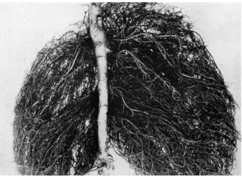
FIG. 1 . Hairy root on apple (Malus sylvestris). This condition is caused by a specific bacterium. (Courtesy of A. J. Riker.)
found in the hairy-root disease of apples (Malus sylvestris) (see Fig. 1 ) . Grieve (30) suggested, as a result of his studies on the Granville wilt disease of tomatoes, that adventitious roots very probably result from disturbances of normal auxin transport in the host as a result of me- chanical blockage rather than from indoleacetic acid elaborated by the bacteria. In the case of Fusarium wilt of tomato, Dimond and Wag- goner (26) indicate that adventitious root formation, like epinasty, may result from the production of ethylene either by the causal fungus or by the cells of the tomato plant as a result of the interaction of the host and pathogen.
1. Modification of Leaves
The curling and distortion of leaves is so characteristic of certain plant diseases that such maladies as leaf roll of potatoes, curly top of sugar beets (Beta vulgaris), and peach leaf curl have been assigned their trivial names because leaf curling is the most conspicuous feature of those diseases. Peach leaf curl, which is caused by Taphrina deformans, has been studied intensively. In that instance the causal fungus infects very young leaves which soon become curled and puckered, have a firm
382 ARMIN C . BRAUN
consistency, and increase greatly in thickness. The host cells which are in the vicinity of the fungus are stimulated to enlarge and, to a lesser extent, to divide. The cells of the palisade parenchyma respond much more actively to the stimulus of the fungus than do the adjacent cells of the spongy parenchyma, and this leads to differential growth with a resulting puckering and curling of the leaf. Such morphological ab- normalities are doubtless due to hormonal imbalances resulting from the infection by the fungus.
2. Frenching
A deformity of tobacco (Nicotiana tabacum) which is commonly known as frenching is found in most tobacco growing regions of the world. An advanced stage of this condition is characterized by a cessa- tion of terminal bud and stem growth. When apical dominance is lost, the buds in the axils of the leaves develop and unusually large numbers of leaves, which may number as many as 300, appear on a plant. Such leaves are characteristically sword- or string-shaped because of failure of the leaf blades to develop. Although the cause of frenching has not been unequivocally established, it is now believed to be a toxicity dis- ease rather than to result from nutritional deficiency. No parasitic or- ganism of any kind has as yet been implicated in this condition.
Steinberg (92, 93) has suggested that frenching of tobacco is caused by a toxic metabolite elaborated by the widely distributed nonpatho- genic soil bacterium Bacillus cereus. The progressive development and type of symptoms produced under controlled conditions in tobacco plants by the toxic metabolite largely paralleled those found in frenched plants in the field. In later studies Steinberg (94) found that the natural isomers of isoleucine and alloisoleucine, when used in concentrations of 2-8 ppm, produced a strapping of leaves of young tobacco plants. This worker found, moreover, that frenching was accompanied by a signifi- cant increase in isoleucine as well as certain other free amino acids in the leaf lamina of field grown plants ( 9 5 ) . The absence of free amino acids in soils that leads to frenching and the marked increase of free isoleucine in frenched leaves indicate that the Bacillus cereus toxin and isoleucine are not identical. It was therefore concluded that the accumu- lation of excessive amounts of free amino acids in strapped leaves was a probable chemical factor involved in the production of the morphological abnormalities observed in frenched tobacco plants.
3. Alterations in Floral Parts
Under the influence of pathogenic agents of various types the sepals, petals, stamens, or pistils of a flower may be altered into structures that
are very different in appearance from those found normally. In the United States there are at least three different viruses—the aster yellows virus, the tomato big bud virus, and the cranberry false blossom virus—
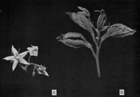
that produce gigantism in the sepals and calyxes of certain solanaceous plant species. The tomato big bud virus appears to affect the sepals of tomato flowers rather specifically (see Fig. 2 ) . These structures en- large greatly under the influence of the virus and fuse to form huge bladderlike structures that may be ten times or more the normal size.
FIG. 2. (A) Flower truss of a healthy tomato plant. (B) A similar truss of the same age infected with tomato big-bud virus. (Courtesy of L. O. Kunkel.) Viruses of this type do not cause gigantism in the sepals of certain other plant species. In Catharanthus roseus (Vinca rosea) such viruses bring about a green coloring of the petals, stamens, and styles. There is, in this instance, a retrograde development of floral parts into foliage leaves.
Metamorphoses of floral parts of higher plants are also accomplished by certain species of fungi. The transformation of stamens into petals commonly follows infection by the rust Puccinia violae on Viola sylves- tris. Stamen primordia found in flowers of Knautia arvensis develop into petallike structures following infection by Peronospora violacea.
Many other such examples could be cited.
384 ARMIN C . BRAUN
Bos ( 7 ) has interpreted the development of the flower and its com- ponent parts on the basis of the phenomenon of antholysis accompany- ing virus infection in Crotalaria. This investigator has suggested on the basis of the homology of reproductive and vegetative parts of a plant and their morphogenetic development that the vegetative and sexual activities in a plant are mutually antagonistic. A growing point can de- velop only into a vegetative shoot or into an inflorescence. During flower initiation, the vegetative characteristics are suppressed and the sexual characters prevail. The reverse is also true. From the manner of ap- pearance of antholysis in virus-infected Crotalaria plants, Bos has con- cluded that flower induction is suddenly stopped and the subsequent de- velopment of floral parts proceeds exclusively in a vegetative manner.
The ultimate result depends on the stage of the development of the primordia in the bud at the time of the suppression of flower induction by the virus. Thus, flowers initiated in succession produce a series of flowers showing increasing antholysis. These in reverse order show a macroscopically recognizable picture of the morphogenesis of the flower and its component parts. Therefore, according to Bos, antholysis sup- ports the theory that the flower must be regarded as a modified leafy branch.
In addition to modifying floral parts, infection with certain fungi re- leases growth restraints and thus overcomes the normally arrested de- velopment of such parts. For example, the anther smut, Ustuago vio- lacea, produces its spores only in the anthers of certain plant species.
When that fungus establishes itself in the female flower of Mehndrium album or M. dioicum, the stamens, which are normally arrested in their development and are found only in rudimentary form in the pistillate flowers, grow to full size and when mature are filled with smut spores rather than with pollen. The nature of the morphogenetic stimulus that overcomes the arrested development of the male sex organs in female flowers remains unknown.
II. Amorphou s Change s i n Growt h Patter n
A. SELF-LIMITING OVERGROWTHS
1. Galls
Localized overgrowths in which the host cells are stimulated to ex- cessive growth by a variety of disease-producing agents are known as galls. There are unusually large numbers of distinct overgrowths of this type to be found in plants. Among the more representative types are the insect and nematode galls, the cedar apples produced by the apple
rust on the juniper (Juniperus), Pteridermium galls on pine (Pinus), the so-called smut "balls/' the black knots on plum and cherry (Prunus spp.), the overgrowths due to certain species of slime molds and chytrids, the galls produced by Pseudomonas savastanoi on the olive (Olea europaea), and the nodules that result in certain species of leguminous plants from infection by bacteria of the genus Rhizobium. It is, of course, not possible to consider in detail in this chapter all the overgrowths of this type, and only a few representative examples have been selected for discussion.
a. Insect galls. Among the most interesting types of self-limiting growth abnormalities found in plants are those that result from the activity of certain of the gall-forming insects. The whole subject of in- sect galls as reviewed in detail by Kiister (44), Ross and Hedicke (79), and more recently by Felt (29) suggests that many of these highly specialized overgrowths represent beautiful examples of dependent dif- ferentiation.
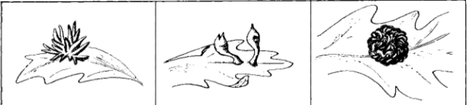
Insect galls may result from either a mechanical or a chemical stimu- lus. Chemical stimulation is probably far more important than mechani- cal irritation in the production of many insect galls, although the chemi- cal stimulus may in some instances be supplemented by directive feeding of the insect. An examination of the literature leaves the un- mistakable impression that highly specific morphogenetic stimuli, of chemical nature and elaborated by insects, are capable of initiating, stimulating, and directing most precisely the development and differen- tiation of plant cells. There is no question about the fact that the morphological form that a gall assumes depends upon the nature of the pathogenic insect. This is evidenced by the fact that the same host species or even the same organ of the same host attacked by different but closely related insect species produces morphologically very different galls. Figure 3 illustrates this point and shows drawings of three galls of quite different morphology induced on leaves of a western species of oak by three closely related species of cynipids. The morphology of the
FIG. 3 . Three morphologically distinct galls produced on leaves of the California white oak by three closely related species of insects. (Drawings by R. J. Man- dlebaum.)
386 ARMIN C . BRAUN
gall is so specific that it is considered by some to be a more reliable criterion for distinguishing between closely related species of insects than are the morphological characters of the adult insects. The morphol- ogy of the gall does not, moreover, appear to be a function of the part of the plant from which it arises. Currant galls of the oak, which are found on both flower stalks and leaves, have similar morphological struc- ture at both points of origin. The same insect species may, moreover, produce the same gall type on different species of plants. The sawfly, Micronematus gallicola, produces bright red galls of similar morphology on four different species of willow (Salix). Findings such as those re- ported above suggest in the strongest possible manner that a large number of highly specific chemical substances are elaborated by insects and that these substances have specific morphogenetic effects on the cells and tissues of a plant. However, only moderate success has thus far been achieved in establishing experimentally the truth of this view.
It is, of course, not possible to describe in detail in a chapter such as this all the diverse morphological structures that have been described for insect galls. Only a few interesting types within each of the larger groupings will be considered. Insect galls have been classified as:
(i) simple galls in which a single animal or colony of animals is limited to a single plant organ, and (ii) compound galls where two or more plant organs may be concerned in the production of the gall. For convenience of description, simple galls have been divided into: ( a ) felt galls,
( b ) mantle galls, and ( c ) solid galls. The felt galls result chiefly from hypertrophied epidermal cells growing out into hairy coverings of vari- ous sorts and shapes. Many felt galls are caused by gall mites. They form cottony growths on limited and sharply defined areas, usually on the undersurface of the leaf. Microscopic studies have shown that in the formation of the felted galls the epidermal cells swell out and be- come transformed into bent and twisted tubes that may be shaped like a club. The mites which produce the felt galls deposit their eggs in elongated cells and their young live on the materials contained within the cells.
The mantle gall is a hollow structure which always has a portion of the surface of the affected organ for its lining. This type of gall, which may arise in various ways and assume a variety of forms, is in effect a chamber formed by hypertrophied growth around the place occupied by the insect. Thus, the insects that give rise to galls of this type live on the surface of the leaves where they multiply and attach their eggs to the epidermis. The growth stimulus produced by the insect excites the leaf tissue to growth which forms a protecting mantle for the animal.
The mantle galls have been divided according to structure into scroll
galls, pocket galls, and covering galls. Scroll galls usually involve the blades and more rarely the petioles of the leaves. As a result of the presence of these insects on a leaf surface the leaf grows more profusely on one side than on the other. This causes the leaf to curl lengthwise and form a scroll or chamber in which the animals are embedded. It is always the side of the leaf on which the insects live that becomes con- cave. In many instances the whole leaf lamina is rolled. In other cases the alteration is restricted to the margins of the leaf.
The pocket galls are closely allied to the scroll forms. In galls of this type the tissue of the leaf lamina or petiole is subjected to the stimulus from the animals with the result that a hollow protuberance arises, the cavity of which serves to protect the insects. Pocket galls exhibit a great variety of form and shape, some being wrinkled or plaited; others, saclike structures attached to the leaf by a very narrow neck. Many of these galls project from both the upper and lower side of the leaf. The pocket gall of the sloe (Prunus spinosa), caused by a gall mite, projects almost as much from the underside as from the upper side of the leaf. On the other hand, a similar gall on the bird cherry (Prunus padus) rises on the upper side as a long pocket but projects on the lower side only as a small wart.
The third type of mantle gall is the covering gall. In this instance the tissue begins to grow at the point at which the insect has settled or where an egg has been fastened to the epidermis. The tissue rises up in the form of a fleshy wall which continues to grow until the animal is roofed in. The cavity in this instance arises from an overarching of the tissue.
The third group of the simple galls is the so-called solid galls. These are produced by insects which pierce the plant tissue and lay their eggs in the wound. The common gall with the mosslike covering occurring on the wild rose (Rosa) and caused by the gall wasp Rhodites rosae is an example of this type. The wasp deposits its pointed or sometimes hooked eggs early in the spring into an undeveloped leaf which is still folded in the bud. The first sign of alteration in leaf structure following insertion of the eggs is the development of numerous hairlike proc- esses. When the eggs hatch, the larvae penetrate deeper into the tissues of a leaf and the stimulated leaf cells grow out into a solid gall contain- ing as many chambers as there are larvae. Hairs continue to form on the exterior until the characteristic structure of the gall results.
The structure of the wall of the larval chamber in the solid galls may differ in the galls induced by different insects. There is, however, always a layer of thin-walled cells immediately surrounding the egg, which is known as the medulla or pith, and an outer layer composed of cortex
388 ARMIN C . BRAUN
and epidermis which surrounds the inner layer like a skin or bark. In many instances there is a third layer inserted between the pith and cortical layer which is composed of very hard cells and which forms a protective layer. The medulla or pith is composed of thin-walled cells rich in nutrients. These cells provide the necessary food for the larvae.
The cells of the gall pith remain capable of division as long as the larvae require food and the cells are replaced almost as soon as they are eaten.
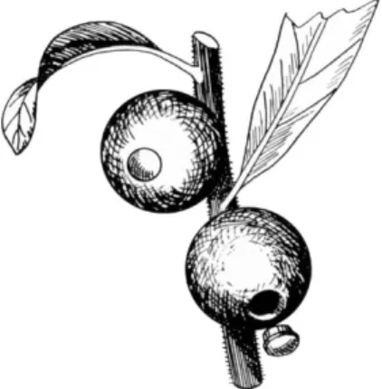
In the solid galls the hard, cortical, and epidermal cell layers are modified in many ways to protect the galls from drying out as well as to protect them from birds and other animal predators. Most solid galls completely enclose the chamber in which the larva dwells. The insect when it emerges must itself make an exit passage from the gall. The gall wasp and certain other insects do this by biting a hole through the gall with their mandibles. In other instances far more subtle methods of escape have been developed. The openings in some galls, which resemble operculate capsules, are especially remarkable. The gall gnat Cecidomyia cerris produces a solid gall on the turkey oak (Quercus ccrris). This gall is so embedded in a leaf that it projects on the upper side as a small pointed cone and on the lower side as a disk covered with a thick coating of hairs. In the autumn a circular piece like a lid which is sharply defined becomes detached from the lower side of the chamber. The operculum falls out of the gall and the larva drops to the ground, where it pupates. Another remarkable example of this type is found in galls produced on the green cortical tissue of young twigs of Schinus longifolia by Cecidoses eremita. This gall is spherical and quite hard. When the time approaches for the larva to leave the gall, a plug with a projecting rim is developed on the side of the gall farthest from its point of attachment to the plant. When the plug drops out, a circular hole is left which leads into the gall chamber, as shown in Fig. 4. It is through this hole that the larva escapes from the gall.
Very little is as yet known concerning the precise mechanism by which the solid galls are produced. Nevertheless, Lewis and Walton (49) reported some very interesting histological and cytological findings dealing with a biologically active substance believed to be involved in the growth and differentiation of cells of a solid gall of witch-hazel (Hamamelis virginiana) known as the cone gall. This gall is produced by the aphid species Hormaphis hamamelidis Fitch. In this instance the stem mother inserts her stylets into and between the cells of an immature leaf and injects very minute droplets of a substance secreted by glands opening into the stylar canal. This stinging process is not to be con- fused with feeding and the sting substance is quite different in its
FIG. 4. Drawing of galls produced on Schinus longifolia by Cecidoses eremita.
This figure is included to illustrate one of the more subtle types of escape mechan- isms used by the insect to leave the gall. As the time approaches for the larva to leave the gall, a plug with a projecting rim is somehow cut out of the gall. The plug drops out at the appropriate time, and the larva escapes. (Drawing by R. J.
Mandlebaum.)
nature from substances found in the salivary secretions. When about 150 stings have been made in a small circular area by the stem mother, the cells in this region at first become etiolated. Almost immediately thereafter cells receiving injected sting material dedifferentiate, undergo rapid mitotic divisions, and then redifferentiate into gall cells rather than typical leaf cells.
The injected sting material consists of a ground substance in which are embedded minute crystalloids. These crystalloids show a reddish- purple cast in such stains as gentian violet, congo red, and acid fuchsin.
They are Feulgen positive. They are, moreover, capable of passing readily from cell to cell as well as of entering cells when they are deposited by the insect in the intercellular spaces. Whether the injection is inter- or intracellular, the end result is the entry of the crystalloids into the nucleolus. There they may fuse to form a large crystalloid which again breaks up into smaller ones as mitosis begins. During nuclear division the crystalloids are distributed to the daughter nuclei where they are again found in the nucleolus. Since these bodies are apparently incapable of self-propagation and since they appear to be used up during growth of the cells, fresh sting material must be injected repeatedly by the stem mother during the entire growth of the gall.
Continued development of the gall is, therefore, dependent upon con- tinued presence of the Feulgen-positive material.
Boysen-Jensen ( 8 ) studied the development of a midge (Mikiola
390 ARMIN C. BRAUN
jcigi) gall on beech (Fagus) leaves. The evidence presented supports the idea that the formation of the gall in this instance is caused by the growth substances given off by the larva. These as yet uncharacterized substances produce cell enlargement and cell division, but not organized growth. It was therefore suggested that cell enlargement and cell divi- sion are regulated by the larva, which moves rapidly about the gall chamber and secretes the growth-promoting substances in definite places, thereby making the gall assume its special form. According to this inter- pretation the growth-promoting substance does not have special organiz- ing properties but the shape of the gall is dependent upon the distribu- tion of a rather nonspecific type of growth substance by the larva. The growth substances are, in other words, tools which are used by the gall larva to model a gall from the cells of a beech leaf. While such a mech- anism as that proposed by Boysen-Jensen may satisfactorily explain the formation of certain simple galls, it is difficult to see how it would explain the development of the highly differentiated and organized cynipid galls.
The compound insect galls originate when several organs of a plant immediately adjacent to one another are concerned in the production of the overgrowth. Such compound galls commonly arise from either vegetative or floral buds. They are so varied in their character that it is difficult to classify them.
The ordinary vegetative bud galls may be of essentially two types.
The first is leafless or more correctly a gall in which the leaves are transformed into knob- or peglike structures which merge into the swollen axis that contains the larva. The second type possesses leaves, the gall being covered by more or less fully developed green foliage leaves or with scalelike bracts.
Certain species of gall gnats lay their eggs in flower buds. The larva hatched from an egg lives in the cavity of the ovary which becomes the larval chamber. The corolla which covers the ovary in the flower bud remains closed and serves as a cap for the larval chamber. The calyx may become inflated, enlarged, and fleshy. The gall in this in- stance resembles a small bulb.
Closely allied to the bud galls are the galls known in Central Europe as "cuckoo-buds." These growths are characterized essentially by the fact that they involve only the base of the shoot, while the upper por- tion of the shoot continues its growth unaltered. The best known and most widely distributed gall of this type is produced by the aphid Chermes abietis on the twigs of the spruce (Ficea) tree. In the early spring the parthenogenic females attach themselves and lay eggs at the base of a young leaf. The larvae that develop from the eggs penetrate
the shoot with their beaks. The shoot swells considerably, as do the bases of the needles and the spruce gall results. The gall contains a series of cavities or chambers in which the larvae live. Usually in late summer the gall dries up, each small cavity opens by a slit, and the winged adults leave the gall. This gall resembles a small fir cone. That a morphogenetic stimulus is elaborated by the spruce-fir aphid is sug- gested by the fact that the aphid attaches itself firmly by its beak to a scale of a fir (Abies) bud and thus can directly influence only a few cells of the young shoot. Nevertheless, thousands of cells of such a shoot are affected and soon begin to assume an altered form and are stimu- lated to growth, indicating that a diffusible stimulus is active.
Cluster galls are bud galls in which the axis is greatly restricted and densely covered with leaf structures. It is between the crowded leaves of these galls that the insects involved pass part or all of their life cycle. Morphologically very different types of galls are produced, depending on whether the free ends of the leaves turn back or remain in contact with each other and whether the axis from which the leaves are derived is more or less restricted. Sometimes galls of this type appear as open rosettes, while at other times the leaves all fold together in a ball like the leaves of a head of cabbage.
Certain of the insect galls are said to bear, superficially at least, remarkable similarities to fruits, berries, nuts, and cones of various plants. The currant gall produced on the male catkins of the oak (Quercus) by Spathegaster baccarum bears a striking resemblance to the red currant berry not only in form and size but also in its red color.
The galls produced on the foliage of the beech by the gall gnat Hormomyia fagi resemble small plums. In this instance there is a hard stony kernel containing the larva and a succulent outer layer that can be compared to the fleshy part of the plum. If the leaf rudiments of the Fistacia shrub are affected by plant lice of the species Pemphigus cornicularius, the rudiment assumes the form of a carpel and becomes fashioned into a hollow body which has the appearance of a pod. This pod is not at all like the fruit of the Fistacia shrub but bears a consider- able resemblance to the pods found on an entirely different plant species, Ceratonia siliqua. Similarly, the type of metamorphoses result- ing from the action of the gall gnat Lasioptera juniperina on the top- most leaves of the juniper assumes a form very much like the fruit of the arborvitae (Thuja).
The distinguishing feature of insect galls in general and the more highly developed cynipid galls in particular is the determinate growth of these structures. Bloch ( 6 ) has suggested that an insect gall is almost comparable in its "determinate" growth to a leaf or a fruit. These galls
392 ARMIN C . BRAUN
are of constant form and size and possess their own polarity and sym- metry. Although the cells of these galls dedifferentiate as a result of the initial stimulus, they again redifferentiate into an orderly rearrange- ment of cells and cell layers which possess a degree of differentiation that is usually not below that of the host. A fascinating field lies open here for exploration at the morphogenetic and biochemical levels.
b. Root nodules. In addition to the insect galls there are a number of other self-limiting overgrowths that are highly organized and special- ized structures. The root nodules found to arise on many species of leguminous plants as a result of infection by bacteria of the genus Rhizobium represent an example of this type. The typical root nodule is composed of four histologically well defined zones. The outermost tissue, or nodular cortex, consists of several layers of parenchymatous cells that originate from the nodular meristem found immediately below the cortex at the distal end of the nodule. The meristem is conspicuous and is composed of small compact rapidly dividing cells. The cortex and meristem are commonly free of bacteria. The provascular tissue of the nodule arises as a result of the radial divisions and differentiation of cells at the periphery of the inner infected cells and the nodular cortex at the time when the nodule is still meristematic. These differentiate later into typical vascular bundles which consist of xylem, phloem fibers, sieve tubes, and companion cells enclosed in parenchymatous tissue and surrounded by an endodermis. The vascular system of the nodule is connected with that of the host and is functional. The central region of the nodule is composed of two types of parenchyma cells, infected and noninfected, and is known as the bacteroid zone. It is in this so- called bacteroid zone that nitrogen fixation occurs. This region of the nodule also contains four very interesting pigments one of which, leghemoglobin, is closely related chemically to blood hemoglobin. At- tempts have been made throughout the years to explain in physiological terms the mechanism involved in the development of the nodule. While it has been possible to produce overgrowths in certain leguminous plants by applying growth substances of the auxin type, it is, never- theless, true that histologically these artificially induced growths do not bear the slightest resemblance to nodules produced under the stimulus of the bacteria. Thimann (104, 105) has shown, however, that auxin production in the nodule roughly parallels the growth of the nodule.
Allen and Allen ( 2 ) pointed out that the nodule is not a shapeless mass of cells but is a well organized structure. Thus, while there appears to be no conflict on the question of increased growth substance content of the nodule, a suitable explanation concerning the role that auxin and
other growth-regulating substances play in directing the organization of the nodule is not yet at hand.
c. Root knot. Another self-limiting overgrowth that shows a degree of cellular specialization is caused by nematodes of the genus Meloidogyne and is known as root knot. The overgrowth produced on roots of many cultivated and wild plant species may appear as scattered tubercles or as extensive swellings which may reach diameters up to 2 inches and involve almost the entire root system.
Numerous studies devoted to the cytology and fine structure have provided an account of the sequence of changes associated with the initiation and development of the knot. Such studies have demonstrated that the larval form of the parasite penetrates the root near the growing point and moves both between and through the cells until becoming sedentary during the latter part of the second developmental stage of the parasite. As feeding begins, the walls of certain host cells dissolve and the contents of such cells coalesce to form multinucleate giant cells or syncytia. Commonly, 6 to 20 nuclei are present in the syncytia al- though Nemec (68) reported more than 500 in a single cell of Cissus
(Vitis) gongylodes. Simultaneously with the dissolution of the cell walls, the nuclei and nucleoli within the syncytia become significantly en- larged. The cytoplasm becomes dense with numerous small vacuoles in place of the single vacuole commonly found in normal cells. These cells are rich in storage material and serve to nourish the larvae in much the same manner as do the nutritive cells of insect galls. Plant cells surrounding the body of the parasite also become enlarged but gen- erally have one nucleus and one vacuole, as do normal cell types. It appears, therefore, that syncytial formation and hypertrophy involve different phenomena.
In a series of papers Owens and his collaborators (71-73, 80) have studied in detail biochemical changes that are induced in host tissues by the root-knot nematode. Work done in this area has been reviewed by Krusberg ( 3 9 ) . The results obtained in these studies tend to demon- strate that the rates of intermediary metabolism are accelerated in the galls especially in pathways leading to the synthesis of nucleic acids and proteins. It was shown that the developing syncytium is a region of intense ribo- and deoxyribonucleic acid synthesis. Incorporation pat- terns for uridine and thymidine were found to be dependent upon the developmental stage of the parasite as well as of the syncytium. DNA synthesis within a syncytium was found to be dependent upon the close association of the feeding nematode whereas RNA synthesis, once ini- tiated, is apparently independent of the parasite. Dropkin (27) has
394 ARMIN C . BRAUN
shown a high positive correlation to exist between gall area and num- bers of larvae in a gall. He concluded that the response of the root to the presence of the nematode is a local one and that the size of the gall ultimately produced is a function of the amount of stimulation provided by each larva present in the overgrowth. Here, then, is another example of a self-limiting and specialized growth, the development of which is dependent upon continued stimulation by the pathogen.
B . NON-SELF-LIMITING OVERGROWTHS 1. Crown Gall1
The non-self-limiting tumors of a transplantable type are of interest because the new growth is composed of persistently altered, more or less randomly proliferating cells that reproduce true to type and against the growth of which there is no adequate control mechanism in a host.
The affected cells thus acquire, as a result of their transformation, a capacity for autonomous growth which permits them to direct their own activities largely irrespective of the laws that govern so precisely the growth of all normal cells within an organism. Theoretically, the acquisition of the capacity of a cell for autonomous growth requires something newly activated and distinctive, something that urges such cell types to continued abnormal and essentially unregulated prolifera- tion. The question as to what makes such tumor cell types unre- sponsive to the normal control mechanisms is fundamental and consti- tutes the ultimate basis of the tumor problem generally.
The tumor problem is basically a problem of growth. A fully auton- omous rapidly growing tumor cell type may, in fact, be described as a highly efficient proliferating system the energy of which is directed largely toward a synthesis of substances required specifically for cell growth and division. The transition from a normal cell to a tumor cell must, therefore, involve a radical reorientation of biosynthetic activities going from the precisely regulated metabolism concerned with differen- tiated function, which is characteristic of a normal resting cell, to one involving the permanently increased synthesis of the nucleic acids, mitotic proteins, and other substances required specifically for cell growth and division. It would appear, therefore, that an understanding of the tumor problem is concerned with the characterization at a biochemical level of the cellular mechanism that is responsible for this major permanent switch in biosynthetic metabolism. In considering that
1 Certain of the studies on crown gall were supported in part by a grant from the Public Health Service, National Cancer Institute, Grant No. PHS CA-06346, and by a grant from the National Science Foundation, Grant No. GB-5350X.
question most of the results to be discussed below were obtained with the use as the experimental test object of the non-self-limiting tumorous disease of plants commonly known as crown gall. This disease is initiated by a tumorigenic principle ( T I P ) elaborated by the specific bacterium Agrobacterium tumefaciens. Other diseases of similar type, such as Black's wound tumor disease which is caused by a typical virus and the Kostoff genetic tumors which regularly occur as a result of irritation in certain interspecific hybrids within the genus Nicotiana, will be consid- ered only insofar as they may contribute to an understanding of certain of the concepts that will be developed here. These diseases have recently been reviewed in detail (1, 5, 16, 17, 21, 23, 33, 3 6 ) .
a. Tumor inception, i. Biology. Plant neoplasms, like the animal tumors, may be initiated by physical, chemical, and biological agencies.
The implication of such findings would appear to be that all affect a common cellular mechanism which, once deranged, urges the cells to continued abnormal growth. This metabolic derangement could in all instances be a reflection of a single type of heritable change or it could conceivably result from any one of several possible types of nuclear or extranuclear modifications. The effectiveness of such agents in elicit- ing tumor formation appears, moreover, to be determined in large part by the hereditary constitution of the cells upon which such agents act.
These tumorigenic agencies, with the possible exception of certain of the viruses, appear to be concerned only with the inception of a tumor and play no role in the continued abnormal and autonomous prolifera- tion of the tumor cell once the cellular transformation has been accom- plished. It is therefore necessary in any comprehensive discussion of the tumor problem to distinguish between the proximate cause which renders the cells neoplastic and the continuing cause or causes that are responsible for the continued autonomous growth of the tumor cell.
Two stages are now recognized in tumor formation in the crown gall disease ( 1 8 ) . The first of these is concerned with the transformation of a normal plant cell to a tumor cell; the second, with the continued autonomous proliferation of transformed cells into a neoplastic growth.
In subsequent studies ( 9 ) two independent events, which have been termed conditioning and induction, have been found to be essential to complete the first or inception phase.
It had long been recognized that irritation accompanying a wound is necessary if tumors are to form in the crown gall disease. The reason for this remained obscure for many years. The necessity of a wound for tumor inception in crown gall was initially conceived of in terms of an entrance site for the bacteria ( 8 8 ) , since it was believed that the inciting organisms were intracellular. This interpretation was ques-
396 ARMIN C . BRAUN
tioned as a result of subsequent studies by others ( 7 8 ) . A specific role for a wound in the inception process in the crown gall disease was reported ( 9 ) within the past decade. It was shown in those studies that host cells must be conditioned or rendered susceptible to trans- formation by irritation accompanying a wound if they are to be trans- formed to tumor cells. This effect was demonstrated by comparing the response of wounded tissue that had been permitted to heal for 48 hours prior to inoculation with the bacteria with the response ob- tained when the bacteria were inoculated directly into previously un- wounded tissue. In both instances the bacteria were allowed to act for only 24 hours at 25° C, a period which is not in itself sufficient to permit the cellular transformation to occur when the bacteria are intro- duced into previously unwounded tissues. In those studies it was found that plants that were wounded 2 days prior to inoculation developed large tumors, while those inoculated at the time of wounding showed no tumorous response. The conditioning process was found to take place gradually, the cells reaching a maximum susceptibility to transforma- tion between the second and third days after a wound is made, while predisposition of the cells to transformation declined again as wound healing progressed toward completion. For example, in Kalanchoe daigremontiana or Catharanthus roseus (Vinca rosea), tumors are not initiated in the first 24 hours or for the most part subsequent to the fifth day following inoculation of the plants with the bacteria. Very small slowly growing tumors are initiated either in 30-34 hours or at the 90-96-hour period following wounding. Moderately fast-growing tumors originate when the cellular transformation occurs at either the 40th hour or 80th hour, while the optimal period for initiation of tumors of the most rapidly growing type is between the 60th and 72nd hours after wounding. It is at that period in the normal wound healing cycle that large numbers of cell divisions are first observed ( 1 9 ) . It is thus just before active cell division occurs in the wound healing cycle that normal cells are converted into tumor cells of the most rapidly grow- ing type. Lipetz (53, 54) studied the effect of temperature on condi- tioning and wound healing in intact plants. He found that in the tem- perature range of 2 50- 3 6 ° C maximum competence occurs earlier at the higher temperatures and that the temporal duration of the population of competent cells is inversely proportional to temperature.
Lippincott and Lippincott (56) reported that when pinto bean (Phaseolus vulgaris) leaves are infected with bacteria the number of competent cells converted into tumor cells increases linearly with time.
It was suggested, moreover, that the conversion of cells in primary
pinto bean leaves into tumor cells is a single particle event at green- house temperatures ( 5 5 ) .
Of interest are studies which demonstrate a striking increase in D N A and protein during the conditioning period. The rise in D N A , which represented an increase of 3 0 - 4 0 % , was reached 48 hours after a wound was made and then declined abruptly, reaching control levels in a 4-day period. These findings were initially interpreted to indicate that the observed increased D N A was TIP itself ( 3 5 ) . More recent studies, however, have demonstrated the rise in D N A to be the conse- quence of wounding and to occur entirely independently of the bacteria and, hence, of the TIP ( 4 3 ) . The role, if any, of this D N A rise in predisposing the cells to transformation during the conditioning period is not clear at this time. If, however, the plant cells are not adequately conditioned, as appears to be the case in the early and late stages of the wound healing process as well as in normal cells not under the influence of a wound, the cellular transformation will not occur despite the fact that many virulent bacteria are in intimate contact with such cells. Predisposition of the host cells to transformation is, then, a transient character, the nature of which has not yet been elucidated.
It would appear that the reorientation in metabolism from that found in a resting cell to that present in a dividing cell is important to the transformation process. It may be that the cellular systems that are directly affected by TIP are exposed and thus rendered vulnerable to transformation in cells that are preparing to divide as a result of irrita- tion accompanying a wound. Once transformed to tumor cells, they are prevented from returning to quiescence as they would during the nor- mal course of wound healing. The pattern of synthesis found in a divid- ing cell thus appears to be perpetuated indefinitely in the tumor cell.
The nature of such heritable changes will be considered later in the discussion.
The role of irritation accompanying a wound, although imperative for the initiation of tumorous growth in the case of the Kostoff genetic tumors and in Black's virus tumor disease, appears to be less specific than it is in the crown gall disease. In those two instances the potential tumor cells do not develop into a neoplastic growth unless such cells are first stimulated to divide as a result of some relatively nonspecific irritant, such as wounding, application of hormones, radiation. The pri- mary tumorigenic agency does not appear, in those instances, to induce quiescent cells to divide but, rather, the induced tumorous state pre- vents cells stimulated to divide as a result of irritation from returning to quiescence as they normally would.
398 ARMIN C . BRAUN
it. Histochemistry. Although the nature of the conditioning process remains uncharacterized, cytological and cytochemical studies have led to interesting findings following the transformation of such conditioned plant cells into crown gall tumor cells by the TIP associated with that disease. Prospective tumor cells in the broad bean (Vicia faba) could first be recognized 24—48 hours after inoculation by marked increases in nuclear and nucleolar volumes that were significantly greater than those found in wounded control stems (76, 103). It was found, further, that in cells containing constant levels of DNA the histone protein was not quantitatively altered as a result of the cellular transformation, but that the amounts of both nucleolar and cytoplasmic RNA and non- histone protein increase significantly by the third day after inoculation over that found in the wounded controls ( 7 6 ) . This would appear to reflect a very early activation of mechanisms concerned in protein synthesis in the incipient tumor cells. Whether such changes are causal or merely secondary effects of more fundamental changes that result during the transformation process is not yet entirely clear ( 7 5 ) . b. Tumor development, i. Physiology. Activation of essential biosyn- thetic systems. Following the transformation of normal plant cells to crown gall tumor cells, the continued abnormal proliferation of the affected cells becomes an automatic process that is entirely independent of any recognizable infectious agent. This aspect of the tumor problem is concerned specifically with growth, and insight into that area would appear to depend largely on an understanding of the physiological and biochemical processes underlying normal cell growth and division. These fundamental growth processes appear to be dependent for their develop- ment in higher plant species upon the quantitative interaction of two growth-regulating substances, the auxins and the cytokinins. The auxins are concerned with cell enlargement, while the cytokinins are mitogenic and act synergistically with the auxins to promote growth accompanied by cell division (32, 9 6 ) .
It has long been recognized that auxin plays a central role in the establishment of growth autonomy in the crown gall tumor cell. Early observations (57) indicated that certain plant species such as the tomato
(Lycopersicon esculentum) respond to infection by virulent crown gall bacteria not only with tumor formation, but also with an epinasty of the leaf petioles, initiation of adventitious roots, inhibition of lateral buds, delayed abscission of senescent leaves, etc., all of which sug- gest that excessive amounts of auxin are synthesized by the develop- ing tumors. Such responses were much less pronounced or entirely absent in plants similarly inoculated with attenuated strains of the inciting bacterium.
The finding some years later (22, 109) that sterile crown gall tumor tissues grow profusely and indefinitely on a simple inorganic salts- sucrose-containing culture medium that does not support the growth of many normal cell types in the absence of an exogenous source of auxin added credence to the belief that crown gall tumor cells synthesize auxin in significant amounts. Other lines of evidence soon appeared which, although of an indirect type, suggested that the rate of growth of a tumor was, in part at least, a function of the amount of auxin synthesized by the cells of the tumor (18, 106). That an auxin can be isolated from crown gall tumor tissues is evidenced from the studies of Kulescha ( 4 0 ) , Nitsch and Nitsch ( 6 9 ) , and Nitsch ( 7 0 ) . That it is required specifically for the growth of the tumor cells is suggested by studies in which antimetabolites were used ( 3 8 ) .
Similarly, the evidence appears now to be conclusive that cytokinins are synthesized by and are required for the continued growth of the crown gall tumor cell. The synergistic effect of auxins and cytokinins in promoting cell division was first suggested by the studies of Steward and Caplin ( 9 6 ) . In that same year Skoog and Tsui (87) reported that tobacco pith parenchyma cells enlarged greatly in size in the presence of an auxin, without, however, dividing. It was later found (32) that such cell types could be made to grow and divide actively if they were treated with both an auxin and coconut milk. Shantz and Steward (83) demonstrated a number of different biologically active substances in coconut milk capable of promoting cell division in carrot phloem tissue.
One of these was identified as 1,3-diphenylurea ( 8 4 ) . Substances pos- sessing comparable biological activity have now been found in many different plant tissues. Miller (61) and Miller et al. (62, 6 3 ) , showed, moreover, that 6-furfurylaminopurine is highly active in promoting cell division in tobacco pith parenchyma cells when used in association with an auxin. That substance, which was synthesized and given the trivial name of kinetin, was initially isolated from heated or aged solu- tions of herring sperm DNA. It does not appear to occur naturally in plants.
Extracts of crown gall tumor tissue have been shown to be a rich source of substances of a cytokinin type which is limiting for cell divi- sion in normal tobacco pith tissue (20) as well as in certain other kinds of plant tissues ( 9 7 ) . Two cytokinins have recently been isolated in pure form and in part chemically characterized from crown gall tumor cells of Catharanthus roseus (Vinca rosea). These substances have been found to be very similar in their chemical, physical, and biological prop- erties. They both contain nicotinamide, a glucose sugar moiety, sulfur in the form of sulfate or sulfonate, one or more methyl groups, and at
400 ARMIN C . BRAUN
least one straight-chain fatty acid. These substances show biological activity in a tobacco pith assay at a concentration of 50 per liter (110). It is interesting to note, moreover, that normal Catharanthus roseus cells stimulated to growth with kinetin synthesize a cytokinin that has physical, chemical, and biological properties that are very similar to if not identical with the tumor cytokinins. These results suggest that kinetin may not itself be a mitotic triggering agent, as is now commonly believed, but rather that it may act indirectly to induce the synthesis by normal cell types of a naturally occurring cytokinin (113).
Studies of the type reported above clearly demonstrate, then, that two growth factors, one of which, an auxin, is concerned with cell en- largement while the other, a cytokinin, promotes cell division when used in association with an auxin, are essential if growth accompanied by cell division is to occur in normal tobacco pith parenchyma cells.
The pith cells have apparently lost, as a result of their maturation, the capacity to produce physiologically effective concentrations of those two growth substances. Since both growth substance-synthesizing sys- tems appear to be solidly blocked in the tobacco pith parenchyma cells, an attempt was made to learn how such cell types would respond when transformed into crown gall tumor cells ( 1 1 ) . If, for example, only the auxin-synthesizing system is activated as a result of the transformation of normal cells to tumor cells, then the altered pith cells should enlarge greatly in size but should not divide. If, on the other hand, the cytokinin-synthesizing system is activated without a corresponding acti- vation of the auxin system, then neoplastic growth should not result because, as indicated above, a cytokinin without auxin is ineffective in promoting growth in pith parenchyma cells. Only if both growth sub- stance-synthesizing systems are permanently activated during the trans- formation of normal cells to tumor cells will a tumor develop in this test system. The results of that study clearly demonstrated that when healing pith parenchyma cells were transformed to crown gall tumor cells a typical crown gall tumor developed. Thus, although normal to- bacco pith cells could not synthesize either an auxin or a cytokinin, after their transformation to tumor cells both substances were produced in greater than regulatory amounts. If that were not true, continued growth accompanied by cell division and, hence, tumor formation would not have resulted in the test system used in that work.
That those two growth-promoting substances were, in fact, actively synthesized by growing tumor tissue was demonstrated by grafting a fragment of sterile tobacco tumor tissue on a fragment of normal pith parenchyma tissue. As the tumor tissue grew, the normal pith paren- chyma cells were stimulated to very active division without, however,
being transformed into tumor cells. It is clear, therefore, that the tumor cells synthesize greater than regulatory amounts of an auxin and a cytokinin. The permanent activation of those two biosynthetic systems with the resulting production of excessive amounts of the growth- promoting substances would appear in itself to be sufficient to account for the continued abnormal and autonomous proliferation of the crown gall tumor cell. Subsequent studies revealed, however, that not only those two but other biosynthetic systems, which represent the en- tire area of metabolism concerned with cell growth and division, become progressively and permanently activated during the transi- tion from a normal plant cell to a fully autonomous crown gall tumor cell ( 1 3 ) .
An experiment such as that described above gives a great deal of information concerning the workings of this system. It demonstrates that as a result of the transition from a normal cell to a fully autonomous tumor cell a series of quite distinct but well defined biosynthetic sys- tems which represent the entire area of metabolism concerned with cell growth and division become progressively and persistently activated.
The degree of activation of those systems determines, moreover, the rate of growth of a crown gall tumor cell.
This study demonstrates, furthermore, why a plant tumor cell grows autonomously. Autonomy, in this instance, finds its explanation in terms of cellular nutrition. The tumor cells have acquired the capacity to synthesize all the growth factors that their normal counterparts require but cannot make for cell growth and division.
Finally, this study shows that as a result of the transition from a normal cell to a tumor cell a profound and persistent switch in the pattern of synthesis occurs, going from the precisely regulated metab- olism which is concerned with differentiated function and which is characteristic of a normal resting cell to one involving the persistently increased synthesis of the nucleic acids, the specialized mitotic proteins and other substances required specifically for cell growth and division.
This new pattern of synthesis is triggered by irritation accompanying a wound. It is maintained in the crown gall tumor cell by virtue of the fact that that cell type has acquired the capacity to synthesize continu- ously the two hormones that regulate cell growth and division. The other metabolites shown to be produced by the tumor cells are required for the continued production of the nucleic acids, the structural and enzymatic proteins and, in the case of myo-inositol, the membrane sys- tems of the cell. It is those metabolites that permit the pattern of syn- thesis concerned with cell growth and division to be expressed.
It is interesting to note that in certain other non-self-limiting tumor-
402 ARMIN C. BRAUN
ous diseases we find that, in the case of the Kostoff genetic tumors, the tumor tissue grows well on a simple chemically defined culture medium that does not support the growth of normal cells of either parent. Similar findings have been reported in the case of Black's wound tumor disease, which is of viral etiology. Thus, in these instances, as in crown gall, the tumor cell acquires a capacity to synthesize all the factors needed for continued cell growth and division.
It is, therefore, clear from the results of studies reported above that, although three different and quite distinct agents can initiate the tumor- ous state in plants, the physiological basis for autonomous growth ap- pears to be similar in all three instances.
If the concept of cellular autonomy involving the permanent activa- tion of biosynthetic systems outlined above is correct in its essential features, it should be possible to reproduce, superficially at least, in vitro not only the morphological but also the histological and cytological abnormalities that characterize the tumorous state in crown gall with the use of certain normal cell types as an experimental test object.
It is clear from an abundant literature on the subject that crown gall tumors may show differences in their morphology depending upon the host species in which they develop. The character of the resulting tumor appears to depend in large measure on three factors ( 1 0 ) . The first of these appears to reflect the degree of cellular transformation achieved during the inception period. High degrees of cellular trans- formation commonly result in tumors that grow very rapidly and cause a pronounced disruption of the normal growth pattern of the host tissue in which they develop. Such tumors are highly disorganized his- tologically, may be poorly vascularized and have no characteristic size or shape. Because of inadequate vascularization they may become necrotic. Tissue cultures of sterile tumor tissues of this type grow very rapidly, are friable, and the cells hang loosely together. In contrast, cells that possess low grades of cellular change grow very slowly and remain localized in a host. Such tumors have a smooth external contour, may be covered with a layer of epidermal cells, and show a higher degree of cellular differentiation than do the fully transformed tumor cells described above. Cells from such tumors grow slowly and com- pactly in a simple inorganic salts-sucrose-containing culture medium.
A second factor that determines the morphological character of the tumor is concerned with the potentialities of the host cells for regenera- tion at the time that the cellular transformation is accomplished. Cells of plant species that possess low competence for regeneration develop into typical unorganized tumors regardless of the degree of alteration
achieved at the time of the cellular transformation. When, on the other hand, pluripotential cells that possess highly developed regener- ative competencies at the time of their alteration are transformed to tumor cells, the character of the resulting tumor is determined by several factors. This may be illustrated by the following type of ex- periment.
When a plant such as tobacco, the cells of which possess highly de- veloped regenerative competencies, is cut through an internode at about the middle of the plant and the cells at both cut surfaces are transformed to tumor cells by a TIP elaborated by a moderately virulent strain of the crown gall bacteria, typical unorganized tumors develop at the basal end of the upper cutting. The other inoculated cut surface, which has become the tip of the lower half of the plant, develops into a complex tumor or teratoma. Such teratomata are composed in part of a chaotic assembly of morphologically highly disorganized tissues and organs. The cells present at the two cut surfaces prior to the time of their separation possessed the same potentialities since they were ad- joining cells in the same stem. Immediately after the cut was made, however, the cells below the point at which the stems had been severed became the apical cells of the basal portion of the original plant, while the cells above the cut became the basal portion of the upper cutting.
Sterile tumor tissue fragments isolated either from teratomata initiated at the cut stem tips or from the unorganized tumors that developed at the basal end of the tobacco cuttings were similar in growth pattern when cultivated in vitro. Cells isolated from either type grew profusely on a simple chemically defined culture medium and retained indefi- nitely a capacity to organize morphologically highly abnormal leaves and buds. Thus, although the sterile tissue fragments were isolated from two morphologically very distinct tumors, their growth patterns in culture were similar. Such sterile fragments, when grafted to the cut stem tips of tobacco plants, developed into typical teratomata.
Similar fragments implanted into internodes of tobacco plants contain- ing functional apical buds developed into crown gall tumors of the typical unorganized type. The restraining influence that a host contain- ing a functional apical bud exercises on the organizational capacity of such pluripotent tumor tissues is doubtless hormonal in nature. These results illustrate, then, that the ability of pluripotent tobacco tumor cells to organize and develop into teratomata is a function of the posi- tion that they occupy in the plant axis. When such pluripotential tobacco cells are transformed to tumor cells by a TIP associated with a highly virulent strain of the inciting bacteria, they lose permanently the capac-
404 ARMIN C . BRAUN
ity to organize morphologically abnormal structures, indicating that the cellular factors concerned with differentiation and organization are com- pletely overwhelmed by the effects of such a TIP.
Cells of a plant species such as Catharanthus roseus (Vinca rosea), which do not possess the regenerative competencies of pluripotential tobacco cells, are altered to tumor cells of a type incapable of organiza- tion by both a TIP-associated with a highly virulent and a moderately virulent strain of the crown gall bacteria. Thus, although the TIP elaborated by a moderately virulent strain is incapable of canceling the organizational capacity of pluripotential tobacco cells, that principle does initiate the development of unorganized tumors when plant cells possessing low competency for regeneration are transformed to tumor cells by it. Therefore, three factors, ( a ) the degree of cellular trans- formation achieved, ( b ) the position that the tumors occupy in the plant axis, and ( c ) the competency of cells for regeneration, are all concerned in determining the morphology of a crown gall tumor.
Histological examinations of typical crown gall tumors commonly show a highly disorganized mass of small rapidly dividing cells and very large single or multinucleate giant cells interspersed among poorly organized groups of vascular elements. Polyploidy and polyteny are characteristic of many such tumors, although Kupila (42) has reported that tumors in the sunflower (Helianthus annuus) are wholly diploid with cells of uniform size.
The morphological as well as the abnormal histological and cytologi- cal picture observed in crown gall tumor tissue can be accounted for in terms of the abnormal growth substance physiology which is character- istic of such tissues. It has been possible to reproduce, under defined experimental conditions and with the use of certain normal cell types as the experimental test object, the several morphological growth pat- terns which include rapid friable and slow compact growth of the un- organized type as well as teratoma-like structures (12, 2 0 ) .
In those studies a correlation was found to exist between the con- centration of an auxin in an otherwise suitable culture medium contain- ing a constant level of a cytokinin and the amount and rate of growth that occurred in normal tobacco pith parenchyma tissue fragments.
When relatively high levels of an auxin of the order of 1 mg of naph- thalene acetic acid per liter were incorporated into a culture medium containing a constant level of cytokinin, the normal pith fragments grew very rapidly and the tissues were friable. The growth pattern of such tissues showed a striking resemblance to crown gall tumor tissue of the most rapidly growing type. When the auxin level was re- duced to 0.05 mg per liter in the cytokinin-containing medium, the
normal tobacco pith cells grew slowly and in a compact manner. They resembled the growth pattern when low grades of neoplastic change are achieved in the crown gall disease. Further reduction of the auxin level to 0.01 mg per liter in the cytokinin-containing culture medium resulted in the very slow and unorganized growth of the pith parenchyma cells for a period of 3—4 weeks. Thereafter, numerous more or less organized structures appeared at the surface of some of the pith tissue fragments.
Such tissues showed a superficial resemblance to crown gall teratoma tissue of tobacco. It is clear, therefore, that a balance of auxins and cytokinins controls not only the processes of cell enlargement and cell division but also organization and cellular differentiation as well, a con- cept that has been so elegantly developed by Skoog (85) and Skoog and Miller ( 8 6 ) .
Studies such as these demonstrate, then, that it is possible to repro- duce under controlled experimental conditions growth patterns that resemble the morphologically distinct types of crown gall tumors that have thus far been described as occurring in tobacco. The artificially stimulated normal pith tissues are, however, self-limiting growths, and when the externally supplied stimuli are removed their growth promptly stops. They are growth substance-stimulated hyperplasias. Crown gall tumor tissue is, on the other hand, autonomous and is itself capable of synthesizing all the growth factors required for its continued abnormal proliferation. The characteristic histological picture involving hyper- plasia and hypertrophy leading to disorganization and loss of function as well as cytological abnormalities such as aberrant nuclear behavior, polyploidy, polyteny, single or multinucleate giant cells, etc., that char- acterize the tumorous state in crown gall have also been reproduced under defined conditions. The histological and cytological abnormalities appear also to reflect the abnormal growth hormone physiology of the tumor tissues.
Patau et al. (74) have suggested that auxin is necessary for D N A doubling as well as for mitosis, whereas a cytokinin is required for mitosis and cell division and possibly also for D N A doubling. Because of the abnormal hormonal relations found in plant tumors, a shift in the pattern of D N A synthesis and mitosis might be expected during tumor growth. This could and probably does lead either to the produc- tion of large nuclei of high D N A content or to the progressive reduction of large nuclei once they have formed. The fact that such artificially stimulated normal cells commonly show histological and cytological characteristics of tumor cells but are themselves self-limiting growths indicates that the observed cellular abnormalities are the result rather than the cause of the tumorous state.