ORIGINAL ARTICLE
Mycoceros antennatissimus gen. et sp. nov.: a mitosporic fungus capturing pollen grains
D. Magyar1 &Z. Merényi2&O. Udvardy1&D. Kajtor-Apatini1&P. Körmöczi3&
A. Fülöp4&Z. Bratek2&L. Kredics3
Received: 22 December 2016 / Revised: 18 January 2017 / Accepted: 27 January 2017
#German Mycological Society and Springer-Verlag Berlin Heidelberg 2017
Abstract Mycoceros antennatissimusgen. et sp. nov. is de- scribed and illustrated from pollen grains deposited on the bark ofElaeagnus angustifoliaandPlatanus×acerifolia in Hungary. This fungus is shown to capture pollen grains by its three-dimensional shape. It clearly shows seasonality and appears to be rare. The following factors determine its ecolog- ical niche: (1) the availability of fresh Pinaceae pollen grains deposited from the air on the bark of a nearby standing angio- sperm tree with (2) water-retaining spongious bark, and (3) rainy weather. Conidia are mainly dispersed by stemflow rain- water, while they hardly become airborne. Direct polymerase chain reaction (PCR) from single conidia made it possible to perform molecular phylogenetic investigation in order to clar- ify its taxonomic relationship within the Ascomycota.
Keywords Hyphomycetes . Orbiliomycetes .Retiarius. Pollen degradation . Molecular phylogeny . Direct PCR
Introduction
In 2007, a fungus was found by the first author on Pinaceae pollen grains deposited in the bark fissures of Russian olive trees (Elaeagnus angustifoliaL.) near (5 m away from) aPicea abies (L.) H. Karst. tree. Free conidia of this fungus were also observed whenAlnus,Carpinus andFagustwigs were washed (Révay and Gönczöl2011, fig. 17–18.). In 2009, the sporulating colonies of this fungus were found again on Pinaceae pollen grains de- posited in the bark fissures of a plane tree (Platanus×acerifolia (Aiton.) Willd.) near (14 m away from) aPicea abiestree. The fungus was repeatedly observed every year on this tree. In 2010, the fungus was found on Pinaceae pollen grains deposited under the bark of a yew (Taxus baccataL.) ca. 500 m from the above- mentioned plane tree, between threePiceatrees (at distances of 1.5, 1.5 and 3 m, respectively).
Pollen parasitism is known in many fungi, including hypho- mycetes, Chytridiomycota and basidiomycetes (Goldstein1960;
Chou and Preece 1968; Warren1972; Hutchison and Barron 1997; Huang et al. 1999; Rodrigues Marques et al. 2013).
Nutrients leached from pollen grains stimulate spore germina- tion, even in plant-pathogenic fungi, which was described as the‘pollen effect’(Fokkema1968;1971a,b). Especially in en- vironments with low nitrogen and phosphorus, utilisation of nu- trients derived from pollen grains is substantial to allow fungi to complete decomposition, e.g. on litter (Stark1972) or in oligo- trophic lakes (Wurzbacher et al. 2014). Hyphomycete species being saprotrophs on pollen grains are common, but specialised parasites of them are rare. The present fungus is specialised to capture pollen grains and is described in a new genus because of its unique morphological and molecular features.
Section Editor: Marc Stadler
This article is part of theBSpecial Issue on ascomycete systematics in honour of Richard P. Korf who died in August 2016^.
Electronic supplementary materialThe online version of this article (doi:10.1007/s11557-017-1275-3) contains supplementary material, which is available to authorized users.
* D. Magyar
magyar.donat@gmail.com
1 National Public Health Center, BudapestAlbert F. s. 2–6, 1437, Hungary
2 Department of Plant Physiology and Molecular Plant Biology, Eötvös Loránd University, BudapestPázmány P. s. 1/c, 1117, Hungary
3 Department of Microbiology, Faculty of Science and Informatics, University of Szeged, SzegedKözép fasor 52, 6726, Hungary
4 Hungarian Meteorological Service, BudapestKitaibel P. s.1, 1024, Hungary
DOI 10.1007/s11557-017-1275-3
Materials and methods
Small bark pieces were collected from adult, livingElaeagnus angustifolia(N47.513685, E19.010648) andPlatanus×acerifolia (N47.476308, E19.091952) trees, at a height of 1.5 m above ground, adjacent to spruce (Picea abies) during spruce pollen season (April–May) in Hungary. The samples were carried to the laboratory in sterile polyethylene bags. During microscopic inves- tigations, deposited pollen grains were recovered from the bark using a wironit needle and a tape-lift method (with MACbond B 1200 pressure-sensitive acrylic strips, MACtac Europe S.A., Brussels). The tape-lift method was described previously in detail (Magyar2008) and successfully used to explore fungal popula- tions inhabiting bark fissures. Digital photomicrographs were tak- en with an Olympus BX-51 microscope at 600× magnification.
Fungal structures were mounted on glass slides with lactic acid in methylene blue for microscopic examination. The type material is deposited in the Hungarian Natural History Museum, Budapest (BP). Other materials are deposited in the first author’s collection (K.M.D.).
DNA extraction and amplification
For monospore DNA isolation, conidia were removed from the substrate with the above-mentioned pressure-sensitive tape, and then the tape was gently pressed to the surface of a pre-dried 2%
Malt Extract Agar (MEA) medium. A 1 × 1-cm block was cut out of the MEA with the conidia and placed onto a microscopic slide. The surface of the agar block was scanned for free and clear conidia under 400× magnification. The conidia were transferred to 20μL Milli-Q water in a polymerase chain reaction (PCR) tube using a wironit needle under 200× magnification. One co- nidium was used to amplify a DNA region from the samples with the direct PCR method including a pre-PCR treatment step like the‘freeze–thaw method’(Bärlocher et al. 2010) in order to disengage the DNA. The phylogenetic analysis was based on two regions of the nuclear ribosomal RNA gene cluster. The internal transcribed spacer (ITS) and the large subunit (LSU) were amplified with the primers ITS1F/ITS7 (Gardes and Bruns1993; Bertini et al.1999) and LROR/LR5 (Vilgalys and Hester1990), respectively. Because we had only conidia avail- able, each sequence was derived from a single conidium and numbered as MD3–MD6. The pre-PCR treatment was as fol- lows: PCR tubes with conidia were put into liquid nitrogen (around−195 °C, for 10 s), thereafter for 20 s into the wells of a PCR machine (Little Genius TC-25/H, Bioier) and heated to 95 °C. This procedure was repeated 8–10 times, followed by a termination step at 95 °C for 3 min and centrifugation (10,000g, 1 min). The PCR master mix was added into these PCR tubes.
PCR was performed in a final volume of 50μL, the components were as follows: 10× DreamTaq Green Buffer (Fermentas) with 20 mM MgCl2(5.0μL); dNTPmix (Fermentas) (2 mM each, 5.0μL); primers for ITS or LSU (0.01 mM each, 1.0μL), Milli-
Q water (17.75 μL); DreamTaq polymerase (Fermentas) (5 unit/μL, 0.25μL). Thermocycling was carried out under the following conditions: 94 °C for 5.5 min, 40 cycles of 94 °C for 18 s, annealing at 51 °C for 30 s, 72 °C for 45 s and a final extension at 72 °C for 7 min. The ABI Prism BigDye™
Terminator Cycle Sequencing Ready Reaction Kit v3.1 (Applied Biosystems) was applied for sequencing. Capillary gel electrophoresis was accomplished by an ABI PRISM 3100 Genetic Analyzer (Applied Biosystems), according to the in- structions of the manufacturer (Biomi Ltd.). The electrophero- grams were visually verified using FinchTV 1.4.0 (Geospiza, Inc.; Seattle, WA, USA; http://www.geospiza.com). The GenBank accession numbers of the sequences from the new species (herb no. BP105172) obtained in this work are: MD3:
KT186372 (ITS), MD4: KT186373 (ITS), MD5: KT186370 (LSU) and MD6: KT186371 (LSU).
Phylogenetic analysis
A BLAST search (Altschul et al. 1997) of the GenBank Nucleotide Database (National Center for Biotechnology Information,http://www.ncbi.nlm.nih.gov) was performed for each sequence obtained in this study and the most closely related taxa were involved in the subsequent phylogenetic analysis. In addition, sequences of known, morphologically similar pollen- capturing fungi [type materials ofRetiarius bovicornutus(IMI 223460) andR. superficiaris(IMI 223459)] were also included (Magyar et al. in preparation). The sequences most similar to our LSU sequence, and some sequences selected from the articles of Li et al. (2005), Spatafora et al. (2006) and Prieto and Wedin (2013), were aligned with the E-INS-I algorithm MAFFT (Katoh and Toh2008). Phylogenetic analysis was conducted with both the ITS and LSU regions, but the ambiguously aligned sites mainly from the divergent ITS region were excluded using trimAl (Capella-Gutiérrez et al.2009). Phylogenetic analysis under the maximum-likelihood (ML) criterion was conducted with the soft- ware RAxML-VI-HPC (Stamatakis2006; Stamatakis et al.2008) on the CIPRES Science Gateway v. 3.1 (Miller et al.2010). The suitable substitution matrix for Bayesian analyses was selected with MrMODELTEST v2.3 (Nylander2004). With regard to the Akaike Information Criterion (AIC), the best-fit likelihood models were found to be the GTR + I + G. Phylogenetic reconstructions performed by MrBayes 3.2.6 (Huelsenbeck and Ronquist2001) ran in four chains with 10,000,000 generations on CIPRES Science Gateway v. 3.1 (Miller et al.2010). The average standard deviation of split frequencies was 0.0026. Every 500th generation was sampled and the first 10% of the trees were discarded as burn- in. A maximum clade credibility (MCC) tree was estimated from the combined posterior sample of trees using TreeAnnotator v1.6.1 (http://beast.bio.ed.ac.uk/TreeAnnotator). The phylogenetic trees were visualised with FigTree v. 1.3.1. (Rambaut2009) and com- pared with each other in the statistical software R (version 3.0.2; R Core Team2016) using the dendextend package (Galili2015).
Air sampling
Airborne spores and pollen were collected using a Hirst-type sampler (Hirst1952), 110 m away from a tree (Platanus) with
Mycoceroscolonies, during a period of 8 years (2009–2016). Air is sucked into the sampler at a rate of 10 L/min through a 2 × 14- mm orifice. Behind the orifice, the air flows over a rotating drum that moves past the inlet at 2 mm/h and is covered with an adhesive-coated, transparent plastic tape. Particles in the air im- pact on the tape to give a time-related sample. Following its removal from the trap, the tape is divided into segments corre- sponding to 24-h periods (48 mm in length). Each segment is mounted between a glass slide and cover slip using a mixture that contains gelatine, glycerol, phenol, distilled water and basic fuch- sine. The samples are then examined by light microscopy and spores and pollen grains are generally identified at 400× or 600×
magnification. Slides were examined along two longitudinal transects. The daily average (00:00–24:00 h) of airborne Pinaceae pollen concentration (pollen/m3) was calculated.
Stemflow sampling
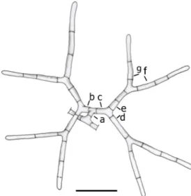
Stemflow rainwater samples were occasionally collected from an Elaeagnus angustifolia tree in a park in Budapest (N47.513685, E19.010648) between 2003 and 2008 and from a Platanus×acerifolia tree in another park (N47.476308, E19.091952) between 2008 and 2014. Depending on the Fig. 1 Mycoceros antennatissimus. Conidial anatomy:amain axis,b,c
arm of the primary dichotomy,d,earms of the secondary dichotomy (secondary arms),f,garms of the tertiary dichotomy (tertiary arms).
Bar = 20μm
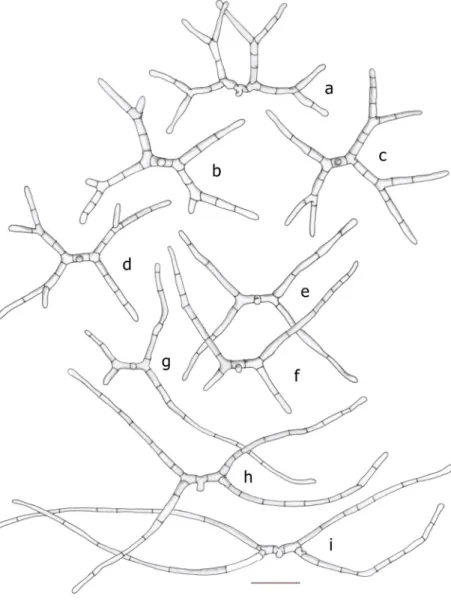
Fig. 2 Mycoceros antennatissimus.a–g Development of conidia.
Bar = 20μm.h–kDevelopment of erect aerial hyphae from colonised pollen grains.
Bar = 20μm
intensity of rainfall, various quantities of water could be col- lected (2–10 mL) into centrifuge tubes. One millilitre of FAA (50% ethanol, 5% glacial acetic acid, 10% formaldehyde) was added to each sample (Ingold1975). Water samples were set- tled; one drop of the sediment was mounted on a microscope slide and allowed to dry. Lactophenol cotton blue was added to the dried sediment to prepare samples for further studies. A total of 11 rain samples were analysed.
Total rainfall (mm) was measured with a rain gauge (Hellmann), daily average temperature (°C) with a Vaisala thermometer (Budapest/Pestszentlőrinc meteorological sta- tion) and moisture of bark of the trees (%) with a moisture meter (Voltcraft FM-200) 24 h after rainfall.
Germination of fungal conidia was observed on 2% MEA with chloramphenicol. Conidia and Pinaceae pollen grains were trans- ferred from a freshly collected tree bark with a sterilised wironit needle onto the surface of MEA. A 1 × 1-cm block was cut out of the MEA with pollen grains and conidia and placed onto a micro- scope slide, then placed and incubated vertically in a glass
container with sterilised distilled water for 48 h. Lactophenol cot- ton blue was added to the MEA block and covered with a cover slip and then photographed under 600× magnification. A simple, three-dimensional physical model was built of plastic rods to see the orientation and attachment positions of conidia on pollen grains.
Results Taxonomy
MycocerosD. Magyar & Z. Merényi, gen. nov.
MycoBank MB 819442
Etymology: from Greek μύκης,mykēs fungus; κερας, keras, literally‘horn’.
Myceliumseptate.Conidiophoresintegrated, micronematous, mononematous, smooth, hyaline. Conidia staurosporous, smooth, hyaline, multicellular. Conidium main axis straight,
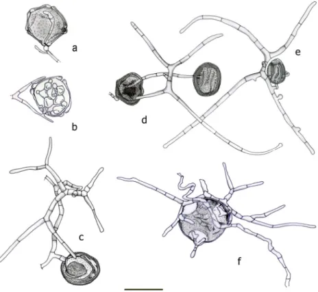
Fig. 3 Mycoceros
antennatissimus.a–iConidia.
Bar = 20μm
short, conidia forming a T-shaped primary dichotomy, each of the primary arms forming a secondary Y-shaped dichotomy, and its secondary arms often forming Y-shaped tertiary dichotomies, resulting in a 9-pike conidium.
Typus:Mycoceros antennatissimus
Mycoceros antennatissimus D. Magyar, sp. nov.
Figures1,2and3.MycoBank MB819443
Etymology:antennatissimus(Latin) referring to the multi- branched form of the conidia.
Myceliumseptate, hyphae 1.2–2.4μm wide.Conidiophores integrated, micronematous, mononematous, smooth, hyaline,
2.3 × 1.8μm.Conidiastaurosporous, smooth, hyaline, multicel- lular. Conidium main axis (Fig.1a) straight, 0(–2)-septate, 2.0–
3.6(–8.5) × 1.2–3.0μm; the first cell slightly enlarged at the base (3.0–4.4μm). The conidia form aT-shaped primary dichotomy.
Above the main axis, the conidium is slightly constricted (Fig.2d, e, Fig.3h). Each of the primary arms [Fig.1b, c, 3.6– 9.2 × 2.0–4.3μm, 1(–3) septate] forms a secondary Y-shaped dichotomy, and its secondary arms [Fig. 1d, e, 13.0–22.7 × 2.0–3.0(–4.2)μm, 1–4 septate, angle between primary and sec- ondary arms: 82–112°, angle between two secondary arms 123–
139°] often formY-shaped tertiary dichotomies (tertiary arms:
Fig. 1f, g, 9.5–39.5 × 1.4–3.0μm, 1–6 septate, angle between
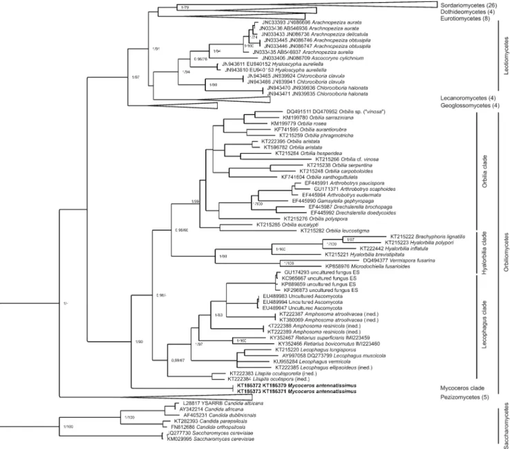
Fig. 4 Bayesian phylogenetic tree of Ascomycota based on trimmed ITS-LSU sequence alignment. Maximum credibility tree resulting from the MrBayes analysis of the trimmed ITS-LSU sequence alignment.
Bayesian posterior probabilities (PP) and maximum likelihood (ML) bootstrap values separated by a slash (/) are provided at the major nodes
in the tree as a measure of support. Only values where PP >0.95 and MP bootstrap support >60% are shown, except in the case of the terminal branches. The collapsed part of the tree represents different fungal classes which are detailed in Supplementary Table 1. The scale bar means 0.2 expected nucleotide changes per site per branch
tertiary arms: 54–109°), resulting in an up to 9-pike conidium.
Tertiary arms are often oriented in the Z-direction; their terminal cells are either tapering to 0.9–1.3μm or cylindrical-rounded (1.3–1.6μm). Tertiary arms are sometimes lacking; in this case, the secondary arms become longer (up to 131μm, Fig.3e–i).
Erect aerial hyphae (up to 135μm) are also present in the colo- nies emerging from the hyphae. Long secondary arms of the conidia are also emerging from the surface of the pollen grains, which are apparently stabilised and supported by the primary and another secondary arm (Fig.2i).
Holotypus:
Herbarium samples (BP 105172) colonies on pollen grains deposited on the bark ofPlatanus×acerifolia, HUNGARY, Budapest, 06 May, 2014, D. Magyar. GenBank Accession numbers: KT186372, KT186373 (ITS) KT186370, KT186371 (LSU).
Further specimens examined:
Herbarium samples no. K.M.D.07/2 colonies on pollen grains deposited on the bark ofElaeagnus angustifolia, HUNGARY, Budapest, Herman O. str., 10 Apr. 2007, D.
Magyar;
Herbarium samples no. K.M.D.07/4 colonies on pollen grains deposited on the bark of Platanus×acerifolia, HUNGARY, Budapest, Albert F. str., 9 May, 2011, D. Magyar;
Herbarium samples no. K.M.D.07/5 colonies from pollen grains deposited on the bark of Platanus×acerifolia, HUNGARY, Budapest, Albert F. str., 12 May, 2011, D.
Magyar;
Herbarium samples no. K.M.D.07/7b colonies on pollen grains deposited on the bark of Platanus×acerifolia, HUNGARY, Budapest, Albert F. str., 06 May, 2016, D.
Magyar.
Mycoceros antennatissimusmarkedly differs from all pre- viously described species. Conidia with secondary dichoto- mies superficially resemble those ofDwayaangam colodena Sokolski & Bérubé,D. dichotomaNawawi andD. quadridens (Drechsler) Subram., but differ in having a shorter, 0(–2)-sep- tate main axis (Drechsler1961; Nawawi1985; Sokolski et al.
2006). It is thought thatM. antennatissimuscaptures pollen grains by means of erect hyphae and conidial branches similar to that described in the two known species ofRetiarius (Olivier1978), from which it differs in having more dichoto- mies of arms. The generic separation is also based on molec- ular data (Fig.4).
The two ITS sequences from MD3 and MD4 conidia and the two LSU sequences from MD5 and MD6 conidia were identi- cal to each other by regions, which is an assurance that there was no contamination. The ITS sequence of Mycoceros antennatissimuscomprises 534 nucleotides (ITS1 204 nt;
5.8S region 157 nt; ITS2 173 nt). In the BLAST search for the ITS region with default settings, the sequence with the highest coverage was an Orbiliaceae sp. (Lilapila oculisporella G. Marson, Baral & E. Weber ined., KT222383) with a query
cover value of 73% and with only 80% similarity. The low coverage implies that mainly the 5.8S region was alignable betweenMycocerosITS and GenBank sequences. This result shows that there is no ITS sequence in the databases that is closely related to our samples. In the BLAST search for the LSU region, the best matches (100% cover) were some uncul- tured Ascomycota from savannah soils (USA, Texas, Vernon) with 92% similarity (EU489983, EU490106 and EU489947).
The conidial morphology indicates that the fungus may belong to the genus Dwayaangam, for which only ITS se- quences of one non-type species,D. colodena, are available in the international nucleotide sequence databases. However, theseDwayaangamITS sequences are not alignable with our ITS sequences, except for the region of 5.8S (156 bp). The pairwise p-distance between the 5.8S regions of them is around 5.8–6.4%, which excludes that they could belong to the same genus. In addition, the sister clade ofDwayaangam colodena, the genus Arachnopeziza(Sokolski et al.2006),
Fig. 5 Mycoceros antennatissimus.aColony with angiosperm pollen.
Bar = 20μm.bPinaceae pollen with penetrating hyphae. Bar = 20μm.c Bark fissure with colonies on accumulated pollen grains. Bar = 200μm
shows a high degree of separation fromMycocerossequences in the phylotree based on ITS-LSU (Fig.4).
Phylogenetic trees revealed similar topologies regardless of the method (Bayesian or maximum likelihood, ML). In the phylotree (Fig.4), our samples represent a separate, highly supported monophyletic cluster within the Orbiliomycetes group. Based on the available sequences (ITS and LSU), it seems thatMycocerossequences form a basal lineage, which is independent of the three major groups of Orbiliomycetes
(Orbilia,HyalorbiliaandLecophagusclades). Therefore, the introduction of a new genus is also supported by molecular methods.
Ecology
Various pollen grains (Alnus,Ambrosia,Betula,Fagus, Morus, Platanusand different Pinaceae) were found in the bark fissures ofElaeagnus,PiceaandPlatanus.Mycoceros colonies were found inElaeagnusandPlatanusbut none in Piceabark fissures. The moisture content of the tree bark was high (13.9–17.0%) in the pollen-loaded bark fissures of a Platanustree. (The fungus preferred the shady side of the tree facing the spruce.) Contrarily, the moisture content of the bark of the nearbyPicea tree was lower (7.7–9.0%). Mycoceros colonies were found on pollen aggregates containing various pollen grains (Alnus, Betula, Corylus, Cupressaceae/
Taxaceae, Pinaceae,Platanus, Fig.5a). Colonies were differ- ent in size, containing 2 to 195 pollen grains, in which Platanusand Pinaceae pollen grains were dominant in having Table 1 Bark sample collections on three known host trees
Elaeagnus Platanus Taxus
Total number of samples 23 55 12
No. of positive samplings 4 16 3
No. of samplings in spring 9 16 2
Sampling periods 2004–2012 2009–2016 2010–2016
Fig. 6 Seasonal occurrence of Mycoceros antennatissimus (samplings are marked with triangles, positive findings are marked withfilled triangles),red lineaverage temperature (°C), black barsprecipitation (mm), grey barsairborne concentration of Pinaceae pollen grains. 2007– 2008: samplings onElaeagnus, 2009–2016: samplings on Platanus
an average of 74.3% and 18.7%, respectively. Extensive at- tempts failed to culture Mycoceros from colonised pollen grains on various artificial media (Malt Extract Agar, Dichloran Rose-Bengal Agar, Potato Dextrose Agar) was un- successful, like other methods (aerated, submerged incubation or incubation of wetted bark with latent colonies, with freshly harvested/freeze-storedPiceapollen grains).
Discussion
Single spore isolation techniques (Goh1999; Choi et al.1999) combined with monospore DNA extraction (Bärlocher et al.
2010; Magyar et al.2016a) are found to be useful in taxonom- ic studies of fungi with poor or no growth on artificial media.
When monospore DNA extraction is carried out, attention should be paid to the cleanness of the selected monospores:
minute spores of common bark-inhabiting species, e.g.
TrichodermaandBeauveria(Magyar, unpublished), could at- tach onto the selected spore and polluted DNA gives mislead- ing results. Our monospore DNA extraction technique is a modification of the‘freeze–thaw method’(Bärlocher et al.
2010) using liquid nitrogen to disrupt spore walls before PCR, by having more repetitions with shorter times and a terminal centrifugation. The primers and PCR temperatures were only slightly different.
The bark of several tree taxa was sampled, including Elaeagnus, Platanus, Quercus, Acer, Robinia, Taxus,
Gymnocladus,Betula,MalusandCarpinus(in descending order of the number of samples collected), but noMycoceroscolonies were found. Only four trees, two (but neighbouring)Elaeagnus, one Taxus and one Platanus, gave positive results for M. antennatissimus. On these trees, the fungus was frequently present, e.g. in the case ofPlatanus, it was found in every year (2009–2016, Table1). Apparently,M. antennatissimusis a rare fungus; however, the frequency of this fungus could be adequate- ly determined if a high number ofElaeagnus,Platanusand Taxustrees with a Pinaceae neighbour were sampled in May.
Although theElaeagnusandPlatanushosts were sampled in all years, noMycoceroscolonies were developed before April and after early June. Apparently, the optimal growth season for this fungus is May (Fig.6). According to Révay and Gönczöl (2011), the other pollen-parasitic hyphomycete, Retiarius bovicornutus, has the highest conidial numbers in April, somewhat declining in May and low or nil in September. The latent period of this fungus is long (i.e. 84%
of the year: summer, winter and early spring). It was suspected that its sporulation is affected by season. It should be noted that, during the extremely warm winter in 2015/2016, Pinaceae pollen appeared in the air andMycoceroswas found on 29th February (onTaxus).
We suppose that the availability of fresh (living) Pinaceae pollen is a key factor in the development ofMycoceros. Host trees always had a neighbour: a pollen-emitting pinaceous tree. Because the pollen grains of Pinaceae are large and heavy, they deposit near the source tree. Within 90 m, 91%
Fig. 7 Mycoceros antennatissimuswith pollen grains.a,bTwo stages of attack:
penetration and development of intracellular mycelium (inAlnus pollen). Conidia with pollen grains of:cFraxinus,dtwo Fraxinus,ePlatanus,fCorylus.
Bar = 20μm
of the pollen grains ofPicea abiescome to rest (Di-Giovanni and Kevan1991). Fresh (living) pollen grains appropriate for Mycoceroscolonisation (Olivier1978) could, therefore, only be found near Pinaceae trees. Despite the availability of other pollen grains before April, e.g. from neighbouringBetulaand Corylustrees, the fungal colonies did not appear. We think that the presence of Pinaceae pollen is important, since this pollen is 2–3 times larger (thus containing more nutrients) than other common pollen grains (BetulaandCorylus) accu- mulating on the bark during spring. It is supposed that the small size ofBetulaandCoryluspollen grains could not pro- vide enough nutrients to enable the development of Mycoceros. However, climate could also be an important fac- tor for starting germination. Temperature and precipitation were measured between 2007 and 2016. The fungus germi- nates in April from pollen grains, when temperature increases rapidly. Although the amount of precipitation was variable (monthly sum 1.4–35.6 mm, SD = 12.7), the fungus appeared even in dry years.
Chemotropic attraction of the fungal germ tubes to fresh Pinaceae pollen grain is plausible (Hutchison and Barron 1997). We also observed directional hyphae growing toward pollen grains. The fungus penetrates into Pinaceae pollen by breaking the exine between sacci at the thinned region (leptoma) (Fig.5b). Angiosperm pollen grains are invaded by the fungus through wall openings (pori and colpi), as also shown by Huang et al. (1999). The mycelia coil around the pollen wall interior. The development of this intracellular my- celium in pollen grains shows toruloid, dichotomous morphol- ogy and it is similar to that illustrated by Olivier (1978, fig. 3f and g; compare it with our Fig.7a, b). Once established in the first (possibly Pinaceae) pollen, the fungus develops conidia and directional hyphae to attack the nearby pollen grains (even small ones, e.g.Platanus pollen, Fig.7e). The colonies are short-lived, and successful observation is limited to 2–3 weeks; thereafter, only moribund conidia are present scarcely until the middle of June. However, pollen grains filled with mycelia are present in the remaining period of the year. It was observed that even small pollen grains are able to preserve Mycocerosmycelia during the latent period of the fungus.
Pollen grains emitted in mid summer, e.g.Ambrosia, were not colonised (unidentified, non-sporulating fungi were ob- served on these pollen grains as well, with penetrating hyphae originating from Pinaceae pollen). Our attempts to induce de- velopment and sporulation ofMycocerosfrom these colonised pollen grains failed constantly. Cultivation of colonised pollen grains on various artificial media was unsuccessful, like other methods (aerated, submerged incubation or incubation of wet- ted bark with latent colonies, with freshly harvested/freeze- storedPiceapollen grains).
Although the host trees were found near to pollen-emitting Piceatrees,Mycoceroswas not present on many other trees sampled (includingElaeagnusandPlatanus), not even when
blooming spruces were close to them. Therefore, it is thought that there are some other unknown factors needed to promote the colonisation of these fungi. It is known that the success of colonisation is determined by the nature and quantities of nu- trients, together with physical features of the substratum, wa- ter availability, relative humidity and temperature (Cannon and Sutton2004). The microclimate of bark fissures may be an important factor, but this needs more study.
NoMycoceroscolonies were found in the bark fissures of Pinaceae trees, although deposits of various pollen grains (in- cluding fresh Pinaceae pollen) were present there and the host Platanuswas in its proximity. Révay and Gönczöl (2011, fig.
17–18) found some free conidia ofM. antennatissimuson various trees but not on Pinaceae, which can be confirmed by our observation. It is thought that the lack ofMycoceros colonisation on Pinaceae trees is due to the low moisture con- tent of the spruce bark. Because of the canopy structure of the
Fig. 8 Mycoceros antennatissimus.aThree-dimensional plastic model with pollen grains (two marked withdashed lines).bClump of different pollen grains attached on the conidia. Bar = 20μm
spruce, raindrops and stemflow rarely reach the bark of the bole. Among the bark cells of spruce there are resin droplets (20–30μm in diameter) keeping the bark dry. Resin droplets were not found in the bark of the host trees. A range of growth inhibitory compounds were also reported from the wood of Pinus spp. (Erdtman 1952; Scheffer and Cowling 1966;
Gunasekera and Webster1983). These factors may inhibit the development ofMycoceroscolonies in the pollen-filled bark fissures of pinaceous trees.
The aged bark ofElaeagnus,PlatanusandTaxusis becom- ing thick with deep crackings. This texture is spongious and water-retaining. Water-filled bark promotes fungal growth on deposited pollen grains. There is a well-defined deposition zone in the bark fissures (Magyar2008), where pollen grains, spores and detritus (microlitter) are accumulated and convert- ed to crown humus. Fungi are living there consuming pollen and microscopical debris of fungal, plant and insect origin.
Such fungi often have radiate conidia apparently adapted to dispersal in stemflow rainwater and anchoring on substrates (Magyar2008; Magyar et al.2016b). Bark fissures represent a habitat with high fungal diversity (Magyar2008), where sea- sonal pollen input can be a substantial factor. In Pinaceae- dominated environments, pollen deposition has been calculat- ed to be in the range of 1–80 kg ha−1y−1(Hutchison and Barron1997). Fungi may have important ecological implica- tions there being the primary consumers of pollen grains.
Further studies are, therefore, needed to understand fungal competition strategies on pollen grains in the accumulation zone of bark fissures.
Olivier (1978) describedRetiarius superficiarisas growing on the adaxial surface of leaves and producing short erect hy- phae by which it traps and becomes parasitic on anemophilous pollen grains. Similar erect hyphae are also present in M. antennatissimus. It is not clear, however, if the pollen grains are trapped directly in the air or just on the bark, after deposi- tion. We suppose that the long arms of conidia and aerial hy- phae could capture pollen grains from the air, similarly to plant trichomes which also increase the capture and retention of pol- len grains (Roda et al.2003). Pollen grains are also washed into the bark fissures by stemflow rainwater (D. Magyar, unpub- lished). We assume that the conidia ofM. antennatissimus could capture pollen grains from the stemflow as well.
Detached conidia are frequently found anchoring on the sur- faces of pollen grains. The three-dimensional model showed that one conidium ofM. antennatissimuscould be in contact with seven pollen grains having a diameter ofPlatanuspollen.
Secondary arms form a space to hold three pollen grains while tertiary arms oriented in the Z-direction allow to catch a further four pollen grains (Fig.8a). Microscopic observations have also verified the position of pollen grains on conidia (Fig.8b).
We frequently observed M. antennatissimus conidia in stemflow rainwater collected fromElaeagnusand Platanus host trees (10–20Mycocerosconidia/mL). It is speculated that
Mycocerosconidia have a two-step dispersal tree-to-tree: first, the conidia are mobilised by stemflow and carried onto the bark surface, and are then carried by splashing rain drops to nearby trees. No doubt that Mycoceros conidia are mainly dispersed by stemflow for short distances, but, as our air sam- plings show, they hardly become airborne, as noMycoceros conidia could be found in our air samples between 2009 and 2016.
Acknowledgements The authors are grateful to Gyula Dura and Anna Páldy, National Institute of Environmental Health for the financial sup- port to complete the DNA analysis, Ágnes Révay, Hungarian Natural History Museum, Budapest for her valuable suggestions, Hans-Otto Baral, Tübingen, Germany for his valuable corrections and Gáti Zsófia for the technical assistance in the molecular methods. PK and LK were supported by grant GINOP-2.3.3-15-2016-00006 (Széchenyi 2020 Programme). ZM was supported by NTP-NFTÖ-16-0216 (National Talent Program of the Ministry of Human Capacities (EMMI), Human Capacities Grant Management Office (EMET) and GINOP- 2.1.1-15- 2015-00115 (Széchenyi 2020 Programme).
References
Altschul SF, Madden TL, Schäffer AA, Zhang J, Zhang Z, Miller W, Lipman DJ (1997) Gapped BLAST and PSI-BLAST: a new gener- ation of protein database search programs. Nucl Acids Res 25:3389– 3402
Bärlocher F, Charette N, Letourneau A, Nikolcheva LG, Sridhar KR (2010) Sequencing DNA extracted from single conidia of aquatic hyphomycetes. Fungal Ecol 3:115–121
Bertini L, Amicucci A, Agostini D, Polidori E, Potenza L, Guidi C, Stocchi V (1999) A new pair of primers designed for amplification of the ITS region inTuberspecies. FEMS Microbiol Lett 173:239– 245
Cannon PF, Sutton BC (2004) Microfungi on wood and plant debris. In:
Mueller GM, Bills GF, Foster MS (eds) Biodiversity of fungi: in- ventory and monitoring methods. Elsevier Academic Press, New York, pp 217–239
Capella-Gutiérrez S, Silla-Martínez JM, Gabaldón T (2009) trimAl: a tool for automated alignment trimming in large-scale phylogenetic anal- yses. Bioinformatics 25:1972–1973
Choi Y-W, Hyde KD, Ho WH (1999) Single spore isolation of fungi.
Fungal Divers 3:29–38
Chou MC, Preece TF (1968) The effect of pollen grains on infections caused byBotrytis cinereaFr. Ann Appl Biol 62:11–22
Di-Giovanni F, Kevan PG (1991) Factors affecting pollen dynamics and its importance to pollen contamination: a review. Can J For Res 21:
1155–1170
Drechsler C (1961) Some clampless Hyphomycetes predacious on nem- atodes and rhizopods. Sydowia 15:9–25
Erdtman H (1952) Phenolic and other extraneous components of conif- erous heartwoods; their relation to taxonomy. In: Wise LE, Jahn EC (eds) Wood chemistry, vol I. Reinhold, New York, pp 661–688 Fokkema NJ (1968) The influence of pollen on the development of
Cladosporium herbarumin the phyllosphere of rye. Neth J Plant Pathol 74:159–165
Fokkema NJ (1971a) Influence of pollen on saprophytic and pathogenic fungi on rye leaves. In: Preece TF, Dickinson CH (eds) Ecology of leaf surface micro-organisms. Academic Press, New York, pp 277– 282
Fokkema NJ (1971b) The effect of pollen in the phyllosphere of rye on colonization by saprophytic fungi and on infection by Helminthosporium sativumand other leaf pathogens. Neth J Plant Pathol 77(Suppl 1):1–60
Galili T (2015) dendextend: an R package for visualizing, adjusting and comparing trees of hierarchical clustering. Bioinformatics 31:3718– 3720
Gardes M, Bruns TD (1993) ITS primers with enhanced specificity for basidiomycetes—application to the identification of mycorrhizae and rusts. Mol Ecol 2:113–118
Goh T-K (1999) Single-spore isolation using a hand-made glass needle.
Fungal Divers 2:47–63
Goldstein S (1960) Degradation of pollen by phycomycetes. Ecology 41:
543–545
Gunasekera SA, Webster J (1983) Inhibitors of aquatic and aero-aquatic hyphomycetes in pine and oak wood. Trans Br Mycol Soc 80:121– 125
Hirst JM (1952) An automatic volumetric spore trap. Ann Appl Biol 39:
257–265
Huang HC, Kokko EG, Erickson RS (1999) Infection of alfalfa pollen by Botrytis cinerea. Bot Bull Acad Sin 40:101–106
Huelsenbeck JP, Ronquist F (2001) MRBAYES: Bayesian inference of phylogenetic trees. Bioinformatics 17:754–755
Hutchison LJ, Barron GL (1997) Parasitism of pollen as a nutritional source for lignicolous Basidiomycota and other fungi. Mycol Res 101:191–194
Ingold CT (1975) An illustrated guide to aquatic and water-borne hypho- mycetes (Fungi Imperfecti) with notes on their biology. Freshwater Biol Assoc Sci Publ 30:1–96
Katoh K, Toh H (2008) Recent developments in the MAFFT multiple sequence alignment program. Brief Bioinform 9:286–298 Li Y, Hyde KD, Jeewon R, Cai L, Vijaykrishna D, Zhang K (2005)
Phylogenetics and evolution of nematode-trapping fungi (Orbiliales) estimated from nuclear and protein coding genes.
Mycologia 97:1034–1046
Magyar D (2008) The tree bark: a natural spore trap. Asp Appl Biol 89:7– 16
Magyar D, Merényi Z, Bratek Z, Baral H-O, Marson G (2016a) Lecophagus vermicolasp. nov., a nematophagous hyphomycete with an unusual hunting strategy. Mycol Progr 15:1137–1144 Magyar D, Vass M, Li DW (2016b) Dispersal strategies of microfungi. In:
Li DW (ed) Biology of microfungi. Springer International Publishing, pp 315–371
Miller MA, Pfeiffer W, Schwartz T (2010) Creating the CIPRES Science Gateway for inference of large phylogenetic trees. In: Proceedings of the Gateway Computing Environments Workshop (GCE), New Orleans, LA, 14 November 2010, pp 1–8
Nawawi A (1985) Some interesting hyphomycetes from water.
Mycotaxon 24:217–226
Nylander JAA (2004) MrModeltest v2. Program distributed by the au- thor. Evolutionary Biology Centre, Uppsala University. Available online at:https://github.com/nylander/MrModeltest2. Accessed 2012
Olivier DL (1978)Retiariusgen. nov.: phyllosphere fungi which capture wind-borne pollen grains. Trans Br Mycol Soc 71:193–201 Prieto M, Wedin M (2013) Dating the diversification of the major line-
ages of Ascomycota (Fungi). PLoS One 8:e65576
R Core Team (2016) R: A language and environment for statistical com- puting. R Foundation for Statistical Computing, Vienna, Austria.
Home page at:https://www.R-project.org/
Rambaut A. (2009) FigTree, ver. 1.3.1. Available online at:http://tree.bio.
ed.ac.uk/software/figtree/
Révay Á, Gönczöl J (2011) Canopy fungi (Bterrestrial aquatic hyphomycetes^) from twigs of living evergreen and deciduous trees in Hungary. Nova Hedwigia 92:303–316
Roda A, Nyrop J, English-Loeb G (2003) Leaf pubescence mediates the abundance of non-prey food and the density of the predatory mite Typhlodromus pyri. Exp Appl Acarol 29:193–211
Rodrigues Marques JP, Amorim L, Bellato Spósito M, Marin D, Appezzato-da-Glória B (2013) Infection of citrus pollen grains by Colletotrichum acutatum. Eur J Plant Pathol 136:35–40
Scheffer TC, Cowling EB (1966) Natural resistance of wood to microbial deterioration. Ann Rev Phytopathol 4:147–170
Sokolski S, Piché Y, Laitung B, Bérubé JA (2006) Streams in Quebec boreal and mixed-wood forests reveal a new aquatic hyphomycete species,Dwayaangam colodenasp. nov. Mycologia 98:628–636 Spatafora JW, Sung GH, Johnson D, Hesse C, O’Rourke B, Serdani M,
Spotts R, Lutzoni F, Hofstetter V, Miadlikowska J, Reeb V, Gueidan C, Fraker E, Lumbsch T, Lücking R, Schmitt I, Hosaka K, Aptroot A, Roux C, Miller AN, Geiser DM, Hafellner J, Hestmark G, Arnold AE, Büdel B, Rauhut A, Hewitt D, Untereiner WA, Cole MS, Scheidegger C, Schultz M, Sipman H, Schoch CL (2006) A five- gene phylogeny of Pezizomycotina. Mycologia 98:1018–1028 Stamatakis A (2006) RAxML-VI-HPC: maximum likelihood-based phy-
logenetic analyses with thousands of taxa and mixed models.
Bioinformatics 22:2688–2690
Stamatakis A, Hoover P, Rougemont J (2008) A rapid bootstrap algo- rithm for the RAxML web servers. Syst Biol 57:758–771 Stark N (1972) Nutrient cycling pathways and litter fungi. Bioscience 22:
355–360
Vilgalys R, Hester M (1990) Rapid genetic identification and mapping of enzymatically amplified ribosomal DNA from severalCryptococcus species. J Bacteriol 172:4238–4246
Warren RC (1972) The effect of pollen on the fungal leaf microflora of Beta vulgarisL. and on infection of leaves byPhoma betae. Neth J Plant Pathol 78:89–98
Wurzbacher C, Rösel S, Rychła A, Grossart H-P (2014) Importance of saprotrophic freshwater fungi for pollen degradation. PLoS One 9:
e94643