Staphylococcus aureus at admission among high- risk Turkish and international patients
MELDA OZDAMAR
pDepartment of Clinical Microbiology, Anadolu Medical Center, Gebze, Kocaeli, Turkey Received: November 9, 2019 • Accepted: January 3, 2020 • Published online: March 09, 2020
ABSTRACT
Objective:The aim of this study was to detect the frequency of methicillin-resistant Staphylococcus aureus(MRSA) colonization at admission in a group of presumably high-risk international or Turkish patients referred to our center for elective operations, some of whom were from countries with an unknown prevalence of MRSA infection or colonization.
Methods:The results of nasal swab screening for MRSA colonization performed using a specific algo- rithm between 2011 and 2018 in a private medical center were retrospectively reviewed. Presence of MRSA was ascertained using culture and/or real-time polymerase chain reaction (real-time PCR).
Results:A total of 3,795 patients were included in the study. More than half of the patients were≤19 years of age (2,094, 55.2%), and MRSA positivity was more common among these patients. Turkish patients constituted 24.5% of the study population. International patients were most frequently referred from Iraq (55.92%), Libya (11.44%), Romania (2.69%), and Bulgaria (1.98%). MRSA positivity was significantly more common among patients referred from other countries when compared to Turkish nationals (11.5% vs. 4.4%,P 50.00001). Countries with the highest prevalence rates of MRSA colo- nization were as follows with decreasing order: United Arab Emirates, 25.0%; Georgia, 23.1%; Russia, 22.7%; Iraq, 13.0%, Romania, 12.7%. Other countries with high number of admitted patients (>70 patients) had the following MRSA rates: Turkey, 4.4%; Libya, 6.0%; Bulgaria, 5.3%.
Conclusions: Although MRSA has a low prevalence in our center, a variation in the rate of MRSA positivity was observed across patients from different countries. Absence hospital acquired contami- nation or outbreaks in our institution may be attributed to the screening algorithm used and un- derscores the importance of risk analysis for patients referred from geographical locations with unknown MRSA frequency, to reduce the risk of transmission.
KEYWORDS
methicillin-resistantStaphylococcus aureus(MRSA), MRSA frequency, nasal colonization, real-time polymerase chain reaction (real-time PCR), international patients
INTRODUCTION
Traditionally, staphylococci have been a major culprit in nosocomial infections. Although a major therapeutic breakthrough had been accomplished in the year 1960, first by the introduction of methicillin, and then by other penicillinase-resistant penicillins into clinical practice, it did not take too long before methicillin resistance among staphylococci was defined [1]. Toward the end of 1970s methicillin-resistant Staphylococcus aureus (MRSA) had already acquired resistance to many of the commonly used antibiotics. Since then, MRSA has eventually become a major global healthcare problem, not only due to challenges asso- ciated with the treatment of multidrug resistant strains, but also due to their potential to cause epidemic nosocomial infections. Following the description of thefirst MRSA epidemic in 1963, an increasing number of epidemic MRSA infections have been reported, and this microorganism has become endemic in many hospitals [2–4].
Acta Microbiologica et Immunologica Hungarica
67 (2020) 1, 73-78
DOI:
10.1556/030.2020.01081
© 2020 Akademiai Kiado, Budapest
ORIGINAL ARTICLE
*Corresponding author. Department of Clinical Microbiology, Anadolu Medical Center, Cumhuriyet Mahallesi, 2255 sokak, no: 3, Gebze, Kocaeli, 41400, Turkey. Tel.:þ90 532 2373820; fax:þ90 2626540544.
E-mail:melda.ozdamar@gmail.com
A recent increase up to 40% has been witnessed in the overall prevalence of methicillin resistance (i.e., among invasive in- fections and asymptomatic colonization patients) in the entire European continent, except for Northern Europe, where the incidence of methicillin resistance has remained low, i.e., below 1% [5–7]. Regarding MRSA rates solely among patients with asymptomatic colonization as screened upon hospital admission for other reasons, several studies from Europe reported rates ranging between 1.2 and 11.6% [8–12]. Similarly, the incidence of MRSA carriage rates shows considerable variation among healthy hospital staff members [13]. In a collaborative study encom- passing several European countries, the reported infection rates of MRSA varied between 8.7% and 20.4% among intensive care unit patients [14], suggesting that medical tourism may represent a potential cause of MRSA outbreaks, especially when it involves patient movements from high to low prevalence areas. In recent years, there has been a dramatic and continuous increase in the number of international patients visiting Turkey for medical treatments, particularly after 2010, from countries such as Iraq, Libya, Russia, and other European countries (Germany, Holland, Romania, Bulgaria, etc.,) [15]. Furthermore, in the past decade, Turkey has been accepting significant number of immigrants or has served as a migration path for very large number of pop- ulations. Compared to other private medical facilities in Turkey, our center has been among the most frequently preferred desti- nation for international patients, and as a result of our facilities unique position, a decision was made to determine the MRSA colonization rate and incidence in this complex and complicated (i.e., potentially high-risk) patient cohort, a proportion of whom come from regions with unknown colonization rates.
The significance of determining the antibacterial suscepti- bility patterns for therapeutic decisions has been well estab- lished. Currently, microbiological cultures and phenotypic sensitivity tests represent the standard techniques used in many laboratories. However, since the turnaround time of pheno- typic tests are at least 24–48 h, significant efforts have been devoted on developing susceptibility tests with faster turn- around times (e.g., immunological or molecular techniques) in the past two decades [16]. Obviously, earlier detection of resistant bacterial strains can have a major impact on the re- covery process, patient survival and healthcare costs [17].
This study was undertaken to detect methicillin-resistant Staphylococcus aureus(MRSA) colonization at admission in a group of presumably high-risk Turkish or international pa- tients referred to our center for elective operations, some of whom were from countries with an unknown prevalence of MRSA infection or colonization. For this purpose, a specific screening algorithm consisting of identification and contact isolation measures was used with the purpose of preventing nosocomial cross-transmission and spread of microorganisms.
MATERIALS AND METHODS
Patients and study design
Results of screening tests for nasal MRSA colonization per- formed at admission in a cohort of patients in our hospital
between 2011 and 2018 were retrospectively analyzed. During this period, a screening algorithm for MRSA was actively implemented to minimize the risk of endemic and epidemic nosocomial infection risk amongst patients and employees in our institution. Contact isolation procedures were used for patients diagnosed with MRSA colonization according to Centers for Disease Control and Prevention-Hospital Infections Control Practices Advisory Committee (CDC-HICPAC) rec- ommendations [18]. The approval of the local institutional re- view board was obtained before the study (ASM-EK-17/69).
MRSA screening algorithm
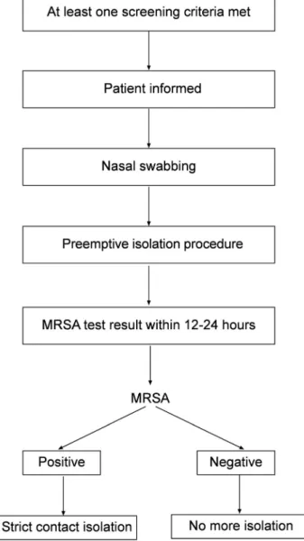
Screening for MRSA was initiated if at least one of the following was present in patient’s history: previous MRSA colonization or infection with in the past year, open wounds, referral from another health facility, current use of chemo- therapy (inpatients or at theirfirst admission), hospitalization within the past 6 months, or presence of central venous catheters (any kind of implantable venous access port, a peripherally inserted central catheter (PICC line), or tunneled
Figure 1.Institutional screening algorithm for MRSA
central venous catheters). As shown inFig. 1, these risk fac- tors are routinely assessed in our institution by the nursing staff upon admission in each patient. Patients were screened using culture (n 5 714) or culture plus real-time PCR (LightCycler MRSA Advanced test) (n5 3,081), depending on availability and cost-efficiency issues. The infection control nurse and the clinical microbiologist are actively involved in conducting the screening procedure, including sampling and conduction of contact isolation round the clock.
Culture techniques
Chromogenic BBL CHROMagar MRSA II (CMRSA) plates were obtained from BD Diagnostics (Sparks, MD). Each chromogenic plate was handled according to the manufac- turer’s package insert instructions. Interpretation of colony size and color on the chromogenic media were confirmed by the microbiology staff members as described by the manu- facturer’s instructions. Mauve colonies of any size morphologically resembling staphylococci were described as MRSA. Uncolored or white colonies were not further investigated for MRSA. All mediums were also stored in the dark before inoculation and during incubation. Quality control testing was performed on each new lot for the plates using a standardized inoculum ofS. aureusATCC 25923.
PCR techniques
LightCycler MRSA Advanced Test, the Food and Drug Administration (FDA)-cleared MRSA detection method, which relies on real-time PCR principle (ROCHE, Minne- sota, USA) was used. Following the mechanical disintegra- tion of the bacterial wall and purification of DNA, target DNA is amplified using the Light Cycler 2.0 Instrument (Roche Diagnostics, Minnesota, USA), and finally hybrid- ized with a specific probe. In this method, detection of the right junction of the orfX region with SCCmec was aimed.
The results were obtained in approximately 1 h. Positive and negative controls were included in each run. The PCR results for the LightCycler MRSA advanced test were interpreted using the LightCycler software. The test was repeated with new samples when specimens yielded invalid results.
Patients positive with either of the methods (culture and/
or real-time PCR) were considered MRSA positive.
Statistical analysis
Data were analyzed using the IBM Statistical Package for Social Sciences v21 (SPSS Inc., Chicago, IL, USA). Normality of continuous data was tested with Kolmogorov–Smirnov test. Mann–Whiney U-test was used to compare continuous data. The distribution of categorical variables was compared
using Pearson chi-square test and Bonferroni correction was made for pairwise comparisons. APvalue less than 0.05 was considered indication of statistical significance.
RESULTS
A total of 3,795 patients (2,266 males, 1,529 females) with a median age of 13.5 years (range 0–97.2 y) were included in the study. Patients were hospitalized in the following de- partments: cardiovascular surgery (61%), orthopedic surgery (12%), medical oncology (16.7%), hemato-oncology (9%), and pediatric surgery (1.3%).
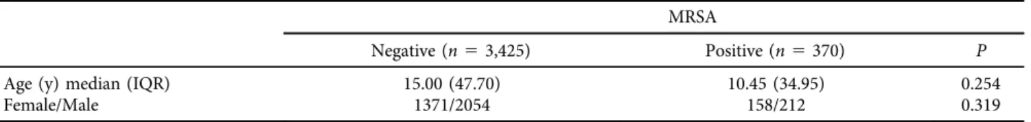
Table 1shows age and gender distribution of patients by MRSA positivity. MRSA positive and negative cases did not differ in terms of age (P5 0.254) and gender (P 50.319) distribution. Table 2 shows distribution of MRSA rates across age groups as well as age distribution of the whole study group. Pediatric patients constituted more than half of the subjects. MRSA positivity was significantly more com- mon among 1–9 year and 10–19-year age groups compared to others (P< 0.01 for both comparisons).
Turkish patients and patients referred from 22 countries were included in this study. Table 3 shows distribution of MRSA positivity rates across patients from different coun- tries. Turkish nationals constitute 24.5% of the cases. Pa- tients were most frequently referred from Iraq (55.92%), Libya (11.44%), Romania (2.69%), and Bulgaria (1.98%).
Countries with the highest prevalence rates of MRSA colo- nization were as follows with decreasing order: United Arab Emirates, 25.0%; Georgia, 23.1%; Russia, 22.7%; Iraq, 13.0%, Romania, 12.7%. Other countries with high number of admitted patients (>70 patients) had the following MRSA rates: Turkey, 4.4%; Libya, 6.0%; Bulgaria, 5.3%.
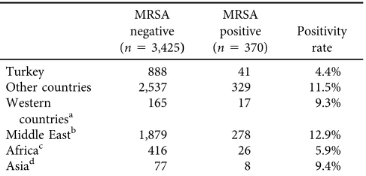
Table 4 shows distribution of MRSA cases by region.
MRSA positivity was significantly more common among patients referred from other countries when compared to Turkish nationals (11.5% vs. 4.4%, P 5 0.00001). Patients from Middle East (P 5 0.00001) and Western countries (P 50.0062) had significantly more frequent MRSA when compared to Turkish nationals. Patients from Middle East had also significantly more frequent MRSA when compared to African patients (P50.00003).
DISCUSSION
This is the first study to examine the frequency of MRSA colonization in a large patient sample from a private health facility in Turkey where there is a high turnover of interna- tional patients as a consequence of medical tourism. In this Table 1.The impact of age, and gender of the patients on MRSA colonization
MRSA
Negative (n53,425) Positive (n5370) P
Age (y) median (IQR) 15.00 (47.70) 10.45 (34.95) 0.254
Female/Male 1371/2054 158/212 0.319
cohort, the rate of nasal MRSA colonization at admission was low, and no hospital acquired contamination or outbreaks occurred during the study period. However, MRSA coloni- zation occurred at a higher frequency, particularly among those from Middle East countries, as compared to the Turkish patients. These results suggest that screening programs may be especially useful in health facilities dealing with a mixed international patient population.
In current practice, contact isolation precautions are determined on the basis of nasal swab MRSA screening re- sults. But this method also may provide clinicians with additional information for predicting the probability of MRSA infection and for earlier tailoring of empiric anti- microbial therapy. MRSA screening offers a rapid, inex- pensive means for medical centers to avoid unnecessary and costly therapy.
Despite a decreasing trend in some countries, the inci- dence of MRSA related infections is still high in many parts
of the world, despite the reported differences between countries and regions [19, 20]. In a study by Fluit et al.
involving 25 university hospitals across Europe, MRSA was found to be responsible for nearly 25% of all nosocomial infections, and the prevalence of MRSA was higher in southern European countries [21]. The highest prevalence rates were detected in hospitals in Portugal (54%), and Italy (43–58%), as opposed to a prevalence rate of only 2% in Switzerland and Holland. In another multicenter study carried out in intensive care units, the highest prevalence of MRSA infections was observed in centers from Italy (81%) and France (78%) [22]. In a study by Buzaid et al. from Libya, MRSA was identified in 31% of 200S. aureusstrains isolated from the wound infections [23].
Relatively few studies examined the MRSA rate among individuals without invasive infection. A recent study from Middle East region examined MRSA frequency among healthcare workers and/or non-healthcare workers. That study from Iraq found MRSA colonization in 13.7% and 4.0%
of healthcare workers and non-healthcare workers, respec- tively; with significantly higher rate among healthcare workers [24]. Several studies examined MRSA rates among patients without invasive infection upon their admission to hospital for other reasons. A study from United Kingdom, found MRSA colonization ranging between 0.8% in obstetrics/gy- necology/neonatology departments and 6.6% in critical care units, among patients without invasive staphylococcal infec- tion [9]. In a multinational large European study, MRSA was identified in 3.8% of surgical ward patients upon admission [11]. On the other hand, relatively high MRSA colonization rates (11.6%) have been reported from palliative care units [8]. Murray et al. evaluated multidrug-resistant microorgan- isms in victims of war in Iraq and Afghanistan between 2005 and 2007 and found high colonization rates for methicillin- resistant Staphylococcus aureus; ESBL-producing Klebsiella pneumoniae, and Acinetobacter baumanni, which were the most commonly recovered pathogens at peri-admission screening cultures [25]. Thesefigures from Middle East and Europe show considerable variation across different hospital Table 3.Distribution of MRSA positivity rates across patients
from different countries
Total number (n)
MRSA rate
%, (n)a
Iraq 2,122 13.0 (276)
Turkey 929 4.4 (41)
Libya 434 6.0 (26)
Romania 102 12.7 (13)
Bulgaria 75 5.3 (4)
Bahrain 30 3.3 (1)
Russia 22 22.7 (5)
Azerbaijan 18 0 (0)
The Republic of Kazakhstan 12 0 (0)
Georgia 13 23.1 (3)
Algeria 7 0 (0)
The Kyrgyz Republic 5 0 (0)
Iran 4 0 (0)
Ukraine 4 0 (0)
England 3 0 (0)
Turkmenistan 3 0 (0)
Uzbekistan 3 0 (0)
United Arab Emirates 4 25.0 (1)
USA 1 0 (0)
Ethiopia 1 0 (0)
Holland 1 0 (0)
Syria 1 0 (0)
The Republic of Tatarstan 1 0 (0)
Total 3,795 9.7 (370)
aNumber of patients colonized with MRSA.
Table 4.Distribution of MRSA positive cases by region MRSA
negative (n53,425)
MRSA positive (n5370)
Positivity rate
Turkey 888 41 4.4%
Other countries 2,537 329 11.5%
Western countriesa
165 17 9.3%
Middle Eastb 1,879 278 12.9%
Africac 416 26 5.9%
Asiad 77 8 9.4%
aUSA, Holland, England, Bulgaria, Romania.
bIraq, Syria, United Arab Emirates, Bahrain.
cLibya, Algeria, Ethiopia.
dThe Republic of Tatarstan, Turkmenistan, Uzbekistan, Iran, Ukraine, The Kyrgyz Republic, The Republic of Kazakhstan, Azerbaijan, Russia, Georgia.
Table 2.Distribution of MRSA rates across age groups Age group Total numbern(%)a MRSA positivity (%)b
<1 y 761 (20.1) 6.8
1–9 y 992 (26.1) 13.0
10–19 341 (9.0) 14.1
20–59 1,158 (30.5) 9.1
≥60 543 (14.3) 6.6
Total 3,795 9.7
aPercent of the age group within all study groups.
bPercent of MRSA positive patients.
settings, and probably across different regions. Similar regional differences ranging between 0% and up to more than 20% is also evident in our study, suggesting a possible role for socioeconomical and cultural variations.
In Turkey, two studies reported 1.2% prevalence of MRSA on hospital admission [26, 27]. Another recent study from Turkey identified MRSA colonization in 6.2% of intensive care unit patients on admission [28]. In our study, MRSA prevalence was 4.4% upon admission in Turkish patients from Istanbul and Kocaeli province. This relatively high rate may be attributed to the profile of our Turkish patients, which are mostly hematology and oncology patients.
Incidence of hospital associated MRSA infection rate differ significantly geographical regions [29]. MRSA has been traditionally regarded as an organism that poses extreme challenges in terms of treatment and eradication as well as representing a major cause of nosocomial epidemics in hos- pitals. Carefully conducted surveillance studies are of utmost importance not only for the early detection of such epidemics, but also for the identification of the possible source(s) of the infection and implementation of appropriate control measures [30]. Collection of MRSA surveillance data by microbiology laboratories is essential for conduction of antimicrobial stew- ardship activities as well as for providing guidance for further investigations whenever epidemics are suspected.
Although MRSA infections are serious, they are also preventable. Most important risk factors for colonization and infection with MRSA include age, underlying diseases, nasal colonization, and indwelling devices such as catheters, and tracheostomy and nasogastric tubes [31]. It has been clearly established that the most common route for the spread of infection is transient contamination of the hands of healthcare workers [32].
Significantly higher rates of MRSA colonization in our patients from Middle East and Africa countries may be related with the poor hygiene both in healthcare facilities and in daily living conditions during and after the war, a high turnover of patients undergoing surgery, inappro- priate use and black market distribution of antibiotics, low socioeconomic status, and overcrowded housing with restricted access to clean water. On the other hand, higher rates of MRSA in patients from Western countries as compared to Turkish patients may be accounted for by the exposure to resistant strains during and after the treatment for complicated oncological conditions in different healthcare settings. Nevertheless, Turkish patients may not be directly compared with international patient groups since they are not matched in terms of age, severity of disease, history, previous antibiotic treatments, and num- ber of previous hospitalizations, etc., Not only regional differences but also clinical characteristics might have led and contributed to this difference as well.
This study has several limitations. Firstly, our electronic database did not contain information on clinical characteris- tics, living conditions, and reasons for including an individual in screening procedure, precluding any causality analysis.
Furthermore, since only prophylactic treatment with vanco- mycin was scheduled for patients with preoperative detection
of MRSA colonization, antibiotic susceptibility testing was not be performed on MRSA strains, and susceptibility testing was only performed whenever an infection occurred.
The primary methodology used to detect MRSA in this study, i.e., real-time PCR screening, may offer a cost-effective option to reduce MRSA infection. Howev- er, several factors should be considered when choosing the most appropriate screening method in specific set- tings and these include the local epidemiology, cost considerations, and infection control policies. Given the complexities of selecting an appropriate screening strat- egy, further research on the cost-effectiveness of MRSA infection control is required.
In conclusion, our findings demonstrated a variable MRSA colonization rate among patients from different countries, geographical regions, and socioeconomic back- grounds. Of interest were the low colonization rates in Turkish patients. Thus, it appears that international patients seeking advanced medical care are more likely to be carriers of MRSA than Turkish patients in our facility, although this may be attributed to clinical profile as well as regional dif- ferences. Health centers involved in the care of international patient populations may benefit from a stratified risk anal- ysis for possible MRSA carrier status, as a potential means to reduce the risk of nosocomial transmission.
Disclosure of interest: The authors report no conflicts of interest. The authors alone are responsible for the content and writing of the paper.
Funding:None.
ACKNOWLEDGMENTS
I appreciate the support of Ipek Deger Karaman, infection control nurse, in implementing the MRSA screening algo- rithm and training the related personnel. I would also thank for the valuable efforts of Prof Dr Salih Turkoglu for improving the paper in terms of scientific quality and flow.
REFERENCES
[1] Jevons MP. “Celbenin” – resistant Staphylococci. Br Med J 1961; 1: 124–5.
[2] Stewart GT, Holt RJ. Evolution of natural resistance to the newer penicillins. Br Med J 1963; 1: 308–11.
[3] Benner EJ, Kayser FH. Growing clinical significance of meth- cillin-resistantStaphylococcus aureus. Lancet 1968; 2: 741–4.
[4] Brumfitt W, Hamilton-Miller J. Methicillin-resistant Staphy- lococcus aureus. N Engl J Med 1989; 320: 1188–96.
[5] Borg MA, Camilleri L, Waisfisz B. Understanding the epide- miology of MRSA in Europe: do we need to think outside the box?. J Hosp Infect 2012; 81: 251–6.
[6] Tiemersma EW, Bronzwaer SL, Lyytikainen O, Degener JE, Schrijnemakers P, Bruinsma N, et al. European antimicrobial
resistance surveillance system, P.: methicillin-resistantStaph- ylococcus aureusin Europe, 1999–2002. Emerg Infect Dis 2004;
10: 1627–34.
[7] Rosdahl VT, Knudsen AM. The decline of methicillin resis- tance among Danish Staphylococcus aureus strains. Infect Control Hosp Epidemiol 1991; 12: 83–8.
[8] Gleeson A, Larkin P, Walsh C, O’Sullivan N. Methicillin- resistant Staphylococcus aureus: prevalence, incidence, risk factors, and effects on survival of patients in a specialist palliative care unit: a prospective observational study. Palliat Med 2016; 30: 374–81.
[9] Otter JA, Herdman MT, Williams B, Tosas O, Edgeworth JD, French GL. Low prevalence of meticillin-resistant Staphylococcus aureus carriage at hospital admission: im- plications for risk-factor-based versus universal screening. J Hosp Infect 2013; 83: 114–21.
[10]Hetem DJ, Derde LP, Empel J, Mroczkowska A, Orczykowska- Kotyna M, Kozinska A, et al. Molecular epidemiology of MRSA in 13 ICUs from eight European countries. J Anti- microb Chemother 2016; 71: 45–52.
[11]Pan A, Lee A, Cooper B, Chalfine A, Daikos GL, Garilli S, et al.
Risk factors for previously unknown meticillin-resistant Staphylococcus aureus carriage on admission to 13 surgical wards in Europe. J Hosp Infect 2013; 83: 107–13.
[12]Neidhart S, Zaatreh S, Klinder A, Redanz S, Spitzmuller R, Holtfreter S, et al. Predictors of colonization with Staphylo- coccus species among patients scheduled for cardiac and or- thopedic interventions at tertiary care hospitals in north- eastern Germany-a prevalence screening study. Eur J Clin Microbiol Infect Dis 2018; 37: 633–41.
[13]VandenBergh MF, Yzerman EP, van Belkum A, Boelens HA, Sijmons M, Verbrugh HA. Follow-up ofStaphylococcus aureus nasal carriage after 8 years: redefining the persistent carrier state. J Clin Microbiol 1999; 37: 3133–40.
[14]Vincent JL, Rello J, Marshall J, Silva E, Anzueto A, Martin CD, et al. International study of the prevalence and outcomes of infection in intensive care units. JAMA 2009; 302: 2323–9.
[15]Kaya S, Yildirim HH, Karsavuran S, Ozer O. Evaluation report on medical tourism in Turkey 2013. Turkish Ministry of Health; 2013. Available from: http://dosyasb.saglik.gov.tr/
Eklenti/535,turkiye-medikal-turizm-degerlendirme-raporu- 2013pdf.pdf. [Accessed 21 October 2019].
[16]Fluit AC, Visser MR, Schmitz FJ. Molecular detection of antimicrobial resistance. Clin Microbiol Rev 2001; 14: 836–71.
[17]Antibiotic resistance threats in the United States: Center for Disease Control; 2013. Available from: https://www.cdc.gov/
drugresistance/pdf/ar-threats-2013-508.pdf. [Accessed 21 October 2019].
[18]Siegel JD, Rhinehart E, Jackson M, Chiarello L, the Healthcare Infection Control Practices Advisory Committee. Guideline for isolation precautions: preventing transmission of infectious agents in healthcare settings; 2007. Available from: https://
www.cdc.gov/infectioncontrol/guidelines/isolation/index.html.
[Accessed 21 October 2019].
[19]European Center for Disease Prevention and Control. Sur- veillance of antimicrobial resistance in Europe; 2017. Available
from: https://www.ecdc.europa.eu/en/publications-data/
surveillance-antimicrobial-resistance-europe-2017. [Accessed 8 December 2019].
[20]Lee AS, de Lencastre H, Garau J, Kluytmans J, Malhotra- Kumar S, Peschel A, et al. Methicillin-resistantStaphylococcus aureus. Nat Rev Dis Primers 2018; 4: 18033.
[21]Fluit AC, Wielders CL, Verhoef J, Schmitz FJ. Epidemiology and susceptibility of 3,051Staphylococcus aureusisolates from 25 university hospitals participating in the European SENTRY study. J Clin Microbiol 2001; 39: 3727–32.
[22]Vincent JL. Microbial resistance: lessons from the EPIC study.
European prevalence of infection. Intensive Care Med 2000;
26(Suppl. 1): S3–8.
[23]Buzaid N, Elzouki AN, Taher I, Ghenghesh KS. Methicillin- resistant Staphylococcus aureus(MRSA) in a tertiary surgical and trauma hospital in Benghazi, Libya. J Infect Dev Ctries 2011; 5: 723–6.
[24]Hussein NR, AssafiMS, Ijaz T. Methicillin-resistantStaphy- lococcus aureus nasal colonisation amongst healthcare workers in Kurdistan region, Iraq. J Glob Antimicrob Resist 2017; 9: 78–81.
[25]Murray CK, Yun HC, Griffith ME, Thompson B, Crouch HK, Monson LS, et al. Recovery of multidrug-resistant bacteria from combat personnel evacuated from Iraq and Afghanistan at a single military treatment facility. Mil Med 2009; 174:
598–604.
[26]Baykam N, Esener H, Ergonul O, Kosker PZ, Cirkin T, Celikbas A, et al. Methicillin-resistant Staphylococcus aureus on hospital admission in Turkey. Am J Infect Control 2009;
37: 247–9.
[27]Oguzkaya-Artan M, Artan C, Baykan Z. Prevalence and risk factors for Staphylococcus aureus and methicillin-resistant Staphylococcus aureusnasal carriage inpatients in a tertiary care hospital’s chest clinic in Turkey. Niger J Clin Pract 2016; 19:
313–7.
[28]Durmaz G, Sanci O, Oz Y, Guven K, Kiremitci A, Aksit F.
Methicillin-resistant S. aureus colonization in intensive care unit patients: early identification and molecular typing. J Infect Dev Ctries 2016; 10: 465–71.
[29]Lindsay JA. Hospital-associated MRSA and antibiotic resis- tance-what have we learned from genomics?. Int J Med Microbiol 2013; 303: 318–23.
[30]Xie X, Dai X, Ni L, Chen B, Luo Z, Yao Y, et al. Molecular epidemiology and virulence characteristics of Staphylococcus aureus nasal colonization in medical laboratory staff: com- parison between microbiological and non-microbiological laboratories. BMC Infect Dis 2018; 18: 122.
[31]Pereira-Franchi EPL, Barreira MRN, Costa N, Fortaleza C, Cunha M. Prevalence of and risk factors associated with the presence of Staphylococcus aureusin the chronic wounds of patients treated in primary health care settings in Brazil. Rev Soc Bras Med Trop 2017; 50: 833–8.
[32]Brans R, Kolomanski K, Mentzel F, Vollmer U, Kaup O, John SM. Colonisation with methicillin-resistant Staphylococcus aureusand associated factors among nurses with occupational skin diseases. Occup Environ Med 2016; 73: 670–5.