PREVALENCE AND MOLECULAR TYPING OF METHICILLIN-RESISTANT STAPHYLOCOCCUS
AUREUS CARRYING PANTON – VALENTINE LEUKOCIDIN GENE
MARYAMRAHIMPOURHESARI1, ALISALEHZADEH1* and REZA KAZEMI
DARSANAKI2
1Department of Biology, Rasht Branch, Islamic Azad University, Rasht, Iran
2Young Researchers and Elites Club, Lahijan Branch, Islamic Azad University, Lahijan, Iran
(Received: 18 November 2016; accepted: 26 May 2017)
Panton–Valentine leukocidin (pvl) toxin is an important virulence factor of Staphylococcus aureus. The main genes arecoaandspafor distinguishing and typing ofS. aureusisolates.The aim of this study was to investigate antibiotic resistance, presence ofmecA andpvlgenes, as well as epidemiological typing of these isolates according to polymerase chain reaction–restriction fragment length polymorphism (PCR-RFLP) method in clinical sample isolated from Rasht city, Iran. A total of 250 clinical samples have been isolated from different hospitals. First, isolates ofS. aureus were identified through microbiological methods and their antibiotic sensitivity was determined by disk diffusion agar based on a standard method of Clinical and Laboratory Standards Institute. DNA was extracted by boiling and presence ofpvl and mecA genes was investigated by PCR using specific primers. To type these isolates, amplification of fragments of coaand spagenes was done and restriction enzyme digestion pattern was determined by PCR-RFLP method. Among the 250 samples, 50 isolates belonged toS. aureusand results of antibiotic sensitivity showed that 68% (34 samples) of isolates were methicillin resistant. Frequency ofmecA and pvlgenes amongS. aureusisolates were 60% (30 samples) and 20% (10 samples). The PCR ofcoagene showed three patterns whereas that ofspagene showed two patterns for enzyme digestion. Result of PCR-RFLP usingHaeIII enzymes forcoagene and Bsp1431 forspagene showed three patterns for enzyme digestion. Recent studies indicated increase in the resistance ofS. aureusto different antibiotics, which is a serious problem in the treatment of infections resulting fromS. aureusin this region.
The result of PCR ofpvlshowed high frequency of this gene in this region, andcoa andspatyping by PCR-RFLP was a useful tool for typing ofS. aureusisolates.
Keywords: Staphylococcus aureus,mecA,pvl,coa,spa, PCR-RFLP
*Corresponding author; E-mails:salehzadehmb@yahoo.com,salehzadeh@iaurasht.ac.ir
Introduction
Staphylococcus aureusis a Gram-positive, facultative anaerobic bacterium, and is a common pathogen in hospital cases. Studies show that it can exist in 20%
of people constantly, while 60% of people are considered as alternative carriers of this bacterium. This bacterium causes an extensive spectrum of diseases, such as endocarditis, osteomyelitis, pneumonia, toxic shock syndrome, boils or abscesses, and the most important way of delivery, this bacterium is caused through polluted hands, specifically in health-care centers [1]. The resistance of S. aureus to antibiotic has special importance, because this bacterium shows drug resistance as compared with other bacteria. Based on geographical region, it has faced significant changes in the pattern of antibiotic sensitivity in previous years. One main problem in the treatment and prevention of infections byS. aureus is the resistance of this bacterium to different antibiotics namely,β-lactams, aminogly- cosides, and macrolides. Infections caused by multiresistant S. aureus leads to higher mortality, longer hospital stay, increased cost of treatment, and possible further dissemination of resistant strains [2]. Studies showed that about 30% to more than 50% ofS. aureusisolates are resistant to methicillin and the reason is related to the presence ofmecA gene [3]. The pathogenicity ofS. aureusinfections is related to various compounds of bacteria level and extracellular protein, such as Panton–Valentine leukocidin (pvl) [4].pvlis one of the important virulence factors in S. aureus and is composed of two protein components S (38 KDa) and F (32 KDa), which are controlled by theLuks/pvandLukf/pvgenes. The product of this gene can cause opening of calcium channels, necrosis, and apoptosis of human leukocytes [5–7]. Positivepvlstrains ofS. aureushave high virulence and are more accompanied by furuncle, skin abscesses, and infections with severe necrosis [8]. Molecular typing ofS. aureusis a useful tool to discriminate isolates during tracing source of infection and to strengthen infection control. It is important to note that a variety ofStaphylococcuscoagulase enzyme causes difference in their antigenicity.Coa gene is the main gene that discriminates isolates ofS. aureus.
The end of 3′repetitive short frequency with the size of 81 bp and their number is different among different strains.Coagene was observed in all theStaphylococcus strains that shows capability of typing for this gene [9]. Protein A is the surface protein and coded byspagene, a region which in a repetitive district, is identified at the end of 3′X region. Repeating region of district X includes 12 sections with length of 24 nucleotides and these 24 nucleotides are a polymorphism region with repetitive and short frequency, and variation of protein A is due to variation in X region [10]. Some types ofS. aureusisolates do not have the ability to retain protein A in their wall, so they release all the proteins produced. This phenomenon
is mainly observed in all methicillin-resistantS. aureus(MRSA) [11]. Regarding the importance of these strains, the aim of this research was to study antibiotic resistance and consider the presence ofmecA gene for confirming MRSA strains and also consider the rate of distribution of pvl gene among clinical strains of S. aureus isolated from hospitals of Rasht, Iran and molecular typing of these isolates based oncoaandspagenes through polymerase chain reaction–restriction fragment length polymorphism (PCR-RFLP) method.
Materials and Methods Bacterial isolates
In this study which lasted over a year and was approved by the committee of research ethics, written informed consent of patients, 250 clinical samples (injury, skin, blood, and urine) from different hospitals (Razi, Poursina, Ghaem, Alzahra, and Ashtiani’s lab) were collected during 2014–2015. Phenotypic identification of strains ofS. aureuswas carried out by test of Gram-staining, catalase, coagulase, DNase, and cultivation on the environment of Baird–Parker agar and Mannitol Salt agar (Merck, Germany) [12].
Antibiotic susceptibility
Microbial sensitivity test was carried out using Kirby–Bauer method and according to the instruction of the Clinical and Laboratory Standards Institute [13].
Sensitivity of isolates of S. aureusagainst antibiotic discs of cefoxitin (10 μg), neomycin (10μg), ciprofloxacin (5μg), penicillin (10 units), gentamicin (10μg), amoxicillin (10μg), chloramphenicol (30μg), and clindamycin (2μg) (MAST, UK) was done in Mueller–Hinton agar (Merck, Germany). In all experiments, standard strain of S. aureus (ATCC 33591) was used as positive control resistant against methicillin (havingmecA gene) and standard strain ofS. aureus(ATCC 49775) as positive control having pvlgene and S. epidermidis (ATCC 12228) was used as negative control.
DNA extraction and PCR
DNA extraction carried out using DNA Extraction Kit (CinnaGen, Tehran, Iran) and consequently PCR method was done. PCR was consistently performed
in a 20μL reaction volume, with each reaction mixture containing 1.0μL of DNA template, 10 mM of each primer, 2.0μL of Taq buffer, 2.5 mM of deoxynucleotide triphosphates, and 2.5 mM of Taq polymerase (CinnaGen, Tehran, Iran).
Sequence of primers used in PCR is shown in TableI.
Thermal cycling was performed in a Prime Thermal Cycler (Techne, Germany) under the following conditions: denaturation for 5 min at 94 °C, 1-min amplification cycles at 94 °C, and additional amplification cycles for 50 s at 55 °C, 1 min at 72 °C, and afinal extension cycle for 10 min at 72 °C. The PCR products were detected by electrophoresis on agarose gels, stained with power load stain, and photographed using a UV transillumination imaging system.
RFLP ofcoagene PCR products
Depending on 81 bp repeats, a strain analysis of PCR-RFLP products was performed withHaeIII restriction enzyme (Thermo Scientific, USA), where 10μL of PCR product ofcoagene was incubated with 6 U of the enzyme at 37 °C for 45 min in a water bath.
RFLP ofspagene PCR products
Five μL of each spa gene amplicon and 10 units of Bsp1431 restriction enzyme (Thermo Scientific, USA) were incubated at 37 °C for 3 h. The PCR products and restriction digest fragments were detected by electrophoresis in 2%
agarose gel. The interpretation criteria for identifying different strains were a single band difference. Unique PCR-RFLP patterns were assigned a genotype. It should be noted that in this step of studying, strain of standardS. aureusATCC 8325/4 was used as control. Statistical analysis was carried out through statistical software of SPSS 22 and χ2 test.
Table I.Sequence of primers used in PCR [14–17]
Gene Primer sequence (5′-3′)
mecA-F TCCAGATTACAACTTCACCAGG
mecA-R CCACTTCATATCTTGTAACG
pvl-F AGAAGATACAAGTAGCGATAAGTG
pvl-R AAGGATTGAAACCACTGTGTAC
coa-F CGAGACCAAGATTCAACAAG
coa-R AAAGAAAACCACTCACATCA
spa-F ATCTGGTGGCGTAACACCTG
spa-R CGCTGCACCTAACGCTAATG
Results
Samples were separated from urine, blood, skin, and wound and using Gram stain, mannitol salt agar, catalase test, coagulase test, 50 isolates ofS. aureuswere separated (TableII).
The sensitivity of S. aureus isolates to the tested antibiotics is shown in Table III. A percentage of 68 were resistant to antibiotic cefoxitin and was considered as MRSA strains. The highest resistance related to antibiotics of penicillin (98%), ampicillin (90%), amoxicillin and trimethoprim (86%) and the least resistance related to antibiotics of vancomycin (sensitive 100%) and cipro- floxacin (sensitive 56%). Total resistance rate of antibiotic in strains separated from urine and wound samples was more than other strains. Resistance to vancomycin was not observed in any strain.
Table II.Distribution ofS. aureusaccording to the type of samples and gender
Type of samples
Female Male Total
% N % N % N
Blood 6 15 8 20 14 35
Trauma 12.8 32 4 10 16.8 42
Skin 11.2 28 6 15 17.2 43
Urine 50 60 28 70 52 130
Table III.Antibiotic susceptibility patterns forS. aureus
Antibiotics
Resistant Intermediate Sensitive
% N % N % N
Cefoxitin 68 34 0 0 32 16
Vancomycin 0 0 0 0 100 50
Ciprofloxacin 42 21 2 1 56 28
Penicillin 98 49 0 0 2 1
Erythromycin 56 28 18 9 26 13
Trimethoprim 86 43 6 3 8 4
Amikacin 42 21 6 3 52 26
Ampicillin 90 45 0 0 10 5
Gentamicin 40 20 6 3 54 27
Amoxicillin 86 43 0 0 14 7
Chloramphenicol 8 4 14 7 78 39
Clindamycin 46 23 12 6 42 21
Amplification ofmecA gene
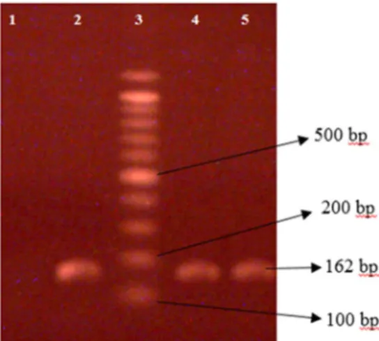
The results showed that the distribution ofmecA gene existed in staphylo- coccus in 60% of samples (30 samples). It should be noted that the rate of distribution ofmecA gene in samples ofS. aureusseparated from wound samples was more than other samples; from 30 samples, 15 samples belonged to wound samples (p<0.032) (Figure1).
Amplification ofpvlgene
For considering existence of pvl gene in strains of separated S. aureus, specific primers of this gene were used and we expected existence of the band 575 bp that its image was observed in electrophorese gel.pvlgene was positive in 20% of samples (10 samples) (Figure2). It is necessary to mention that there was not meaningful relationship between existence of pvl gene, type of sample (p<0.046), and mecA gene among strains (p<0.015).
Amplification ofcoaandspagenes
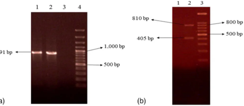
For considering existence ofcoagene andspagene in strains of separated S. aureus, specific primers of these genes were used and we expected existence of 575 bp band for both genes. The result showed that each has two patterns of
Figure 1.1% agarose gel electrophoresis analysis of PCR amplification products ofmecA gene of 162 bp extracted fromS. aureus. Lane 1: negative control (no DNA template); lanes 2, 4: positive isolates; lane 3: DNA molecular size marker (100 bp ladder); lane 5: positive control (mecA-positive
strain ATCC 33591)
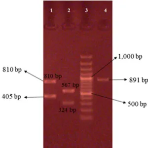
amplification and size of product PCR of genes was varied. PCR products ofcoa gene were 891 bp in some isolates, 810 and 405 bp in others. PCR products ofspa gene were 1,200 bp in some isolates, 1,296 and 240 bp in others (Figures3and4).
PCR-RFLP of coaandspagenes
For being certain about different genetic content of PCR bands, RFLP technique was used. Regarding research in computer (in silico) oncoaandspa
Figure 2.1% agarose gel electrophoresis analysis of PCR ofpvlgene. Lanes 1–5: positive isolates;
lane 6: positive control (pvl-positive strain ATCC 49775); lane 7: DNA molecular size marker (100 bp ladder); lane 8: negative control
Figure 3.Representative 2% agarose gel electrophoresis ofcoagene PCR products where 3 (b) and 4 (a) are DNA molecular size markers (100 bp ladder). (a) Isolates 1 and 2 showing single band,
(b) isolate 2 showing two bands
genes in database GenBank, enzymes HaeIII and Bsp1431 were used that has proper restriction sites oncoaandspa genes (Figures5and 6).
Discussion
Distribution of resistance against different antibiotics among strains of S. aureushas created many problems in different parts of the world. Unfortunately, there is little information on the resistance rate ofS. aureusstrains against different antibiotics in some cities of Iran. In this study, 68% of S. aureusstrains were isolated from patients who are resistant to cefoxitin (MRSA) and this rate is more than that of studies conducted by some researchers. These results showed that the frequency of MRSA strains in the subjects is more related to infectology and surgical departments. Potential danger of the distribution of MRSA strains in a special care section has been paid attention due to the occurrence of more problems in the hospitals, various medical manipulation, and extensive consump- tion of antibiotics. Study of Zamani et al. [18], on 70 samples ofS. aureus, showed that 50% were MRSA and considering antibiotic-resistance pattern, they showed resistance to tetracycline (74.2%), co-trimoxazole (68.5%), erythromycin (68.5%), and ceftazidime (51.4%). Studies by Moradi et al. [19], on 104 samples of S. aureus, showed that the highest rate of sensitivity was toward vancomycin (96.2%), chloramphenicol (88.2%), rifampin (81.7%), and the lowest rate of resistance was toward cefoxitin (40.4%). In a study by Lepsanovic et al. [20],
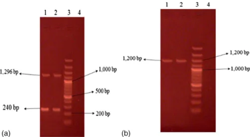
Figure 4.Representative 2% agarose gel electrophoresis ofspagene PCR products where lane 3 is DNA molecular size marker (100 bp ladder). (a) Isolates 1 and 2 showing two bands, (b) isolates 1
and 2 showing single band
all MRSA strains from hospitalized patients were resistant to one or more non-β-lactam antibiotics while 52% were multiresistant. In isolates from healthy people, 16% were sensitive to all non-β-lactam antibiotics and 40% were
Figure 6.Restriction analysis ofspagene withBsp1431 restriction enzyme. Column 2 is DNA molecular size marker (100 bp ladder). Isolate 1 showing four bands ofspagene, isolate 3 showing
three bands ofspagene, and isolate 4 showing three bands ofspagene PCR product Figure 5.Restriction analysis ofcoagene withHaeIII restriction enzyme. Column 3 is DNA molecular size marker (100 bp ladder), isolates 1–4 showing 1 band ofcoagene that remained uncut,
and isolate 2 showing two bands ofcoagene PCR product
multiresistant. Study by Haghgoo et al. [21], on 13 samples ofS. aureus, isolated from the cultivation of blood of patients kept in Shahid Madani Hospital in Tabriz, Iran, showed that the highest resistance was toward methicillin and ceftriaxone (31%). In a study by Karasartova et al. [22], 43 phages isolated from 48 MRSA were investigated for carrying toxin genes including thesak, eta, lukf-pv, sea, selp, sek, seg, seq chp, and scn virulence genes using PCR and Southern blot. The results indicate that prophages encode a significant proportion of MRSA virulence factors. Distribution of strains of cefoxitin-resistantS. aureusin this study is more than that in the mentioned studies.pvl is a virulence factor that is carried by a bacteriophage and it can be transferred to otherStaphylococcusstrains. Positive strains ofpvl have high virulence and are responsible for severe infections like infection of bone joint and necrosis pneumonia. Therefore, rapid distinguishing and on time control of thepvl-positive strains is necessary. This is thefirst study to determine the frequency ofpvlgene in clinical samples from Rasht, Iran. In this study, it was shown that from 50 isolates ofS. aureus, 20% (10 samples) carried this gene and were more frequent in samples of urine and also it was shown that no significant relationship existed betweenpvl andmecA genes among the strains.
Molla-abbaszadeh et al. [23], considered the distribution ofpvlgene in strains of S. aureusand from 100 strains, 18 (18%) were reported to be positive, regarding the existence ofpvlgene and from these subjects, 94.4% MRSA and 56.6% were methicillin-sensitiveStaphylococcus aureus(MSSA). Khosravi et al. [17] reported distribution ofpvlgene in MRSA strains was 7.2% and in MSSA strains, it was 33.3% [17]. Recently, more studies have been conducted on positivepvlstrains of MRSA, whereas positive pvl infections of MSSA play important role in the distribution of positivepvlstrains. About 60% of the total positive strains forpvlin England in the past 5 years were sensitive to methicillin [24]. A study of Cupane et al. [25] showed that that 75% of strains ofS. aureushadpvlgene and most of them (60.7%) were MRSA. In a study by Osman et al. [26], among 210S. aureus samples, pvl gene was observed in 58%. Brown et al. [8] considered 1,055 S. aureusregarding existence ofpvlgene, 377 strains (35.7%) had this gene, which is most resistant to methicillin. In a study of Lima et al. [27], on strains of MRSA isolated from patients with cysticfibrosis showed that almost half of the strains carriedpvlgene. In this study, distribution ofpvlgene in MRSA strains was more that 60% of the samples of positivepvl(six samples) in strains MRSA and 40%
(4 samples) in MSSA strains. The distribution of pvl gene in the study of Molla-abbaszadeh et al. [23] and Khosravi et al. [17] was more than that of the study of Cupane et al. [25] and Osman et al. [26], and in the study of Lima et al.
[27], it was less, and it seems that the distribution of positive pvl gene was more in MRSA strains in all the mentioned studies. Coa gene exists in all Staphylococcus strains and shows capability of typing for this gene; therefore,
it is a main gene for distinguishing S. aureus. In this study, three types of coa genes were observed among the strains that there was band of 405 bp in most strains. Existence of more than one band shows more than one allele incoagene, which shows that the strain produces different variants of this protein. Polymor- phism existing incoagene resulted from deletion and insertion at the end of 3′of coagene and gene sizes changes in this way. Himabindu et al. [28] showed that PCR product ofcoagene has three band patterns and most isolates had 812 bp, whereas in this study, band of 405 bp existed in most strains. According to studies of Lawrence et al. [29], typing ofcoagene creates common band of 402 bp, which is similar to the present studies in which 405 bp band occurred in most strains. In this study, the PCR product of some strains was not cut byHaeIII enzyme and it showed that they do not have cutting site forHaeIII. Study of Lawrence et al. [29]
demonstrated that band of 402 bp byHaeIII enzyme was changed to bands 176, 146, and 81 bp. In case that in our studies, band of 405 bp have not been cut by HaeIII enzyme. In a study by Karahan et al. [30], in Turkey, it was shown that from 200 strains ofS. aureus, 161 (80.5%) had polymorphism incoagene. About 83.9% of these strains had a band with size of 500–1,400 bp and 16.1% had two pieces. Size ofspagene based on its X region is polymorphic and has repetitive sequence of 24 bp; this number and sequence in different strains are different. Size ofspagene in this study was created in the 3′end with different sizes, which were observed to be 1,200, 1,296, and 240 bp in gels. Inexistence of spagene was observed in three strains. Study by Shakeri et al. [10] showed thatspagene was not present in 5% of the strains. Schmitz et al. [31] showed thatfive types ofspagene were reported among strains of S. aureus. They reported that the number of repetitive sequences of X region of spa gene is related to the epidemiological compatibility. Strains having shorter length cannot attach to the epithelium of nose; therefore, exit through breath, caught, and sneezed out through the nose. It should be noted that this is thefirst study conducted on molecular typing of clinical isolates ofS. aureusbased oncoaandspagenes samples, from hospitals of Rasht, Iran; this shows difference of genetic patterns. In some cases, some similarities are observed in different sections, which probably show a kind of bacterial transfer between the staff and hospitals.
Conclusions
The results of this study showed increasing resistance of clinical samples ofS. aureusto different antibiotics. The resistance rate of S. aureusto cefoxitin was high and on the other hand, its resistance toward other antibiotics, such as β-lactam, aminoglycosides, macrolides, and quinolone, is high. Thepvlgene shows
relatively high frequency in this region as compared with other places in the world.
Since infection with the bacteria,S. aureusis very prevalent and toxin-producing bacteria create problems at the society level, so it is necessary to use proper diagnostic method, especially molecular methods to identify virulence factors.
Conflict of Interest No conflict of interest associated with this work.
References
1. Masalha, M., Borovok, I., Schreiber, R., Aharonowitz, Y., Cohen, G.: Analysis of transcription of the Staphylococcus aureus aerobic class Ib and anaerobic class III ribonucleotide reductase genes in response to oxygen. J Bacteriol183, 7260–7272 (2001).
2. Tarai, B., Das, P., Kumar, D.: Recurrent challenges for clinicians: Emergence of methicillin- resistant, vancomycin resistance, and current treatment options. J Lab Physicians5, 71–78 (2013).
3. Shorr, A. F.: Epidemiology of Staphylococcal resistance. Clin Infect Dis45, 171–176 (2007).
4. Lina, G., Piemont, Y., Godail-Gamot, F., Bes, M., Peter, M. O., Gauduchon, V.:
Involvement of Panton–Valentine leukocidin-producingStaphylococcus aureusin primary skin infections and pneumonia. Clin Infect Dis29, 1128–1132 (1999).
5. Supersac, G., Prevost, G., Piemont, Y.: Sequencing of leucocidin R fromStaphylococcus aureusP83 suggests that Staphylococcal leucocidins and gamma-hemolysin are members of a single, two-component family of toxins. Infect Immun61, 580–587 (1993).
6. Clark, J.: A brief review of Panton–Valentine leukocidin producing staphylococcal infections in the intensive therapy unit. Curr Anaesth Crit Care19, 330–332 (2008).
7. Colin, D. A., Mazurier, I., Sire, S., Finck-Barbancon, V.: Interaction of the two components of leukocidin from Staphylococcus aureus with human polymorphonuclear leukocyte membranes: Sequential binding and subsequent activation. Infect Immun 62, 3184– 3190 (1994).
8. Brown, M. L., O’Hara, F. P., Close, N. M., Mera, R. M., Miller, L. A., Suaya, J. A.:
Prevalence and sequence variation of Panton–Valentine leukocidin in methicillin-resistant and methicillin-susceptible Staphylococcus aureus strains in the United States. J Clin Microbiol50, 86–90 (2012).
9. Simpson, K. H., Bowden, G., Hook, M., Anvari, B.: Measurement of adhesive forces between individualStaphylococcus aureusMSCRAMMs and protein-coated surfaces by use of optical tweezers. J Bacteriol185, 2031–2035 (2003).
10. Shakeri, F., Shojai, A., Golalipour, M., Alang, S. R., Vaez, H., Ghaemi, E. A.:Spadiversity among MRSA and MSSA strains ofStaphylococcus aureusin north of Iran. Int J Microbiol 2010, Article ID 351397 (2010).
11. Ravathi, G., Puri, J., Jain, B. K.: Bacteriology of burns. Burns24, 347–349 (1998).
12. Lu, S. Y., Chang, F. Y., Cheng, C. C., Lee, K. D., Huang, Y. C.: Methicillin-resistant Staphylococcus aureusnasal colonization among adult patients visiting emergency depart- ment in a Medical Center in Taiwan. PLoS One6, e18620 (2011).
13. Clinical and Laboratory Standards Institute: Performance Standards for Antimicrobial Susceptibility Testing, Vol. 17. Clinical and Laboratory Standards Institute, Wayne, PA, 2007.
14. Ghaznavi-Rad, E., Nor Shamsudin, M., Sekawi, Z., Belkum, A., Neela, V.: A simplified multiplex PCR assay for fast and easy discrimination of globally distributed staphylococcal cassette chromosomemectypes in meticillin-resistantStaphylococcus aureus. Med Micro- biol59, 1135–1139 (2010).
15. Moghadam, S., Havaei, A.: Prevalence of methicillin-resistant Staphylococcus aureus carrying Panton–Valentine leukocidin gene in cutaneous infections in the City of Isfahan. J Med Bacteriol19, 9–16 (2012).
16. Mostafa, S.: Molecular typing of methicillin resistantStaphylococcus aureusbyspagene polymorphism. Afr J Microbiol Res7, 755–759 (2013).
17. Khosravi, A. D., Hoveizavi, H., Farshadzadeh, Z.: The prevalence of genes encoding leukocidins inStaphylococcus aureusstrains resistant and sensitive to methicillin isolated from burn patients in Taleghani Hospital, Ahvaz, Iran. Burns38, 247–251 (2012).
18. Zamani, A., Sadeghian, S., Ghaderkhani, J., Alikhani, M. Y., Najafimosleh, M., Taghi Goodarzi, M.: Detection of methicillin-resistance (mec-A) gene inStaphylococcus aureus strains by PCR and determination of antibiotic susceptibility. Ann Microbiol57, 273–276 (2007).
19. Moradi, N., Javadpour, S., Karmostaji, A.: Reduced sensitivity ofStaphylococcus aureusto vancomycin. Hormozgan Uni Med Sci15, 169–177 (2011).
20. Lepsanovic, Z., Jeremic, L. P., Lazic, S., Cirkovic, I.: High prevalence and resistance patterns of community-associated methicillin-resistant Staphylococcus aureus in the Pomoravlje Region, Serbia. Acta Microbiol Immunol Hung63, 83–92 (2016).
21. Haghgoo, S., Moaddab, S., Rafi, A.: Study of antibiotic resistance pattern ofStaphylococ- cus aureus strains isolated from blood cultures in Tabriz Shahid Madani Hospital. J Jundishapur3, 383–390 (2012).
22. Karasartova, D., Cavusoglu, Z. B., Turegun, B., Ozsan, M. T.,¸
Sahin, F.: Identification of virulence genes carried by bacteriophages obtained from clinically isolated methicillin- resistantStaphylococcus aureus. Acta Microbiol Immunol Hung63, 433–447 (2016).
23. Molla-abbaszadeh, H., Mobayen, H., Mirzaei, H.: Identification of Panton–Valentine leukocidin (pvl) genes inStaphylococcus aureusisolated from in-patients of Emam Reza and Shohada Hospitals of Tabriz by real-time PCR. Iran J Med Microbiol6, 72–80 (2013).
24. Otokunefor, K., Sloan, T., Kearns, A. M., James, R.: Molecular characterization and Panton–Valentine leukocidin typing of community-acquired methicillin-sensitiveStaphy- lococcus aureusclinical isolates. J Clin Microbiol50, 3069–3072 (2012).
25. Cupane, L., Pugacova, N., Berzina, D., Cauce, V., Gardovska, D., Miklasevics, E.: Patients with Panton–Valentine leukocidin positive Staphylococcus aureus infections run an increased risk of longer hospitalisation. Int J Mol Epidemiol Genet3, 48–55 (2012).
26. Osman, N. A. M., Alrayah, I. E., Mohamed, Y. M., El-Eragi, A. M., Eldirdery, M. M., Salih, M. A.: Molecular study of Panton–Valentine leukocidin genes amongStaphylococ- cus aureusclinical isolates in Khartoum State, Sudan. Am J Microbiol Res3, 107–111 (2015).
27. Lima, D. F., Brazao, N. B., Folescu, T. W., Neves, F. P., Ferreira, A. G., Santos, E. A., Marques, E. A., Leao, R. S.: Panton–Valentine leukocidin (PVL) gene carriage among Staphylococcus aureusstrains withSCCmectypes I, III, IV, and V recovered from cystic fibrosis pediatric patients in Brazil. Diagn Microbiol Infect Dis78, 59–62 (2014).
28. Himabindu, M., Muthamilselvan, D. S., Bishi, D. K., Verma, R. S.: Molecular analysis of coagulase gene polymorphism in clinical isolates of methicilin resistant Staphylococcus aureusby restriction fragment length polymorphism based genotyping. Am J Infect Dis5, 163–169 (2009).
29. Lawrence, C., Cosseron, M., Mimoz, O.: Use of coagulase gene typing method for detection of carrier of methicillin resistantStaphylococcus aureus. J Antimicrob Chemother 37, 687–696 (1996).
30. Karahan, M., Nuri, M., Cetinkaya, B.: Investigation of virulence genes by PCR in Stapylococcus aureusisolates originated from subclinical bovine mastitis in Turkey. Pak Vet J31, 249–253 (2011).
31. Schmitz, F., Steiert, M., Tichy, H. V.: Typing of methicillin-resistant Staphylococcus aureusisolates from Dusseldorf by six genotypic methods. J Med Microbiol47, 341–351 (1998).
![Table I. Sequence of primers used in PCR [14 – 17]](https://thumb-eu.123doks.com/thumbv2/9dokorg/1398927.117006/4.714.186.575.131.287/table-i-sequence-primers-used-pcr.webp)