Epidemiology and resistance trends of Staphylococcus aureus isolated from vaginal samples: a 10-year retrospective study in Hungary
Márió Gajdács1,2 ✉, Edit Urbán3
1Department of Pharmacodynamics and Biopharmacy, Faculty of Pharmacy, University of Szeged, Szeged, Hungary. 2Institute of Clinical Microbiology, Faculty of Medicine, University of Szeged, Szeged, Hungary. 3Department of Public Health, Faculty of Medicine, University of Szeged, Szeged, Hungary.
Introduction
The vaginal flora is a complex microbial environment, consist- ing of a multitude of microbial species in variable quantities and proportions (1, 2). This collection of microorganisms is very dis- tinct from other anatomical regions of the human body. In pre- menarcheal girls, Lactobacillus species are infrequent or minor constituents of the vaginal microbiota, with other facultative and strict anaerobes being in abundance. This corresponds to a neutral or slightly alkaline vaginal pH during early childhood. In healthy adult women, the microbial diversity is greatly reduced, with an abundance of Lactobacillus species, responsible for the acidification (due to lactic acid and H2O2 production) of this en- vironment (3, 4). The colonization of the vaginal flora by lacto- bacilli is influenced by their adhesion levels to vaginal epithelial cells; in addition, some of the nutrients are derived from these dead epithelial cells of the female hosts (and others can be found in some secretions of the glands in the lower reproductive tract) (5, 6). Following puberty, the thicker stratified epithelium and higher levels of glycogen in the reproductive tract of females also favor Lactobacillus colonization (2–4). In addition to lactobacilli, other microorganisms commonly identified as commensals in the
female genital tract include Staphylococcus aureus, Streptococcus agalactiae (group B Streptococcus), Enterococcus faecalis, Escher- ichia coli, and Candida albicans (7–9). After menopause, estrogen levels drop in the female body, dramatically affecting the vaginal microbiota; the pH rises to 6.0 to 8.0, corresponding to the reduc- tion or elimination of lactobacilli and the increased colonization of the vaginal tract by members of the Enterobacteriaceae family, Gram-positive cocci, and Bacteroides and Prevotella species (fe- cal flora) (2–6). However, if the normal vaginal flora is disrupted, these bacteria become pathogenic, causing vaginal discharge (often with a putrid odor) and other symptoms such as itching, which is detrimental to patients’ quality of life (QoL) (10, 11). If there is a multitude of bacterial species present in the vaginal flora, that usually corresponds to a disrupted, pathological state of the vaginal microbiome.
Aerobic vaginitis (AV) was first characterized in 2002 as a dis- tinct form of vaginal inflammation from bacterial vaginosis (BV) (12–14). Although both conditions are characterized by the disrup- tion of the female genital Lactobacillus flora, BV is a non-inflam- matory condition, distinguished by the high quantity of anaerobic bacteria, whereas in AV there is significant inflammation and it is mainly caused by aerobic/facultative anaerobic bacteria (13–14).
Abstract
Introduction: The vaginal flora is a complex microbial environment. The disruption of this niche usually leads to a pathological state and symptoms in patients. Aerobic vaginitis is a distinct form of vaginal inflammation, mainly caused by aerobic/facultative anaerobic bacteria (Staphylococcus aureus, Streptococcus agalactiae, and members of the Enterobacteriaceae). This study de- scribes the prevalence and antibiotic susceptibility patterns of S. aureus isolated from vaginal samples from females at a tertiary- care teaching hospital in Hungary.
Methods: This retrospective study was carried out using data collected at the Albert Szent-Györgyi Clinical Center (University of Szeged) corresponding to a 10-year period (2008–2017). Antimicrobial susceptibility testing was performed using the disk diffu- sion method and gradient diffusion, using EUCAST interpretative standards. Methicillin-resistant S. aureus (MRSA) was detected on mannitol salt agar using cefoxitin disks, the PBP2' Latex Agglutination Test Kit, and matrix-assisted laser desorption/ionization time-of-flight (MALDI-TOF) mass spectrometry.
Results: The median age of affected patients was 31 years. Most (93.95%) of the samples received were vaginal swabs. A total of 3,356 individual isolates were recorded (335.6 ± 89.10/year, range: 213–480 isolates). In 91.4% of samples, S. aureus was the only pathogen isolated. The highest levels of resistance were detected against erythromycin (11.11 ± 3.65%, range: 6.76–17.17%) and clindamycin (10.85 ± 3.36%, range: 6.49–15.54%), whereas resistance rates against doxycycline, ciprofloxacin, chloramphenicol, sulfamethoxazole-trimethoprim, and gentamicin were much lower (0–4.48%). Susceptibility to cefoxitin was observed in 97.79%
of the isolates; 74 strains were MRSA. All MRSA strains were susceptible to the antibiotics used for therapy for multidrug-resistant Gram-positive infections.
Conclusions: A slow and steady increase in resistance levels could be observed (mainly corresponding to MRSA isolates). Although the present resistance trends are still advantageous (compared to European resistance levels) and do not hinder adequate therapy, continuous surveillance of resistance levels is recommended. Macrolides and clindamycin should be used with caution, and, if available, only when susceptibility to these drugs has been verified.
Keywords: Staphylococcus aureus, epidemiology, antimicrobial resistance, vaginal samples
Received: 3 June 2019 | Returned for modification: 6 August 2019 | Accepted: 9 September 2019
The main causes of AV include overpopulation of the vaginal flora with S. aureus, S. agalactiae, or E. coli, typically concurring with an increased inflammatory response or epithelial atrophy (or both) (15). The diagnosis of AV should be performed on the ba- sis of wet mount microscopy, and it should be differentiated from BV on the basis of Nugent’s method or Gram-staining (12–13, 16).
Antimicrobial susceptibility testing results aid the choice of the appropriate antibiotic; however, therapy on the basis of culture results only is not recommended. The prevalence of AV is around 8 to 11% in pregnant women, and 5 to 24% in women reporting complaints regarding the reproductive system (12–17).
Infections of the vagina have been associated with a significant- ly increased risk of preterm labor and low birth weight; in addi- tion, other sequela may include pelvic inflammatory disease (PID), which can lead to ectopic pregnancies, tubal infertility, and other dysfunctions of the female reproductive system (18, 19). The role of bacterial infections in the vagina has also been described in the progression of cervical dysplasia and the transmission/acquisition of various viral infections (HIV and herpes simplex virus 2) (20).
The epidemiology of genitourinary pathogens varies signifi- cantly with respect to the healthcare institution in question and the geographical localization (21). The emerging threat of methi- cillin-resistant S. aureus (MRSA) infections is a grave concern for clinicians (22). This study describes the prevalence and antibiotic susceptibility patterns of S. aureus, a significant pathogen in AV, isolated from vaginal samples at a tertiary-care teaching hospital in Hungary during a 10-year study period (2008–2017).
Methods
Study design, data collection
This retrospective study was carried out using data collected be- tween January 1st, 2008 and December 31st, 2017 at the microbi- ology laboratory of the Albert Szent-Györgyi Clinical Center. This teaching hospital annually serves more than 400,000 patients in the Southern Great Plain of Hungary, according to the national in- surance data (23). An electronic search in the records of the Med- Bakter laboratory information system (LIS) for samples processed at our laboratory that were positive for S. aureus was conducted by the authors.
For the purposes of data analyses, the 10-year study period was divided into two 5-year periods (2008–2012 and 2013–2017, respec- tively). In the data analysis, the authors included samples with 105 or more colony-forming units (CFU) for S. aureus (except in cases where international guidelines recommend otherwise). Only the first isolate per patient was included in the study. In addition, S.
aureus isolates with divergent susceptibility patterns were consid- ered different isolates. In addition, patient data were collected re- garding demographic characteristics (age and reason for sample submission as indicated on the request forms for microbiological analysis). The study was deemed exempt from an ethics review by the Institutional Review Board and informed consent was not required because data anonymity was maintained.
Identification of relevant isolates
Sample processing was carried out according to guidelines in rou- tine clinical bacteriology. All culture media (5% sheep blood agar, chocolate agar, and eosin methylene blue agar) were incubated at 37 °C for 24 to 48 hours in a 5% CO2 atmosphere. If S. aureus
was detected from the relevant samples, the plates were passed on for further processing. Between 2008 and 2012, presumptive phenotypic (biochemical reaction-based) methods and VITEK 2 ID (bioMérieux, Marcy-l’Étoile, France) were used for bacte- rial identification, whereas after 2013 this was complemented by matrix-assisted laser desorption/ionization time-of-flight mass spectrometry (MALDI-TOF MS; Bruker Daltonik GmbH, Germany) (24). The methodology of sample preparation for MALDI-TOF MS measurements was described elsewhere (25).
Antimicrobial susceptibility testing
Antimicrobial susceptibility testing (AST) was performed using the Kirby–Bauer disk diffusion method and, when appropriate, E-test (Liofilchem, Abruzzo, Italy) on Mueller–Hinton agar (MHA) plates. In addition, for the verification of discrepant results, VITEK 2 AST (bioMérieux, Marcy-l’Étoile, France) was also utilized. The interpretation of the results was based on EUCAST breakpoints (http://www.eucast.org). S. aureus ATCC 29213 and S. aureus ATCC 43300 were used as quality control strains.
To evaluate the resistance trends of isolated strains, erythromy- cin (ERI), chloramphenicol (CHL), ciprofloxacin (CIP), clindamy- cin (CLI), doxycycline (DOX), gentamicin (GEN), and sulfameth- oxazole/trimethoprim (SXT) were chosen as indicator antibiotics, based on the local antibiotic utilization data method). MRSA was detected using mannitol salt agar (MSA) using cefoxitin (FOX) disks (< 22 mm zone diameter) and PBP2' Latex Agglutination Test Kit (Thermo Fisher Scientific Hungary GmbH, Budapest, Hungary).
After 2013, a combined MALDI-TOF MS and PBP2' latex agglutina- tion protocol was introduced in our laboratory (26). MRSA-positive isolates were considered to be resistant to all β-lactam antibiotics.
In the case of MRSA-positivity, susceptibility testing for additional antibiotics (vancomycin, VAN; linezolid, LZD; daptomycin, DAP;
mupirocin, MUP; and fusidic acid, FZA) was performed (22). Dur- ing data analysis, intermediate results were grouped with and re- ported as resistant. Inducible CLI resistance was detected using the D test, and these strains were also reported as resistant.
Statistical analysis
Descriptive statistical analysis (including means or medians with ranges and percentages to characterize data) was performed us- ing Microsoft Excel 2013 (Microsoft Corp., Redmond, WA). Statisti- cal analyses were performed with SPSS software version 24 (IBM SPSS Statistics for Windows 24.0, IBM Corp., Armonk, NY), using the χ2 test, Student’s t-test, and Mann–Whitney U test. The nor- mality of variables was tested using Shapiro–Wilk tests. P values
< 0.05 were considered statistically significant.
Results
Demographic characteristics, sample types
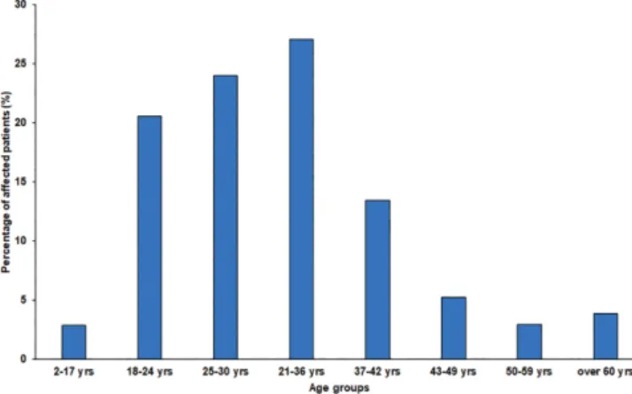
The median age of affected patients was 31 years in both the first (2008–2012) and second (2013–2017) half of the study period (range 2008–2012: 2–91 years, range 2013–2017: 9–83 years; p > 0.05), and the detailed age distribution of patients is presented in Figure 1.
Most (93.95%) of the samples received were vaginal swabs, and 5.62% were high cervical swabs, 0.29% were explanted intrauter- ine devices (IUDs), and 0.14% were urethral swabs.
The main indications for the sample submission associated
with samples positive for S. aureus included vaginitis/vulvitis (49.79%) and high-risk pregnancy (31.02%); less common indica- tions included nonspecific abdominal pain (1.73%), symptoms of menopause (1.31%), suspicion of a urinary tract infection (0.66%), unspecified pathology of the cervix (0.39%), amenorrhea/dysmen- orrhea (0.33%), polycystic ovary syndrome (PCOS) or other cysts of the female genitourinary tract (0.30%), endometriosis (0.30%), or other nonspecific reasons (14.84%).
Distribution of S. aureus isolates among vaginal samples from women
During the 10-year surveillance period (January 1st, 2008 – De- cember 31st, 2017), the Institute of Clinical Microbiology received 4,012 samples from outpatient clinics and inpatient departments that turned out to be positive for S. aureus; after data consolida- tion, 3,356 individual isolates were recorded (335.6 ± 89.10/year, range: 213–480 isolates; highest in 2016, lowest in 2008) during the study period (the detailed isolation frequency is presented in Fig. 2). A considerable but not significant increase (p = 0.182) was observed in the isolation frequency in the second part of the study period (296.80 ± 58.81 vs. 374.40 ± 103.15). In 91.4% of samples, S.
aureus was the only pathogen isolated.
Antibiotic susceptibility trends among S. aureus strains in the study period
The resistance levels of the individual S. aureus isolates are sum- marized in Table 1. The highest levels of resistance were detected
against ERI (11.11 ± 3.65%, range: 6.76–17.17%) and CLI (10.85 ± 3.36%, range: 6.49–15.54%), whereas resistance rates against DOX (2.19 ± 2.10%, range: 0–4.48%), CIP (1.50 ± 1.04%, range:
0–3.41%), CHL (0.67 ± 0.40%, range: 0–1.90%), SXT (0.31 ± 0.81%, range: 0–1.61%), and GEN (0.20 ± 0.14%, range: 0–0.59%) were much lower in comparison. Statistically significant differences during the two halves of the study period could only be observed in the case of CLI (p < 0.001), and no such difference was noted for other antibiotics (p = 0.138, p = 0.685, p = 0.345, p = 0.147, p
= 0.693, and p = 0.292 for ERI, CHL, CIP, DOX, GEN, and SXT, re- spectively). Susceptibility to FOX was observed in 97.79% of the isolates over the 10-year study period.
Overall, 74 strains were detected that were MRSA-positive (FOX resistant and positive for PBP2' latex agglutination; the first iso- late was recovered in 2012), representing 2.21 ± 1.91% of the iso- lates (range 0–5.28%, highest in 2017). In addition to MRSA-pos- itivity, 56 (75.7%) strains were also resistant to ERI, 38 (51.4%) to CLI, 31 (41.9%) to DOX, 11 (14.9%) to CIP, and 6 (8.1%) to GEN. No MRSA-positive strains were resistant to CHL and SXT; in addition, all MRSA strains were susceptible to the antibiotics specifically used for the therapy for MDR Gram-positive infections or MRSA- decolonization (namely VAN, LZD, DAP, MUP, and FZA).
Discussion
This study reports on the epidemiological trends and resistance levels of S. aureus in gynecological samples in the Southern Great Plain of Hungary over a long surveillance period (10 years). To date, this is the first and longest such study in Hungary. There are
Table 1 | Table 1. Percentage of S. aureus resistant isolates to the tested antibiotics.
Antibiotics
Study year ERI CHL CIP CLI DOX GEN SXT MRSA*
2008 8.45 % 0.00 % 0.00 % 7.98 % 6.10 % 0.00 % 0.00 % 0.00 %
2009 11.40 % 0.65 % 1.63 % 9.45 % 0.65 % 0.33 % 2.61 % 0.00 %
2010 6.76 % 0.54 % 0.27 % 6.49 % 2.43 % 0.54 % 0.27 % 0.00 %
2011 8.86 % 0.74 % 0.74 % 8.12 % 2.95 % 0.00 % 0.37 % 0.00 %
2012 9.29 % 1.24 % 3.41 % 8.36 % 3.72 % 0.31 % 0.00 % 3.41 %
2013 13.00 % 0.45 % 0.90 % 11.21 % 4.48 % 0.00 % 0.00 % 1.79 %
2014 12.50 % 0.82 % 2.45 % 11.14 % 0.27 % 0.27 % 0.00 % 3.53 %
2015 17.17 % 1.30 % 1.96 % 15.43 % 1.09 % 0.00 % 0.00 % 2.83 %
2016 7.29 % 0.63 % 1.88 % 14.79 % 0.21 % 0.00 % 0.42 % 3.13 %
2017 16.42 % 0.29 % 1.76 % 15.54 % 0.00 % 0.59 % 0.00 % 5.28 %
10-year average 11.11 % 0.67 % 1.50 % 10.85 % 2.19 % 0.20 % 0.37 % 2.21 %
SD ± 3.65 % 0.40 % 1.04 % 3.36 % 2.10 % 0.24 % 0.81 % 1.91 %
*Represents FOX resistant and PBP2’ latex agglutination-positive isolates.
ERI = erythromycin, CHL = chloramphenicol, CIP = ciprofloxacin, CLI = clindamycin, DOX = doxycycline, GEN = gentamicin, SXT =sulfamethoxazole/trimethoprim, MRSA = methicillin-resistant S. aureus, SD = standard deviation.
Values in boldface represent peak resistance levels.
Figure 1 | Age distribution of affected patients during study period. Figure 2 | Frequency of Staphylococcus aureus isolates in vaginal samples from outpatients and inpatients during the 10-year study period.
References
1. Mendling W. Vaginal microbiota. Adv Exp Med Biol. 2019;902:83–93.
2. Martin DH. The microbiota of the vagina and its influence on women’s health and disease. Am J Med Sci. 2012;343:2–9.
3. Miller EA, Beasley DE, Dunn RR, Archie EA. Lactobacilli dominance and vaginal pH: why is the human vaginal microbiome unique? Front Microbiol. 2016;7:1936.
4. Vásquez A, Jakobsson T, Ahnré S, Forsum U, Molin G. Vaginal lactobacillus flora of healthy Swedish women. J Clin Microbiol. 2002;40:2746–9.
5. Boris S, Suárez JE, Vázquez F, Barbés C. Adherence of human vaginal lactoba- cilli to vaginal epithelial cells and interaction with uropathogens. Infect Immun.
1998;66:1985–9.
6. Zárate G, Nader-Marcias ME. Influence of probiotic vaginal lactobacilli on in vitro adhesion of urogenital pathogens to vaginal epithelial cells. Lett Appl Microbiol.
2006;43:174–80.
7. Larsen B, Monif GRG. Understanding the bacterial flora of the female genital tract. Clin Infect Dis. 2001;32:e67–e77.
8. Rose WA, McGowin CL, Spagnuolo RA, Eaves-Pyles TD, Popov VL, Pyles RB. Com- mensal bacteria modulate innate immune responses of vaginal epithelial cell multilayer cultures. PlosOne. 2012;7:e032728.
9. Smith WL, Hedges SR, Mordechai E, Adelson ME, Trama JP, Gygax SE, et al. Cer- vical and vaginal flora specimens are highly concordant with respect to bacte- rial vaginosis–associated organisms and commensal lactobacillus species in women of reproductive age. J Clin Microbiol. 2014;52:3078–81.
10. Spence D, Melville C. Vaginal discharge. BMJ. 2007;335:1147–51.
11. Mitchell H. Vaginal discharge—causes, diagnosis, and treatment. BMJ. 2004;
328:1306.
12. Donders GGG, Bellen G, Grinceviciene S, Ruban K, Vieriea-Baptista P. Aerobic vaginitis: no longer a stranger. Res Microbiol. 2017;168:845–58.
13. Sun X, Qui H, Jin Y. Highly efficient treatment of aerobic vaginitis with simple acidic buffered gels: the importance of pH and buffers on the microenvironment of vaginas. Int J Pharmaceutics. 2017;52:175–82.
scant data available in the literature regarding the susceptibility patterns of S. aureus isolates from gynecological samples, and therefore additional data are necessary to provide a comprehen- sive picture of resistance trends (15, 22, 27–28). As a general rule, the susceptibility patterns were advantageous, and the resistance rates were below 20% in the case of every antibiotic tested. How- ever, a slow and steady increase in resistance levels could be ob- served (corresponding to MRSA isolates, which are resistant to all β-lactam antibiotics, apart from fifth-generation cephalosporins), especially since 2012, when the first MRSA isolate was reported (22, 29–30). In a similar study in the same geographical region and time period (2008–2017), the resistance trends of S. aureus were described from male STI samples; more than 98% of isolates were methicillin-susceptible, and the highest levels of resistance were detected against macrolides (15–28%) and clindamycin (13–30%) (31). S. aureus as a urinary pathogen was also characterized in the southern part of Hungary: in these studies, 9.8 to 11.6% of iso- lates were MRSA between 2013 and 2017, and a numerically in- creasing tendency was observed (unpublished results). Based on the results of the European Antimicrobial Resistance Surveillance Network (EARS-Net), the European average for the percentage of MRSA was 16.9%, showing a decreasing tendency with large in- ter-country variation (1.0–44.0%) (32). Regarding Hungary, MRSA levels have been over 20% since the 2010s, and were fluctuating between 21 and 27% in the last 3 years of surveillance (2014–2017) (32, 33). Of course, the resistance situation is not as dramatic in the S. aureus isolates from vaginal samples (with the highest lev- els of MRSA detected around 5%) compared to invasive isolates;
nevertheless, the obvious increase in resistance should be noted.
There is no generally accepted clinical strategy for therapy for AV; however, several proposals have been published. The dis- tinction between AV and BV is crucial for the choice of appropri- ate therapy (3–13). Metronidazole has no effect on AV, unlike in BV and Trichomonas vaginalis infections, for which this drug is routinely used (6–9). Clindamycin may be considered as a valid therapeutic option (especially in pregnancy), whereas fluoroqui- nolones are recommended in non-pregnant females because their effect on the vaginal microbiota (Lactobacillus) seems to be mini- mal (3–13). However, antibiotic therapy should be complemented if the inflammation (topical steroids) or atrophy (estrogen) is pro- nounced. In addition, the use of vaginal probiotics should also be encouraged. Regarding the use of estrogen, in some patient popu- lations (breast cancer patients and postmenopausal women) its use is contraindicated; in these cases, a very low dose of local es-
triol should be used in combination with probiotics (3–13). Based on our results, the therapy for these genitourinary infections will not be hindered by antibiotic resistance for now; however, close surveillance should be performed for S. aureus isolates from all anatomical sites to monitor the changes in resistance trends (22).
On the other hand, macrolides and CLI should be used with cau- tion, and, if available, only when susceptibility to these drugs has been verified.
Limitations of this study must be acknowledged. First, the design of the study is retrospective and we could not access the medical records of the individual patients affected by these in- fections. For this reason, the correlation between the existence of relevant risk factors and underlying illnesses (apart from age) and the isolation of S. aureus could not be assessed. There is a risk of selection bias because studies describing the prevalence of various infectious diseases and resistance trends are mainly from tertiary-care centers, which generally correspond to patients with more severe conditions or underlying illnesses. Finally, a molecular characterization of the resistance determinants in the individual isolates was not performed (i.e., mecA or mecC genes), only to the extent of FOX resistance and latex agglutination tests.
Conclusions
AV is a distinct form of vaginal inflammation from BV, predom- inantly caused by S. aureus, S. agalactiae, and members of the Enterobacteriaceae family, which are abundantly present in case of vaginal dysbiosis. Because S. aureus is a significant pathogen in AV, the aim of this study was to evaluate its relevance and fre- quency in vaginal samples at our tertiary-care teaching hospital during a 10-year study period (2008–2017). The highest levels of resistance were associated with CLI and macrolides (around 10%), whereas MRSA-levels were below 5% up until 2017. Con- tinuous monitoring of resistance trends and the introduction of antimicrobial stewardship is recommended in the therapy for AV.
Acknowledgements
The authors would like to thank Tünde Deák and Erika Karasz for their excellent laboratory assistance during the routine diagnostic work. Márió Gajdács was supported by the National Youth Excel- lence Scholarship (Grant Number NTP-NTFÖ-18-C-0225) and the ESCMID Mentorship and Observership Programme.
14. Bagnall P, Rizollo D. Bacterial vaginosis—a practical review. JAAPA 2017;30:15–
21.
15. Sangeetha KT, Golia S, Vasudha CL. A study of aerobic bacterial pathogens as- sociated with vaginitis in reproductive age group women (15–45 years) and their sensitivity pattern. Int J Med Sci. 2015;3:2268–73.
16. Mastromarino P, Vitali B, Mosca L. Bacterial vaginosis: a review on clinical trials with probiotics. New Microbiol. 2013;36:229–38.
17. Geng N, Wu W, Fan A, Han C, Wnag C, Wang Y, et al Analysis of the risk factors for aerobic vaginitis: a case-control study. Gynecol Obstetric Invest. 2016;81:
148–54.
18. Wen A, Srinivasan U, Goldberg D, Owen J, Marrs CF, Misha D, et al. Selected vaginal bacteria and risk of preterm birth: an ecological perspective. J Infect Dis.
2014;209:1087–94.
19. Fettweis JM, Serrano MG, Brooks JP, Edwards DJ, Girerd PH, Parikh HI, et al. The vaginal microbiome and preterm birth. Nature Med. 2019; 25:1012–21.
20. Ling Z, Liu X, Luo Y, Wu X, Yuan L, Tong X, et al. Associations between vaginal pathogenic community and bacterial vaginosis in Chinese reproductive-age women. Plos One. 2013;8:e76589.
21. Gajdács M, Urbán E. Epidemiological trends and resistance associated with Stenotrophomonas maltophilia bacteremia: a 10-year retrospective cohort study in a tertiary-care hospital in Hungary. Diseases. 2019;7:41.
22. Gajdács M. The continuing threat of methicillin-resistant Staphylococcus au- reus. Antibiotics. 2019;8:52.
23. Hospital bed count and patient turnover report 2017. National Health Insurance Fund of Hungary; [cited 2019 May 8]. Available from: http://www.neak.gov.hu/
felso_menu/szakmai_oldalak/publikus_forgalmi_adatok/gyogyito_megelozo_
forgalmi_adat/fekvobeteg_szakellatas/korhazi_agyszam.html.
24. Gajdács M, Spengler G, Urbán E. Identification and antimicrobial susceptibility testing of anaerobic bacteria: Rubik’s cube of clinical microbiology? Antibiotics.
2017;6:25.
25. Nagy E, Becker S, Kostrzewa M, Barta N, Urbán E. The value of MALDI-TOF MS for the identification of clinically relevant anaerobic bacteria in routine laborato- ries. J Med Microbiol. 2012;61:1393–400.
26. Ábrók M, Lázár A, Szécsényi M, Deák, J, Urbán, E. Combination of MALDI-TOF MS and PBP2' latex agglutination assay for rapid MRSA detection. J Microbiol Methods. 2018;144:122–4.
27. Guinan ME, Dan BB, Guidotti RJ, Reingold AL, Schmid GP, Bettoli EJ, Lossick JG, Shands KN, Kramer MA, Hargrett NT, Anderson RL, Broome CV. Vaginal coloniza- tion with Staphylococcus aureus in healthy women: a review of four studies. Ann Intern Med. 1982;96:944–7.
28. Mumtaz S, Ahmad M, Aftab I, Akhtar N, ul Hassan M, Hamid A. Aerobic vaginal pathogens and their sensitivity pattern. J Ayub Med Coll Abbottabad. 2008;20:
113–7.
29. Gajdács M. The concept of an ideal antibiotic: implications for drug design. Mol- ecules. 2019;24:892.
30. Gajdács M. [Extra deaths due to pandrug resistant bacteria: a survey of the lit- erature]. Egészségfejlesztés. 2019;60:31–6. Hungarian.
31. Gajdács M. [Epidemiology and susceptibility patters of Staphylococcus aureus isolates from STI samples of male patients (2008–2017)]. Magyar Urológia.
2019;31:66–8. Hungarian.
32. European Antimicrobial Resistance Surveillance Network (EARS-Net). [cited 2019 Jul 11]. Available from: https://ecdc.europa.eu/en/about-us/partnerships- and-networks/disease-and-laboratory-networks/ears-net.
33. Conceição T, Aires-de-Sousa M, Füzi M, Tóth A, Pászti J, Ungvári E, et al. Replace- ment of methicillin-resistant Staphylococcus aureus clones in Hungary over time: a 10-year surveillance study. Clin Microbiol Infect. 2007;13:971–9.