antibiotics
Article
Comparative Epidemiology and Resistance Trends of Proteae in Urinary Tract Infections of Inpatients and Outpatients: A 10-Year Retrospective Study
MárióGajdács1,2,* and Edit Urbán3
1 Department of Pharmacodynamics and Biopharmacy, Faculty of Pharmacy, University of Szeged, 6720 Szeged, Eötvös utca 6., Hungary
2 Institute of Clinical Microbiology, Faculty of Medicine, University of Szeged, 6725 Szeged, Semmelweis utca 6., Hungary
3 Department of Public Health, Faculty of Medicine, University of Szeged, 6720 Szeged, Dóm tér 10., Hungary
* Correspondence: gajdacs.mario@pharm.u-szeged.hu; Tel.:+36-62-341-330
Received: 15 May 2019; Accepted: 9 July 2019; Published: 11 July 2019
Abstract:Compared with infections caused by other bacterial pathogens, urinary tract infections (UTIs) caused byProteaeare often more severe and associated with a higher rate of recurrence, sequelae, and pyelonephritis. The aim of this retrospective study was to assess and compare the prevalence of UTIs caused by different species of theProteaetribe (namelyProteus,MorganellaandProvidenciaspecies) and the antibiotic resistance levels isolated from inpatients and outpatients in a primary- and tertiary-care teaching hospital in the Southern Great Plain of Hungary, during a 10-year study period. To evaluate the resistance trends of isolated strains, amoxicillin/clavulanic acid, ceftriaxone, meropenem, ertapenem, gentamicin, ciprofloxacin, and fosfomycin were chosen as indicator antibiotics, based on local antibiotic utilization data. Members ofProteaewere more frequently isolated in the case of inpatients (7.20±1.74% vs. 5.00±0.88%;p=0.0031),P. mirabiliswas the most frequently isolated member of the group. The ratio of resistant strains to sulfamethoxazole/trimethoprim, ciprofloxacin, ceftriaxone, and fosfomycin was significantly higher in the inpatient group. In the case of amoxicillin/clavulanic acid, ceftriaxone, ciprofloxacin, and sulfamethoxazole/trimethoprim, the ratio of resistant isolates was markedly higher between 2013–2017 (p<0.01). Resistance developments ofProteae, coupled with their intrinsic non-susceptibility to several antibiotics (tetracyclines, colistin, nitrofurantoin) severely limits the number of therapeutic alternatives, especially for outpatients.
Keywords: urinary tract infection; UTI; antibiotic; resistance; indicator; epidemiology; fosfomycin;
Morganella;Proteus;Providencia
1. Introduction
Urinary tract infections (UTIs) are one of the most common infections in both community (accounting for 10–30% of infections in primary care) and hospital settings (30–40%) [1–3]. In addition to their high incidence and diverse spectrum of etiological agents, these infections are often associated with recurrence, complications, and sequelae, corresponding to a decreased quality of life (QoL) for the affected patients [1–5]. For this reason, UTIs should be considered as an important factor of morbidity.
The most common causative agents of UTIs areEscherichia coli,Klebsiellaspp.,Enterococcus faecalis, members of the Proteae tribe (see below), Pseudomonas aeruginosa, Group B streptococci (GBS), Staphylococcus saprophyticus, andS. aureus, in addition toCandidaspp.; however, the distribution of etiological agents in UTIs have changed considerably, both in nosocomial and community settings [1–6].
Although there have been developments in novel antimicrobial drugs in the last decade, the treatment of UTIs is an increasingly complex challenge for clinicians [7]. The growing levels of antibiotic
Antibiotics2019,8, 91; doi:10.3390/antibiotics8030091 www.mdpi.com/journal/antibiotics
Antibiotics2019,8, 91 2 of 13
resistance (especially in Gram-negative bacteria) severely limits the available treatment options in these pathologies [8]. Nevertheless, with the emergence of extended-spectrumβ-lactamase (ESBL) and carbapenemase-producing strains in urinary pathogens, multidrug resistance (MDR) is a growing concern because, when coupled with other inherent and acquired resistance mechanisms, these pathogens may be resistant to a broad range of antibiotics [9–12].
Most members of theMorganellaceaefamily (including generaArsenophonus,Cosenzaea,Moellerella, Morganella,Photorhabdus,Proteus,Providencia, andXenorhabdus) are peritrichous Gram-negative rods, that are ubiquitous in the environment [13–15]. Taxonomically,Morganellaceaeare found in the order Enterobacterales(Gammaproteobacteria), which has been recently re-organized based on comparative genomic analyses [16]. All human pathogenic members of theMorganellaceaefamily are found in the Proteaetribe, comprising of the generaProteus(includingP. hauseri,P. mirabilis,P. myxofaciens,P. penneri, P. vulgaris, and several yet unnamed genospecies),Morganella(includingM. morganii subsp. morganiiand M. morganii subsp. sibonii), andProvidencia(includingP. alcalifaciens,P. heimbachae,P. rettgerii,P. rustigianii, andP. stuartii) [13,14]. Species ofProteaeare widespread in the environment and are normal inhabitants of the gut microbiota, in addition, they are the third most frequent causative agents of UTIs, especially in nosocomial settings [2–4,17]. They are characterized by strong urease production from urine (lowering the pH, which leads to tissue damage, scarring, and stone formation, through the composition of struvite and apatite crystals via precipitation of Ca2+and Mg2+ions), additionally, they possess virulence factors crucial for the pathogenesis of UTIs (IgA protease, hemolysin, cytotoxins, and fimbriae) [13,14,17–19].
In contrast toE. coli,Proteaeare more frequently isolated in patients with complicated UTIs (i.e., the presence of urinary catheters and functional or anatomical abnormalities) and are frequently associated with pyelonephritis, recurrence and prolonged treatment [20–23]. They are intrinsically resistant to several groups of antibiotics (nitrofurantoin, tetracyclines, and colistin; this intrinsic resistance may be used as a presumptive identification marker for these organisms), they produce variousβ-lactamases (penicillinases, AmpC-β-lactamases) and have an intrinsic reduced susceptibility to imipenem, making the management of these infections even more difficult [13,14,17–19,24]. It is generally accepted that P. mirabilishas the most advantageous resistance trends among theProteaegroup, whileP. vulgarisand Morganellaspecies have somewhat higher, andProvidenciaspecies have the highest rates of resistance to the relevant antibiotics. Indeed, these differences are frequently used in routine microbiology laboratories for their phenotypic identification/differentiation [13,14,17–19,24].
Since the 2000s, several national and global (e.g., the SENTRY Antimicrobial Surveillance Program or the Study for Monitoring Antimicrobial Resistance Trends; SMART) surveillance reports have evaluated and published the resistance trends of various Gram-positive and Gram-negative bacteria [25–27]. Nevertheless, there are very few studies available that specifically investigated the epidemiology and resistance rates of UTIs caused by the tribeProteaeas a whole, despite a trend of increased resistance in these pathogens having been observed in recent years, with the emergence of ESBL-producingProteusspecies as a particular concern [28–30]. The epidemiology and antibiotic susceptibility-patterns of urinary tract pathogens vary greatly by region, and, therefore, the assessment of local data is essential to evaluate trends over time and to reflect on the national situation compared to international data [31]. Additionally, knowledge of the relevant antibiotic susceptibility patterns of the major bacterial pathogens for UTIs is of utmost importance to allow for the optimal choice for antibiotic therapy [32–34].
With this in mind, the aim of this study was to assess and compare the prevalence of UTIs caused by different species of the Proteaetribe (namelyProteus, MorganellaandProvidenciaspecies) from inpatients and outpatients and the antibiotic resistance levels isolated at the Albert Szent-Györgyi Clinical Center (Szeged, Hungary) retrospectively, during a 10-year study period.
Antibiotics2019,8, 91 3 of 13
2. Results
2.1. Demographic Characteristics, Sample Types
The median age of affected patients was 75 years (range: 0.5–98) in the inpatient group with a female-to-male ratio of 0.83; the age distribution of patients was the following: 8.56% 0–5 years, 6.03%
6–14 years, 18.36% 15–65 years, and 67.05% over 65 years of age. In the outpatient group, the median age was 57 years (range: 0.5–98) with a female-to-male ratio of 0.91; the age distribution of patients was the following: 26.19% 0–5 years, 9.87% 6–14 years, 23.42% 15–65 years, and 40.52% over 65 years of age.
The difference in the age distribution of the two patient groups was statistically significant (p<0.0001).
Most (99.3%) of the samples received from outpatient clinics were voided (midstream) urine, while the majority (69.38%) from the inpatient departments were catheter-specimen urine; midstream urine (28.89%) and samples obtained through suprapubic bladder aspiration (1.73%) were less numerous.
2.2. Distribution of Proteae among Inpatient and Outpatient Urine Samples
During the 10-year surveillance period (1st of January 2008–31st of December 2017), the Institute of Clinical Microbiology received 21,150 urine samples from outpatient clinics and 19,325 positive samples from inpatient departments that turned out to be positive for a significant urinary pathogen.
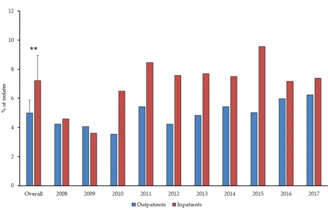
From the outpatients, 1058Proteaeisolates were obtained and 1392 from the inpatients. Members of the Proteaetribe were more frequently isolated in the case of inpatients (7.20±1.74% (range: 3.52–9.56%, lowest in 2009, highest in 2015) vs. 5.00±0.88% (range: 3.55–6.25%, lowest in 2010, highest in 2017) of all positive urine samples;p=0.0031) (Figure1.). P. mirabiliswas the most frequently isolated member of the group (inpatients: 81.54±2.76% (range: 77.94–85.81%, lowest in 2008, highest in 2012);
outpatients: 82.49±4.76% (range: 74.63–92.11%, lowest in 2010, highest in 2012);p>0.05), followed by P. vulgaris(inpatients: 13.24±8.94% (range: 3.38–32.15%, lowest in 2012, highest in 2009); outpatients:
10.82±6.45% (range: 3.95–23.88%, lowest in 2012, highest in 2010);p>0.05),M. morganii subsp. morganii (inpatients: 5.07±3.57% (range: 0–12.42%, lowest in 2011, highest in 2013); outpatients: 5.03±3.29%
(range: 0–9.23%, lowest in 2009, highest in 2015);p>0.05) andProvidenciaspp., includingP. stuartiiand P. rettgerii(inpatients: 1.83±2.72% (range: 0–8.39%, highest in 2017); outpatients: 0.91±0.89% (range:
0–2.63%, highest in 2016);p>0.05). There were no statistically significant differences in the distribution of isolates species from inpatient and outpatient samples; however, the ratio of non-Proteusisolates was much higher in the second half (2013–2017) of the study period in both patient groups (p=0.018 andp=0.029, respectively). In 7.28% of inpatients and 8.33% of outpatients, co-infection with another urinary pathogen was detected (Table1.), mainly associated with patients over 65 years of age and urinary catheters (in the case of inpatients). The distribution of co-isolated pathogens corresponds to the most frequently isolated bacteria in UTIs (e.g.,E. coli,K. pneumoniae,E. faecalis). Co-pathogens were not found in urine samples withProvidenciaspp.
Antibiotics2019,8, 91 4 of 13
Antibiotics 2019, 8, x 4 of 13
Figure 1. Ratio of Proteae in positive urine samples of inpatients and outpatients during the study period.
Table 1. Frequency of co-isolation and species distribution in inpatient and outpatient samples.
Isolated co-pathogen Setting Proteus spp. Morganella spp.
Escherichia coli Inpatient 41 5
Outpatient 29 3
Enterococcus faecalis Inpatient 31 3
Outpatient 22 2
Klebsiella pneumoniae Inpatient 23 1
Outpatient 7 2
Pseudomonas aeruginosa Inpatient 8 0
Outpatient 9 0
Enterobacter aerogenes Inpatient 2 0
Outpatient 2 0
Candida albicans Inpatient 0 0
Outpatient 2 0
Acinetobacter baumannii Inpatient 0 0
Outpatient 0 1
2.3. Antibiotic Susceptibility Trends among Proteae
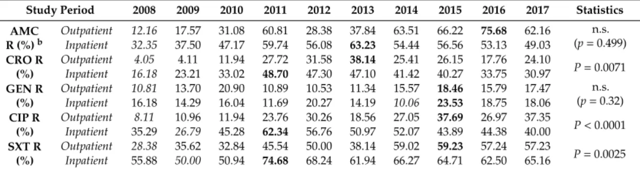
The resistance trends of isolated Proteae for amoxicillin/clavulanic acid (AMC) (in the case of P.
mirabilis only), ceftriaxone (CRO), gentamicin (GEN), ciprofloxacin (CIP), and sulfamethoxazole- trimethoprim (SXT) during the 10-year surveillance period are presented in Table 2. and Figure 2.
The ratio of resistant strains in the inpatient group was significantly higher to SXT, CIP, and CRO (p
= 0.0025, p < 0.0001, and p = 0.0071, respectively), but not in the case of GEN (p > 0.05). In contrast, AMC resistance in P. mirabilis was numerically, but not significantly higher (p = 0.499) in outpatient samples (going as high as 63.51–75.68% in the second half of the study period, while this ratio was 49.03–63.23% in the inpatient group). No statistical or numerical differences were observed among the resistance rates of various Proteae species (i.e., Proteus vs. Providencia vs. Morganella; p > 0.05)
Table 2. Percentage of resistant strains to indicator antibiotics from inpatient and outpatient departments (2008–2017) a.
Figure 1.Ratio ofProteaein positive urine samples of inpatients and outpatients during the study period.
Table 1.Frequency of co-isolation and species distribution in inpatient and outpatient samples.
Isolated co-pathogen Setting Proteusspp. Morganellaspp.
Escherichia coli Inpatient 41 5
Outpatient 29 3
Enterococcus faecalis Inpatient 31 3
Outpatient 22 2
Klebsiella pneumoniae Inpatient 23 1
Outpatient 7 2
Pseudomonas aeruginosa Inpatient 8 0
Outpatient 9 0
Enterobacter aerogenes Inpatient 2 0
Outpatient 2 0
Candida albicans Inpatient 0 0
Outpatient 2 0
Acinetobacter baumannii Inpatient 0 0
Outpatient 0 1
2.3. Antibiotic Susceptibility Trends among Proteae
The resistance trends of isolatedProteaefor amoxicillin/clavulanic acid (AMC) (in the case of P. mirabilisonly), ceftriaxone (CRO), gentamicin (GEN), ciprofloxacin (CIP), and sulfamethoxazole- trimethoprim (SXT) during the 10-year surveillance period are presented in Table2. and Figure2.
The ratio of resistant strains in the inpatient group was significantly higher to SXT, CIP, and CRO (p=0.0025,p<0.0001, andp=0.0071, respectively), but not in the case of GEN (p>0.05). In contrast, AMC resistance inP. mirabiliswas numerically, but not significantly higher (p=0.499) in outpatient samples (going as high as 63.51–75.68% in the second half of the study period, while this ratio was 49.03–63.23% in the inpatient group). No statistical or numerical differences were observed among the resistance rates of variousProteaespecies (i.e.,Proteusvs.Providenciavs.Morganella; p>0.05).
Antibiotics2019,8, 91 5 of 13
Table 2.Percentage of resistant strains to indicator antibiotics from inpatient and outpatient departments (2008–2017)a.
Study Period 2008 2009 2010 2011 2012 2013 2014 2015 2016 2017 Statistics AMC
R (%)b
Outpatient 12.16 17.57 31.08 60.81 28.38 37.84 63.51 66.22 75.68 62.16 n.s.
(p=0.499) Inpatient 32.35 37.50 47.17 59.74 56.08 63.23 54.44 56.56 53.13 49.03
CRO R (%)
Outpatient 4.05 4.11 11.94 27.72 31.58 38.14 25.41 26.15 17.76 24.10
P=0.0071 Inpatient 16.18 23.21 33.02 48.70 47.30 47.10 41.42 40.27 33.75 30.97
GEN R (%)
Outpatient 10.81 13.70 20.90 10.89 10.53 11.34 15.57 18.46 15.79 17.47 n.s.
(p=0.32) Inpatient 16.18 14.29 16.04 11.69 20.27 14.19 10.06 23.53 18.75 18.06
CIP R (%)
Outpatient 8.11 10.96 11.94 23.76 30.26 18.56 27.05 37.69 26.97 37.35
P<0.0001 Inpatient 35.29 26.79 45.28 62.34 56.76 50.97 52.07 43.89 44.38 40.00
SXT R (%)
Outpatient 28.38 35.62 32.84 45.54 50.00 38.14 59.02 59.23 57.24 57.23
P=0.0025 Inpatient 55.88 50.00 50.94 74.68 68.24 61.94 66.27 64.71 62.50 65.16
aValues initalicsrepresent the lowest (baseline) resistance levels, boldface (peak) values correspond to the highest resistance levels in the study period;bCorresponding toP. mirabilissusceptibility; n.s.: not significant.
Antibiotics 2019, 8, x 5 of 13
Study period 2008 2009 2010 2011 2012 2013 2014 2015 2016 2017 Statistics AMC R
(%)b
Outpatient 12.16 17.57 31.08 60.81 28.38 37.84 63.51 66.22 75.68 62.16 n.s.
(p = 0.499) Inpatient 32.35 37.50 47.17 59.74 56.08 63.23 54.44 56.56 53.13 49.03
CRO R (%)
Outpatient 4.05 4.11 11.94 27.72 31.58 38.14 25.41 26.15 17.76 24.10
P = 0.0071 Inpatient 16.18 23.21 33.02 48.70 47.30 47.10 41.42 40.27 33.75 30.97
GEN R (%)
Outpatient 10.81 13.70 20.90 10.89 10.53 11.34 15.57 18.46 15.79 17.47 n.s.
(p = 0.32) Inpatient 16.18 14.29 16.04 11.69 20.27 14.19 10.06 23.53 18.75 18.06
CIP R (%)
Outpatient 8.11 10.96 11.94 23.76 30.26 18.56 27.05 37.69 26.97 37.35
P < 0.0001 Inpatient 35.29 26.79 45.28 62.34 56.76 50.97 52.07 43.89 44.38 40.00
SXT R (%)
Outpatient 28.38 35.62 32.84 45.54 50.00 38.14 59.02 59.23 57.24 57.23
P = 0.0025 Inpatient 55.88 50.00 50.94 74.68 68.24 61.94 66.27 64.71 62.50 65.16
The high incidence of strains resistant to ceftriaxone (>30% of isolates in the inpatient samples, while ranging between 17.76–38.14% for outpatient samples in the second half of the study period) peaked in 2011 (48.70%) has been consistent since 2010. Isolates originating from outpatient departments showed similar growing trends in the survey period. Ciprofloxacin resistance peaked in 2011 for the inpatient group (62.34%) and has remained above 40% since 2010; while, in the outpatient group, the peak occurred in 2015 (37.69%). The highest levels of resistance overall were recorded for SXT: Resistance in inpatient isolates peaked in 2011 (74.68%) and has never gone below 50% in the inpatient group; in the outpatient group, the peak occurred in 2015 (59.23%) and resistance levels were around 50% since 2011.
The temporal nature of resistance development could also be identified between the first (2008–
2012) and second (2013–2017) half of the study period (p < 0.01) in the case of AMC, CRO CIP, and SXT, while the resistance levels of GEN has remained the lowest and relatively constant (compared to the other tested drugs, with 1.5–1.8-fold variation between the baseline and peak resistance) in the 10-year surveillance period.
Figure 2.Resistance levels ofProteaeoriginating from inpatient and outpatient urinary tract infections AMC resistance levels correspond toP. mirabilisisolates only.
The high incidence of strains resistant to ceftriaxone (>30% of isolates in the inpatient samples, while ranging between 17.76–38.14% for outpatient samples in the second half of the study period) peaked in 2011 (48.70%) has been consistent since 2010. Isolates originating from outpatient departments showed similar growing trends in the survey period. Ciprofloxacin resistance peaked in 2011 for the inpatient group (62.34%) and has remained above 40% since 2010; while, in the outpatient group, the peak occurred in 2015 (37.69%). The highest levels of resistance overall were recorded for SXT:
Resistance in inpatient isolates peaked in 2011 (74.68%) and has never gone below 50% in the inpatient group; in the outpatient group, the peak occurred in 2015 (59.23%) and resistance levels were around 50% since 2011.
The temporal nature of resistance development could also be identified between the first (2008–2012) and second (2013–2017) half of the study period (p<0.01) in the case of AMC, CRO CIP, and SXT, while the resistance levels of GEN has remained the lowest and relatively constant (compared to the other tested drugs, with 1.5–1.8-fold variation between the baseline and peak resistance) in the 10-year surveillance period.
Antibiotics2019,8, 91 6 of 13
No meropenem- or ertapenem-resistant isolates were recovered during the 10-year study period.
Fosfomycin (FOS) susceptibility testing was performed for 17.77% of outpatient and 21.23% of inpatient isolates, respectively. Overall, the ratio of resistant strains was 30.33% (outpatients) and 18.69%
(inpatients) (p=0.023).
3. Discussion
Compared with UTIs caused by other relevant bacterial pathogens, UTIs caused byProteaeare often more severe and associated with a higher incidence of recurrence, sequelae, and pyelonephritis [13,14].
In addition, the majority of bloodstream infections caused byProteaemembers originate from a UTI (urosepsis), frequently associated with urinary catheters and underlying conditions [35]. Increased resistance levels to antimicrobial agents are not only leading to changes in treatment guidelines and issues in the clinic, but also to poor prognoses, decreased QoL and an increase in the mortality rate of patients, especially in nosocomial settings [1–6]. Treatment ofProteaeinfections is especially challenging due to the various intrinsic resistance mechanisms they possess [13,14]. Other last-resort drugs used in severe infections with Gram-negative pathogens, like tigecycline and colistin are not useful in the therapy ofProteae-associated pathologies [36,37]. Several reports on the emergence and spread MDR Proteusand related species have also been published, which is further cause for concern [38,39]. This study presents the changing epidemiology and resistance trends ofProteaeassociated with urinary tract infections (UTIs) in Hungary over a long surveillance period (10 years), demonstrating a steady increase in the resistance levels regarding various antibiotics. To the best of our knowledge, this is the first and longest-spanning study reporting on the prevalence and susceptibility patterns ofProteae(and UTIs caused by these uropathogens by proxy) in Hungary.
The members of theProteaetribe were causative agents in UTIs in around 5% of cases for outpatient and 7% in inpatient settings, and, therefore, their clinical relevance should not be disregarded [13,14,35].
Based on the results of this retrospective survey, the most prevalent isolate at our tertiary-care center remainsP. mirabilis; however, a noteworthy increase (~5–7-times higher) was observed in the isolation rate ofMorganellaandProvidenciaspecies in the second half of the study period. Their higher prevalence in hospitalized patients and catheter-associated infections, a slight male dominance (~1.1–1.5-times) and the advanced age (over 65 years of age) of many affected patients is in line with the findings in the literature [13,14,17–19,24]. There is great variation in the species-distribution and susceptibilities of urinary tract pathogens in various parts of the globe, and, therefore, continuous and strict surveillance is recommended [40]. Based on relevant studies in the same geographical region (Southern Great Plain of Hungary), members ofProteae(especiallyP. mirabilis) are the third most commonly isolated microorganisms (E. coliandKlebsiellaspp. were the most common, while afterProteae, non-fermenting Gram-negative bacteria, members of the CES group (Citrobacter-Enterobacter-Serratia) andCandidaspp.
were the most prevalent) [41,42]. In another study from Hungary (concentrating on the region near the capital)Proteaewere isolated from 3–9% of urine samples; the resistance rates of these pathogens were the following: fluoroquinolones 10–50%, aminoglycosides 0–38%, ceftriaxone 0–9%, SMX/TMP 20–53%, and fosfomycin 0–14% [31]. Global reports suggest that overall,P. mirabilisis the fourth most common pathogen in UTIs, with an estimated prevalence of 2–2.8% [3]. The global SMART study, published by Morrissey et al., reportedP. mirabilisin 3.6% of isolates (fourth most common);
in this study, the ESBL-positivity for these bacteria were ~8% in the US, ~12% in Europe and>40%
in some parts of Asia [25]. In a separate arm of the SMART study in Latin America, Ponce-de-Leon et al. reported 2% ofProteusisolates as ESBL-positive, while ceftriaxone, levofloxacin, and amikacin susceptibilities were 92%, 89%, and 100%, respectively [27]. In a study by Stefaniuk et al. in Poland, this was further verified, as the reported prevalence ofProteusspp. was 7.6% (2.8% in uncomplicated infections, while 13.5% in complicated UTIs), with a similar pattern of resistance trends as in the present study [5]. In a study from New York State, Rank et al. reportedProteaeas the group fourth most common uropathogens, with the following susceptibility rates: SMX/TMP 87.0%, ciprofloxacin 84.0%, ceftazidime 93.0%, and tobramycin 95.0% [43]. In contrast, Yang et al. reportedProteaeas the
Antibiotics2019,8, 91 7 of 13
third most common UTI pathogen group in a tertiary-care hospital in China; the resistance rates of these isolates were 8.6–9.8% for amikacin, 12.1–14.6% for levofloxacin [33].
The role of the diagnostic bacteriology laboratories is to supply clinically relevant information in a precise and timely manner, which should be reciprocated by the feedback of the physicians, starting from the sample submission, followed by information regarding the symptoms of the patient and the clinical presentation [44]. Our local resistance results reflect the global pattern of increasing drug resistance inProteaetowards a variety of antimicrobial agents. It also highlights the importance of continuous surveillance at a local level in order to guide treatment recommendations for local use [43]. The results of this study suggest that—in most cases—empiric therapy forProteaeUTIs is not recommended, as the resistance rates (both for inpatient and outpatient samples) was over 20% for all of the tested drugs. Based on international guidelines, empiric antibiotic therapy is not recommended with a drug, where local resistance rates exceed 20% (in some cases 10%) [24,45]. In addition, the consultation with a hospital cumulative antibiogram (if it exists) is of the utmost importance, before the final choice of antimicrobial therapy [46]. The resistance developments ofProteae, coupled with their intrinsic non-susceptibility to several drug groups, severely limits the number of therapeutic alternatives, especially for outpatients. As a general rule, fosfomycin (if susceptibility is confirmed) still represents a safe and viable alternative for the therapy of these infections [47].
The indole-negativeP. mirabilisstrains are generally more susceptible to the relevant antibiotics than P. vulgaris(and other related species), Morganellaspp., and Providencia spp [13,14,17–19,24];
however, this difference in resistance rates was not demonstrated in our present study. Gentamicin resistance has remained relatively constant in the surveillance period, while other antibiotics, which are more frequently used in primary care (especially amoxicillin/clavulanic acid, ciprofloxacin, sulfamethoxazole/trimethoprim, and fosfomycin) or are available in oral formulations, resistance levels show a pronounced and statistically significant increase [48]. Resistance rates for amoxicillin/clavulanic acid inP. mirabiliswere two times higher in inpatient and six times higher in outpatient samples from baseline resistance values. Ciprofloxacin resistance rates were two times higher in inpatients and 4.5 times higher in outpatient from baseline resistance levels. Similarly, high levels of fluoroquinolone resistance have been reported forProteaein regions where there are no restrictions and they are still considered to be first-line agents [49–52]. Quantitatively, antibiotic utilization levels in Hungary are relatively good; however, the qualitative analysis reveals a much bleaker picture: The use of broad-spectrum antimicrobials (including fluoroquinolones) is significantly higher, which may correspond to the development of local resistance trends [48,53,54]. Interestingly, while the ratio of resistant isolates from inpatient samples was significantly higher, the increase in the ratio of resistant strains over time was actually more pronounced in the isolates from outpatient samples.
Based on the results of our study, the most concerning development is the resistance rates to third-generation cephalosporins (during our study ceftriaxone was chosen as an indicator; however, the group also includes cefotaxime, ceftriaxone, ceftazidime, and cefoperazone) [55]. Ceftriaxone-resistance rates have increased by three times in inpatient samples, and by almost ten times in outpatient samples.
Resistance to β-lactam antibiotics is a severe issue in Proteae infections, especially in vulnerable patient groups (e.g., pregnant women, children), where some other therapeutic alternatives are inappropriate due to their teratogenicity. Resistance to third-generation cephalosporins (16.18–48.70%
for inpatients and 4.05–38.14% for outpatients) inProteaeis mainly mediated through the production of ESBLs or AmpCβ-lactamases [13,14,17–19,24]. Until recently, the detection of extended-spectrum beta-lactamase (ESBL)-producing isolates inProteaewas only recommended from blood culture isolates, and for public health purposes by EUCAST; however, this recommendation has been revised in 2017 (http://www.eucast.org/resistance_mechanisms/). Since this study reports on the resistance trends ofProteaebetween 2008–2017, the exact (molecular) identification of theβ-lactam (i.e., ceftriaxone) resistance mechanism was, therefore, not performed during the study period. Additionally, this task was mainly carried out by a reference laboratory in Hungary for monetary considerations, and for invasive isolates only. Although the exact ratio of ESBL-positive CRO-resistant strains is unknown,
Antibiotics2019,8, 91 8 of 13
there is literature data available, reporting that the ESBL-positivity rates are one of the lowest inProteae (Klebsiella>Escherichia>Enterobacter>Proteus>Morganella>Providencia) [56]. In Hungary, since the 2000s, the most prevalent (>95%) type of ESBL-enzymes are theblaCTX-M-type beta-lactamases, which practically ousted most of the other types of ESBL-enzymes [57].
When it comes to carbapenems, it is well-known that imipenem is only marginally effective againstProteae. No meropenem-resistant strains were detected, therefore, in an inpatient setting (or through the utilization of outpatient parenteral antimicrobial therapy; OPAT) meropenem may be a safe option for now [58]. However, hospitalization in itself also carries a number of risks (e.g., acquiring nosocomial infections), especially in severely debilitated, immunocompromised patients. Nevertheless, this selection pressure will probably aid the emergence and global spread of carbapenemase-producing Enterobacteriaceae(CPE) even further, adding to the crisis of antimicrobial resistance [10]. However, if global resistance developments are any indication, Hungary will not avoid the increasing prevalence of MDRProteae.
Some limitations of this study must be acknowledged. Firstly, the design of this study is retrospective and, due to the inability to access the medical records of the individual patients affected, the correlation between the existence of relevant risk factors and underlying illnesses (apart from age, inpatient/outpatient status, and catheterization) andProteaeUTIs could not be assessed.
The age-associated incidence in isolation ofProteaemay also reflect (at least in part) the high rate of bacteriuria in the elderly population. Furthermore, phenotypic verification of the causes ofβ-lactam (ceftriaxone) resistance (AmpC- or ESBL-enzymes, porin loss, changes in permeability or other) or the molecular characterization of the genetic background of resistance (by PCR or sequencing) in the individual isolates was not performed, due to financial constraints, and previous local guidelines.
There is a risk of selection bias, as most studies describing the prevalence of infectious diseases are tertiary-care centers, which generally corresponds to patients with more severe conditions or underlying illnesses.
4. Materials and Methods
4.1. Study Design, Data Collection
This retrospective study was carried out using microbiological data collected from the period between the 1st of January 2008 and 31st of December 2017 at the Albert Szent-Györgyi Clinical Center, a university-affiliated (University of Szeged), primary- and tertiary-care teaching hospital in the Southern Great Plain of Hungary. The Clinical Center has a bed capacity of 1820 beds (1465 active and 355 chronic beds, respectively) and annually serves more than 400,000 patients in the region (based on the data of the National Health Insurance Fund) (Figure3) [59]. An electronic search in the records of the MedBakter laboratory information system (LIS) for urine samples submitted with the suspicion of a urinary tract infection was conducted by the authors (M.G. and E.U.). Samples with clinically significant colony counts forProteae(105<CFU/mL; however, this was subject to interpretation, based on the information provided on the request forms for microbiological analysis and relevant international guidelines) were included in the data analysis. Isolates were considered separate if they occurred more than 14 days apart or isolates with different antibiotic-susceptibility patterns were detected [60].
In addition, patient data limited to demographic characteristics (age and sex) were also collected.
Antibiotics2019,8, 91 9 of 13
Antibiotics 2019, 8, x 9 of 13
Figure 3. Study site in Hungary (Southern Great Plain of Hungary: in green; Albert Szent-Györgyi Clinical Center, Szeged: in red).
4.2. Identification of Isolates
10 µL of each un-centrifuged urine sample was cultured on UriSelect chromogenic agar plates (Bio-Rad, Berkeley, CA, USA) with a calibrated loop, according to the manufacturer’s instructions and incubated at 37 °C for 24–48 hours, aerobically. If the relevant pathogens presented in significant colony count, the plates were passed on for further processing. Between 2008–2012, presumptive phenotypic (biochemical reaction-based) methods and VITEK 2 Compact Automated ID/AST System (bioMérieux, Marcy-l'Étoile, France) were used for bacterial identification, and, after 2013, this was complemented by matrix-assisted laser desorption/ionization time-of-flight mass spectrometry (MALDI-TOF MS; Bruker Daltonik Gmbh. Gr.). The methodology of sample preparation for MALDI- TOF MS measurements was described elsewhere [61]. Mass spectrometry was performed by the Microflex MALDI Biotyper (Bruker Daltonics, Germany) in positive linear mode across the m/z range of 2 to 20 kDa; for each spectrum, 240 laser shots at 60 Hz in groups of 40 shots per sampling area were collected. The MALDI Biotyper RTC 3.1 software (Bruker Daltonics, Germany) and the MALDI Biotyper Library 3.1 were used for spectrum analysis.
4.3. Antimicrobial Susceptibility Testing
Antimicrobial susceptibility testing (AST) was performed using the Kirby–Bauer disk diffusion method and E-test (Liofilchem, Abruzzo, Italy) on Mueller–Hinton agar (MHA) plates. In addition, for the verification of discrepant results, VITEK 2 Compact Automated ID/AST System (Gr- AST card) (bioMérieux, Marcy-l'Étoile, France) was also utilized. The interpretation of the results was based on EUCAST breakpoints (http://www.eucast.org). Staphylococcus aureus ATCC 29213, Enterococcus faecalis ATCC 29212, Proteus mirabilis ATCC 35659, Escherichia coli ATCC 25922 and Pseudomonas aeruginosa ATCC 27853 were used as quality control strains.
To evaluate the resistance trends of isolated strains, amoxicillin/clavulanic acid (AMC), ceftriaxone (CRO), meropenem (MER), ertapenem (ERT), gentamicin (GEN), ciprofloxacin (CIP), sulfamethoxazole/trimethoprim (SXT), and fosfomycin (FOS) were chosen as indicator antibiotics, based on local antibiotic utilization data [62–65]. AMC resistance data was collected for P. mirabilis only (as most other members of Proteae are intrinsically resistant) [14]. Susceptibility testing data for fosfomycin (FOS) was available from 2012; FOS susceptibility testing was not routinely performed,
Figure 3. Study site in Hungary (Southern Great Plain of Hungary: in green; Albert Szent-Györgyi Clinical Center, Szeged: in red).
4.2. Identification of Isolates
10µL of each un-centrifuged urine sample was cultured on UriSelect chromogenic agar plates (Bio-Rad, Berkeley, CA, USA) with a calibrated loop, according to the manufacturer’s instructions and incubated at 37◦C for 24–48 hours, aerobically. If the relevant pathogens presented in significant colony count, the plates were passed on for further processing. Between 2008–2012, presumptive phenotypic (biochemical reaction-based) methods and VITEK 2 Compact Automated ID/AST System (bioMérieux, Marcy-l'Étoile, France) were used for bacterial identification, and, after 2013, this was complemented by matrix-assisted laser desorption/ionization time-of-flight mass spectrometry (MALDI-TOF MS; Bruker Daltonik Gmbh. Gr.). The methodology of sample preparation for MALDI-TOF MS measurements was described elsewhere [61]. Mass spectrometry was performed by the Microflex MALDI Biotyper (Bruker Daltonics, Germany) in positive linear mode across the m/z range of 2 to 20 kDa; for each spectrum, 240 laser shots at 60 Hz in groups of 40 shots per sampling area were collected. The MALDI Biotyper RTC 3.1 software (Bruker Daltonics, Germany) and the MALDI Biotyper Library 3.1 were used for spectrum analysis.
4.3. Antimicrobial Susceptibility Testing
Antimicrobial susceptibility testing (AST) was performed using the Kirby–Bauer disk diffusion method and E-test (Liofilchem, Abruzzo, Italy) on Mueller–Hinton agar (MHA) plates. In addition, for the verification of discrepant results, VITEK 2 Compact Automated ID/AST System (Gr- AST card) (bioMérieux, Marcy-l'Étoile, France) was also utilized. The interpretation of the results was based on EUCAST breakpoints (http://www.eucast.org).Staphylococcus aureusATCC 29213,Enterococcus faecalis ATCC 29212,Proteus mirabilisATCC 35659,Escherichia coliATCC 25922 andPseudomonas aeruginosa ATCC 27853 were used as quality control strains.
To evaluate the resistance trends of isolated strains, amoxicillin/clavulanic acid (AMC), ceftriaxone (CRO), meropenem (MER), ertapenem (ERT), gentamicin (GEN), ciprofloxacin (CIP), sulfamethoxazole/trimethoprim (SXT), and fosfomycin (FOS) were chosen as indicator antibiotics, based on local antibiotic utilization data [62–65]. AMC resistance data was collected for P. mirabilis only (as most other members of Proteae are intrinsically resistant) [14]. Susceptibility testing data for fosfomycin (FOS) was available from 2012; FOS susceptibility testing was not routinely performed,
Antibiotics2019,8, 91 10 of 13
only in cases of extensive drug resistance or per request of the clinicians [66]. During data analysis, intermediately-susceptible results were grouped with and reported as resistant.
4.4. Statistical Analysis
Descriptive statistical analysis (including means or medians with ranges and percentages to characterize data) was performed using Microsoft Excel 2013 (Redmond, WA, Microsoft Corp.).
Statistical analyses were performed with SPSS software version 24 (IBM SPSS Statistics for Windows 24.0, Armonk, NY, IBM Corp.), using theχ2-test, Student’s t-test and Mann–Whitney U test. The normality of variables was tested using Shapiro–Wilk tests. P values<0.05 were considered statistically significant.
Author Contributions:M.G. conceived and designed the study. E.U. was the senior microbiologist and performed the identification of the bacterial isolates during the study period. M.G. and E.U. performed data collection and analysis, wrote and revised the full paper.
Funding:This research received no external funding. Part of the APC was funded by MDPI.
Acknowledgments:The authors would like to thank Tünde Deák and Erika Karasz for their excellent laboratory assistance during the routine diagnostic work. M.G. was supported by the National Youth Excellence Scholarship [Grant Number NTP-NTFÖ-18-C-0225] and the ESCMID Mentorship and Observership Programme.
Conflicts of Interest:The authors declare no conflict of interest, monetary or otherwise.
List of abbreviations
AMC:amoxicillin/clavulanic acid; AST:antimicrobial susceptibility testing;CFU:colony-forming unit;CPE:
carbapenem-resistant Enterobacteriaceae; CIP: ciprofloxacin; CRO: ceftriaxone; ESBL: extended-spectrum β-lactamase;EUCAST:European Committee for Antimicrobial Susceptibility Testing;FOS:fosfomycin;GBS:
Group BStreptococcus;GEN:gentamicin;ID:identification;LIS:laboratory information system;MALDI-TOF MS:matrix-assisted laser desorption/ionization time-of-flight mass spectrometry;MDR:multidrug-resistance;
OPAT:outpatient parenteral antibiotic therapy; PCR:polymerase chain reaction; QoL:quality-of-life; SXT:
sulfamethoxazole-trimethoprim;UTI:urinary tract infection
References
1. Gupta, K.; Hooton, T.M.; Naber, K.G.; Wullt, B.; Colgan, R.; Miller, L.G.; Moran, G.J.; Nicolle, L.E.; Raz, R.;
Schaeffer, A.J.; et al. International clinical practice guidelines for the treatment of acute uncomplicated cystitis and pyelonephritis in women: A 2010 update by the Infectious diseases society of America and the European society for microbiology and infectious diseases.Clin. Infect. Dis.2011,52, e103–e120. [CrossRef] [PubMed]
2. Wiedemann, B.; Heisig, A.; Heisig, P. Uncomplicated urinary tract infections and antibiotic resistance-epidemiological and mechanistic aspects.Antibiotics2014,3, 341–352. [CrossRef] [PubMed]
3. Flores-Mireles, A.L.; Walker, J.N.; Caparon, M.; Hultgren, S.J. Urinary tract infections: Epidemiology, mechanisms of infection and treatment options.Nat. Rev. Microbiol. 2015,13, 269–284. [CrossRef] [PubMed]
4. Hooton, T.M.; Bradley, S.F.; Cardenas, D.D.; Colgan, R.; Geerlings, S.E.; Rice, J.C.; Saint, S.; Schaeffer, A.J.;
Tambayh, P.A.; Tenke, P.; et al. Diagnosis, prevention, and treatment of catheter-associated urinary tract infection in adults: 2009 international clinical practice guidelines from the infectious diseases society of America.Clin. Infect. Dis.2010,50, 625–663. [CrossRef] [PubMed]
5. Stefaniuk, E.; Suchocka, U.; Bosacka, K.; Hryniewicz, W. Etiology and antibiotic susceptibility of bacterial pathogens responsible for community-acquired urinary tract infections in Poland.Eur. J. Clin. Microbiol.
Infect. Dis.2016,35, 1363–1369. [CrossRef] [PubMed]
6. Achkar, J.M.; Fries, B.C. Candida infections of the genitourinary tract.Clin. Microbiol. Rev.2010,23, 253–273.
[CrossRef] [PubMed]
7. Calzi, A.; Grignolo, S.; Caviglia, I.; Calevo, M.G.; Losurdo, G.; Piaggio, G.; Bandettini, R.; Castagnola, E.
Resistance to oral antibiotics in 4569 Gram-negative rods isolated from urinary tract infection in children.
Eur. J. Pediatr.2016,175, 1219–1225. [CrossRef]
8. Gajdács, M. The concept of an ideal antibiotic: Implications for drug design. Molecules2019, 24, 892.
[CrossRef]
9. Spengler, G.; Kincses, A.; Gajdacs, M.; Amaral, L. New roads leading to old destinations: Efflux pumps as targets to reverse multidrug resistance in bacteria.Molecules2017,22, 468. [CrossRef]
Antibiotics2019,8, 91 11 of 13
10. Codjoe, F.S.; Donkor, E.S. Carbapenem resistance: A review.Med. Sci.2018,6, 1. [CrossRef]
11. Dhillon, R.H.-P.; Clark, J. ESBLs: A clear and present danger? Crit. Care Res. Pract. 2012,2012, 625170.
[CrossRef] [PubMed]
12. Gajdács, M. [Extra deaths due to pandrug resistant bacteria: A survey of the literature].Egészségfejlesztés 2019,60, 31–36.
13. O’Hara, C.M.; Brenner, F.W.; Miller, J.M. Classification, identification, and clinical significance of Proteus, Providencia, and Morganella.Clin. Microbiol. Rev.2000,13, 534–546. [CrossRef] [PubMed]
14. Manos, J.; Belas, R. The genera Proteus, Providencia, and Morganella. InThe Prokaryotes: Volume 6:
Proteobacteria: Gamma Subclass; Dworkin, M., Falkow, S., Rosenberg, E., Schleifer, K.-H., Stackebrandt, E., Eds.; Springer: New York, NY, USA, 2006; pp. 245–269. ISBN 978-0-387-30746-6.
15. Taxonomy of the family Morganellaceae Adeolu et al. 2016. Available online:https://www.namesforlife.
com/10.1601/tx.29308(accessed on 14 May 2019). [CrossRef]
16. Adeolu, M.; Alnajar, S.; Naushad, S.; Gupta, R.S. Genome-based phylogeny and taxonomy of the
“Enterobacteriales”: Proposal forEnterobacteralesord. nov. divided into the families Enterobacteriaceae, Erwiniaceaefam. nov.,Pectobacteriaceaefam. nov.,Yersiniaceaefam. nov.,Hafniaceaefam. nov.,Morganellaceae fam. nov., andBudviciaceaefam. nov.Int. J. Syst. Evol. Microbiol.2016,66, 5575–5599. [PubMed]
17. Braunstein, H.; Tomasulo, M. Identification of Proteus morganii and distinction from other Proteus species.
Am. J. Clin. Pathol.1978,70, 905–908. [CrossRef] [PubMed]
18. Pignato, S.; Giammanco, G.M.; Grimont, F.; Grimont, P.A.D.; Giammanco, G. Molecular characterization of the Genera Proteus, Morganella, and Providencia by Ribotyping.J. Clin. Microbiol.1999,37, 2840–2847.
[PubMed]
19. Barnaud, G.; Arlet, G.; Danglot, C.; Philippon, A. Cloning and sequencing of the gene encoding the AmpC beta-lactamase of Morganella morganii.FEMS Microbiol. Lett.1997,148, 15–20. [CrossRef]
20. Penner, J.; Allerberger, F.; Dierich, M.P.; Pfaller, W.; Hager, J. In vitro experiments on catheter-related infections due to gram-negative rods.Chemotherapy1993,39, 336–354. [CrossRef] [PubMed]
21. Maharjan, G.; Khadka, P.; Shilpakar, G.S.; Chapagain, G.; Dhungana, G.R. Catheter-associated urinary tract infection and obstinate biofilm producers.Can. J. Infect. Dis. Med. Microbiol.2018,2018, 7624857. [CrossRef]
[PubMed]
22. Cortese, Y.J.; Wagner, V.E.; Tierney, M.; Devine, D.; Fogarty, A. Review of catheter-associated urinary tract infections and in vitro urinary tract models.J. Healthc. Eng.2018,2018, 2986742. [CrossRef] [PubMed]
23. Jacobsen, S.M.; Stickler, D.J.; Mobley, H.L.T.; Shirtliff, M.E. Complicated catheter-associated urinary tract infections due to Escherichia coli and Proteus mirabilis. Clin. Microbiol. Rev. 2008,21, 26–59. [CrossRef]
[PubMed]
24. Mazzariol, A.; Bazaj, A.; Cornaglia, G. Multi-drug-resistant Gram-negative bacteria causing urinary tract infections: A review.J. Chemother.2017,29, 2–9. [CrossRef] [PubMed]
25. Morrissey, I.; Hackel, M.; Badal, R.; Bouchillon, S.; Hawser, S.; Biedenbach, D. A review of ten years of the study for monitoring antimicrobial resistance trends (SMART) from 2002 to 2011.Pharmaceuticals2013,6, 1335–1346. [CrossRef] [PubMed]
26. Sader, H.S.; Farrell, D.J.; Flamm, R.K.; Jones, R.N. Antimicrobial susceptibility of Gram-negative organisms isolated from patients hospitalised with pneumonia in US and European hospitals: Results from the SENTRY Antimicrobial Surveillance Program, 2009–2012. Int. J. Antimicrob. Agents2014,43, 328–334. [CrossRef]
[PubMed]
27. Ponce-de-Leon, A.; Rodríguez-Noriega, E.; Morfín-Otero, R.; Cornejo-Juárez, D.P.; Tinoco, J.C.;
Martínez-Gamboa, A.; Gaona-Tapia, C.J.; Guerrero-Almeida, M.L.; Martin-Onraët, A.; Vallejo Cervantes, J.L.;
et al. Antimicrobial susceptibility of gram-negative bacilli isolated from intra-abdominal and urinary-tract infections in Mexico from 2009 to 2015: Results from the study for monitoring antimicrobial resistance trends (SMART).PLoS ONE2018,13, e0198621. [CrossRef] [PubMed]
28. Laupland, K.B.; Parkins, M.D.; Ross, T.; Pitout, J.D.D. Population-based laboratory surveillance for tribe Proteeae isolates in a large Canadian health region. Clin. Microbiol. Infect. 2007,13, 683–688. [CrossRef]
[PubMed]
29. Meier, S.; Weber, R.; Zbinden, R.; Ruef, C.; Hasse, B. Extended-spectrum β-lactamase-producing Gram-negative pathogens in community-acquired urinary tract infections: An increasing challenge for antimicrobial therapy.Infection2011,39, 333–340. [CrossRef]
Antibiotics2019,8, 91 12 of 13
30. Luzzaro, F.; Perilli, M.; Amicosante, G.; Lombardi, G.; Belloni, R.; Zollo, A.; Bianchi, C.; Toniolo, A.
Properties of multidrug-resistant, ESBL-producing Proteus mirabilis isolates and possible role of beta-lactam/beta-lactamase inhibitor combinations.Int. J. Antimicrob. Agents2001,17, 131–135. [CrossRef]
31. Magyar, A.; Köves, B.; Nagy, K.; Dobák, A.; Arthanareeswaran, V.K.A.; Bálint, P.; Wagenlehner, F.; Tenke, P.
Spectrum and antibiotic resistance of uropathogens between 2004 and 2015 in a tertiary care hospital in Hungary.J. Med. Microbiol.2017,66, 788–797. [CrossRef] [PubMed]
32. Takhar, S.S.; Moran, G.J. Diagnosis and management of urinary tract infection in the emergency department and outpatient settings.Infect. Dis. Clin. N. Am.2014,28, 33–48. [CrossRef] [PubMed]
33. Yang, B.; Yang, F.; Wang, S.; Wang, Q.; Liu, Z.; Feng, W.; Sun, F.; Xia, P. Analysis of the spectrum and antibiotic resistance of uropathogens in outpatients a. tertiary hospital.J. Chemother. 2018,30, 145–149. [CrossRef]
[PubMed]
34. Gajdács, M.; Paulik, E.; Szabó, A. [The opinions of community pharmacists related to antibiotic use and resistance] (article in Hungarian).Acta Pharm. Hung.2018,88, 249–252.
35. Kim, B.-N.; Kim, N.J.; Kim, M.-N.; Kim, Y.S.; Woo, J.-H.; Ryu, J. Bacteraemia due to tribe Proteeae: A review of 132 cases during a decade (1991–2000).Scand. J. Infect. Dis.2003,35, 98–103. [CrossRef] [PubMed]
36. Abbo, L.M.; Hooton, T.M. Antimicrobial stewardship and urinary tract infections.Antibiotics2014,3, 174–192.
[CrossRef] [PubMed]
37. Munoz-Davila, M.J. Role of old antibiotics in the era of antibiotic resistance. Highlighted nitrofurantoin for the treatment of lower urinary tract infections.Antibiotics2014,3, 39–48. [CrossRef]
38. Cohen-Nahum, K.; Saidel-Odes, L.; Riesenberg, K.; Schlaeffer, F.; Borer, A. Urinary tract infections caused by multi-drug resistant Proteus mirabilis: Risk factors and clinical outcomes. Infection2010, 38, 41–46.
[CrossRef]
39. Yeika, E.V.; Foryoung, J.B.; Efie, D.T.; Nkwetateba, E.A.; Tolefac, P.N.; Ngowe, M.N. Multidrug resistant Proteus mirabilis and Escherichia coli causing fulminant necrotising fasciitis: A case report.BMC Res. Notes 2018,11, 322. [CrossRef]
40. Wagenlehner, F.; Tandogdu, Z.; Bartoletti, R.; Cai, T.; Cek, M.; Kulchavenya, E.; Köves, B.; Naber, K.;
Perepanova, T.; Tenke, P.; et al. The global prevalence of infections in urology study: A long-term, worldwide surveillance study on urological infections.Pathogens2016,5, 10. [CrossRef]
41. Gajdács, M.; Dóczi, I.;Ábrók, M.; Lázár, A.; Burián, K. Epidemiology of candiduria and Candida urinary tract infections in inpatients and outpatients: Results from a 10-year retrospective survey.Cent. Eur. J. Urol.
2019,72, 209–214.
42. Gajdács, M.; Urbán, E. Resistance trends and epidemiology of citrobacter-enterobacter-serratia in urinary tract infections of inpatients and outpatients (RECESUTI): A 10-year survey.Medicina2019,55, 285. [CrossRef]
43. Rank, E.L.; Lodise, T.; Avery, L.; Bankert, E.; Dobson, E.; Dumyati, G.; Hassett, S.; Keller, M.; Pearsall, M.;
Lubowski, T.; et al. Antimicrobial susceptibility trends observed in urinary pathogens obtained from New York State.Open Forum Infect. Dis.2018,5, ofy297. [CrossRef] [PubMed]
44. Gajdács, M.; Spengler, G.; Urbán, E. Identification and antimicrobial susceptibility testing of anaerobic bacteria: Rubik’s cube of clinical microbiology?Antibiotics2017,6, 25. [CrossRef] [PubMed]
45. Kang, C.I.; Kim, J.; Park, D.W.; Kim, B.N.; Ha, U.S.; Lee, S.J.; Yeo, J.K.; Min, S.K.; Lee, H.; Wie, S.H.
Clinical practice guidelines for the antibiotic treatment of community-acquired urinary tract infections.
Infect. Chemother.2018,50, 67–100. [CrossRef] [PubMed]
46. Moehring, R.W.; Hazen, K.C.; Hawkins, M.R.; Drew, R.H.; Sexton, D.J.; Anderson, D.J. Challenges in preparation of cumulative antibiogram reports for community hospitals. J. Clin. Microbiol. 2015, 53, 2977–2982. [CrossRef] [PubMed]
47. Abbott, I.J.; Meletiadis, J.; Belghanch, I.; Wijma, R.A.; Kanioura, L.; Roberts, J.A.; Peleg, A.Y.; Mouton, J.W.
Fosfomycin efficacy and emergence of resistance among Enterobacteriaceae in an in vitro dynamic bladder infection model.J. Antimicrob. Chemother.2018,73, 709–719. [CrossRef]
48. Stewardson, A.J.; Vervoort, J.; Adriaenssens, N.; Coenen, S.; Godycki-Cwirko, M.; Kowalczyk, A.;
Huttner, B.D.; Lammens, C.; Malhotra-Kumar, S.; Goossens, H.; et al. Effect of outpatient antibiotics for urinary tract infections on antimicrobial resistance among commensal Enterobacteriaceae: A multinational prospective cohort study.Clin. Microbiol. Infect.2018,24, 972–979. [CrossRef]
49. Trautner, B.W. Fluoroquinolones for urinary tract infection and within-household spread of resistant Enterobacteriaceae: The smoking gun.Clin. Microbiol. Infect.2018,24, 929–930. [CrossRef]
Antibiotics2019,8, 91 13 of 13
50. Catry, B.; Latour, K.; Bruyndonckx, R.; Diba, C.; Geerdens, C.; Coenen, S. Characteristics of the antibiotic regimen that affect antimicrobial resistance in urinary pathogens.Antimicrob. Resist. Infect. Control2018,7, 76. [CrossRef]
51. Latour, K.; Jans, B.; Coenen, S.; Preal, R.; Catry, B. Antibiograms of consecutive urinary tract samples in elderly.Antimicrob. Resist. Infect. Control2013,2, P22. [CrossRef]
52. Owumi, W.; Banaei, N.; Shortliffe, L.D. Adult and pediatric intra-institutional trends of ciprofloxacin susceptibility in E. coli positive urinary cultures.Antibiotics2014,3, 163–173. [CrossRef]
53. Adriaenssens, N.; Coenen, S.; Versporten, A.; Muller, A.; Vankerckhoven, V.; Goossens, H.; ESAC Project Group. European surveillance of antimicrobial consumption (ESAC): Quality appraisal of antibiotic use in Europe.J. Antimicrob. Chemother.2011,66(Suppl. 6), vi71–vi77. [CrossRef] [PubMed]
54. Elseviers, M.M.; Ferech, M.; Vander Stichele, R.H.; Goossens, H.; ESAC Project Group. Antibiotic use in ambulatory care in Europe (ESAC data 1997–2002): Trends, regional differences and seasonal fluctuations.
Pharmacoepidemiol. Drug Saf.2007,16, 115–123. [CrossRef] [PubMed]
55. Kong, K.-F.; Schneper, L.; Mathee, K. Beta-lactam antibiotics: From antibiosis to resistance and bacteriology.
APMIS2010,118, 1–36. [CrossRef] [PubMed]
56. Rupp, M.E.; Fey, P.D. Extended spectrum beta-lactamase (ESBL)-producing Enterobacteriaceae:
Considerations for diagnosis, prevention and drug treatment.Drugs2003,63, 353–365. [CrossRef] [PubMed]
57. Cantón, R.; González-Alba, J.M.; Galán, J.C. CTX-M enzymes: Origin and diffusion.Front. Microbiol.2012,3, 110. [CrossRef] [PubMed]
58. Neuwirth, C.; Siébor, E.; Duez, J.M.; Péchinot, A.; Kazmierczak, A. Imipenem resistance in clinical isolates of Proteus mirabilis associated with alterations in penicillin-binding proteins.J. Antimicrob. Chemother.1995,36, 335–342. [CrossRef]
59. National Health Insurance Fund of Hungary.Hospital Bed Count and Patient Turnover Report 2017; National Health Insurance Fund of Hungary: Budapest, Hungary, 2017.
60. Gajdács, M.; Urbán, E. Epidemiological trends and resistance associated with Stenotrophomonas maltophilia bacteremia: A 10-year retrospective cohort study in a tertiary-care hospital in Hungary.Diseases2019,7, 41.
[CrossRef]
61. Ábrók, M.; Lázár, A.; Szécsényi, M.; Deák, J.; Urbán, E. Combination of MALDI-TOF MS and PBP2’ latex agglutination assay for rapid MRSA detection.J. Microbiol. Methods2018,144, 122–124. [CrossRef]
62. Benk˝o, R.; Matuz, M.; Hajdú, E.; Bor, A.; Doró, P.; Viola, R.; Soós, G. [Antibiotic use in the Hungarian hospitals in the last two decades (1996–2015)].ORVOSI HETILAP2016,157, 1839–1846. [CrossRef]
63. Juhász, Z.; Benk˝o, R.; Matuz, M.; Viola, R.; Soós, G.; Hajdú, E. [Treatment practice of acute cystitis on the basis of national prescription data].ORVOSI HETILAP2014,155, 590–596. [CrossRef]
64. Matuz, M.; Benk˝o, R.; Hajdú, E.; Viola, R.; Soós, G. [Evaluation of ambulatory antibiotic use in Hungary using drug-specific quality indicators].ORVOSI HETILAP2013,154, 947–956. [CrossRef] [PubMed]
65. Gajdács, M.; Paulik, E.; Szabó, A. [The attitude of community pharmacists towards their widening roles in the prevention and treatment of infectious diseases in the southeast region of Hungary] (article in Hungarian).
Gyógyszerészet2019,63, 26–30.
66. Gajdács, M.;Ábrók, M.; Lázár, A.; Burián, K. [Susceptibility patterns of extended-spectrum beta-lactamase- producing (ESBL) urinary pathogens: Single-center experience.] (article in Hungarian).Gyógyszerészet2019, in press.
©2019 by the authors. Licensee MDPI, Basel, Switzerland. This article is an open access article distributed under the terms and conditions of the Creative Commons Attribution (CC BY) license (http://creativecommons.org/licenses/by/4.0/).