antibiotics
Communication
Resistance Levels and Epidemiology of
Non-Fermenting Gram-Negative Bacteria in Urinary Tract Infections of Inpatients and Outpatients
(RENFUTI): A 10-Year Epidemiological Snapshot
MárióGajdács1,* , Katalin Burián2,3and Gabriella Terhes3
1 Department of Pharmacodynamics and Biopharmacy, Faculty of Pharmacy, University of Szeged, Eötvös utca 6, 6720 Szeged, Hungary
2 Department of Medical Microbiology and Immunobiology, Faculty of Medicine, University of Szeged, Dóm tér 10, 6720 Szeged, Hungary
3 Institute of Clinical Microbiology, Faculty of Medicine, University of Szeged, Semmelweis utca 6, 6725 Szeged, Hungary
* Correspondence: gajdacs.mario@pharm.u-szeged.hu; Tel.:+36-62-341-330
Received: 20 August 2019; Accepted: 7 September 2019; Published: 9 September 2019
Abstract:Background:Urinary tract infections (UTIs) are one of the most common infections in the human medicine, both among outpatients and inpatients. There is an increasing appreciation for the pathogenic role of non-fermenting Gram-negative bacteria (NFGNBs) in UTIs, particularly in the presence of underlying illnesses. Methods: The study was carried out using data regarding a 10-year period (2008–2017). The antimicrobial susceptibility testing was performed using the disk diffusion method, E-tests, and broth microdilution. Results: NFGNB represented 3.46%±0.93%
for the outpatients, while 6.43%±0.81% of all positive urine samples for the inpatients (p<0.001).
In both groups,Pseudomonasspp. (78.7% compared to 85.1%) andAcinetobacterspp. (19.6% compared to 10.9%), were the most prevalent. TheAcinetobacterresistance levels were significantly higher in inpatients isolates (pvalues ranging between 0.046 and<0.001), while the differences in the resistance levels ofPseudomonaswas not as pronounced. Theβ-lactam-resistance levels were between 15–25%
and 12–28% for theAcinetobacterandPseudomonasspp., respectively. 4.71% ofAcinetobacterand 1.67%
ofPseudomonaswere extensively drug resistant (XDR); no colistin-resistant isolates were recovered.
Conclusions: Increasing resistance levels of theAcinetobacterspp. from 2013 onward, but not in the case of thePseudomonasspp. Although rare, the drug resistant NFGNB in UTIs present a concerning therapeutic challenge to clinicians with few therapeutic options left.
Keywords: urinary tract infection; UTI; antibiotic; resistance; epidemiology; non-fermenting;
Acinetobacter;Pseudomonas;Stenotrophomonas
1. Introduction
Urinary tract infections (UTIs) are the second most common type of infections in the human medicine in the United States and Europe and the third most common (following respiratory tract infections and gastrointestinal infections) infectious pathologies worldwide, representing an important factor of morbidity and mortality, both among outpatients and hospitalized patients (in the latter group, they may represent 25–50% of infections overall) [1–3]. UTIs are a considerable economic burden for healthcare institutions and national economies; additionally, they also have a substantial economic impact, as they result in lost working days [4,5]. In fact, the annual cost of UTIs in the US has been estimated to be more than 3.5 billion US dollars [6]. The principal causes of uncomplicated
Antibiotics2019,8, 143; doi:10.3390/antibiotics8030143 www.mdpi.com/journal/antibiotics
and community-associated UTIs are the members of theEnterobacteriaceaefamily (or more recently, the Enterobacteralesorder), namely theEscherichia coliandKlebsiellaspp. in the highest numbers, while the CES [Citrobacter-Enterobacter-Serratia] group and members of theProteaetribe are represented in the lesser numbers [1–3,7–13]; nevertheless, the etiological spectrum of nosocomial infections is more diverse, with non-fermenters,Staphylococcus aureus,S. saphrophyticus,Enterococcusspp., andCandida spp. in the higher numbers [2,14–18].
Non-fermenting Gram-negative bacteria (NFGNB) are a heterogenous group ofProteobacteria, which are characterized by the inability to ferment sugars to generate energy for their vital cellular functions. NFGNB include (in a decreasing order of prevalence) Pseudomonas,Acinetobacter, the Burkholderia cepacia complex(BCC),Stenotrophomonas(Xanthomonas)maltophilia, in addition to some less frequently isolated genera, such asAchromobacter,Alcaligenes,Brevimundas,Elisabethkingia,Flavobacterium andRalstoniaamong others [19,20]. Some less prevalent members, such asB. malleiandB. pseudomallei even possess the relevance as bioterrorism agents [21,22]. These microorganisms are ubiquitous in nature, especially in aquatic environments and on abiotic surfaces, in addition to being associated with plants pathologies [19,23]. In humans, they are most frequently isolated from respiratory tract samples (they are especially important in cystic fibrosis patients) and from invasive infections (bacteremia, sepsis), however, the pathogenic role of these microorganisms has been described in a variety of other clinical situations [24–26]. They are extremely prevalent in opportunistic infections, affecting severely immunocompromised, debilitated patients over 60 years of age [19,20,24,26–28]. Interpretation of the NFGNB-positivity may be tricky for clinical microbiologists, as their true significance (contaminant, colonizer or true pathogen) should be ascertained based on the patient’s symptoms and the presence of relevant risk factors [29,30]. The introduction of the matrix-assisted laser desorption/ionization time-of-flight mass spectrometry (MALDI-TOF MS) in routine laboratories has revolutionized clinical microbiology diagnostics [31,32]. The use of this technology (based on measuring the spectra of conserved, ribosomal proteins of relevant microorganisms) has brought forward a significant change in the detection of NFGNB as well, allowing for the timely and precise identification of several species, which previously only could be differentiated by the use of molecular methods (e.g., polymerase chain reaction) [33–35].
There is an increasing appreciation for the pathogenic role of NFGNB in urinary tract infections, particularly in children and in patients, who are present with underlying factors that predispose them to the development of complicated UTIs, e.g., developmental abnormalities, obstruction, vesicourethral reflux, Type II diabetes, immunosuppression (corresponding to diseases or iatrogenic factors, such as the therapeutic use of corticosteroids, mycophenolate mofetil or methotrexate), cancer or others [36–38].
Additionally, the urinary catheterization is one of the most important factors, predisposing patients to the development of UTIs [2,18]. The NFGNB possess lipopolysaccharide (LPS), various adhesins, flagella, pili, and they are characterized by the production of biofilms, cytotoxins (exotoxin A, exoenzyme S), and toxic pigments (pyoverdine, pyocyanin, pyomelanine), proteases, hemolysin, and siderophores;
all these virulence factors may have a role in the pathogenesis of UTIs, especially if the infection occurs through the intraluminal (catheter-associated) route [36,39–43]. In addition, there is extensive literature regarding the proclivity of NFGNB as multidrug resistant (MDR) pathogens. The therapy of MDR UTIs is a serious concern for clinicians, as there are few therapeutic options available, especially if some agents are further excluded due to intrinsic resistance mechanisms [36,38–40,44,45]. The etiological spectrum and the prevalence of individual pathogens in UTIs may vary significantly in different geographical regions or healthcare settings. In addition, treating physicians, armed with the knowledge of regional epidemiological (prevalence) and non-susceptibility levels, may choose the appropriate antimicrobial therapy for their patients more effectively [46,47]. The aim of this study was to report the prevalence and the temporal changes in the susceptibility levels of NFGNB in the urinary tract infections of inpatients and outpatients, using the methods of analytical epidemiology at a tertiary-care center in Hungary retrospectively, during a 10-year study period (2008–2017).
2. Results
2.1. Demographic Characteristics, Sample Types
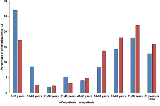
The median age of affected patients was 67 years (range: 0.2–99; median2008-2012: 64 years; range:
0–95; median2013-2017: 69; range: 0–99;p>0.05) in the outpatient group with a female-to-male ratio of 0.48 (32.3% female), while in the inpatient group, these values were 56 years (range: 0.7–95 years;
median2008-2012: 45 years; range: 0–88; median2013-2017: 62 years; range: 0–95;p=0.032) and 0.59 (37.2%
female), respectively. The detailed age distribution of the patients in both affected patient groups is presented in Figure1. The difference in the age distribution of the inpatient and outpatient groups was statistically significant (p=0.0013). Among the affected patients, the age groups of 10 years or younger (outpatients: 27.0%, inpatients: 17.2%) and patients over 60 years of age (outpatients: 44.9%, inpatients: 56.1%) were the most numerous.
Antibiotics 2019, 8, x 3 of 13
2. Results
2.1. Demographic Characteristics, Sample Types
The median age of affected patients was 67 years (range: 0.2–99; median2008-2012: 64 years; range:
0–95; median2013-2017: 69; range: 0–99; p > 0.05) in the outpatient group with a female-to-male ratio of 0.48 (32.3% female), while in the inpatient group, these values were 56 years (range: 0.7–95 years;
median2008-2012: 45 years; range: 0–88; median2013-2017: 62 years; range: 0–95; p = 0.032) and 0.59 (37.2%
female), respectively. The detailed age distribution of the patients in both affected patient groups is presented in Figure 1. The difference in the age distribution of the inpatient and outpatient groups was statistically significant (p = 0.0013). Among the affected patients, the age groups of 10 years or younger (outpatients: 27.0%, inpatients: 17.2%) and patients over 60 years of age (outpatients: 44.9%, inpatients: 56.1%) were the most numerous.
Figure 1. Age distribution of the affected patients in the outpatient and inpatient group.
During the 10-year study period, the Institute of Clinical Microbiology received 21,150 urine samples from outpatient clinics and 19,325 samples from inpatient departments that turned out to be positive for a significant urinary pathogen. All samples (100%; n = 731) received from outpatient clinics were voided (midstream) urine, while the sample distribution from the inpatient departments was the following: Catheter-specimen urine (72.4%), midstream urine (24.4%), first-stream urine (2.3%), and samples obtained through a suprapubic bladder aspiration (0.8%).
2.2. Distribution of Non-fermenting Gram-negative Bacteria in Urine Samples
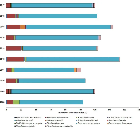
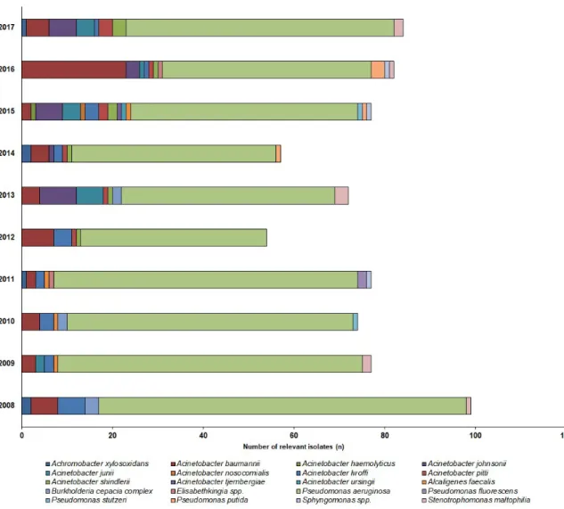
731 NFGNB isolates were obtained from outpatients (73.1 ± 11.9/year; range: 54–99) and 1229 from inpatients (122.9 ± 15.6/year; range: 104–144), corresponding to n = 649 outpatients and n = 1084 inpatients. Thus, out of the positive urine samples, these pathogens represented 3.46% ± 0.93% (range:
2.52–5.53%) for outpatients, while 6.43% ± 0.81% (range: 5.61–7.84%) of all positive urine samples for inpatients; (p < 0.001). In both groups, the Pseudomonas spp. (outpatients: 78.7%; inpatients: 85.1%;
mainly P. aeruginosa, >99%) and Acinetobacter spp. (outpatients: 19.6%, inpatients: 10.9%), were the most prevalent, while the other species, e.g., S. maltophilia, Alcaligenes spp., B. cepacia complex, Elisabethkingia spp., Sphynogomonas spp.) were in a minority (inpatients: 4%, outpatients: 1.9%). The epidemiology and detailed species distribution of the samples in both patient groups are presented
Figure 1.Age distribution of the affected patients in the outpatient and inpatient group.
During the 10-year study period, the Institute of Clinical Microbiology received 21,150 urine samples from outpatient clinics and 19,325 samples from inpatient departments that turned out to be positive for a significant urinary pathogen. All samples (100%;n=731) received from outpatient clinics were voided (midstream) urine, while the sample distribution from the inpatient departments was the following: Catheter-specimen urine (72.4%), midstream urine (24.4%), first-stream urine (2.3%), and samples obtained through a suprapubic bladder aspiration (0.8%).
2.2. Distribution of Non-fermenting Gram-negative Bacteria in Urine Samples
731 NFGNB isolates were obtained from outpatients (73.1 ± 11.9/year; range: 54–99) and 1229 from inpatients (122.9±15.6/year; range: 104–144), corresponding ton=649 outpatients and n=1084 inpatients. Thus, out of the positive urine samples, these pathogens represented 3.46%±0.93%
(range: 2.52–5.53%) for outpatients, while 6.43%±0.81% (range: 5.61–7.84%) of all positive urine samples for inpatients; (p<0.001). In both groups, thePseudomonasspp. (outpatients: 78.7%; inpatients:
85.1%; mainlyP. aeruginosa,>99%) andAcinetobacterspp. (outpatients: 19.6%, inpatients: 10.9%), were the most prevalent, while the other species, e.g.,S. maltophilia,Alcaligenesspp.,B. cepaciacomplex, Elisabethkingiaspp.,Sphynogomonasspp.) were in a minority (inpatients: 4%, outpatients: 1.9%). The epidemiology and detailed species distribution of the samples in both patient groups are presented in Figure2(inpatients) and Figure3(outpatients). In the inpatient group, 14 different species of NFGNB were isolated (median: 6; range: 4–8), while in the outpatient group, the species distribution was more diverse, with 20 different species (median: 11; range: 4–15) detected.
Antibiotics 2019, 8, x 4 of 13
in Figure 2 (inpatients) and Figure 3 (outpatients). In the inpatient group, 14 different species of NFGNB were isolated (median: 6; range: 4–8), while in the outpatient group, the species distribution was more diverse, with 20 different species (median: 11; range: 4–15) detected.
Figure 2. Frequency and species distribution of non-fermenting Gram-negative bacterial (NFGNB) isolates in the outpatient samples (2008—2017).
Figure 2.Frequency and species distribution of non-fermenting Gram-negative bacterial (NFGNB) isolates in the outpatient samples (2008—2017).
Antibiotics 2019, 8, x 5 of 13
Figure 3. Frequency and species distribution of non-fermenting Gram-negative bacterial (NFGNB) isolates in the inpatient samples (2008—2017).
2.3. Antibiotic Resistance Levels Among Urinary Non-fermenting Gram-negative Bacteria
The resistance levels of Acinetobacter and Pseudomonas isolates against the relevant antibiotics are presented in Tables 1 and 2, respectively. To identify temporal developments in the resistance levels, the 10-year study period was divided into two five-year periods (2008–2012 and 2013–2017, respectively). The level of resistance in the Acinetobacter species was significantly higher in (p values ranging between 0.046 and <0.001) in the isolates originating from inpatients, in both study periods (excluding SMX/TMP resistance in the second half of the study period), the ratio of resistant isolates was 3–10-times higher between 2008–2012, while 3–5-times higher during 2013–2017. The differences in the resistance levels of Pseudomonas spp. was not as pronounced: While in the first part of the study period, there was a significant difference among the inpatient/outpatient isolates (p values ranging between 0.033–0.045; excluding amikacin resistance), this difference was shown only for gentamicin (p = 0.043), imipenem (p = 0.036), and meropenem (p = 0.029) in the second half of the study period;
the ratio of the resistant isolates was 1.2–1.4-times higher between 2008–2012, while 0.8–2.2-times higher during 2013–2017. A significant increase in the resistance levels of aminoglycosides, fluoroquinolones, and SMX/TMP was demonstrated for the Acinetobacter spp. between 2008–2012 and 2013–2017, while similar trends were identified for imipenem, meropenem, and ceftazidime, in the case of the Pseudomonas spp. (p < 0.01). Based on the susceptibility-patterns of the individual isolates,
Figure 3.Frequency and species distribution of non-fermenting Gram-negative bacterial (NFGNB) isolates in the inpatient samples (2008—2017).
2.3. Antibiotic Resistance Levels Among Urinary Non-fermenting Gram-negative Bacteria
The resistance levels ofAcinetobacterandPseudomonasisolates against the relevant antibiotics are presented in Tables1and2, respectively. To identify temporal developments in the resistance levels, the 10-year study period was divided into two five-year periods (2008–2012 and 2013–2017, respectively).
The level of resistance in theAcinetobacterspecies was significantly higher in (pvalues ranging between 0.046 and<0.001) in the isolates originating from inpatients, in both study periods (excluding SMX/TMP resistance in the second half of the study period), the ratio of resistant isolates was 3–10-times higher between 2008–2012, while 3–5-times higher during 2013–2017. The differences in the resistance levels ofPseudomonasspp. was not as pronounced: While in the first part of the study period, there was a significant difference among the inpatient/outpatient isolates (pvalues ranging between 0.033–0.045;
excluding amikacin resistance), this difference was shown only for gentamicin (p=0.043), imipenem (p=0.036), and meropenem (p=0.029) in the second half of the study period; the ratio of the resistant isolates was 1.2–1.4-times higher between 2008–2012, while 0.8–2.2-times higher during 2013–2017.
A significant increase in the resistance levels of aminoglycosides, fluoroquinolones, and SMX/TMP was demonstrated for theAcinetobacterspp. between 2008–2012 and 2013–2017, while similar trends were identified for imipenem, meropenem, and ceftazidime, in the case of thePseudomonasspp. (p<0.01).
Based on the susceptibility-patterns of the individual isolates, 9.66% of theAcinetobacterspp. and 8.54% of thePseudomonasspp. were multidrug resistant (MDR), while 4.71% of theAcinetobacterspp.
and 1.67% of thePseudomonasspp. were extensively drug resistant (XDR), during the 10-year period
overall. No colistin-resistantAcinetobacterorPseudomonasisolates were recovered from the urinary isolates during the study period. Resistance trends of the urinaryS. maltophiliawere the following:
Among eight outpatient isolates, six were susceptible to SMX/TMP, five to levofloxacin, four to colistin and two to amikacin in the inpatient group, among 16 isolates, 12 were susceptible to SMX/TMP, 10 to levofloxacin, eight to amikacin and seven to colistin.
Table 1. Percentage of resistantAcinetobacterstrains to relevant antibiotics from the inpatient and outpatient departments (2008–2017).
Tested Antibiotics
2008–2012 2013–2017
Outpatients Inpatients Statisticsa Outpatients Inpatients Statisticsa Amikacin 5.7% (n=4) 57.0% (n=39) p<0.001 10.95% (n=8) 51.0% (n=34) p<0.001 Gentamicin 7.1% (n=6) 59.3% (n=40) p<0.001 10.95% (n=8) 41.6% (n=28) p<0.001 Tobramycin 6.3% (n=5) 37.4% (n=25) p=0.022 10.95% (n=8) 26.9% (n=19) p=0.036 Ciprofloxacin 10.0% (n=8) 61.1% (n=41) p<0.001 17.8% (n=12) 43.7% (n=29) p<0.001 Levofloxacin 7.1% (n=6) 53.7% (n=36) p<0.001 16.4% (n=11) 38.6% (n=26) p<0.001 Imipenem 5.7% (n=4) 16.7% (n=11) p=0.041 8.2% (n=6) 24.7% (n=16) p=0.019 Meropenem 5.7% (n=4) 22.2% (n=15) p=0.046 6.8% (n=5) 20.8% (n=14) p=0.028 SMX/TMPb 11.4% (n=10) 46.3% (n=31) p<0.001 27.4% (n=20) 23.4% (n=15) n.s.
Colistin 0% (n=3) 0% (n=2) - 0% (n=8) 0% (n=11) -
a Comparison of resistance levels among isolates originating from outpatients and inpatients;
bsulfamethoxazole/trimethoprim; Statistical analyses were performed using the Student’s t-test;pvalues<0.05 were considered statistically significant, n.s.: Not significant.
Table 2.Percentage of resistantPseudomonasstrains to relevant antibiotics from inpatient and outpatient departments (2008–2017).
Tested Antibiotics
2008–2012 2013–2017
Outpatients Inpatients Statisticsa Outpatients Inpatients Statisticsa Amikacin 18.3% (n=52) 22.1% (n=116) n.s. 13.1% (n=38) 13.1% (n=69) n.s.
Gentamicin 31.1% (n=89) 47.4% (n=247) p=0.043 13.1% (n=38) 25.9% (n=135) p=0.043 Tobramycin 28.6% (n=82) 44.2% (n=231) p=0.038 18.2% (n=52) 22.7% (n=119) n.s.
Ciprofloxacin 34.5% (n=99) 51.2% (n=268) p=0.033 31.6% (n=91) 38.2% (n=200) n.s.
Levofloxacin 39.4% (n=113) 54.8% (n=286) p=0.033 33.9% (n=98) 41.5% (n=217) n.s.
Imipenem 10.9%(n=31) 22.8% (n=119) p=0.042 16.2% (n=47) 28.3% (n=148) p=0.036 Meropenem 12.7% (n=36) 24.7% (n=129) p=0.04 11.9% (n=34) 26.3% (n=138) p=0.029 Ceftazidime 9.6% (n=29) 23.1% (n=121) p=0.036 13.0% (n=37) 15.1% (n=79) n.s.
Cefepime 14.9% (n=43) 23.3% (n=122) p=0.045 9.5% (n=27) 12.1% (n=63) n.s.
Piperacillin/
tazobactam 11.2% (n=32) 21.9% (n=115) p=0.045 16.9% (n=48) 18.4% (n=96) n.s.
Colistin 0% (n=2) 0% (n=3) - 0% (n=10) 0% (n=12) -
aComparison of resistance levels among isolates originating from outpatients and inpatients; Statistical analyses were performed using the Student’s t-test;pvalues<0.05 were considered statistically significant, n.s.: Not significant.
3. Discussion
Non-fermenting Gram-negative bacteria present a concerning therapeutic challenge to clinicians, due to their increasing levels of resistance to several classes of antibiotics, ultimately leading to MDR, XDR or even pandrug-resistant (PDR) isolates, leading to prolonged therapy, sequelae, and excess mortality in the affected patient population [36,38–41,43–45,48–53]. While the most worrisome reports in the international literature have emerged regarding drug resistantA. baumannii, due to its much higher prevalence, the relevance ofP. aeruginosais not negligible, as this microorganism also has the proclivity of becoming multiple drug resistant [36,41,44,49]. Resistance in these pathogens may arise due to intrinsic non-susceptibility mechanisms, they may be acquired (mutations or through plasmids/integrons) or they may develop during prolonged therapy, which was initially effective [36,38–41,43–45,48,49]. The mechanism of resistance include porin loss and mutations affecting outer membrane permeability (β-lactam antibiotics), alterations in target sites (aminoglycosides, fluoroquinolones), energy-dependent
efflux pumps (fluoroquinolones), in addition to the production of drug-inactivating enzymes (e.g., AmpC-β-lactamases, carbapenemases) [36,38–41,43–45,48,49,54,55]. In some cases, these resistance mechanisms affect the susceptibility of individual antibiotics differently (even in the same group); this is the reason why some isolates may be resistant to meropenem, but not imipenem, or resistant to amikacin, but not tobramycin.S. maltophiliainfections are also a serious concern, as this microorganism is intrinsically resistant to a wide range of antimicrobial drugs, and data on clinical effectiveness is only available for sulfamethoxazole/trimethoprim and fluoroquinolones [56–59].
The epidemiological characteristics of this region, regarding other Gram-negative urinary pathogens has already been described previously:E. coliwas the most prevalent (~57% for outpatients and ~42%
for inpatients), followed byKlebsiellaspp. (~8% compared to ~13%) [60],Proteus-Morganella-Providencia species (~5% compared to ~7%) [61], and the CES group [Citrobacter-Enterobacter-Serratiaspecies]
(~3% compared to ~3%) [62]. Thus, it can be concluded that NFGNB in the UTIs should not be neglected as important pathogens from an epidemiological standpoint, as their recorded prevalence was higher than of microorganisms in the CES group, and it was on par with members of theProteae tribe [39]. Interestingly, the abovementioned group of bacteria are often grouped together by clinicians as “SPACE” pathogens (Serratia, Sseudomonas, Scinetobacter, Sitrobacter and Snterobacterspp.), as all of these bacteria possess AmpC-typeβ-lactamases in their chromosomes [63,64]. In our present study, there was a marked increase detected in the resistance levels of theAcinetobacterspp. in the second half of the study period (from 2013 onward), while this trend was not as pronounced in the case of thePseudomonasspp., theβ-lactam-resistance levels were between 15–25% among the Acinetobacterspecies, while for the Pseudomonasspp., the β-lactam-resistance levels were 12–28%
and the aminoglycoside resistance was 13–25%. The increase in the ratio of resistant NFGNB isolates severely limits the therapeutic options available for clinicians in the infections, which is especially true for vulnerable patient populations (e.g., neonates, children, pregnant women) as some of the possible alternative drugs (fluoroquinolones, aminoglycosides) are contraindicated due to their debilitating side effects or teratogenicity [36,41,44,49,65]. In some cases, physicians have no choice but to use agents with pronounced toxicities (e.g., colistin), or newer agents with significantly higher prices (e.g., ceftazidime-avibactam, delafloxacin) [66,67]. The introduction of such novel antimicrobial drugs in the last decade may temporarily prevent the situation of untreatable infections, however, it is unknown when will they become a part of mainstream therapeutic protocols, due to financial considerations [66,68]. In addition to underlying patient factors and drug hypersensitivity, national/institutional drug availability and the local resistance profile of urinary pathogens should influence the choice of antibiotic therapy [69–71].
The purpose of the present study was to report on the importance of non-fermenting Gram-negative bacteria in urinary tract infections at the southern region of Hungary over a long surveillance period (10 years), in a clear and concise fashion. To the best of our knowledge, this is the longest-spanning and most detailed study originating from Hungary. The data in this study may aid the creation of a national surveillance system for urinary tract pathogens and to ascertain the relevance of non-fermenters as important uropathogens. Some limitations of this study should be noted: The retrospective design and the inability to access the medical records of the individual patients affected by these infections hindered the authors from assessing the correlation of the relevant risk factors and underlying pathologies with the NFGNB UTIs. The selection bias is a characteristic of such epidemiological studies, as most of these reports are originated from tertiary-care centers, corresponding to patients with more severe conditions or underlying illnesses [72]. Lastly, the molecular characterization of resistance determinants in the mentioned isolates was not performed, non-susceptibility was characterized by phenotypic methods only.
4. Materials and Methods
4.1. Study Location and Design, Data Collection
The present retrospective microbiological study was carried out using data collected, corresponding to the time period between 1 January 2008–31 December 2017, at the Institute of Clinical Microbiology, University of Szeged. This clinical microbiology laboratory serves the Albert Szent-Györgyi Clinical Center, which is an 1820-bed primary- and tertiary-care teaching hospital in the Southern Great Plain of Hungary (population: 401,500 people; 2017) [73]. Data collection was performed electronically, in the records of the laboratory information system (LIS), corresponding to urine samples positive for the NFGNB, based on the criteria below.
Samples with clinically significant colony counts for NFGNB (>105CFU/mL; however, this was subject to interpretation by the senior clinical microbiologists, based on the information provided on the clinical request forms for the microbiological analysis and international guidelines) that were positive for the nitrite and leukocyte-esterase tests were included in the data analysis. Only the first isolate per patient was included in the study; however, isolates with different antibiotic-susceptibility patterns from the same patient were considered as different individual isolates. To evaluate the demographic characteristics of these infections, patient data was also collected, which was limited to sex, age at the sample submission, and inpatient/outpatient status. The study was deemed exempt from ethics review by the Institutional Review Board, and informed consent was not required as data anonymity was maintained.
4.2. Identification of Isolates
Ten microliters of each uncentrifuged urine sample was cultured on UriSelect chromogenic agar (Bio-Rad, Berkeley, CA, USA) and blood agar (bioMérieux, Marcy-l’Étoile, Lyon, France) plates with a calibrated loop, according to the manufacturer’s instructions, and incubated at 37◦C for 24–48 h, aerobically. In the period between 2008–2012, presumptive, biochemical reaction-based methods and VITEK 2 Compact ID/AST (bioMérieux, Marcy-l’Étoile, France) were used for bacterial identification;
from 2013 onward, the MALDI-TOF MS (Bruker Daltonik Gmbh., Billerica, MA., USA) was introduced to the workflow of the Department of Bacteriology. Mass spectrometry was performed by the Microflex MALDI Biotyper (Bruker Daltonics, Germany) instrument, using the MALDI Biotyper RTC 3.1 software (Bruker Daltonics, Germany) and the MALDI Biotyper Library 3.1 for the spectrum analysis. The sample preparation, methodology, and the technical details of the MALDI-TOF MS measurements were described elsewhere [74].
4.3. Susceptibility Testing of Relevant Isolates
Antimicrobial susceptibility testing for thePseudomonasandAcinetobacterspecies was performed using the Kirby-Bauer disk diffusion method and E-tests (Liofilchem, Abruzzo, Italy) on the Mueller-Hinton agar (MHA) plates in the case of piperacillin-tazobactam, ceftazidime, cefepime, imipenem, meropenem, ciprofloxacin, levofloxacin, gentamicin, tobramycin, amikacin, and sulfamethoxazole-trimethoprim (SMX/TMP), taking into account the intrinsic resistance mechanisms of the NFGNB and the local antibiotic utilization data [44,75]. In addition, for the verification of discrepant results, the VITEK 2 Compact ID/AST (bioMérieux, Marcy-l’Étoile, France) was also utilized.
Colistin susceptibility was performed using the broth microdilution method in a cation-adjusted Mueller-Hinton broth (MERLIN Diagnostik). Colistin susceptibility testing was not routinely performed, only per request of the clinicians. Susceptibility testing for theS. maltophilia was performed for sulfamethoxazole-trimethoprim, levofloxacin, colistin, amikacin, and tigecycline, according to a protocol previously described [57]. The interpretation of the results was based on EUCAST breakpoints (http://www.eucast.org). TheS. aureusATCC 29213,E. faecalisATCC 29212,Proteus mirabilisATCC 35659,E. coliATCC 25922,P. aeruginosaATCC 27853,A. baumanniiATCC 19606, andS. maltophilia ATCC 13637 were used as quality control strains. Intermediate results were grouped with and reported
as resistant. Classification of the isolates as a multidrug resistant (MDR) or extensively drug resistant (XDR) was based on the EUCAST Expert Rules [76].
4.4. Statistical Analyses
Statistical analyses, including the descriptive analysis (means or medians with ranges and percentages to characterize data) and statistical tests (Student’s t-test [for data on resistance levels] and Mann-Whitney U test [for epidemiological data]) were performed with the SPSS software version 24 (IBM SPSS Statistics for Windows 24.0, IBM Corp., Armonk, NY, USA,). The normality of variables was tested using Shapiro–Wilk tests [for epidemiological and resistance data]. pvalues<0.05 were considered statistically significant.
5. Conclusions
Urinary tract infections are principally caused by members of the Enterobacterales (E. coli, Klebsiellaspp., CES species andProteae), non-fermenting Gram-negative bacteria are emerging as important causative agents of UTIs, primarily affecting elderly, hospitalized patients (characterized by co-morbidities, catheterization), both in high- and low-income countries. The emergence of drug resistance in these pathogens should be closely monitored, due to their proclivity to becoming MDR and their plasticity in drug resistance mechanisms. The present report aims to summarize the results of a long-term surveillance study of resistance levels in NFGNB originating from urine samples.
Although the levels of extensively drug resistant isolates was relatively low in the southern region of Hungary (<5%), an increase in the levels of non-susceptibility to the respective antibiotics (especially in case ofAcinetobacterspp.) was shown. For public health purposes, the continuous surveillance of resistance trends in these pathogens (both in urinary tract infections and from invasive samples) is of utmost importance.
Author Contributions:M.G. conceived and designed the study. G.T. performed the data collection and analysis, wrote and revised the full paper. K.B. wrote and revised the full paper.
Funding:M.G. was supported by the National Youth Excellence Scholarship (Grant Number NTP-NTFÖ-18-C-0225) and the ESCMID Mentorship and Observership Programme.
Conflicts of Interest:The authors declare no conflict of interest, monetary or otherwise.
Abbreviations
AST: Antimicrobial susceptibility testing; CES:Citrobacter-Enterobacter-Serratia; EUCAST: European Committee on Antimicrobial Susceptibility Testing; ID: Identification; LIS: Laboratory information system; LPS:
Lipopolysaccharide; MHA: Mueller-Hinton agar; NFGNB: Non-fermenting Gram-negative bacteria; MALDI-TOF MS: Matrix-assisted laser desorption/ionization time-of-flight mass spectrometry; MDR: Multidrug resistant;
SMX/TMP: Sulfamethoxazole/trimethoprim; SPACE:Serratia-Pseudomonas-Acinetobacter-Citrobacter-Enterobacter;
US: United States; UTI: Urinary tract infections; XDR: Extensively drug resistant.
References
1. Flores-Mireles, A.L.; Walker, J.N.; Caparon, M.; Hultgren, S.J. Urinary tract infections: Epidemiology, mechanisms of infection and treatment options.Nat. Rev. Microbiol. 2015,13, 269–284. [CrossRef] [PubMed]
2. Hooton, T.M.; Bradley, S.F.; Cardenas, D.D.; Colgan, R.; Geerlings, S.E.; Rice, J.C.; Saint, S.; Schaeffer, A.J.;
Tambayh, P.A.; Tenke, P.; et al. Diagnosis, Prevention, and Treatment of Catheter-Associated Urinary Tract Infection in Adults: 2009 International Clinical Practice Guidelines from the Infectious Diseases Society of America.Clin. Infect. Dis.2010,50, 625–663. [CrossRef] [PubMed]
3. Gupta, K.; Hooton, T.M.; Naber, K.G.; Wullt, B.; Colgan, R.; Miller, L.G.; Moran, G.J.; Nicolle, L.E.; Raz, R.;
Schaeffer, A.J.; et al. International clinical practice guidelines for the treatment of acute uncomplicated cystitis and pyelonephritis in women: A 2010 update by the Infectious Diseases Society of America and the European Society for Microbiology and Infectious Diseases.Clin. Infect. Dis.2011,52, e103–e120. [CrossRef]
[PubMed]
4. Wiedemann, B.; Heisig, A.; Heisig, P. Uncomplicated urinary tract infections and antibiotic resistance-epidemiological and mechanistic aspects.Antibiotics2014,3, 341–352. [CrossRef] [PubMed]
5. Foxman, B. Epidemiology of urinary tract infections: Incidence, morbidity, and economic costs.Dis. Mon.
2003,49, 53–70. [CrossRef] [PubMed]
6. Simmering, J.E.; Tang, F.; Cavanaugh, J.E.; Polgreen, L.A.; Polgreen, P.M. The Increase in Hospitalizations for Urinary Tract Infections and the Associated Costs in the United States, 1998–2011.Open Forum. Infect. Dis.
2017,4, ofw281. [CrossRef] [PubMed]
7. Cullen, I.M.; Manecksha, R.P.; McCullagh, E.; Ahmad, S.; O’Kelly, F.; Flynn, R.; McDermott, T.E.D.; Murphy, P.;
Grainger, R.; Fennell, J.P.; et al. An 11-year analysis of the prevalent uropathogens and the changing pattern of Escherichia coli antibiotic resistance in 38,530 community urinary tract infections, Dublin 1999-2009.Ir. J.
Med. Sci.2013,182, 81–89. [CrossRef]
8. Calzi, A.; Grignolo, S.; Caviglia, I.; Calevo, M.G.; Losurdo, G.; Piaggio, G.; Bandettini, R.; Castagnola, E.
Resistance to oral antibiotics in 4569 Gram-negative rods isolated from urinary tract infection in children.
Eur. J. Pediatr.2016,175, 1219–1225. [CrossRef]
9. Stefaniuk, E.; Suchocka, U.; Bosacka, K.; Hryniewicz, W. Etiology and antibiotic susceptibility of bacterial pathogens responsible for community-acquired urinary tract infections in Poland.Eur. J. Clin. Microbiol.
Infect. Dis.2016,35, 1363–1369. [CrossRef]
10. Adeolu, M.; Alnajar, S.; Naushad, S.; Gupta, R.S. Genome-based phylogeny and taxonomy of the
“Enterobacteriales”: Proposal forEnterobacteralesord. nov. divided into the families Enterobacteriaceae, Erwiniaceaefam. nov.,Pectobacteriaceaefam. nov.,Yersiniaceaefam. nov.,Hafniaceaefam. nov.,Morganellaceae fam. nov., andBudviciaceaefam. nov.Int. J. Syst. Evol. Microbiol.2016,66, 5575–5599.
11. Pignato, S.; Giammanco, G.M.; Grimont, F.; Grimont, P.A.D.; Giammanco, G. Molecular Characterization of the Genera Proteus, Morganella, and Providencia by Ribotyping.J. Clin. Microbiol.1999,37, 2840–2847.
[PubMed]
12. Davin-Regli, A.; Pagès, J.-M. Enterobacter aerogenes and Enterobacter cloacae; versatile bacterial pathogens confronting antibiotic treatment.Front. Microbiol.2015,6, 392. [CrossRef] [PubMed]
13. Choi, S.-H.; Kim, Y.S.; Chung, J.-W.; Kim, T.H.; Choo, E.J.; Kim, M.-N.; Kim, B.-N.; Kim, N.J.; Woo, J.H.;
Ryu, J. Serratia bacteremia in a large university hospital: Trends in antibiotic resistance during 10 years and implications for antibiotic use.Infect. Control. Hosp. Epidemiol.2002,23, 740–747. [CrossRef] [PubMed]
14. Magyar, A.; Köves, B.; Nagy, K.; Dobák, A.; Arthanareeswaran, V.K.A.; Bálint, P.; Wagenlehner, F.; Tenke, P.
Spectrum and antibiotic resistance of uropathogens between 2004 and 2015 in a tertiary care hospital in Hungary.J. Med. Microbiol.2017,66, 788–797. [CrossRef] [PubMed]
15. Takhar, S.S.; Moran, G.J. Diagnosis and management of urinary tract infection in the emergency department and outpatient settings.Infect. Dis. Clin. North. Am.2014,28, 33–48. [CrossRef] [PubMed]
16. Behzadi, P.; Behzadi, E.; Ranjbar, R. Urinary tract infections and Candida albicans.Cent. European J. Urol.
2015,68, 96–101. [CrossRef] [PubMed]
17. Gajdács, M.; Dóczi, I.;Ábrók, M.; Lázár, A.; Burián, K. Epidemiology of candiduria and Candida urinary tract infections in inpatients and outpatients: Results from a 10-year retrospective survey.Cent. European J. Urol.2019,72, 209–214. [PubMed]
18. Maharjan, G.; Khadka, P.; Siddhi Shilpakar, G.; Chapagain, G.; Dhungana, G.R. Catheter-Associated Urinary Tract Infection and Obstinate Biofilm Producers. Can. J. Infect. Dis. Med. Microbiol. 2018,2018, 7624857.
[CrossRef] [PubMed]
19. Clinical Microbiology Procedures Handbook, 4th ed.; Leber, A.L. (Ed.) ASM Press: Washington, DC, USA, 2016;
ISBN 978-1-55581-880-7.
20. Chawla, K.; Vishwanath, S.; Munim, F.C. Nonfermenting Gram-negative Bacilli other than Pseudomonas aeruginosa and Acinetobacter Spp. Causing Respiratory Tract Infections in a Tertiary Care Center.J. Glob.
Infect. Dis.2013,5, 144–148. [PubMed]
21. Gilad, J.; Schwartz, D.; Amsalem, Y. Clinical Features and Laboratory Diagnosis of Infection with the Potential Bioterrorism Agents Burkholderia Mallei and Burkholderia Pseudomallei.Int. J. Biomed. Sci.2007,3, 144–152.
[PubMed]
22. Rajkumari, N.; Mathur, P.; Gupta, A.K.; Sharma, K.; Misra, M.C. Epidemiology and outcomes of Stenotrophomonas maltophilia and Burkholderia cepacia infections among trauma patients of India:
A five year experience.J. Infect. Prev.2015,16, 103–110. [CrossRef] [PubMed]
23. Sobel, J.D.; Kaye, D. Urinary Tract Infections. InMandell, Douglas, and Bennett’s Principles and Practice of Infectious Diseases (Eighth Edition), 8th ed.; Bennett, J.E., Dolin, R., Blaser, M.J., Eds.; Elsevier Inc.: Amsterdam, The Netherlands, 2015; pp. 886–913. ISBN 978-1-4557-4801-3.
24. Enoch, D.A.; Birkett, C.I.; Ludlam, H.A. Non-fermentative Gram-negative bacteria.Int. J. Antimicrob. Agents 2007,29, S33–S41. [CrossRef]
25. Hotta, G.; Matsumura, Y.; Kato, K.; Nakano, S.; Yunoki, T.; Yamamoto, M.; Nagao, M.; Ito, Y.;
Takakura, S.; Ichiyama, S. Risk factors and clinical charasteristics of Stenotrophomonas maltophilia bacteremia:
A comparison with bacteremia due to other glucose-non fermenters.Kansenshogaku Zasshi2013,87, 596–602.
[CrossRef] [PubMed]
26. Tohamy, S.T.; Aboshanab, K.M.; El-Mahallawy, H.A.; El-Ansary, M.R.; Afifi, S.S. Prevalence of Multidrug-Resistant Gram-Negative Pathogens Isolated from Febrile Neutropenic Cancer Patients with Bloodstream Infections in Egypt and New Synergistic Antibiotic Combinations. Available online:https://www.dovepress.com/prevalence-of-multidrug-resistant-gram-negative-pathogens-isolated- fro-peer-reviewed-fulltext-article-IDR(accessed on 20 August 2019).
27. Samonis, G.; Karageorgopoulos, D.E.; Maraki, S.; Levis, P.; Dimopoulou, D.; Spernovasilis, N.A.;
Kofteridis, D.P.; Falagas, M.E. Stenotrophomonas maltophilia infections in a general hospital: Patient characteristics, antimicrobial susceptibility, and treatment outcome.PLoS ONE2012,7, e37375. [CrossRef]
[PubMed]
28. Gales, A.C.; Jones, R.N.; Forward, K.R.; Liñares, J.; Sader, H.S.; Verhoef, J. Emerging importance of multidrug-resistant Acinetobacter species and Stenotrophomonas maltophilia as pathogens in seriously ill patients: Geographic patterns, epidemiological features, and trends in the SENTRY Antimicrobial Surveillance Program (1997–1999).Clin. Infect. Dis.2001,32, S104–S113. [CrossRef] [PubMed]
29. Del Toro, M.D.; Rodríguez-Bano, J.; Herrero, M.; Rivero, A.; García-Ordoñez, M.A.; Corzo, J.; Pérez-Cano, R.
Grupo Andaluz para el Estudio de las Enfermedades Infecciosas Clinical epidemiology of Stenotrophomonas maltophilia colonization and infection: A multicenter study.Medicine (Baltimore)2002,81, 228–239. [CrossRef]
[PubMed]
30. Gajdács, M.; Urbán, E. Prevalence and Antibiotic Resistance of Stenotrophomonas maltophilia in Respiratory Tract Samples: A 10-Year Epidemiological Snapshot. Health Serv. Res. Manag. Epidemiol. 2019, 6, 2333392819870774.
31. Schubert, S.; Kostrzewa, M. MALDI-TOF MS in the Microbiology Laboratory: Current Trends.Curr. Issues Mol. Biol.2017,23, 17–20. [CrossRef]
32. Nagy, E.MALDI-TOF Mass Spectrometry in Microbiology; Kostrzewa, M., Schubert, S., Eds.; Caister Academic Press: Norfolk, UK, 2016; ISBN 978-1-910190-41-8.
33. Fernández-Olmos, A.; García-Castillo, M.; Morosini, M.-I.; Lamas, A.; Máiz, L.; Cantón, R. MALDI-TOF MS improves routine identification of non-fermenting Gram negative isolates from cystic fibrosis patients.
J. Cyst. Fibros.2012,11, 59–62. [CrossRef]
34. Schaumann, R.; Knoop, N.; Genzel, G.H.; Losensky, K.; Rosenkranz, C.; Stîngu, C.S.; Schellenberger, W.;
Rodloff, A.C.; Eschrich, K. Discrimination of Enterobacteriaceae and Non-fermenting Gram Negative Bacilli by MALDI-TOF Mass Spectrometry.Open Microbiol. J.2013,7, 118–122. [CrossRef]
35. Gautam, V.; Sharma, M.; Singhal, L.; Kumar, S.; Kaur, P.; Tiwari, R.; Ray, P. MALDI-TOF mass spectrometry:
An emerging tool for unequivocal identification of non-fermenting Gram-negative bacilli.Indian J. Med. Res.
2017,145, 665–672. [PubMed]
36. Jiménez-Guerra, G.; Heras-Cañas, V.; Gutiérrez-Soto, M.; Del Pilar Aznarte-Padial, M.; Expósito-Ruiz, M.;
Navarro-Marí, J.M.; Gutiérrez-Fernández, J. Urinary tract infection by Acinetobacter baumannii and Pseudomonas aeruginosa: Evolution of antimicrobial resistance and therapeutic alternatives.
J. Med. Microbiol.2018. [CrossRef] [PubMed]
37. Zarakolu, P.; Hasçelik, G.; Unal, S. Antimicrobial susceptibility pattern of nosocomial gram negative pathogens: Results from MYSTIC study in Hacettepe University Adult Hospital (2000–2004).Mikrobiyol. Bul.
2006,40, 147–154. [PubMed]
38. Labarca, J.A.; Salles, M.J.C.; Seas, C.; Guzmán-Blanco, M. Carbapenem resistance in Pseudomonas aeruginosa and Acinetobacter baumannii in the nosocomial setting in Latin America. Crit. Rev. Microbiol. 2016, 42, 276–292. [PubMed]
39. Mittal, R.; Aggarwal, S.; Sharma, S.; Chhibber, S.; Harjai, K. Urinary tract infections caused by Pseudomonas aeruginosa: A minireview.J. Infect. Public Health2009,2, 101–111. [CrossRef] [PubMed]
40. Frontiers|Editorial: Pseudomonas and Acinetobacter: From Drug Resistance to Pathogenesis|Cellular and Infection Microbiology. Available online:https://www.frontiersin.org/articles/10.3389/fcimb.2018.00068/full (accessed on 20 August 2019).
41. Asif, M.; Alvi, I.A.; Rehman, S.U. Insight into Acinetobacter baumannii: Pathogenesis, global resistance, mechanisms of resistance, treatment options, and alternative modalities. Infect. Drug Resist. 2018, 11, 1249–1260. [CrossRef] [PubMed]
42. Abbott, I.J.; Slavin, M.A.; Turnidge, J.D.; Thursky, K.A.; Worth, L.J. Stenotrophomonas maltophilia: Emerging disease patterns and challenges for treatment. Expert Rev. Anti. Infect. Ther.2011,9, 471–488. [CrossRef]
[PubMed]
43. Van Delden, C. Virulence Factors in Pseudomonas Aeruginosa. In Virulence and Gene Regulation;
Ramos, J.-L., Ed.; Springer: Boston, MA, USA, 2004; pp. 3–45, ISBN 978-1-4419-9084-6.
44. McGowan, J.E. Resistance in nonfermenting gram-negative bacteria: Multidrug resistance to the maximum.
Am. J. Med.2006,119, S29–S36; discussion S62–S70. [CrossRef] [PubMed]
45. Nicasio, A.M.; Kuti, J.L.; Nicolau, D.P. The current state of multidrug-resistant gram-negative bacilli in North America.Pharmacotherapy2008,28, 235–249. [CrossRef] [PubMed]
46. Abbo, L.M.; Hooton, T.M. Antimicrobial Stewardship and Urinary Tract Infections.Antibiotics (Basel)2014, 3, 174–192. [CrossRef]
47. Morrissey, I.; Hackel, M.; Badal, R.; Bouchillon, S.; Hawser, S.; Biedenbach, D. A Review of Ten Years of the Study for Monitoring Antimicrobial Resistance Trends (SMART) from 2002 to 2011.Pharmaceuticals (Basel) 2013,6, 1335–1346. [CrossRef] [PubMed]
48. Navon-Venezia, S.; Ben-Ami, R.; Carmeli, Y. Update on Pseudomonas aeruginosa and Acinetobacter baumannii infections in the healthcare setting. Curr. Opin. Infect. Dis. 2005,18, 306–313. [CrossRef]
[PubMed]
49. Kempf, M.; Rolain, J.-M. Emergence of resistance to carbapenems in Acinetobacter baumannii in Europe:
Clinical impact and therapeutic options.Int. J. Antimicrob. Agents2012,39, 105–114. [CrossRef] [PubMed]
50. Dobbin, C.; Maley, M.; Harkness, J.; Benn, R.; Malouf, M.; Glanville, A.; Bye, P. The impact of pan-resistant bacterial pathogens on survival after lung transplantation in cystic fibrosis: Results from a single large referral centre.J. Hosp. Infect.2004,56, 277–282. [CrossRef] [PubMed]
51. Hadjiliadis, D.; Steele, M.P.; Chaparro, C.; Singer, L.G.; Waddell, T.K.; Hutcheon, M.A.; Davis, R.D.; Tullis, D.E.;
Palmer, S.M.; Keshavjee, S. Survival of lung transplant patients with cystic fibrosis harboring panresistant bacteria other than Burkholderia cepacia, compared with patients harboring sensitive bacteria. J. Heart Lung Transplant.2007,26, 834–838. [CrossRef] [PubMed]
52. Gajdács, M. Extra deaths due to pandrug resistant bacteria: A survey of the literature.Egészségfejlesztés2019, 60, 31–36.
53. Sader, H.S.; Jones, R.N. Antimicrobial susceptibility of uncommonly isolated non-enteric Gram-negative bacilli.Int. J. Antimicrob. Agents2005,25, 95–109. [CrossRef] [PubMed]
54. Codjoe, F.S.; Donkor, E.S. Carbapenem Resistance: A Review.Med. Sci (Basel)2017,6, 1. [CrossRef] [PubMed]
55. Mikuniya, T.; Kato, Y.; Ida, T.; Maebashi, K.; Monden, K.; Kariyama, R.; Kumon, H. Treatment of Pseudomonas aeruginosa biofilms with a combination of fluoroquinolones and fosfomycin in a rat urinary tract infection model.J. Infect. Chemother.2007,13, 285–290. [CrossRef] [PubMed]
56. Adegoke, A.A.; Stenström, T.A.; Okoh, A.I. Stenotrophomonas maltophilia as an Emerging Ubiquitous Pathogen: Looking Beyond Contemporary Antibiotic Therapy.Front. Microbiol.2017,8, 2276. [CrossRef]
57. Gajdács, M.; Urbán, E. Epidemiological Trends and Resistance Associated with Stenotrophomonas maltophilia Bacteremia: A 10-Year Retrospective Cohort Study in a Tertiary-Care Hospital in Hungary.Diseases2019, 7, 41.
58. Denton, M.; Kerr, K.G. Microbiological and clinical aspects of infection associated with Stenotrophomonas maltophilia.Clin. Microbiol. Rev.1998,11, 57–80. [CrossRef] [PubMed]
59. Ko, J.-H.; Kang, C.-I.; Cornejo-Juárez, P.; Yeh, K.-M.; Wang, C.-H.; Cho, S.Y.; Gözel, M.G.; Kim, S.-H.;
Hsueh, P.-R.; Sekiya, N.; et al. Fluoroquinolones versus trimethoprim-sulfamethoxazole for the treatment of Stenotrophomonas maltophilia infections: A systematic review and meta-analysis.Clin. Microbiol. Infect.
2019,25, 546–554. [CrossRef] [PubMed]
60. Gajdács, M.;Ábrók, M.; Lázár, A.; Burián, K. Comparative Epidemiology and Resistance Trends of Common Urinary Pathogens in a Tertiary-Care Hospital: A 10-Year Surveillance Study. Medicina2019, 55, 356.
[CrossRef]
61. Gajdács, M.; Urbán, E. Comparative Epidemiology and Resistance Trends of Proteae in Urinary Tract Infections of Inpatients and Outpatients: A 10-Year Retrospective Study.Antibiotics2019,8, 91. [CrossRef]
62. Gajdács, M.; Urbán, E. Resistance Trends and Epidemiology of Citrobacter-Enterobacter-Serratia in Urinary Tract Infections of Inpatients and Outpatients (RECESUTI): A 10-Year Survey.Medicina (Kaunas)2019,55, 285.
[CrossRef]
63. MacDougall, C. Beyond Susceptible and Resistant, Part I: Treatment of Infections Due to Gram-Negative Organisms with Inducibleβ-Lactamases.J. Pediatr. Pharmacol. Ther.2011,16, 23–30.
64. ur Rahman, S.; Ali, T.; Ali, I.; Khan, N.A.; Han, B.; Gao, J. The Growing Genetic and Functional Diversity of Extended Spectrum Beta-Lactamases. Available online:https://www.hindawi.com/journals/bmri/2018/
9519718/(accessed on 20 August 2019).
65. Bookstaver, P.B.; Bland, C.M.; Griffin, B.; Stover, K.R.; Eiland, L.S.; McLaughlin, M. A Review of Antibiotic Use in Pregnancy.Pharmacotherapy2015,35, 1052–1062. [CrossRef] [PubMed]
66. Behzad, P.; Issakhanian, L. Antimicrobial Agents and Urinary Tract Infections. Available online: http:
//www.eurekaselect.com/172819/article(accessed on 20 August 2019).
67. Gupta, S.; Govil, D.; Kakar, P.N.; Prakash, O.; Arora, D.; Das, S.; Govil, P.; Malhotra, A. Colistin and polymyxin B: A re-emergence.Indian J. Crit. Care Med.2009,13, 49–53. [PubMed]
68. Gajdács, M. The Concept of an Ideal Antibiotic: Implications for Drug Design. Molecules2019,24, 892.
[CrossRef]
69. Artero, A.; Alberola, J.; Eiros, J.M.; Nogueira, J.M.; Cano, A. Pyelonephritis in pregnancy. How adequate is empirical treatment?Rev. Esp. Quimioter.2013,26, 30–33. [PubMed]
70. Catry, B.; Latour, K.; Bruyndonckx, R.; Diba, C.; Geerdens, C.; Coenen, S. Characteristics of the antibiotic regimen that affect antimicrobial resistance in urinary pathogens. Antimicrob. Resist. Infect. Control2018, 7, 76. [CrossRef] [PubMed]
71. Latour, K.; Jans, B.; Coenen, S.; Preal, R.; Catry, B. Antibiograms of consecutive urinary tract samples in elderly.Antimicrob. Resist. Infect. Control2013,2, 22. [CrossRef]
72. Al-Hasan, M.N.; Eckel-Passow, J.E.; Baddour, L.M. Influence of referral bias on the clinical characteristics of patients with Gram-negative bloodstream infection.Epidemiol. Infect.2011,139, 1750–1756. [CrossRef]
[PubMed]
73. Hospital Bed Count and Patient Turnover Report 2017. National Health Insurance Fund of Hungary. Available online: http://www.neak.gov.hu/felso_menu/szakmai_oldalak/publikus_forgalmi_
adatok/gyogyito_megelozo_forgalmi_adat/fekvobeteg_szakellatas/korhazi_agyszam.html (accessed on 20 August 2019).
74. Gajdács, M.; Urbán, E. The relevance of anaerobic bacteria in brain abscesses: A ten-year retrospective analysis (2008–2017).Infect. Dis. (London)2019,51, 779–781. [CrossRef] [PubMed]
75. Benk˝o, R.; Matuz, M.; Hajdú, E.; Bor, A.; Doró, P.; Viola, R.; Soós, G. Antibiotic use in the Hungarian hospitals in the last two decades (1996–2015).Orv. Hetil.2016,157, 1839–1846. [CrossRef] [PubMed]
76. Leclercq, R.; Cantón, R.; Brown, D.F.J.; Giske, C.G.; Heisig, P.; MacGowan, A.P.; Mouton, J.W.; Nordmann, P.;
Rodloff, A.C.; Rossolini, G.M.; et al. EUCAST expert rules in antimicrobial susceptibility testing.
Clin. Microbiol. Infect.2013,19, 141–160. [CrossRef]
©2019 by the authors. Licensee MDPI, Basel, Switzerland. This article is an open access article distributed under the terms and conditions of the Creative Commons Attribution (CC BY) license (http://creativecommons.org/licenses/by/4.0/).