ORIGINAL ARTICLE
Comprehensive genetic testing in children with a clinical diagnosis of ARPKD identifies phenocopies
Tamás Szabó1&Petronella Orosz1,2&Eszter Balogh2,3&Eszter Jávorszky2,3&István Máttyus2&Csaba Bereczki4&
Zoltán Maróti4&Tibor Kalmár4&Attila J Szabó2,5&George Reusz2&Ildikó Várkonyi2&Erzsébet Marián6&
Éva Gombos7&Orsolya Orosz7&László Madar7&György Balla1&János Kappelmayer7&Kálmán Tory2,3&
István Balogh7
Received: 9 January 2018 / Revised: 12 May 2018 / Accepted: 29 May 2018
#IPNA 2018 Abstract
BackgroundAutosomal recessive polycystic kidney disease (ARPKD) is genetically one of the least heterogeneous ciliopathies, resulting primarily from mutations ofPKHD1. Nevertheless, 13–20% of patients diagnosed with ARPKD are found not to carry PKHD1mutations by sequencing. Here, we assess whetherPKHD1copy number variations or second locus mutations explain these cases.
Methods Thirty-six unrelated patients with the clinical diagnosis of ARPKD were screened forPKHD1point mutations and copy number variations. Patients without biallelic mutations were re-evaluated and screened for second locus mutations targeted by the phenotype, followed, if negative, by clinical exome sequencing.
Results Twenty-eight patients (78%) carriedPKHD1point mutations, three of whom on only one allele. Two of the three patients harbored in trans either a duplication of exons 33–35 or a large deletion involving exons 1–55. All eight patients withoutPKHD1 mutations (22%) harbored mutations in other genes (PKD1(n= 2),HNF1B(n= 3),NPHP1,TMEM67,PKD1/TSC2). Perinatal respiratory failure, a kidney length > +4SD and early-onset hypertension increase the likelihood ofPKHD1-associated ARPKD.
A patient compound heterozygous for a second and a last exon truncatingPKHD1mutation (p.Gly4013Alafs*25) presented with a moderate phenotype, indicating that fibrocystin is partially functional in the absence of its C-terminal 62 amino acids.
ConclusionsWe found all ARPKD cases withoutPKHD1point mutations to be phenocopies, and none to be explained by biallelicPKHD1copy number variations. Screening for copy number variations is recommended in patients with a heterozygous point mutation.
Keywords Polycystic kidney . CNV . Duplication . Phenocopy . Second locus mutation
Tamás Szabó and Petronella Orosz equally contributed to this work Electronic supplementary materialThe online version of this article (https://doi.org/10.1007/s00467-018-3992-5) contains supplementary material, which is available to authorized users.
* Kálmán Tory
tory.kalman@med.semmelweis-univ.hu
* István Balogh balogh@med.unideb.hu
1 Department of Pediatrics, Faculty of Medicine, University of Debrecen, Debrecen, Hungary
2 Ist Department of Pediatrics, Semmelweis University Budapest, Bókay J. u. 53., Budapest 1083, Hungary
3 MTA-SE Lendulet Nephrogenetic Laboratory, Budapest, Hungary
4 Department of Pediatrics, University of Szeged, Szeged, Hungary
5 MTA-SE Pediatrics and Nephrology Research Group, Budapest, Hungary
6 Department of Pediatrics, Szabolcs-Szatmár-Bereg Jósa András County Hospital, Nyíregyháza, Hungary
7 Division of Clinical Genetics, Department of Laboratory Medicine, Faculty of Medicine, University of Debrecen, 4032 Debrecen, Nagyerdei krt. 98., Debrecen, Hungary
https://doi.org/10.1007/s00467-018-3992-5
Introduction
Autosomal recessive polycystic kidney disease (ARPKD) be- longs to the family of primary cilia-related diseases. Though more than 70 genes have been identified in ciliopathies [1], the genetic homogeneity of ARPKD makes it unique: it is princi- pally caused by mutations ofPKHD1[2]. Recently, mutations of a second gene,DZIP1L,have been identified [3] and pro- moter mutation ofPMM2was recently described in polycystic kidney disease and hyperinsulinemic hypoglycemia [4].
PKHD1is expressed in the renal collecting ducts and in the hepatic bile ducts. The encoded protein, fibrocystin, has a receptor-like structure, and plays a role in maintaining calcium homeostasis [5]. Fibrocystin interacts with polycystin-2, a cal- cium channel, and is also regulated by polycystin-1 [6–9].
Their role in a common pathway seems to explain the similar renal manifestations of patients with biallelicPKD1orPKD2 mutations [10,11]. The expression of fibrocystin—besides other ciliary proteins—is regulated by HNF1B. Its haploinsuf- ficiency is a common cause of cystic kidney dysplasia, diag- nosed often as a hyperechogenic kidney in utero, which can also mimic perinatally a mild form of ARPKD [12–14]. The clinical presentation of severe ARPKD is highly specific: it is typically diagnosed in utero, with oligohydramnios and ex- tremely enlarged, hyperechogenic kidneys. Thirty percent of the affected children die perinatally because of secondary pul- monary hypoplasia [15]. The diagnosis of mild cases can however be challenging [15]. In these cases, kidneys can be even normal sized, though not smaller than the height- matched median [16]. Hepatic fibrosis (HF) is an obligate feature in ARPKD, secondary to defective remodeling of the ductal plate and hyperplastic biliary ducts [17–19].
Nevertheless, its severity is highly variable, and in most cases, it is difficult to detect by ultrasound scan [18–20]. The asso- ciation of hepatic fibrosis to cystic kidneys is common in ciliopathies, most typically inNPHP3- andTMEM67-associ- ated nephronophthisis [21,22]. Since these can also result in above-average sized hyperechogenic kidneys, their clinical differential diagnosis can also be challenging. Overlapping phenotypes secondary to the influence of second loci or epi- genetic factors can further complicate the genetic diagnosis [16,23,24].
Genetic confirmation of the diagnosis is demanding for several reasons:PKHD1is one of the largest genes consisting of 67 exons in its largest transcript, with more than 700 known mutations (URL:http://www.humgen.rwth-aachen.de) [25].
Furthermore, PKHD1 copy number variations (CNVs), which are difficult to detect by sequencing, have been described in some cases [15,25–27]. Finally, the cumulative allele frequency of its mutations is the highest among all ciliopathy genes, resulting in a carrier frequency of 1.5% in the Caucasian population [28]. Therefore, a heterozygous PKHD1mutation is a relatively common incidental finding,
and does not necessarily mean the trans-association of an un- detected mutation in an affected patient.
Despite the principal role of PKHD1 in ARPKD, no PKHD1 mutations are identified in 13–20% of the cases by sequencing [15,16,27,29–33]. Here we wanted to find the reason for these cases: whether they result from undetected PKHD1mutations or from second locus mutations. We there- fore aimed to identify the causal mutations in all cases within a cohort of 36 patients diagnosed with ARPKD by sequencing and multiplex ligation-dependent probe amplification (MLPA) analysis ofPKHD1, followed, if negative, by muta- tional screening of second locus mutations and by clinical exome sequencing. We show that the cases negative for PKHD1point mutations are phenocopies.
Materials and methods
PatientsThirty-six unrelated patients from four Hungarian pediatric nephrology centers were included, based on the following criteria: (1) hyperreflective kidneys with microcysts (< 2 cm in diameter) on ultrasound, (2) a kidney length above the 50th percentile (http://radiology-universe.org/calculator/pediatric- kidney-sizes/calculator.php) on at least one side, (3) a trans- mission compatible with autosomal recessive inheritance, (4) no urinary tract malformation and (5) no extra-renal and - hepatic involvement suggestive of other ciliopathies. None of the families was known to be consanguineous. Parents and patients gave informed written consent.
Screening forPKHD1mutations
Genomic DNA was isolated from peripheral blood by stan- dard methods. The exons and intronic junctions were am- plified using the primers described by Losekoot et al. [34]
and Sanger sequenced on an ABI Prism 310 Genetic Analyzer (Thermo Fischer Scientific, Waltham, MA, USA). Patients without biallelic point mutations were sub- sequently screened for CNVs by MLPA, performed accord- ing to the manufacturer’s instructions (MRC-Holland, Amsterdam, the Netherlands). Briefly, DNA was denatured and hybridized overnight with the probe mixes P341 and P342. Hybridized probes were ligated, and amplified with 5′-labeled fluorescent primers. Following separation on an ABI Prism 310 Genetic Analyzer, copy numbers were cal- culated based on the normalized peak heights following intra- and intersample normalization. Parental samples were screened for the identified mutations to confirm segregation and trans-heterozygosity.
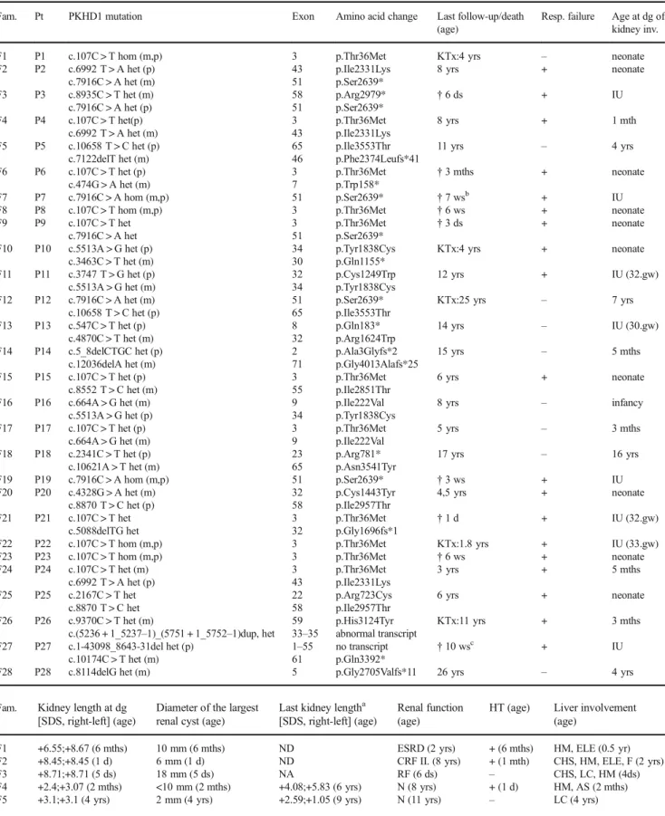
Table 1 Clinical characteristics of patients with PKHD1 mutations.aBefore end-stage renal disease;bBorn on 29. gw,cborn on 31. gw; AS: abnormal structure, CHS: cholestasis, CRF: chronic renal failure, d(s): day(s), dg: diagnosis, Diam.: diameter, ELE: elevated liver enzymes, ESRD: end-stage renal disease, F: fibrosis, Fam: family, gw: gestational week, het: heterozygous, HM: hepatomegaly, hom: homozygous, HSM: hepatosplenomegaly, HT:
hypertension; inv.: involvement, IU: intrauterine; KTx: kidney transplant, LC: liver cysts, LTx: liver transplant m: maternal, mth(s): month(s), N: normal, NA: not applicable, ND: no data, p: paternal, P/Pt: patient, Resp: respiratory, RF: renal failure, SDS: standard deviation score, w(s): week(s), yr(s):
year(s)
Fam. Pt PKHD1 mutation Exon Amino acid change Last follow-up/death
(age)
Resp. failure Age at dg of kidney inv.
F1 P1 c.107C > T hom (m,p) 3 p.Thr36Met KTx:4 yrs – neonate
F2 P2 c.6992 T > A het (p) 43 p.Ile2331Lys 8 yrs + neonate
c.7916C > A het (m) 51 p.Ser2639*
F3 P3 c.8935C > T het (m) 58 p.Arg2979* †6 ds + IU
c.7916C > A het (p) 51 p.Ser2639*
F4 P4 c.107C > T het(p) 3 p.Thr36Met 8 yrs + 1 mth
c.6992 T > A het (m) 43 p.Ile2331Lys
F5 P5 c.10658 T > C het (p) 65 p.Ile3553Thr 11 yrs – 4 yrs
c.7122delT het (m) 46 p.Phe2374Leufs*41
F6 P6 c.107C > T het (p) 3 p.Thr36Met †3 mths + neonate
c.474G > A het (m) 7 p.Trp158*
F7 P7 c.7916C > A hom (m,p) 51 p.Ser2639* †7 wsb + IU
F8 P8 c.107C > T hom (m,p) 3 p.Thr36Met †6 ws + neonate
F9 P9 c.107C > T het 3 p.Thr36Met †3 ds + neonate
c.7916C > A het 51 p.Ser2639*
F10 P10 c.5513A > G het (p) 34 p.Tyr1838Cys KTx:4 yrs + neonate
c.3463C > T het (m) 30 p.Gln1155*
F11 P11 c.3747 T > G het (p) 32 p.Cys1249Trp 12 yrs + IU (32.gw)
c.5513A > G het (m) 34 p.Tyr1838Cys
F12 P12 c.7916C > A het (m) 51 p.Ser2639* KTx:25 yrs – 7 yrs
c.10658 T > C het (p) 65 p.Ile3553Thr
F13 P13 c.547C > T het (p) 8 p.Gln183* 14 yrs – IU (30.gw)
c.4870C > T het (m) 32 p.Arg1624Trp
F14 P14 c.5_8delCTGC het (p) 2 p.Ala3Glyfs*2 15 yrs – 5 mths
c.12036delA het (m) 71 p.Gly4013Alafs*25
F15 P15 c.107C > T het (p) 3 p.Thr36Met 6 yrs + neonate
c.8552 T > C het (m) 55 p.Ile2851Thr
F16 P16 c.664A > G het (m) 9 p.Ile222Val 8 yrs – infancy
c.5513A > G het (p) 34 p.Tyr1838Cys
F17 P17 c.107C > T het (p) 3 p.Thr36Met 5 yrs – 3 mths
c.664A > G het (m) 9 p.Ile222Val
F18 P18 c.2341C > T het (p) 23 p.Arg781* 17 yrs – 16 yrs
c.10621A > T het (m) 65 p.Asn3541Tyr
F19 P19 c.7916C > A hom (m,p) 51 p.Ser2639* †3 ws + IU
F20 P20 c.4328G > A het (m) 32 p.Cys1443Tyr 4,5 yrs + neonate
c.8870 T > C het (p) 58 p.Ile2957Thr
F21 P21 c.107C > T het 3 p.Thr36Met †1 d + IU (32.gw)
c.5088delTG het 32 p.Gly1696fs*1
F22 P22 c.107C > T hom (m,p) 3 p.Thr36Met KTx:1.8 yrs + IU (33.gw)
F23 P23 c.107C > T hom (m,p) 3 p.Thr36Met †6 ws + neonate
F24 P24 c.107C > T het (m) 3 p.Thr36Met 3 yrs + 5 mths
c.6992 T > A het (p) 43 p.Ile2331Lys
F25 P25 c.2167C > T het 22 p.Arg723Cys 6 yrs + neonate
c.8870 T > C het 58 p.Ile2957Thr
F26 P26 c.9370C > T het (m) 59 p.His3124Tyr KTx:11 yrs + 3 mths
c.(5236 + 1_5237–1)_(5751 + 1_5752–1)dup, het 33–35 abnormal transcript
F27 P27 c.1-43098_8643-31del het (p) 1–55 no transcript †10 wsc + IU
c.10174C > T het (m) 61 p.Gln3392*
F28 P28 c.8114delG het (m) 5 p.Gly2705Valfs*11 26 yrs – 4 yrs
Fam. Kidney length at dg [SDS, right-left] (age)
Diameter of the largest renal cyst (age)
Last kidney lengtha [SDS, right-left] (age)
Renal function (age)
HT (age) Liver involvement (age)
F1 +6.55;+8.67 (6 mths) 10 mm (6 mths) ND ESRD (2 yrs) + (6 mths) HM, ELE (0.5 yr)
F2 +8.45;+8.45 (1 d) 6 mm (1 d) ND CRF II. (8 yrs) + (1 mth) CHS, HM, ELE, F (2 yrs)
F3 +8.71;+8.71 (5 ds) 18 mm (5 ds) NA RF (6 ds) – CHS, LC, HM (4ds)
F4 +2.4;+3.07 (2 mths) <10 mm (2 mths) +4.08;+5.83 (6 yrs) N (8 yrs) + (1 d) HM, AS (2 mths)
F5 +3.1;+3.1 (4 yrs) 2 mm (4 yrs) +2.59;+1.05 (9 yrs) N (11 yrs) – LC (4 yrs)
Screening for second locus CNVs
Patients without biallelic PKHD1 mutations were re- evaluated based on their most recent phenotype, and were screened for second locus mutations accordingly.
Deletions of NPHP1 and HNF1B were tested by QMPSF analysis as described previously [12, 35].
Similarly, continuous gene deletion of TSC2 and PKD1 was screened by QMPSF analysis, with the primers listed in Suppl. Table 1, according to the protocol of NPHP1-QMPSF [35] with the differences detailed in Suppl. Methods. The deletion of PKD1 was validated with MLPA, using the SALSA MLPA probemix P352 PKD1-PKD2, performed according to the manufacturer’s i n s t r u c t i o n s ( M R C - H o l l a n d , A m s t e r d a m , t h e Netherlands).
Screening for second locus (PKD1,PKD2,HNF1B andTMEM67) small-scale mutations
Coding exons and intronic junctions of HNF1B and TMEM67were Sanger sequenced. A dominant polycystic kidney panel was designed using the Ion AmpliSeq Designer version 4.2.1 (Thermo Fischer Scientific, Waltham, MA). Sample enrichment was performed by the Ion AmpliSeq Library Kit (Thermo Fischer Scientific).
Samples were barcoded and sequenced on IonTorrent 316 chip (Thermo Fischer Scientific). More than 95% of the target sequence was covered at least 50-fold. Putative disease-causing genetic variants were validated by Sanger sequencing.
Clinical exome sequencing
Clinical exome sequencing was performed as described pre- viously [36]. More than 95% of the target sequence was cov- ered at least 20-fold. Reads were mapped against the human NCBI37/hg19 reference genome. DNA alignment and se- quence variant analysis were carried out using NextGene Software version 2.4.2 (SoftGenetics, State College, PA).
Data from the proband was filtered for coding and splicing region mutations of known/putative genes associated with cystic kidney diseases.
Results
PKHD1mutations
Of the 36 unrelated patients with a clinical diagnosis of ARPKD, 27 (75%) were found to carry biallelicPKHD1mu- tations (Table1). Among them, 25 patients carried biallelic point mutations (P1–25) and two were compound heterozy- gous for a point mutation and either a duplication of exons 33–
35 (P26) or a large deletion encompassing exons 1 to 55 (P27). Furthermore, one patient (P28) was found to carry a single heterozygous frameshift mutation. NoPKHD1 muta- tion was found in 8 families (Table2, Fig.1).
Two mutations, p.Thr36Met and p.Ser2639* were found frequently, in 15/54 (28%) and in 8/54 (15%) of the mutated alleles, respectively. Seven mutations were novel: besides the duplication of exons 33–35 and the deletion of exons 1–55, three truncating mutations (c.5_8delCTGC, p.Ala3Glyfs*2;
Table 1 (continued)
F6 +12.82;+12.82 (4 ds) 3 mm (4 ds) +8.96;+8.96 (3 mths) ESRD (3 mths) + (1 d) mild HM (4 ds)
F7 +2.42;+2.42 (4 ds) ND ND RF (4 ws) – HM (4 ds)
F8 +14.42;+14.42 (4 ds) 5 mm (1 mth) ND RF (6 ws) + (1 d) –
F9 +6.47;+5.09 (1 d) 4 mm (1 d) NA NA – mild HM, ELE, AS (1 d)
F10 +12.36;+12.36 (1 d) 4 mm (3 ds) +7.88;+7.88 (3 yrs) ESRD (4 yrs) + (1 w) LTx:4 yrs F11 +9;+9 (1 mth) 6 mm (1 mth) +8.77;+6.59 (12 yrs) CRF IV. (12 yrs) + (3 mths) ELE (1 mth) F12 +4.85;+4.85 (10 yrs) 5 mm (10 yrs) +4.46;+4.46 (18.5 yrs) ESRD (25 yrs) + (10 yrs) LTx:25 yrs
F13 +0.05;+1.8 (3 mths) 6 mm (8 yrs) +5.15;+7.5 (14 yrs) N (14 yrs) – –
F14 +2.59;+3.8 (5 mths) 1 mm (9 yrs) +8.28;+10.08 (15 yrs) CRF II. (15 yrs) + (3 yrs) –
F15 +9.69;+9.69 (5 mths) 9 mm (5 mths) +19.16;+19.16 (6 yrs) CRF III. (6 yrs) + (1 d) LC (1 d), AS, HM (3 yrs)
F16 +5.24;+5.24 (3 yrs) 4 mm (3 yrs) +3.83;3.83 (8 yrs) N (8 yrs) – –
F17 +3.8;+3.8 (4 mths) 10 mm (4 mths) +6.21;+6.21 (5 yrs) N (5 yrs) – –
F18 +1.59;+3.64 (16 yrs) 11 mm (16 yrs) ND CRF II. (17 yrs) – AS (16 yrs)
F19 +12.36;+12.36 (1 d) 8 mm (1 d) +16.79;+16.79 (1 w) RF (1 w) + (1 d) –
F20 +4.01;+4.01 (4 mths) 3 mm (4 mths) +10.31;9.1(4.5 yrs) CRF II. (4.5 yrs) + (1d) AS (4 mths), F (10 mths), HSM (1 yr)
F21 +18.87;+18.87 (1 d) 4 mm (1 d) NA NA ND –
F22 +4.79;+4.79 (8 ds) 1 mm (8 ds) +21.95;+21.95 (1 yr) ESRD (1 yr) + (1 d) F (1 d), ELE (3 mths)
F23 +15.09;+15.09 (2 ds) 4 mm (2 ds) ND RF (6 ws) + (1 d) –
F24 +0.2;+0.2 (6 mths) 4 mm (6 mths) −1.75;-1.75 (3 yrs) CRF II (1.5 yrs) – –
F25 +5.92;+5.92 (5 mths) 8 mm (5 mths) +10.6;+10.6 (6 yrs) ESRD (6 yrs) + (1 yr) HSM (5 mths), LC (3 yrs)
F26 +9.24;+4.48 (3mths) 10 mm (3 mths) ND ESRD (11 yrs) + (3 mths) LC, HM (3 mths)
F27 +4,1;+4,1 (1 d) ND ND RF (10 ws) + (1 d) –
F28 +7.26;+3.26 (4 yrs) 10 mm (4 yrs) +3.73;+3.73 (26 yrs) CRF II. (26 yrs) + (17 yrs) F, HM, ELE (4 yrs)
Table2Clinicalcharacteristicsofpatientswithsecondlocusmutations.a Beforeend-stagerenaldisease;b PatienthadGBSperinatalsepsis;c P36andP37areasiblingpairandforthecalculations, genotypeinformationfromonlyoneofthemwasused.d:day,dg:diagnosis,CRF:chronicrenalfailure,ELE:elevatedliverenzymes,ESRD:end-stagerenaldisease,F:fibrosis,Fam:family,gw: gestationalweek,het:heterozygous,hom:homozygous,HT:hypertension,inv.:involvement,IU:intrauterine,KTx:kidneytransplant,m:maternal,mth(s):month(s),N:normal,NA:notapplicable,p: paternal,P/Pt:patient,Resp:respiratory,SDS:standarddeviationscore,w(s):week(s),yr(s):year(s) Fam.PtSecondlocus mutationExonAminoacid changeLast follow- up(age) Resp. failureAgeatdg ofkidney inv.
Kidneylength atdg[SDS, right-left](age) Diameter ofthelargest renalcyst(age) Lastkidney lengtha [SDS, right-left](age) Renal function (age)
HT(age)Liver involvement (age) F29P29PKD1c.12310_ 12313delGTTAhet (denovo)
5p.Val4104Phefs*934yrs–IU+3.26;+3.26 (7mths)3mm(7mths)+2.17;+3.23 (4yrs)N(4yrs)–– F30P30PKD1c.920delThet (denovo)45p.Phe307Serfs*273.5yrs–IU(32.gw)+2.19;+0.24 (1d)15mm(1d)+6.93;+0.95 (3.5yrs)N(3.5yrs)–– F31P31PKD1/TSC216:g. (?_2088232)_ (2134081_?)del het.(denovo)
TSC2:41–42, PKD1:1–46–6.5yrs–6ws+0.44;+0.54 (3.5yrs)10mm(2mths)+0.42;+0.42 (6.5yrs)N(6.5yrs)–– F32P32HNF1B(?_-1)_(*1_?)del het(denovo)1–9–9yrs–IU(16.gw)+1.32;+1.32 (2mths)8mm(2mths)+2.82;+2.82 (9yrs)N(9yrs)–– F33P33HNF1B(?_-1)_(*1_?)del het(denovo)1–9–6yrs–neonate+1.62;+1.62 (1d)9mm(5yrs)+0.15;+0.5 (6yrs)N(6yrs)–– F34P34HNF1B(?_-1)_(*1_?)del het(denovo)1–9–11yrs–IU−0.1;+0.23 (3mths)3mm(3mths)+0.53;+0.53 (10yrs)CRFII. (10yrs)–ELE(7yrs) F35P35NPHP1(?_-1)_(*1_?)del het(m)1–20–KTx: 12.5 yrs
–11.5yrs+0.42;+0.42 (11.5yrs)10mm (11.5yrs)NAESRD (11.5yrs)+(11.5yrs)ELE (11.5yrs) NPHP1c.(1810+1_ 1811–1)_(*1_?)del(p)18–20-– F36P36 cTMEM67c.1843T>C hom18p.Cys615ArgKTx: 9yrs+b IU+1.29;+1.8 (1mth)9mm(1mth)−4.74;-4.74 (9yrs)ESRD (9yrs)–– F36P37c 20yrs–3mths+1.51;+1.51 (3mths)2mm(9yrs)−2.88;-2.78 (18yrs)ESRD (19yrs)–ELE(11yrs), F(20yrs)
c . 5 0 8 8 d e l T G , p . G l y 1 6 9 6 f s * 1 ; c . 1 2 0 3 6 d e l A , p.Gly4013Alafs*24) and two missense mutations ( c . 4 3 2 8 G > A , p . C y s 1 4 4 3 Ty r a n d c . 1 0 6 2 1 A > T, p.Asn3541Tyr). We considered these two latter to also be pathogenic, because neither is reported in the gnomAD data- base, both affect amino acids conserved in mammals and are predicted to be pathogenic by Polyphen-2 (score: 1.0 and 0.981, respectively) and MutationTaster.
Second locus mutations
All eight families withoutPKHD1mutations were found to carry second locus mutations (Table2, Fig.1). Three children diagnosed with hyperechogenic, normal-sized kidneys in utero or in infancy carried a de novo deletion of HNF1B. The renal morphology of two children became suggestive of ADPKD between 2 and 4 years of age. They both harbored de novoPKD1mutations. One patient (P31) was diagnosed with
tuberous sclerosis at the age of 4 years, and carried a de novo TSC2/PKD1 deletion. Finally, the phenotype of a patient (P35) diagnosed at the age of 11 years with end-stage renal disease was suggestive of juvenile nephronophthisis. He was compound heterozygous for a complete and a partial deletion of NPHP1, as described recently [35]. A sibling pair (P36, P37), diagnosed with hyperechogenic kidneys and hepatic fibrosis, was homozygous for the frequentTMEM67missense mutation, p.Cys615Arg [21] (for the calculations, genotype information from only one of them was used). They had no neurological involvement. The patient with a heterozygous PKHD1mutation (P28) was not found to carry a second locus mutation, even by clinical exome sequencing, which also well covered the coding regions ofNPHP2, NPHP3,andWDR19/
NPHP13. The mutational screening procedure is summarized in Fig.1.
Phenotype of patients withPKHD1and second locus mutations
Of the 27 patients withPKHD1mutations, 19 children (70%) developed perinatal respiratory failure, and nine (33%) died perinatally (< 3 months of age) (Table1). In contrast, only one of the nine patients (from eight families) with second locus mutations had a transient perinatal respiratory failure, second- ary to an infection (Table2). Among the perinatal cases who survived beyond that period, all but one of the ten children withPKHD1mutations developed hypertension by the age of 1 year, compared with none of the six patients with second locus mutations (Table1). A mean kidney length above + 4 SD at diagnosis was also specific for PKHD1-associated ARPKD: 19 of 27 patients (70%) with biallelicPKHD1mu- tations, but none of the nine patients with second locus muta- tions had such an enlarged kidney. Based on these data, the phenotype of the patient with a single heterozygousPKHD1 mutation (P28) is highly suggestive of a moderatePKHD1- associated ARPKD: she developed hypertension at an early stage, as well as highly enlarged kidneys and hepatic fibrosis.
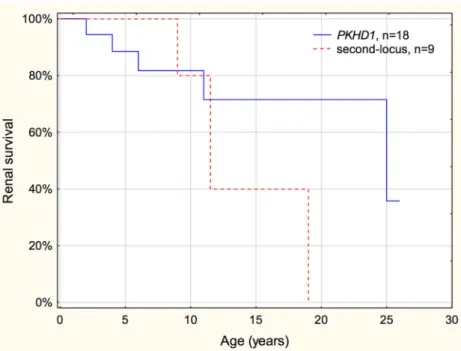
There was no difference in the renal survival between patients withPKHD1mutations who survived the perinatal period and the heterogeneous group of patients with second locus muta- tions (Tables1and2, Fig.2).
Discussion
Here we aimed to investigate the reason for the high propor- tion of ARPKD cases negative forPKHD1point mutations.
OurPKHD1-positive rate of 78%—including a patient with a single heterozygous mutation—corresponds well to previous reports [15,16,27,29,32,33]. We first assessed the potential role of PKHD1CNVs, but found no biallelic CNVs in the PKHD1negative cases. This finding was in accordance with Fig. 1 Mutational screening procedure in children with the clinical
diagnosis of autosomal recessive polycystic kidney disease (ARPKD)
the low prevalence ofPKHD1CNVs in other cohorts [15,16, 26,27,30,31]. We only found a three-exon duplication and a large deletion in two out of three patients with a single hetero- zygousPKHD1point mutation. Duplications are extremely rare; here, we present only the second such case [26]. These results emphasize that screening for CNVs is primarily impor- tant in patients with a heterozygousPKHD1point mutation.
To identify all mutations of the coding regions, we used the most sensitive but also labor- and time-consuming methods of Sanger sequencing and MLPA analysis. However, excluding the causal role ofPKHD1intronic and promoter mutations or rearrangements in patients with or without a single heterozy- gous mutation is especially challenging by direct genetic tests.
Therefore, we aimed to identify the causal mutations in all patients with a clinical diagnosis of ARPKD, and thus exclude the causal role ofPKHD1indirectly. We found all patients withoutPKHD1mutations to carry causal mutations in second loci, by re-evaluation of the phenotype and targeted mutation screening, indicating that 22% of the initial clinical ARPKD diagnoses were false. We failed to identify in the third child with a heterozygousPKHD1mutation a second locus muta- tion, even following clinical exome sequencing that also cov- ered the promoter region ofPMM2[4]. This is in accordance with the phenotype which is strongly suggestive ofPKHD1- associated ARPKD. Her case thus points on the difficulty in identifying some PKHD1mutations even by the combined approach of sequencing and MLPA, and suggests a potential role of an intronic or a regulatoryPKHD1mutation.
The successful identification of second locus mutations in negative cases by direct genetic tests emphasizes the impor- tance of their thorough re-phenotyping. The differential diag- nosis of mild and moderate forms without extremely enlarged
kidneys and respiratory failure can be challenging at diagno- sis. Within this cohort, an infant-onset hypertension was high- ly suggestive ofPKHD1-associated ARPKD.
In accordance with the literature, the phenotype of patients with PKHD1mutations strongly correlated with the causal mutations; no patient survived the perinatal period with biallelic loss-of-function mutations [16, 37, 38].
Interestingly, a patient, compound heterozygous for a second and a last exon truncating mutation, p.Gly4013Alafs*24 (P14), presented with a moderate phenotype: she was diag- nosed at the age of 5 months and had a normal GFR at the age of 9 years, indicating that the loss of the C-terminal 62 amino acids of fibrocystin does not cause complete loss of function.
The intracellular C-terminal part of fibrocystin consists of 192 amino acids and is known to modulate the mTOR pathway [39]. It also contains the ciliary targeting sequence (p.3876_3893CLVCCWLKRSKSRKTKPE), that remains unaffected in the Gly4013Alafs*24 fibrocystin [40]. Along the same lines as the hypomorphic nature of this C-terminal truncation, mice lacking the last exon (exon 67), which en- codes the nuclear localization signal and the polycystin 2 binding domain, develop a normal phenotype [41].
In conclusion, we found one quarter of the ARPKD cases to be phenocopies, caused by second locus muta- tions. Our data suggest that perinatal respiratory failure, a kidney length > + 4 SD and early-onset hypertension increase the likelihood of PKHD1-associated ARPKD.
The phenotype of cases that are negative on PKHD1 sequencing should first be re-evaluated. We recommend screening for PKHD1 CNVs in patients with a heterozy- gous point mutation and in families with an unequivocal phenotype.
Fig. 2 Renal survival of patients withPKHD1and second locus mutations. No difference was found in the renal progression between patients withPKHD1 and second locus mutations (p= 0.51). Among patients with second locus mutations, those withPKD1andHNF1B mutations were young (≤ 11 years), and patients with nephronophthisis reached end- stage renal disease between 9 and 19 years of age, giving a worse renal survival curve than generally expectable
Funding This work was supported by OTKA K109076 and Ministry of National Economy, Hungary GINOP-2.3.2-15-2016-00039 (to István Balogh, Zoltán Maróti and Tibor Kalmár), MTA-SE Lendulet Research Grant (LP2015-11/2015) of the Hungarian Academy of Sciences and NKFIA/OTKA K109718, KH125566 (to Kálmán Tory).
Compliance with ethical standards
Conflict of interest The authors declare that they have no conflict of interest.
Ethical approval All procedures performed in studies involving human participants were in accordance with the ethical standards of the institu- tional and/or national research committee and with the 1964 Helsinki declaration and its later amendments or comparable ethical standards.
Parents and patients gave informed written consent.
References
1. Kurschat CE, Muller RU, Franke M, Maintz D, Schermer B, Benzing T (2014) An approach to cystic kidney diseases: the clini- cian’s view. Nat Rev Nephrol 10:687–699
2. Ward CJ, Hogan MC, Rossetti S, Walker D, Sneddon T, Wang X, Kubly V, Cunningham JM, Bacallao R, Ishibashi M, Milliner DS, Torres VE, Harris PC (2002) The gene mutated in autosomal reces- sive polycystic kidney disease encodes a large, receptor-like pro- tein. Nat Genet 30:259–269
3. Lu H, Galeano MCR, Ott E, Kaeslin G, Kausalya PJ, Kramer C, Ortiz-Bruchle N, Hilger N, Metzis V, Hiersche M, Tay SY, Tunningley R, Vij S, Courtney AD, Whittle B, Wuhl E, Vester U, Hartleben B, Neuber S, Frank V, Little MH, Epting D, Papathanasiou P, Perkins AC, Wright GD, Hunziker W, Gee HY, Otto EA, Zerres K, Hildebrandt F, Roy S, Wicking C, Bergmann C (2017) Mutations in DZIP1L, which encodes a ciliary-transition- zone protein, cause autosomal recessive polycystic kidney disease.
Nat Genet 49:1025–1034
4. Cabezas OR, Flanagan SE, Stanescu H, Garcia-Martinez E, Caswell R, Lango-Allen H, Anton-Gamero M, Argente J, Bussell AM, Brandli A, Cheshire C, Crowne E, Dumitriu S, Drynda R, Hamilton-Shield JP, Hayes W, Hofherr A, Iancu D, Issler N, Jefferies C, Jones P, Johnson M, Kesselheim A, Klootwijk E, Koettgen M, Lewis W, Martos JM, Mozere M, Norman J, Patel V, Parrish A, Perez-Cerda C, Pozo J, Rahman SA, Sebire N, Tekman M, Turnpenny PD, Hoff WV, Viering D, Weedon MN, Wilson P, Guay-Woodford L, Kleta R, Hussain K, Ellard S, Bockenhauer D (2017) Polycystic kidney disease with hyperinsulinemic hypoglycemia caused by a promoter mutation in phosphomannomutase 2. J Am Soc Nephrol 28:2529–2539 5. Nagano J, Kitamura K, Hujer KM, Ward CJ, Bram RJ, Hopfer U,
Tomita K, Huang C, Miller RT (2005) Fibrocystin interacts with CAML, a protein involved in Ca2+ signaling. Biochem Biophys Res Commun 338:880–889
6. Yamaguchi T, Hempson SJ, Reif GA, Hedge AM, Wallace DP (2006) Calcium restores a normal proliferation phenotype in human polycystic kidney disease epithelial cells. J Am Soc Nephrol 17:
178–187
7. Wu M, Yu S (2016) New insights into the molecular mechanisms targeting tubular channels/transporters in PKD development.
Kidney Dis (Basel) 2:128–135
8. Liu W, Murcia NS, Duan Y, Weinbaum S, Yoder BK, Schwiebert E, Satlin LM (2005) Mechanoregulation of intracellular Ca2+
concentration is attenuated in collecting duct of monocilium- impaired orpk mice. Am J Physiol Ren Physiol 289:F978–F988 9. Siroky BJ, Ferguson WB, Fuson AL, Xie Y, Fintha A, Komlosi P,
Yoder BK, Schwiebert EM, Guay-Woodford LM, Bell PD (2006) Loss of primary cilia results in deregulated and unabated apical calcium entry in ARPKD collecting duct cells. Am J Physiol Ren Physiol 290:F1320–F1328
10. Vujic M, Heyer CM, Ars E, Hopp K, Markoff A, Orndal C, Rudenhed B, Nasr SH, Torres VE, Torra R, Bogdanova N, Harris PC (2010) Incompletely penetrant PKD1 alleles mimic the renal manifestations of ARPKD. J Am Soc Nephrol 21:1097–1102 11. Losekoot M, Ruivenkamp CA, Tholens AP, Grimbergen JE,
Vijfhuizen L, Vermeer S, Dijkman HB, Cornelissen EA, Bongers EM, Peters DJ (2012) Neonatal onset autosomal dominant polycys- tic kidney disease (ADPKD) in a patient homozygous for a PKD2 missense mutation due to uniparental disomy. J Med Genet 49:37– 40
12. Bellanne-Chantelot C, Clauin S, Chauveau D, Collin P, Daumont M, Douillard C, Dubois-Laforgue D, Dusselier L, Gautier JF, Jadoul M, Laloi-Michelin M, Jacquesson L, Larger E, Louis J, Nicolino M, Subra JF, Wilhem JM, Young J, Velho G, Timsit J (2005) Large genomic rearrangements in the hepatocyte nuclear factor-1beta (TCF2) gene are the most frequent cause of maturity- onset diabetes of the young type 5. Diabetes 54:3126–3132 13. Hiesberger T, Shao X, Gourley E, Reimann A, Pontoglio M,
Igarashi P (2005) Role of the hepatocyte nuclear factor-1beta (HNF-1beta) C-terminal domain in Pkhd1 (ARPKD) gene tran- scription and renal cystogenesis. J Biol Chem 280:10578–10586 14. Williams SS, Cobo-Stark P, Hajarnis S, Aboudehen K, Shao X,
Richardson JA, Patel V, Igarashi P (2014) Tissue-specific regulation of the mouse Pkhd1 (ARPKD) gene promoter. Am J Physiol Ren Physiol 307:F356–F368
15. Denamur E, Delezoide AL, Alberti C, Bourillon A, Gubler MC, Bouvier R, Pascaud O, Elion J, Grandchamp B, Michel-Calemard L, Missy P, Zaccaria I, Le Nagard H, Gerard B, Loirat C, Societe Francaise de F, Barbet J, Beaufrere AM, Berchel C, Bessieres B, Boudjemaa S, Buenerd A, Carles D, Clemenson A, Dechelotte P, Devisme L, Dijoud F, Esperandieu O, Fallet C, Gonzales M, Hillion Y, Jacob B, Joubert M, Kermanach P, Lallemand A, Laquerriere A, Laurent N, Liprandi A, Loeuillet L, Loget P, Martinovic J, Menez F, Narcy F, Roux JJ, Rouleau-Dubois C, Sinico M, Tantau J, Wann AR (2010) Genotype-phenotype correlations in fetuses and neo- nates with autosomal recessive polycystic kidney disease. Kidney Int 77:350–358
16. Bergmann C, Senderek J, Windelen E, Kupper F, Middeldorf I, Schneider F, Dornia C, Rudnik-Schoneborn S, Konrad M, Schmitt CP, Seeman T, Neuhaus TJ, Vester U, Kirfel J, Buttner R, Zerres K, Apn (2005) Clinical consequences of PKHD1 mutations in 164 patients with autosomal-recessive polycystic kidney disease (ARPKD). Kidney Int 67:829–848
17. Gunay-Aygun M, Font-Montgomery E, Lukose L, Tuchman Gerstein M, Piwnica-Worms K, Choyke P, Daryanani KT, Turkbey B, Fischer R, Bernardini I, Sincan M, Zhao X, Sandler NG, Roque A, Douek DC, Graf J, Huizing M, Bryant JC, Mohan P, Gahl WA, Heller T (2013) Characteristics of congenital hepatic fibrosis in a large cohort of patients with autosomal recessive poly- cystic kidney disease. Gastroenterology 144:112–121 e112 18. Shneider BL, Magid MS (2005) Liver disease in autosomal reces-
sive polycystic kidney disease. Pediatr Transplant 9:634–639 19. Turkbey B, Ocak I, Daryanani K, Font-Montgomery E, Lukose L,
Bryant J, Tuchman M, Mohan P, Heller T, Gahl WA, Choyke PL, Gunay-Aygun M (2009) Autosomal recessive polycystic kidney disease and congenital hepatic fibrosis (ARPKD/CHF). Pediatr Radiol 39:100–111
20. Zerres K, Mucher G, Becker J, Steinkamm C, Rudnik-Schoneborn S, Heikkila P, Rapola J, Salonen R, Germino GG, Onuchic L,
Somlo S, Avner ED, Harman LA, Stockwin JM, Guay-Woodford LM (1998) Prenatal diagnosis of autosomal recessive polycystic kidney disease (ARPKD): molecular genetics, clinical experience, and fetal morphology. Am J Med Genet 76:137–144
21. Otto EA, Tory K, Attanasio M, Zhou W, Chaki M, Paruchuri Y, Wise EL, Wolf MT, Utsch B, Becker C, Nurnberg G, Nurnberg P, Nayir A, Saunier S, Antignac C, Hildebrandt F (2009) Hypomorphic mutations in meckelin (MKS3/TMEM67) cause nephronophthisis with liver fibrosis (NPHP11). J Med Genet 46:
663–670
22. Tory K, Rousset-Rouviere C, Gubler MC, Moriniere V, Pawtowski A, Becker C, Guyot C, Gie S, Frishberg Y, Nivet H, Deschenes G, Cochat P, Gagnadoux MF, Saunier S, Antignac C, Salomon R (2009) Mutations of NPHP2 and N PH P3 in infantile nephronophthisis. Kidney Int 75:839–847
23. Arbeiter A, Buscher R, Bonzel KE, Wingen AM, Vester U, Wohlschlager J, Zerres K, Nurnberger J, Bergmann C, Hoyer PF (2008) Nephrectomy in an autosomal recessive polycystic kidney disease (ARPKD) patient with rapid kidney enlargement and in- creased expression of EGFR. Nephrol Dial Transplant 23:3026–
3029
24. Rossetti S, Harris PC (2007) Genotype-phenotype correlations in autosomal dominant and autosomal recessive polycystic kidney disease. J Am Soc Nephrol 18:1374–1380
25. Stenson PD, Mort M, Ball EV, Evans K, Hayden M, Heywood S, Hussain M, Phillips AD, Cooper DN (2017) The human gene mu- tation database: towards a comprehensive repository of inherited mutation data for medical research, genetic diagnosis and next- generation sequencing studies. Hum Genet 136:665–677 26. Miyazaki J, Ito M, Nishizawa H, Kato T, Minami Y, Inagaki H,
Ohye T, Miyata M, Boda H, Kiriyama Y, Kuroda M, Sekiya T, Kurahashi H, Fujii T (2015) Intragenic duplication in the PKHD1 gene in autosomal recessive polycystic kidney disease. BMC Med Genet 16:98
27. Bergmann C, Kupper F, Schmitt CP, Vester U, Neuhaus TJ, Senderek J, Zerres K (2005) Multi-exon deletions of the PKHD1 gene cause autosomal recessive polycystic kidney disease (ARPKD). J Med Genet 42:e63
28. Sweeney WE Jr, Avner ED (2011) Diagnosis and management of childhood polycystic kidney disease. Pediatr Nephrol 26:675–692 29. Krall P, Pineda C, Ruiz P, Ejarque L, Vendrell T, Camacho JA, Mendizabal S, Oliver A, Ballarin J, Torra R, Ars E (2014) Cost- effective PKHD1 genetic testing for autosomal recessive polycystic kidney disease. Pediatr Nephrol 29:223–234
30. Bergmann C, Senderek J, Sedlacek B, Pegiazoglou I, Puglia P, Eggermann T, Rudnik-Schoneborn S, Furu L, Onuchic LF, De Baca M, Germino GG, Guay-Woodford L, Somlo S, Moser M, Buttner R, Zerres K (2003) Spectrum of mutations in the gene for autosomal recessive polycystic kidney disease (ARPKD/PKHD1).
J Am Soc Nephrol 14:76–89
31. Obeidova L, Seeman T, Elisakova V, Reiterova J, Puchmajerova A, Stekrova J (2015) Molecular genetic analysis of PKHD1 by next- generation sequencing in Czech families with autosomal recessive polycystic kidney disease. BMC Med Genet 16:116
32. Sharp AM, Messiaen LM, Page G, Antignac C, Gubler MC, Onuchic LF, Somlo S, Germino GG, Guay-Woodford LM (2005) Comprehensive genomic analysis of PKHD1 mutations in ARPKD cohorts. J Med Genet 42:336–349
33. Gunay-Aygun M, Tuchman M, Font-Montgomery E, Lukose L, Edwards H, Garcia A, Ausavarat S, Ziegler SG, Piwnica-Worms K, Bryant J, Bernardini I, Fischer R, Huizing M, Guay-Woodford L, Gahl WA (2010) PKHD1 sequence variations in 78 children and adults with autosomal recessive polycystic kidney disease and con- genital hepatic fibrosis. Mol Genet Metab 99:160–173
34. Losekoot M, Haarloo C, Ruivenkamp C, White SJ, Breuning MH, Peters DJ (2005) Analysis of missense variants in the PKHD1-gene in patients with autosomal recessive polycystic kidney disease (ARPKD). Hum Genet 118:185–206
35. Javorszky E, Moriniere V, Kerti A, Balogh E, Piko H, Saunier S, Karcagi V, Antignac C, Tory K (2017) QMPSF is sensitive and specific in the detection of NPHP1 heterozygous deletions. Clin Chem Lab Med 55:809–816
36. Orosz O, Rajta I, Vajas A, Takacs L, Csutak A, Fodor M, Kolozsvari B, Resch M, Senyi K, Lesch B, Szabo V, Berta A, Balogh I, Losonczy G (2017) Myopia and late-onset progressive cone dystrophy associate to LVAVA/MVAVA exon 3 interchange haplotypes of opsin genes on chromosome X. Invest Ophthalmol Vis Sci 58:1834–1842
37. Furu L, Onuchic LF, Gharavi A, Hou X, Esquivel EL, Nagasawa Y, Bergmann C, Senderek J, Avner E, Zerres K, Germino GG, Guay- Woodford LM, Somlo S (2003) Milder presentation of recessive polycystic kidney disease requires presence of amino acid substitu- tion mutations. J Am Soc Nephrol 14:2004–2014
38. Rossetti S, Torra R, Coto E, Consugar M, Kubly V, Malaga S, Navarro M, El-Youssef M, Torres VE, Harris PC (2003) A com- plete mutation screen of PKHD1 in autosomal-recessive polycystic kidney disease (ARPKD) pedigrees. Kidney Int 64:391–403 39. Wang S, Wu M, Yao G, Zhang J, Zhou J (2014) The cytoplasmic
tail of FPC antagonizes the full-length protein in the regulation of mTOR pathway. PLoS One 9:e95630
40. Follit JA, Li L, Vucica Y, Pazour GJ (2010) The cytoplasmic tail of fibrocystin contains a ciliary targeting sequence. J Cell Biol 188:
21–28
41. Outeda P, Menezes L, Hartung EA, Bridges S, Zhou F, Zhu X, Xu H, Huang Q, Yao Q, Qian F, Germino GG, Watnick T (2017) A novel model of autosomal recessive polycystic kidney questions the role of the fibrocystin C-terminus in disease mechanism. Kidney Int 92:1130–1144