Kvantitative RT-PCR- based microRNA profiling and immunohistochemistry expression analysis in
urogenital abnormalities of childhood
Ph.D. thesis
Zsófia Magyar MD
Semmelweis University Doctoral School of Clinical Medicine
Supervisor: Éva Görbe, MD, Ph.D.
Official reviewers: Márta Katona, MD, Ph.D.
Ágnes Jermendy, MD, Ph.D.
Head of the Final Examination Committee:
György Reusz, MD, DSc Members of the Final Examination Committee:
Tibor Ertl, MD, DSc Miklós Szabó, MD, Ph.D.
Budapest 2020
1 1 INTRODUCTION
The congenital anomalies of kidney and urinary tract (CAKUT) are the major causes of end-stage kidney disease in childhood. The complications of congenital hydronephrosis are well-known. The early recognition and adequate, individualized therapy are based on the better knowledge of the pathogenesis. The outcome of CAKUT has improved lot in the past few years, mostly thanks to the prenatal ultrasonography examinations and to the modern renal replacement therapies (RRT) (dialysis and kidney transplantation).
Disorders of the development of nephrons can lead to formation of Wilms tumor.
Wilms tumor is the most common solid tumor in childhood. MicroRNAs can have a role in the pathogenesis of Wilms tumor. The microRNA profile of this cancer can have impact on the response to the chemotherapy.
1.1 Congenital anomalies of kidney and urinary tract
1.1.1 Development of kidney and urinary tract and kidney function
In the course of kidney development, coordinated interactions between mesenchymal and epithelial cells turn into highly specialized vascular network, tubular structures and stromal elements. In case of mammalia, the urogenital tract develops in three steps: firstly pronephros then mesonephros and at the end (at 5-6 th gestational age) metanephros (permanent kidney) evolve. The metanephros consists of epithelial cells of ureteric bud and mesenchymal cells.
Accurate and aligned working of several genes take part in the nephrogenesis. The main signaling pathways are GDNF/RET, FGF (Fibroblast Growth Factor), TGFβ (Transforming Growth Factor β (BMP)) superfamily, Sonic hedgehog (SHH), Canonical WNT/β- Catenin and Notch. The role of WT-1-gen and Renin-angiotensine -aldosterone system was also described in the formation of glomeruli.
The urine production can be vizualized from 5-8th gestational age. The glomerular filtration rate (GFR) increases progressively during fetal period until the 35th week of pregnancy and after that it continues in the postnatal life. The GFR reachs the adult value around one year of age. The procession of nephrogenesis perform until the 35 -36th gestational age. By this time, approximately 1 million (250,000-1,500.000) nephron per kidney developed.
2 1.1.2 Obstructive uropathies
Hydronephrosis (HN) is defined as a distension of the renal pyelon and/or the calyces, which can associate with parenchymal changing. Pyelectasis is a milder form of hydronephrosis: only the pyelon is dilated.
The antenatal or fetal hydronephrosis (ANH) is the most common fetal diagnosis, it occurs in 1-5% of pregnancies.
In childhood, the obstructive uropathies are congenital in most cases. The etiology of ANH includes: transient hydronephrosis (41-88%), ureteropelvic junction obstruction (UPJO) (10-30%), vesicoureteral reflux (VUR) (10-20%), ureterovesical junction obstruction (UVJO)/megaureters (5-10%), multicystic dysplastic kidney (4-6%), posterior urethral valve (PUV) or urethral atresia (UA) (1-2 %). 5-7 % of ANH are ureterocele, ectopic ureter and ureter duplex. The Prune-belly syndrome (or Triad syndrome or Eagle-Barrett syndrome), the cystic kidneys, the congenital ureter stricture and megalourethra are rare.
The initial response to an acute obstruction of ureter is the increasing of the pressure and the increasing of the vascular resistance of the obstructive kidney. It is mediated by the renin- angiotensine -aldosterone system. This is followed by an interstitial inflammatory response that is characterized by macrophag infiltration and tubular dilatation. Several cytokines regulate the macrophags’ function. The mechanical stretching of epithelial cells stimulates the apoptosis induced by TGFβ1 és TNFα (Tumor Necrosis Factor α) in tubular cells. Several molecules inhibit the tubular apoptosis: eg.
iNOS (inducible nitrogen-monoxid sintase), EGF (epidermal growth factor) and IGF-1 (insulin-like growth factor). After that, TGFβ1 has a central role in the formation of interstitial fibrosis.
Most cases of hydronephrosis are recognizable antenatally. One of the measured parameters is the antero-posterior diameter (APD) of the pyelon. Univocal limit does not exsists between normal and pathological APD. By ultrasonography, the amount of amniotic fluid, the appearence of renal cortex, the echogenicity of the kidneys, the presence of corticomedullary differentiation, the dilatation of bladderm the thickness of bladder wall can be assessed. Furthermore, the intrarenal resistance index can be calculated.
3
Prenatally, several biomarkers can be measured from fetal serum, urine, and amniotic fluid (β2- microglobulin, N-acetyl-β-D-glucoseaminidase (NAG), urinary calcium, sodium, chlorid, urinary osmolality, total protein, cystatin C), but to our knowledge, to assess the renal function in the future, none of them is enough reliable. In certain cases, prenatal intervention is available (vesicoamnial shunt insertion- VAS).
PLUTO study did not confirm the positive effect of long-term renal survival after vesicoamnial shunt insertion.
The postnatal management of hydronephrosis includes performing abdominal ultrasonography on the 3rd postnatal day and sonocystography or miction cystourethrography (MCUG) for excuding VUR (VUR is the cause of the cases in 15%) and performing MAG-3 diuretic renography in 4-6 weeks of age.
To classify hydronephrosis, several grading system exsists. Currently, grading system of Society for Fetal Urology (SFU) is used widely which confines five grades according to the postnatal appearance of pyleon, calices, and renal parenchyma.
The complications of hydronephrosis are urinary tract infection, pyelonephritis, pyonephros, sepsis, hypertony, kidney stone, renal failure and sometimes bladder dysfunction, impaired bladder emptying.
1.1.3 Vascular endothelial growth factor (VEGF)
VEGF is a major angiogen factor and has a critial role in vasculogenesis and vascular permeability and in the normal development of the glomeruli. Its expression is tightly regulated. Its expression is induced by hypoxia too (by HIFα1).
In animal research, experimental urinary obstruction was associated with mechanical streching of tubular epithelial cells and activation of renin-angiotensin system, leading to the upregulation of cytokines and growth factors (VEGF, TNFα).
Tubulointerstitial nephritis was induced in an experimental model of obstructive uropathy.
4 1.2 Wilms tumor
Wilms tumor (or nephroblastoma) is the most common childhood abdominal malignancy (5% of the childhood malignancies). It is the most common renal tumor under 15 years (15% of the cases).
Wilms tumor is an embryonal kidney tumor, it develops because of the abnormal proliferation of early metanephrogen cells. This undifferentiated mesenchyma is called
’nephrogen rest’(NR). It can be the precursor of Wilms tumor.
Several genes can have a role in the formation of Wilms tumor. Mutation of WT-1 gene is present in 15-20% of the tumors. Somatic and germ cell mutations occurs. Certain syndromes are associated with higher incidence of Wilms tumor (WAGR-, Denys-Drash- , Beckwith-Wiedemann, Perlman syndrome). Regarding the literature, mutations of β- Catenin coding CTNNB1, a WTX gene, a p53 gene, a DICER1 gene can also have a role in the pathogenesis. In approximately 70% of cases, epigenetic alterations occurs.
Deregulations of microRNAs were observed in many types of malignant disease, including Wilms tumor. MicroRNAs are endogenous, approximately 22 nucleotides- long, non- coding RNAs which regulate the gene expression. MicroRNAs can be tumorsuppressors or oncogens as well. Up- or down-regulation of microRNAs can cause chemoresistence.
In Europe, SIOP WT 2001 protokoll is used for diagnostics and therapy. The protocol consists of four to six weeks of pre-operative chemotherapy (vincristin, actinomycin), which is followed by surgical resection (except before 6 months of age).
The postoperative chemotherapy and radiotherapy depend on the histological type of the tumor.
One of the most reliable prognostic factor is the histological type of Wilms tumor.
Typically, the classic Wilms tumor is triphasic; it is classified by the predominant cell type: blastemal, epithelial, stromal or mixed. All the types can transform into anaplastic type which is high grade, has poorer prognosis but it is more sensitive to the cytostatics.
In 2/3 of cases, the tumors show regression after chemotharapy administration.
Blastemal type is less responsive to chemotherapy and it is associated with higher risk.
The survival of patiens diagnosed with Wilms tumor has improved a lot in recent years.
With favourable histologic types, the overall survival is 80-90%, in contrast, regarding the blastemal type: it is only 65%.
5 2 AIMS
2.1 Hydronephrosis and VEGF
Our aim was to get to know the pathogenesis of hydronephrosis more accurately.
In animal studies, the experimental urinary obstruction was associated with mechanical stretching of tubular epithelial cells which finally leads to the upregulation of cytokines and growth factors (VEGF, TNFα).
I was looking for the answers to the following questions:
1. Does the expression of VEGF increase in obstructive uropathies in humans?
2.2 Wilms tumor and microRNA
In children, regarding Wilms tumor, the blastemal type is one of the most common histological subtypes. Subtype showig blastemal predominance is less responsive to chemotherapy and has less favourable prognosis. In Europe, the treatment protocol (SIOP WT 2001) starts with preoperative chemotherapy. Understanding differences between tumor types at molecular level is the key to identify factors underlying chemo- responsiveness. Deregulation of microRNAs can have a role in the pathogenesis of Wilms tumor and can influence the response to chemotherapy.
Our goals:
1. Are the FFPE (formalin-fixed, paraffin.embedded) samples of Wilms tumor reliable microRNA sources?
2. To determine the microRNA profile of blastemal Wilms tumors. Which microRNAs are up- and downregulated in tumors compared with tumor-free region? Is the downregulation characteristic as in literature?
3. Does the microRNA profile of blastemal Wilms tumor correlate with the clinical course?
4. What kind of significance can the knowledge of microRNA profile have? Is it possible to conclude from the microRNA profile of blastemal Wilms tumor to the chemo- responsiveness? Does the chemotherapy change the microRNA profile of blastemal Wilms tumor?
6 3 METHODS
3.1 Hydronephrosis and VEGF 3.1.1 Patients and methods
We investigated he expression of VEGF in 14 different cases of urinary tract obstruction in children after surgical treatment (the diagnosis based on the antero- posterior diameter (APD) of pyelon). The mean age at surgery was 7.1± 4.1 years. The male/famale ratio was 10/4. In 4 infants, after prenatal diagnosis, there was a postnatal evaluation of hydronephrosis. In 5 cases, the patients had abdominal pain and in 3 patients urinary tract infection occurred before the diagnosis. In 2 newborn infants, the routine ultrasound examination found congenital obstruction.
All the 14 cases had grade IV-V hydronephrosis. (It corresponds to hydronephrosis grade III-IV according to the Society of Fetal Urology grading system).
3.1.2 Hystology samples
In 11 cases, the expression of VEGF was measured in the tissue of the ureteropelvic junction. The tissues had been taken because of ureteropelvic junction obstruction and the surgical treatment was pyeloplasty by Anderson- Heynes. In 7 cases the obstruction was on the left side.
In three cases, the VEGF expression was evaluated in hystology samples which had been taken from the lower third of the ureter. Out of this three patients, in one case the surgical treatment was ureter- neoimplantation because of ureterovesical stenosis. In another case, the patient showed vesicoureteral reflux and grade IV hydronephrosis, treated by ureteronephrectomy. Hydronephrosis and hydroureter affecting both sides caused by posterior urethral valve was diagnosed in one case and the treatment was vesicostomy and bilateral ureter-neoimplantation.
The control group consisted of ureter tissue samples, which was taken from 10 fetuses, mean age 20.6 weeks of gestation (± 2.2 SD), that did not show any kidney and urinary tract malformations on autopsy examinations. The induced abortion was performed because of abnormalities incompatible with life, as requested by the parents.
We used placenta as a test tissue (VEGF is surely positive).
7 3.1.3 Immunohystochemistry
VEGF protein was evaluated by specific immunological method in situ in the ureter tissues. The samples after surgical treatment was fixed in formaline then washed, dehydrated and embedded into paraffin (Paraplast). The paraffin-embedded specimens were sliced (4 um), dewaxed, and hydrated, incubated in citrate buffer solution for antigen recovery (56 min at 91 ºC) at Semmelweis University, 1st Department of Pathology and Experimental Cancer Research. The endogen peroxidase was blocked by 3%-H2O2 for 30minutes, then the aspecific bounding was inhibited by 2,5% ready-to-use normal blocking serum for 20 minutes.
Monoclonal rabbit anti- human VEGF clone VG1 antibody (Dako, Denmark) was used for 48 minutes at 37 ºC in a 1:30 dilution for immunohistochemistry. Finally, the slides were incubated with VentanaUltraview Universal HRP (DAB 3,3’- Diaminobenzidine chromogen) secondary antibody. This antibody binds specifically to the primer antibody and carries a switch molecule to the HRP enzymes (horseradish peroxidáz). After adding DAB, in the presence of peroxidase, DAB forms brownish precipitate. We used a BenchMark Ultra automatic staining machine.
The immunostaining results were evaluated and scored in cooperation with pathologists who lacked knowledge of the clinicopathological outcomes of the patients.
Digital images were manually scored according to staining intensity and morphology.
Positive expression of VEGF was identified by the presence of brown-yellow granules in the cytoplasm. In 5 random high-power fields, a positive slide had ≥30% positive cells, and negative slide had <30% positive cells and no color staining.
Ethics permission was obtained from authorities (ETT TUKEB 387/2013, 21300- 4/2013/EKU), and the patients were informed and signed a consent.
3.2 Wilms tumor and microRNA 3.2.1 Patients and methods
Our clinical sample consisted of eight Hungarian patients (from Hungarian Childhood Tumor Registry) who were treated at the 2nd Department of Paediatrics, Semmelweis University. They aged between 1 and 8 years (median age: 2.5 years) at the time of diagnosis. Criteria for inclusion were the presence of Wilms tumor with a
8
prominent blastemal component and an available archived FFPE (formalin-fixed, paraffin-embedded) sample younger than 6 years.
Most cases were diagnosed at routine abdominal ultrasonography screening or from observation of common features (palpable abdominal mass, abdominal pain, haematuria, fever, anaemia). In one case (Patient 7), the tumor was discovered during the follow-up of polycystic kidneys, while in another patient (Patient 5), abdominal ultrasonography was performed upon noting obesity and Cushingoid features (later the patient found to have cortisol-producing islets in the Wilms tumor tissue).
The therapy was performed according to the SIOP WT 2001 protocol. After 4-6 weeks of preoperative chemotherapy, the tumor regression was judged by using MRI. It was followed by surgical resection. Patient 7 was initially misdiagnosed for renal cell carcinoma, so preoperative chemotherapy was not applyed before surgical resection in this case.
Most patients had no relapse during 2-5 years of follow-up period. They are in remission. Only Patient 4 showed a relapse 1 year after the initial diagnosis, and received an autologous bone marrow transplant after completing relapse protocol. Currently (one year after the administration of the last regimen) she is in remission.
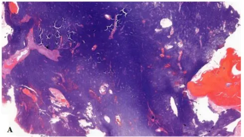
1. figure Blastemal Wilms tumor from one of our samples (Patient 2) (10x) At 10x magnification (A) (1. figure), diffuse tumor tissue can be seen with necrotic elements.
9
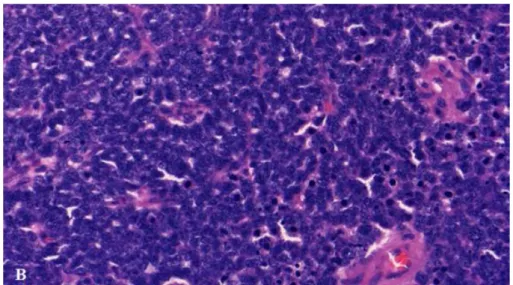
2. figure Blastemal Wilms tumor from one of our samples (Patient 2) (400x) At 400x magnification (B) (2. figure), undifferentiated cells are visible, many of them are mitotic or necrotic. Nuclei are hyperchromatic and cytoplasm is almost completely absent.
Laboratory methods
FFPE samples of eight children with blastemal Wilms tumor were analyzed. We used two FFPE samples per patient: one from the same surgical sample, but a tumor-free region. Tumor samples did not contain anaplastic foci and nephrogenic rests, only blastemal component
From each slide which had been sliced by microtome to 20µm previously, we extracted microRNAs (through several steps including deparaffination, adding protease, whole RNA isolation, adding DNAse, RNA cleaning) using a miRNeasy FFPE Kit by Quiagen (Cat. No.: 217504). All FFPE samples were younger than 6 years.
We produced cDNA from isolated microRNAs (by reverse transcription) using miScript II RT Kits (Quiagen, Cat. No.: 218160). After cDNA was produced, we stored them at -20 °C.
3.2.2 Pilot experiment
We run two 96-well miScript miRNA PCR Arrays (byQuiagen, Cat.No.:
331221MIHS-112ZF), originally designed for human prostate cancer but suitable for other types of genitourinary malignancy. We used primers for 80 mature microRNAs and 4 microRNA precursors suggested as relevant in tumorigenesis, along with 6 endogenous
10
and 6 exogenous controls. Using a Roche LightCycler 96 PCR instrument, we performed expression analysis of appropriately diluted tumor and control cDNA samples from the same patient (Patient 1).
We applied the ΔΔCt method and calculated fold changes as described in the literature. We used a single endogenous control gene to ensure that results are comparable between the pilot arrays and later PCR experiments. Our chosen control gene was small nucleolar RNA U61 (SNORD61).
Based on initial results, four microRNAs were selected for further investigation in the remaining patients: miR-34c-5p, miR-184, miR-194-5p and miR-203a.
3.2.3 Kvalitative Real-Time-PCR with the four chosen primers
We analyzed the four selected microRNAs (miR-34c-5p, miR-184, miR-194-5p miR-203a) in our eight patients diagnosed with blastemal Wilms tumor.
We used Quiagen’s miScript Primer Assays (Cat. No.: MS000033705, MS00003640, MS00006727, MS00003766, MS00033705) for each of the chosen microRNAs, and an assay for SNORD61 as control. Median Ct values of technical triplicates were used.
Ethics permission was obtained from the authorities (SE RKEB szám: 27/2020).
11 4 RESULTS
4.1 Hydronephrosis and VEGF
In case of 14 patients diagnosed with hydronephrosis, the time of surgical procedure differed. The clinical data of our patients are demonstrated in 1. Table.
1. Table Demographic and clinical data
Type of obstruction Ureteropelvic obstruction
Ureterovesical obstruction
Posterior Urethral Valve
Vesico-ureteral reflux
Number of patient 11 1 1 1
Age at surgical procedure
56.4 months (±51.4
SD) 6 months 12 months 60 months
Grade of
Hydronephrosis Grade IV-V Grade IV-V Grade IV-V Grade V
Surgical treatment Pyelonplasty (by Anderson-Heynes)
Ureter- neoimplantation
Vesicostomy éand ureter neoimpl.
Nephrectomy on the right side Tissue samples Ureteropelvic
obstruction Ureter Ureter Ureter
Expresssion of VEGF No color staining, negatíve
No color staining, negatíve
No color staining, negatíve
No color staining, negatíve
3. Figure Positive expression of VEGF in placental tissue
The 3. Figure shows positive expression of VEGF identified by the presence of brown- yellow granules (arrow) in the cytoplasm of endothelial cells in placental tissue (20x).
4. Figure Expression of VEGF can not be visualized in ureteropelvic junction tissue
As demonstrated at 4. Figure, on the mucosal surface of the ureteropelvic junction tissue in a 5 years old boy, there is no positive cells and no color staining. The slide is negative for the brown-yellow granules in the cytoplasm.
12
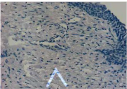
5. Figure The control group showed no increase in expression of VEGF in the ureteropelvic junction tissue
In our control group (human fetuses without urinary tract malformation), in the subserosal layer of the fetal ureteropelvic tissue, there is no positive cells in the fetal tissue (5.
Figure).
4.2 Wilms tumor and microRNA
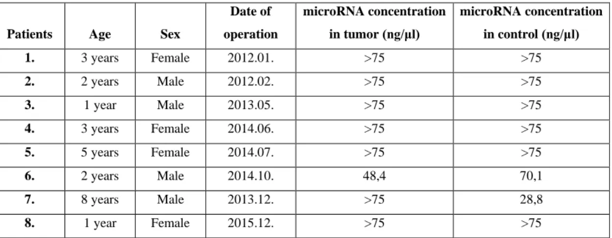
MicroRNA yields were adequate from all FFPE samples (2. Table).
2. Table Enrolled patients with microRNA concentrations obtained from their samples
Patients Age Sex
Date of operation
microRNA concentration in tumor (ng/μl)
microRNA concentration in control (ng/μl)
1. 3 years Female 2012.01. >75 >75
2. 2 years Male 2012.02. >75 >75
3. 1 year Male 2013.05. >75 >75
4. 3 years Female 2014.06. >75 >75
5. 5 years Female 2014.07. >75 >75
6. 2 years Male 2014.10. 48,4 70,1
7. 8 years Male 2013.12. >75 28,8
8. 1 year Female 2015.12. >75 >75
4.2.1 Pilot experiment
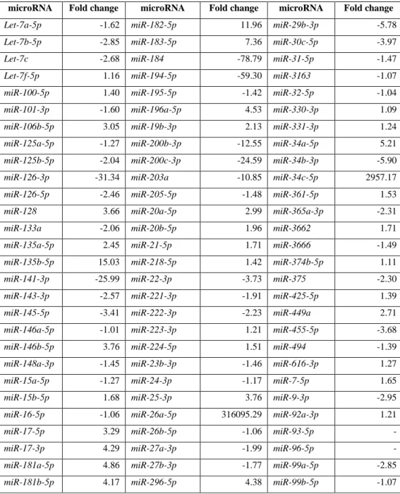
The expression of 4 precursors and 80 mature microRNAs was observed between Wilms tumor and the control sample by PCR Array from Patient 1. We used ΔΔCt method. The relative changing of gene expression (’fold cange’) was calculated by 2−ΔΔ𝐶𝑡 formula. The 3. Table demonstrates the fold changes.
13
3. Table Fold change in microRNA expressions (in tumor tissue compared to a tumor-free region of the same sample) in Patient 1, obtained by PCR Array.
microRNA Fold change microRNA Fold change microRNA Fold change
Let-7a-5p -1.62 miR-182-5p 11.96 miR-29b-3p -5.78
Let-7b-5p -2.85 miR-183-5p 7.36 miR-30c-5p -3.97
Let-7c -2.68 miR-184 -78.79 miR-31-5p -1.47
Let-7f-5p 1.16 miR-194-5p -59.30 miR-3163 -1.07
miR-100-5p 1.40 miR-195-5p -1.42 miR-32-5p -1.04
miR-101-3p -1.60 miR-196a-5p 4.53 miR-330-3p 1.09
miR-106b-5p 3.05 miR-19b-3p 2.13 miR-331-3p 1.24
miR-125a-5p -1.27 miR-200b-3p -12.55 miR-34a-5p 5.21
miR-125b-5p -2.04 miR-200c-3p -24.59 miR-34b-3p -5.90
miR-126-3p -31.34 miR-203a -10.85 miR-34c-5p 2957.17
miR-126-5p -2.46 miR-205-5p -1.48 miR-361-5p 1.53
miR-128 3.66 miR-20a-5p 2.99 miR-365a-3p -2.31
miR-133a -2.06 miR-20b-5p 1.96 miR-3662 1.71
miR-135a-5p 2.45 miR-21-5p 1.71 miR-3666 -1.49
miR-135b-5p 15.03 miR-218-5p 1.42 miR-374b-5p 1.11
miR-141-3p -25.99 miR-22-3p -3.73 miR-375 -2.30
miR-143-3p -2.57 miR-221-3p -1.91 miR-425-5p 1.39
miR-145-5p -3.41 miR-222-3p -2.23 miR-449a 2.71
miR-146a-5p -1.01 miR-223-3p 1.21 miR-455-5p -3.68
miR-146b-5p 3.76 miR-224-5p 1.51 miR-494 -1.39
miR-148a-3p -1.45 miR-23b-3p -1.46 miR-616-3p 1.27
miR-15a-5p -1.27 miR-24-3p -1.17 miR-7-5p 1.65
miR-15b-5p 1.68 miR-25-3p 3.76 miR-9-3p -2.95
miR-16-5p -1.06 miR-26a-5p 316095.29 miR-92a-3p 1.21
miR-17-5p 3.29 miR-26b-5p -1.06 miR-93-5p -
miR-17-3p 4.29 miR-27a-3p -1.99 miR-96-5p -
miR-181a-5p 4.86 miR-27b-3p -1.77 miR-99a-5p -2.85
miR-181b-5p 4.17 miR-296-5p 4.38 miR-99b-5p -1.07
As demonstrated in 3. Table, we failed to detect miR-93-5p and miR-96-5p in the control sample. The overexpression of miR-26 with an unrealistic magnitude was considered an artifact. (According to the literature, it should be underexpressed).
14
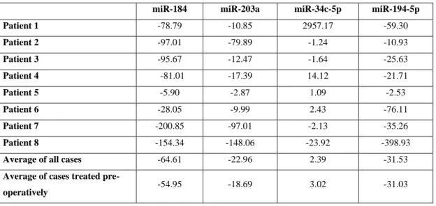
4. Table shows the summary of ’fold changes’ of the four microRNAs in our eight patients, as well as the average ’fold chang’e for each microRNA.
4. Table Fold change in microRNA expression (in tumor tissues compared to tumor-free regions of the same samples) in all patients
miR-184 miR-203a miR-34c-5p miR-194-5p
Patient 1 -78.79 -10.85 2957.17 -59.30
Patient 2 -97.01 -79.89 -1.24 -10.93
Patient 3 -95.67 -12.47 -1.64 -25.63
Patient 4 -81.01 -17.39 14.12 -21.71
Patient 5 -5.90 -2.87 1.09 -2.53
Patient 6 -28.05 -9.99 2.43 -76.11
Patient 7 -200.85 -97.01 -2.13 -35.26
Patient 8 -154.34 -148.06 -23.92 -398.93
Average of all cases -64.61 -22.96 2.39 -31.53
Average of cases treated pre-
operatively -54.95 -18.69 3.02 -31.03
As Patient 7 did not receive preoperative chemotherapy, an average was also calculated with the omission of this case for easier comparability.
15
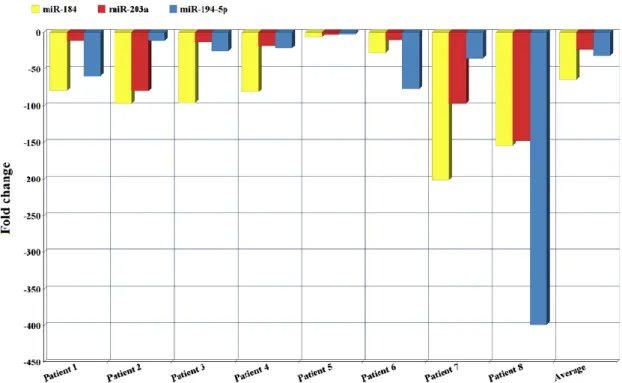
6. Figure shows the underexpression of selected microRNAs.
Patient 1, 2, 3 and Patient 5, 6, 7, 8 stayed in remission while Patient 4 had a relapse.
Patient 5 had an extraodinarily looking tumor which was immunohystochemically confirmed to be cortisol-producing.
Patient 1, 2, 3, 4, 5, 6 showed regression after chemotherapy in a different rate.
Patient 7 did not received preoperative chemotherapy because the initial diagnose was incorrect (renalcell carcinoma). Patient 8 showed no response to chemotherapy at all.
6. Figure Fold change in miR-184, miR-203, miR-194-5p in all studied patients
16 5 CONCLUSIONS
By examining the VEGF expression in hydronephrotic patients and the microRNA profile in children diagnosed with blastemal Wilms tumor, the following conclusions can be derived:
1. To our knowledge, our study is the first evaluation of VEGF expression in humans. The ureter tissue samples from 14 infants with obstructive uropathy showed no increased VEGF activity. Taking account of the limitations of this study, the role of VEGF as useful biomarker in diagnostics and therapy is ambiguous. We believe that our pilot study can be applied to further studies.
2. Archived FFPE samples from children diagnosed with blastemal Wilms tumor which are younger than 6 years are useful microRNA sources. MicroRNA yields were adequate from all FFPE samples. Utilizing FFPE archives as microRNA sources few years later may have clinical value in Wilms tumor.
3. As we examined the FFPE samples, we observed microRNA downregulations in three microRNAs from the selected four microRNAs in blastemal Wilms tumor. miR- 184 and miR-203a were found to be downregulated in all samples which seems to be a new finding. Despite the small sample size of our study, results were suggestive for having a tumorsuppressor role in the pathogenesis of Wilms tumor.
4. However our study has a small sample size, we suggest that the preoperative chemotherapy affects the microRNA profile of blastemal Wilms tumor and knowing microRNA profile of Wilms tumors will help to differentiate between chemoresistant and chemosensitive Wilms tumors.
17
6 BIBLIOGRAPHY OF THE CANDIDATE’S PUBLICATIONS 6.1 Publications related to the doctoral thesis
1. Zsófia Magyar1, Julianna Schönleber, Miklós Romics, Ervin Hruby, Bálint Nagy, Bálint Sulya, Artúr Beke, Ágnes Harmath, Judit Jeager, János Rigó Jr, Éva Görbe.
Expression of VEGF in neonatal urinary obstruction: does expression of VEGF predict hydronephrosis? Medical Science Monitor, 2015 May, 8;21:1319-23.
Doi: 10.12659/MSM.894133.
IF: 1,405
2. Gergely Buglyó1, Zsófia Magyar1, Éva Romicsné Görbe, Rita Bánusz, Monika Csóka, Tamás Micsik, Zsanett Berki, Péter Varga P, Zoltán Sápi, Bálint Nagy.
Quantitative RT-PCR-based miRNA profiling of blastemal Wilms' tumors from formalin- fixed paraffin-embedded samples.
Gergely Buglyó and Zsófia Magyar contributed equally to this work.
Journal of Biotechnology, 2019 Jun, 10;298:11-15.
Doi: 10.1016/j.jbiotec.2019.04.001. Epub 2019 Apr 5.
IF: 3,163
6.2 Publications not related to the topic of the doctoral thesis
1. Péter Varga, Botond Berecz, Barbara Pete, Tímea Kollár, Zsófia Magyar , Judit Jeager, Éva Romics Görbe, János Rigó Jr, József Gábor Joó, Ákos Gasparics.
Trends in mortality and morbidity in infants under 500 grams birthweight: observations from our neonatal intensive care unit (NICU).
Medical Science Monitor. 2018 Jun 29;24:4474-4480. Doi: 10.12659/MSM.907652.
IF: 1,980
18
2. Péter Varga, Judit Jeager, Ágnes Harmath, Botond Berecz, Tímea Kollár, Barbara Pete, Zsófia Magyar, János Rigó J Jr, Éva Romicsné Görbe.
Changes in the outcome for infants, with birth weight under 500 grams, at our department (First Department of Obstetrics and Gynecology, Semmelweis University, Budapest).
Orvosi Hetilap, 2015 Mar 8;156(10):404-8.
Doi: 10.1556/OH.2015.30101.
IF: 0,291
3. Péter Varga, Botond Berecz, Ákos Gasparics, Zsófia Dombi, Zsuzsanna Varga, Judit Jeager, Zsófia Magyar, János Rigó Jr, József Gábor Joó, László Kornya.
Morbidity and mortality trends in very-very low birth weight premature infants in light of recent changes in obstetric care.
European Journal of Obstetrics and Gynecology Reproductive Biology. 2017 Jan 23;211:134-139. Doi: 10.1016/j.ejogrb.2017.01.051.
IF: 1,809
4. Vörös Krisztián, Magyar Zsófia, Kalabay László
Pszichoszociális problémákkal társult magasvérnyomás-betegség beállítása Bibliogr.: p. 14. - Abstr. Hun.
Magyar Családorvosok Lapja. - ISSN 1789-607X. - 2012. 6. Sz., p. 11-14.
5. Magyar Zsófia, Mester-Holczinger Veronika, Dezsőfi Antal Ártalmatlan-e a Gilbert-kór?
Gyermekgyógyászat 2017; 68. évfolyam, 2. szám, p. 76-78.