MicroRNA Expression Profiling in Adrenal Myelolipoma
Abel Decmann,1 P ´al Perge,1 G ´abor Ny´ır}o,2 Ott ´o Darvasi,3 Istv ´an Lik ´o,3
Katalin Borka,4 Tam ´as Micsik,5 Zsuzsanna T ´oth,1 Irina Bancos,6 Raffaele Pezzani,7 Maurizio Iacobone,8 Attila Pat ´ocs,3 and Peter Igaz1,2
1Second Department of Medicine, Faculty of Medicine, Semmelweis University, 1088 Budapest, Hungary;
2MTA-SE Molecular Medicine Research Group, Hungarian Academy of Sciences and Semmelweis University, 1088 Budapest, Hungary;3Hereditary Endocrine Tumors Research Group, Hungarian Academy of Sciences and Semmelweis University, 1088 Budapest, Hungary;4Second Department of Pathology, Faculty of Medicine, Semmelweis University, 1091 Budapest, Hungary;5First Department of Pathology and Experimental Cancer Research, Faculty of Medicine, Semmelweis University, 1085 Budapest, Hungary;
6Division of Endocrinology, Diabetes, Metabolism and Nutrition, Department of Internal Medicine, Mayo Clinic, Rochester, Minnesota 55905;7Endocrinology Unit, Department of Medicine, University of Padua, 35128 Padova, Italy; and8Minimally Invasive Endocrine Surgery Unit, Department of Surgery, Oncology and Gastroenterology (DISCOG), University of Padova, 35128 Padova, Italy
Introduction: Adrenal myelolipoma (AML) is the second most common and invariably benign primary adrenal neoplasm. Due to the variable proportion of fat and hematopoietic elements and its often large size, it can cause differential diagnostic problems. Several reports confirmed the utility of miRNAs in the diagnosis of tumors, but miRNA expression in AML has not yet been investigated.
Materials and Methods:Next-generation sequencing (NGS) was performed on 30 formalin-fixed, paraffin-embedded (FFPE) archived tissue samples [10 each of AML, adrenocortical adenoma (ACA), and adrenocortical carcinoma (ACC)]. Validation was performed by real-time quantitative reverse transcription polymerase chain reaction on a cohort containing 41 further FFPE samples (15 AML, 14 ACA, and 12 ACC samples). Circulating miRNA counterparts of significantly differentially expressed tissue miRNAs were studied in 33 plasma samples (11 each of ACA, ACC, and AML).
Results:By NGS, 256 significantly differentially expressed miRNAs were discovered, and 8 of these were chosen for validation. Significant overexpression ofhsa-miR-451a,hsa-miR-486-5p,hsa-miR-363- 3p, andhsa-miR-150-5pwas confirmed in AML relative to ACA and ACC.hsa-miR-184,hsa-miR-483-5p, andhsa-miR-183-5pwere significantly overexpressed in ACC relative to ACA but not to AML. Cir- culatinghsa-miR-451aandhsa-miR-363-3p were significantly overexpressed in AML, whereas cir- culatinghsa-miR-483-5pandhsa-miR-483-3pwere only significantly overexpressed in ACC vs ACA.
Conclusions: We have found significantly differentially expressed miRNAs in AML and adreno- cortical tumors. Circulatinghsa-miR-451amight be a promising minimally invasive biomarker of AML. The lack of significantly different expression ofhsa-miR-483-3pandhsa-miR-483-5pbetween AML and ACC might limit their applicability as diagnostic miRNA markers for ACC.(J Clin Endocrinol Metab103: 3522–3530, 2018)
A
drenal neoplasms are common. Among adrenal tumors, adrenocortical adenomas (ACAs) are the most frequent, constituting 60% to 70% of adrenalincidentalomas. Adrenal myelolipoma (AML) is the second most common primary adrenal tumor, repre- senting 6% to 16% of all adrenal incidentalomas (1, 2).
ISSN Print 0021-972X ISSN Online 1945-7197 Printed in USA
Copyright © 2018 Endocrine Society
Received 14 April 2018. Accepted 15 June 2018.
First Published Online 21 June 2018
Abbreviations: ACA, adrenocortical adenoma; ACC, adrenocortical carcinoma; AML, adrenal myelolipoma; AUC, area under the curve; FC, fold change; FFPE, formalin fixed, paraffin embedded; NGS, next-generation sequencing.
3522 https://academic.oup.com/jcem J Clin Endocrinol Metab, September 2018, 103(9):3522–3530 doi: 10.1210/jc.2018-00817
Downloaded from https://academic.oup.com/jcem/article/103/9/3522/5041928 by Semmelweis Egyetem user on 15 December 2021
AML is an invariably benign tumor that is composed of adipose tissue and extramedullary hematopoietic elements.
The pathogenesis of AML is unclear (3, 4). AMLs are often large tumors with an average size of 10.2 cm at diagnosis (3). On the other hand, adrenocortical carcinoma (ACC) is an uncommon disease with an annual incidence of 0.5 to 2 per million (5–8) and a poor prognosis, with a 5-year survival rate of,15% in stage IV (9, 10).
Because of their large size, it might be occasionally challenging to distinguish AML from other adrenal tu- mors, especially ACCs, which also often present with a large size (3, 8). Although the presence of macroscopic fat is pathognomonic for AML, the variability in the content of fat and hematopoietic elements in AML could lead to an indeterminate appearance on imaging, and even in- tense 18F-fluorodeoxyglucose uptake on positron emis- sion tomography–CT due to the hemopoietic elements was reported (11). Moreover, the age distribution of AML is similar to that of ACC, with a peak incidence in the fifth and sixth decades (9).
Mature miRNAs are short, 19- to 25-nucleotide-long single-stranded noncoding RNA molecules that are in- volved in the regulation of gene expression mostly at the posttranscriptional level. miRNAs are expressed in a tissue-specific fashion and secreted in body fluids (12).
Several studies have shown that miRNAs can be useful biomarkers in different diseases, including various neo- plasms. Recent studies, including ours, have reported significant differences in tissue and circulating miRNA expression of patients with ACA and ACC (13–17). To our knowledge, the miRNA expression profile of adrenal myelolipoma has not been investigated. With this in mind, we hypothesized that miRNA profiling in AML might lead to the identification of biomarkers that could be used in challenging diagnostic situations.
Materials and Methods
Tissue collection and ethics approval
A total of 71 histologically proven formalin-fixed, paraffin-embedded (FFPE) archived tissue samples were used (Table 1). The discovery cohort contained 30 samples (10 ACA, 10 ACC and 10 AML samples), and the in- dependent validation cohort contained another 41 FFPE samples (15 AML, 14 ACA, and 12 ACC samples). A total of 33 independent preoperative EDTA-anticoagulated plasma samples from patients with histologically proven adrenal tumors (11 samples each of ACA, ACC, and AML) were used for the analysis of circulating miRNA. Preoperative bio- chemical testing for hormonal evaluation involved basal cortisol, ACTH, aldosterone, renin activity, dehydroepian- drosterone sulfate, urinary catecholamines, and low-dose dexamethasone test (cutoff: 1.8 mg/dL). The study was ap- proved by the Ethical Committee of the Hungarian Health Council. All experiments were performed in accordance with
relevant guidelines and regulations, and informed consent was obtained from the involved patients.
Sample processing and RNA isolation
Total RNA was isolated from all the FFPE samples by the RecoverAll Total Nucleic Acid Isolation Kit for FFPE (Thermo Fisher Scientific, Waltham, MA). Total RNA from plasma was isolated by the miRNeasy Serum/Plasma Kit (Qiagen GmbH, Hilden, Germany). As a spike-in control for purification ef- ficiency, 5 mL of 5 nM Syn-cel-miR-39 miScript miRNA Mimic (Qiagen GmbH) was added before the addition of acid- phenol/chloroform. Total RNA was stored at 280°C until further processing.
miRNA expression profiling from tissue samples by next-generation sequencing
The cDNA library was made from total RNA by the QIAseq miRNA Library Kit (Qiagen GmbH) according to the in- structions of the manufacturer. The library was prepared for sequencing according to the instructions of the MiSeq Reagent Kit v3 (Illumina, San Diego, CA). Next-generation sequencing (NGS) was performed by Illumina MiSeq (Illumina). FASTQ files were used in the primary data analysis procedure. Qiagen online analysis software was applied. Primary analysis included the trimming of adapters using cutadapt (Marcel Martin, Technical University, Dortmund, Germany). Reads with ,16 bp insert sequences or with ,10 bp Unique Molecular Index were dis- carded. Alignment of reads was performed using bowtie (John Hopkins University, Baltimore, MD), and miRbase V21 was used for miRNAs. After DESeq2 normalization (18), secondary analysis revealed significantly differently expressed miRNAs.
Validation of individual miRNAs
RNA was reverse-transcribed using the TaqMan microRNA Reverse Transcription Kit (Thermo Fisher Scientific) and in- dividual TaqMan miRNA assays (CN: 4427975; Thermo Fisher Scientific) for tissue and plasma samples. Selected miRNAs were hsa-miR-451a (ID: 001141), hsa-miR-486-5p (ID: 001278),hsa-miR-363-3p(ID: 001271),hsa-miR-150-5p (ID: 000473),hsa-miR-184(ID: 000485),hsa-miR-483-5p(ID:
002338),hsa-miR-483-3p(ID: 002339), andhsa-miR-183-5p (ID: 002269). The internal control wasRNU48(ID: 001006) for tissue samples and cel-miR-39 (ID: 000200) for plasma samples. Quantitative real-time PCR was performed by the TaqMan Fast Universal PCR Master Mix (2x) (CN: 4352042;
Thermo Fisher Scientific) on a Quantstudio 7 Flex Real-Time PCR System (Thermo Fisher Scientific) according to the man- ufacturer’s protocol for TaqMan miRNA assays with minor modifications. Negative control reactions contained no cDNA templates. Samples were always run in triplicate. For data evaluation, we used the dCt method (delta Ct value equals target miRNA’s Ct minus internal control miRNA’s Ct) using Microsoft Excel 2016 (Microsoft, Redmond, WA).
Statistical analysis
Statistical power analysis was performed with a statistical power and sample size calculator (Tempest Technologies, Helena, MT). Real-time quantitative PCR data were analyzed by GraphPad Prism 7.00 (GraphPad Software, La Jolla, CA).
For differentiating between ACA, ACC, and AML groups, ANOVA or Kruskal-Wallis test was used according to the result
Downloaded from https://academic.oup.com/jcem/article/103/9/3522/5041928 by Semmelweis Egyetem user on 15 December 2021
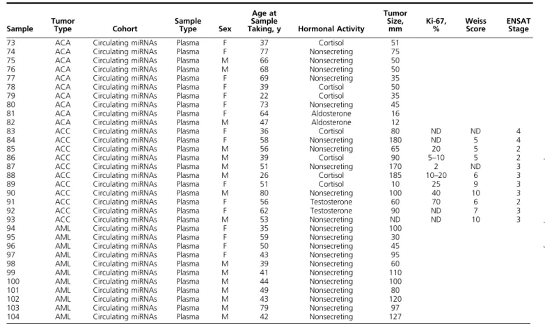
Table 1. Characteristics of the Tumor and Plasma Samples Studied
Sample Tumor
Type Cohort Sample
Type Sex
Age at Sample
Taking, y Hormonal Activity
Tumor Size,
mm Ki-67,
% Weiss
Score ENSAT Stage
1 ACA Discovery FFPE F 55 Nonsecreting 25
2 ACA Discovery FFPE M 62 Nonsecreting 40
3 ACA Discovery FFPE M 44 Cortisol 85
4 ACA Discovery FFPE M 62 Nonsecreting 40
5 ACA Discovery FFPE M 50 Nonsecreting 40
6 ACA Discovery FFPE F 57 Nonsecreting 3
7 ACA Discovery FFPE F 64 Nonsecreting 38
8 ACA Discovery FFPE M 55 Nonsecreting 60
9 ACA Discovery FFPE M 44 Cortisol 85
10 ACA Discovery FFPE F 36 Aldosterone 30
11 ACC Discovery FFPE F 70 Nonsecreting 120 ,20 ND 4
12 ACC Discovery FFPE M 43 Nonsecreting 120 ND 6 4
13 ACC Discovery FFPE F 39 Cortisol 90 10 7 2
14 ACC Discovery FFPE F 58 Nonsecreting 115 ND ND 2
15 ACC Discovery FFPE F 53 Aldosterone, cortisol 90 40–50 6 2
16 ACC Discovery FFPE F 72 Nonsecreting 10 4–5 ND 3
17 ACC Discovery FFPE F 46 Nonsecreting 200 42 7 4
18 ACC Discovery FFPE F 50 Nonsecreting ND ND 5 2
19 ACC Discovery FFPE F 54 Nonsecreting 70 20 3 4
20 ACC Discovery FFPE F 55 Nonsecreting 60 20–30 5 4
21 AML Discovery FFPE M 68 Nonsecreting 80
22 AML Discovery FFPE F 66 Nonsecreting 70
23 AML Discovery FFPE F 66 Nonsecreting 35
24 AML Discovery FFPE F 35 Nonsecreting 60
25 AML Discovery FFPE F 55 Nonsecreting 60
26 AML Discovery FFPE F 58 Nonsecreting 80
27 AML Discovery FFPE M 70 Nonsecreting 90
28 AML Discovery FFPE F 37 Nonsecreting 80
29 AML Discovery FFPE M 42 Nonsecreting 80
30 AML Discovery FFPE M 61 Nonsecreting 50
31 ACA Validation FFPE F 55 Nonsecreting 90
32 ACA Validation FFPE M 60 Nonsecreting 30
33 ACA Validation FFPE F 52 Aldosterone 20
34 ACA Validation FFPE M 59 Nonsecreting 35
35 ACA Validation FFPE F 41 Aldosterone 30
36 ACA Validation FFPE M 51 Aldosterone 45
37 ACA Validation FFPE F 48 Aldosterone 10
38 ACA Validation FFPE F 68 Aldosterone 20
39 ACA Validation FFPE F 43 Aldosterone 15
40 ACA Validation FFPE F 84 Nonsecreting 90
41 ACA Validation FFPE F 58 Nonsecreting 40
42 ACA Validation FFPE F 56 Cortisol 25
43 ACA Validation FFPE M 25 Nonsecreting 70
44 ACA Validation FFPE M 64 Nonsecreting 100
45 ACC Validation FFPE F 57 Nonsecreting 60 20–30 5 4
46 ACC Validation FFPE F 62 Cortisol 78 5 5 2
47 ACC Validation FFPE F 61 Nonsecreting 100 20 5 4
48 ACC Validation FFPE F 48 Nonsecreting 120 30 6 4
49 ACC Validation FFPE F 69 Nonsecreting 110 10–20 6 3
50 ACC Validation FFPE M 25 Cortisol 120 10 6 4
51 ACC Validation FFPE M 79 Nonsecreting 86 10 5 3
52 ACC Validation FFPE F 71 Nonsecreting 80 ND ND 4
53 ACC Validation FFPE M 17 Nonsecreting 110 10–15 3 2
54 ACC Validation FFPE F 61 Cortisol 80 20–30 5 4
55 ACC Validation FFPE M 28 Nonsecreting ND ND ND 4
56 ACC Validation FFPE F 47 Nonsecreting 140 20–25 4 4
57 AML Validation FFPE F 36 Nonsecreting 100
58 AML Validation FFPE F 55 Nonsecreting 135
59 AML Validation FFPE M 51 Nonsecreting 30
60 AML Validation FFPE F 62 Nonsecreting 40
61 AML Validation FFPE F 54 Nonsecreting 60
62 AML Validation FFPE F 35 Nonsecreting 50
63 AML Validation FFPE M 46 Nonsecreting 60
64 AML Validation FFPE F 54 Nonsecreting 45
65 AML Validation FFPE F 38 Nonsecreting 110
66 AML Validation FFPE M 60 Nonsecreting 80
67 AML Validation FFPE F 29 Nonsecreting 50
68 AML Validation FFPE F 42 Nonsecreting 110
69 AML Validation FFPE F 44 Nonsecreting 40
70 AML Validation FFPE F 71 Nonsecreting 50
71 AML Validation FFPE M 60 Nonsecreting 45
72 ACA Circulating miRNAs Plasma M 62 Cortisol 40
(Continued)
Downloaded from https://academic.oup.com/jcem/article/103/9/3522/5041928 by Semmelweis Egyetem user on 15 December 2021
of the Shapiro-Wilk normality test. miRNAs that could be used potentially as minimally invasive biomarkers in adrenal neo- plasms underwent receiver operating characteristic analysis.
Pvalues,0.05 were considered significant.
Pathway analysis
The potential targets of miRNAs were investigated using Diana Tools mirPath v.3 (Diana Lab Tools, University of Thessaly, Thessaly, Greece). For target prediction, Targetscan (Whitehead Institute, Cambridge, MA) was used.
Results
miRNA expression profiling by NGS
NGS was performed on 30 FFPE samples. Individu- al miRNAs are listed in Supplemental Table 1. In total, 256 significantly differentially expressed miRNAs were found. From the top-ranked overexpressed miRNAs in AML listed in Supplemental Table 2, we have selected hsa-miR-451a [fold change (FC) to ACC: 14.7; P , 0.0001], hsa-miR-486-5p (FC: 14.1;P ,0.0001), hsa- miR-363-3p (FC: 6;P, 0.0001), and hsa-miR-150-5p (FC: 6.7; P , 0.0001) to validate. These miRNAs are significantly upregulated in AML compared with ACA and ACC.hsa-miR-483-3p(FC: 47.3;P,0.0001),hsa- miR-184(FC: 14.5;P,0.0001),hsa-miR-483-5p(FC:
18.2;P, 0.0001), and hsa-miR-183-5p (FC: 9.5;P, 0.0001) were significantly upregulated in ACC compared with AML and ACA (FC and P values compared with AML). NGS data are available under the Gene Expres- sion Omnibus (GEO) accession number GSE112804.
Validation of significantly differentially expressed miRNAs by real-time quantitative reverse
transcription polymerase chain reaction
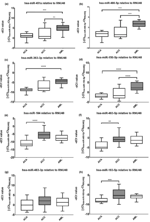
In total, 41 independent FFPE samples were subjected to validation. miRNAs with significantly higher ex- pression in AML relative to ACA and ACC by NGS were successfully validated by real-time quantitative reverse transcription polymerase chain reaction: hsa-miR-451, hsa-miR-486-5p,hsa-miR-363-3p, andhsa-miR-150-5p were significantly overexpressed in AML compared with ACA and ACC (Fig. 1).hsa-miR-363-3pwas significantly overexpressed in AML compared only with ACA, but a tendency of upregulation can be seen relative to ACC.
However, the validation of significantly overexpressed miRNAs in ACC compared with AML and ACA by real- time quantitative PCR was only partly successful, as we could only observe significant overexpression of three miRNAs (hsa-miR-184,hsa-miR-483-5p, andhsa-miR- 183-5p) in ACC compared with ACA but not with AML.
Table 1. Characteristics of the Tumor and Plasma Samples Studied (Continued)
Sample Tumor
Type Cohort Sample
Type Sex
Age at Sample
Taking, y Hormonal Activity
Tumor Size,
mm Ki-67,
% Weiss
Score ENSAT Stage
73 ACA Circulating miRNAs Plasma F 37 Cortisol 51
74 ACA Circulating miRNAs Plasma F 77 Nonsecreting 75
75 ACA Circulating miRNAs Plasma M 66 Nonsecreting 50
76 ACA Circulating miRNAs Plasma M 68 Nonsecreting 50
77 ACA Circulating miRNAs Plasma F 69 Nonsecreting 35
78 ACA Circulating miRNAs Plasma F 39 Cortisol 50
79 ACA Circulating miRNAs Plasma F 22 Cortisol 35
80 ACA Circulating miRNAs Plasma F 73 Nonsecreting 45
81 ACA Circulating miRNAs Plasma F 64 Aldosterone 16
82 ACA Circulating miRNAs Plasma M 47 Aldosterone 12
83 ACC Circulating miRNAs Plasma F 36 Cortisol 80 ND ND 4
84 ACC Circulating miRNAs Plasma F 58 Nonsecreting 180 ND 5 4
85 ACC Circulating miRNAs Plasma M 56 Nonsecreting 65 20 5 2
86 ACC Circulating miRNAs Plasma M 39 Cortisol 90 5–10 5 2
87 ACC Circulating miRNAs Plasma M 51 Nonsecreting 170 2 ND 3
88 ACC Circulating miRNAs Plasma M 26 Cortisol 185 10–20 6 3
89 ACC Circulating miRNAs Plasma F 51 Cortisol 10 25 9 3
90 ACC Circulating miRNAs Plasma M 80 Nonsecreting 100 40 10 3
91 ACC Circulating miRNAs Plasma F 56 Testosterone 60 70 6 2
92 ACC Circulating miRNAs Plasma F 62 Testosterone 90 ND 7 3
93 ACC Circulating miRNAs Plasma M 53 Nonsecreting ND ND 10 3
94 AML Circulating miRNAs Plasma F 35 Nonsecreting 100
95 AML Circulating miRNAs Plasma F 59 Nonsecreting 30
96 AML Circulating miRNAs Plasma F 50 Nonsecreting 45
97 AML Circulating miRNAs Plasma F 43 Nonsecreting 95
98 AML Circulating miRNAs Plasma M 39 Nonsecreting 60
99 AML Circulating miRNAs Plasma M 41 Nonsecreting 110
100 AML Circulating miRNAs Plasma M 44 Nonsecreting 100
101 AML Circulating miRNAs Plasma M 49 Nonsecreting 80
102 AML Circulating miRNAs Plasma M 43 Nonsecreting 120
103 AML Circulating miRNAs Plasma M 79 Nonsecreting 97
104 AML Circulating miRNAs Plasma M 42 Nonsecreting 127
Abbreviations: ENSAT, European Network for the Study of Adrenal Tumors; F, female; M, male; ND, no data.
Downloaded from https://academic.oup.com/jcem/article/103/9/3522/5041928 by Semmelweis Egyetem user on 15 December 2021
We have not observed significant differences in the expres- sion of hsa-miR-483-3pamong the groups studied. Most notably, the expression ofhsa-miR-483-5pwas similar in ACC and AML samples. Statistical power analysis showed that with these 41 samples, the power of our study is.99%.
miRNA expression analysis in plasma samples Having found significantly differentially expressed miRNAs in tissue samples, we extended our study to plasma samples searching for potential minimally
invasive circulating miRNA markers.
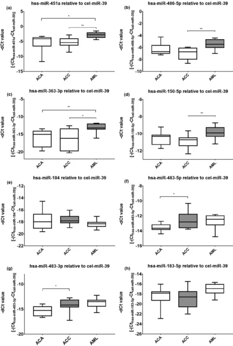
Significant overexpression ofhsa-miR- 451a and hsa-miR-363-3p in AML compared with both ACA and ACC was found (Fig. 2). The expression of hsa-miR-486-5p and hsa-miR-150-5p was only significantly upregulated in AML compared with ACC but not with ACA.
On the other hand, no significant differences in the expression of hsa- miR-184 and hsa-miR-183-5p were noted. hsa-miR-483-3p and hsa-miR- 483-5pwere significantly overexpressed in ACC relative to ACA but not to AML. Statistical power analysis showed that with the 11 samples per group, the power of our study is 0.9985.
Diagnostic performance of miRNAs Circulating miRNAs that could be potentially used as minimally invasive biomarkers underwent receiver operating characteristic analysis.hsa-miR-451aand hsa-miR-483-3pshowed the highest area under curve (AUC) value. Forhsa-miR- 451a, when AML samples were com- pared with ACA samples, the AUC was 0.88, and when AML samples were compared with ACC samples, the AUC value was 0.91 (Fig. 3). By selecting 3.676 as the cutoff point, both sensitivity and specificity were 81.82% for differentiat- ing AML and ACA. For differentiating AML and ACC, sensitivity was 90.91%
and specificity was 81.82% by setting the cutoff point to 3.994. The negative pre- dictive value of overexpressed hsa-miR- 451a to rule out ACC was 83.33%, whereas its positive predictive value to confirm AML was 90%.
Circulating hsa-miR-483-3p per- formed best in distinguishing ACC from ACA with an AUC value of 0.88. By setting the cutoff point to 14.42, sensitivity was 81.82%, whereas specificity was 90.91%.
Pathway analysis
Among the predicted targets of hsa-miR-451a, hsa- miR-486-5p, hsa-miR-363-3p, and hsa-miR-150-5p, mRNAs coding for proteins involved in fatty acid metabolism, degradation, and biosynthesis were found (3-oxoacyl-ACP synthase, mitochondrial; enoyl-CoA,
Figure 1.Overexpressed tissue miRNAs in AML and ACC by quantitative reverse
transcription polymerase chain reaction. Mean6SD of2dCt values of selected miRNAs: (a) hsa-miR-451a, (b) hsa-miR-486-5p, (c) hsa-miR-363-3p, (d) hsa-miR-150-5p, (e) hsa-miR-184, (f) hsa-miR-483-5p, (g) hsa-miR-483-3p, and (h) hsa-miR-183-5p. **P,0.01. ***P,0.001.
****P,0.0001. ANOVA or Kruskal-Wallis and Tukey or Dunn multiple-comparisons test. Gray shading represents the candidate miRNA.
Downloaded from https://academic.oup.com/jcem/article/103/9/3522/5041928 by Semmelweis Egyetem user on 15 December 2021
hydratase/3-hydroxyacyl CoA dehydrogenase; cyto- chrome P450, family 4, subfamily A, member 22).
P value was,0.0001 for all the three genes (Table 2).
Discussion
Adrenal myelolipoma is an invariably benign tumor, but it might cause differential diagnostic problems leading to unnecessary procedures. In our study, we have identified miRNA markers specific for AML in tissue and plasma
samples. To our knowledge, this is the first report on the miRNA expres- sion profile of AML. Based on the results of NGS, miRNAs hsa-miR- 451a, hsa-miR-486-5p, hsa-miR-363- 3p, and hsa-miR-150-5p performed best in the diagnosis of AML and were able to differentiate AML from ACA and ACC. On the other hand, the al- ready reported ACC-associated miRNAs hsa-miR-184 (19, 20), hsa-miR-483-5p (15, 17), and hsa-miR-483-3p were the most highly ranked overexpressed miRNAs in ACC. Overexpression of hsa-miR-183-5p has not yet been re- ported in ACC and represents a novel finding, to our knowledge.
Three of four tissue miRNAs were confirmed by quantitative reverse tran- scription polymerase chain reaction to be significantly overexpressed in AML relative to ACA and ACC (hsa-miR- 451a, hsa-miR-486-5p, and hsa-miR- 150-5p). In concert with previous findings (16, 21, 22), we have found that tissuehsa-miR-483-5pwas signif- icantly overexpressed in ACC relative to ACA, but no difference of expres- sion relative to AML has been observed.
Whereas a tendency ofhsa-miR-483-3p overexpression in ACC was noted, this has not reached statistical signifi- cance in our cohort of patients. Over- expression of bothhsa-miR-483-5pand hsa-miR-483-3p has been previously described in ACC (23, 24).
Regarding circulating miRNAs, we demonstrated that hsa-miR-451a andhsa-miR-363-3pwere significantly overexpressed in AML relative to ACA and ACC. In addition, hsa-miR-486- 5p and hsa-miR-150-5p were signifi- cantly overexpressed in AML but only compared with ACC and not with ACA. In concordance to previous studies (13, 15, 17, 20), we have observed a significant overexpression of plasma hsa-miR-483-5p andhsa-miR-483-3pin patients with ACCs, but we could not detect a significant difference of these in expression between AML and ACC.
Tissue and circulating hsa-miR-483-5p has been considered the best marker of adrenocortical malignancy to date (13, 15, 17). The noted lack of significance be- tween ACC and AML in the expression of both tissue and
Figure 2. Overexpressed circulating plasma miRNAs in AML and ACC by quantitative reverse transcription polymerase chain reaction. Mean6SD of2dCt values of selected miRNAs: (a) hsa-miR-451a, (b) hsa-miR-486-5p, (c) hsa-miR-363-3p, (d) hsa-miR-150-5p, (e) hsa-miR-184, (f) hsa-miR-483-5p, (g) hsa-miR-483-3p, and (h) hsa-miR-183-5p. *P,0.05. **P,0.01.
ANOVA or Kruskal-Wallis and Tukey or Dunn multiple-comparisons test. Gray shading represents the candidate miRNA.
Downloaded from https://academic.oup.com/jcem/article/103/9/3522/5041928 by Semmelweis Egyetem user on 15 December 2021
plasmahsa-miR-483-5pandhsa-miR-483-3pis clinically relevant because it might represent a limitation in the use of these markers.
It is intriguing that there has been no significant difference in the tissue expression of hsa-miR-184, hsa-miR-483-3p,hsa-miR-483-5p, andhsa-miR-183-5p between ACC and AML, whereas three of these four miRNAs were significantly overexpressed in ACC vs ACA. Although it is pure hypothesis at present, the similar miRNA expression between ACC and AML might indicate some common step in their pathogenesis.
Pathway analysis revealed that the significantly overex- pressed miRNAs of AML are mostly linked to fatty acid metabolism. The miRNAs overexpressed in AML have been reported to be involved in several tumors.hsa-miR- 451a was reported to be overexpressed in pancreatic ductal adenocarcinoma (25) and papillary thyroid car- cinoma (26) but downregulated in lung adenocarci- noma (27) and melanoma (28). According to the cellular context, the same miRNA can behave as an overex- pressed oncogene or downregulated tumor suppressor in different tissues (14).hsa-miR-486-5p is mostly down- regulated in different tumors and classified as a tumor suppressor (29, 30). Bothhsa-miR-363-3p (31, 32) and hsa-miR-150-5p (33, 34) are mostly downregulated in various tumors. It seems that the overexpressed miRNAs in AML are mostly downregulated in other tumors. AML
might thus represent a unique tissue context. Red blood cells are known to harborhsa-miR-451andhsa-miR-486- 5p(35); moreover,hsa-miR-451 seems to be involved in erythropoiesis (36).
hsa-miR-451andhsa-miR-486-5pare among the most abundant miRNAs in the blood of healthy individuals (37), and their overexpression in AML might thus be related to the presence of extramedullary hematopoiesis.hsa-miR- 363that was found to be overexpressed in our AML samples was associated with the regulation of adipogenesis (38).
Tissue and plasma miRNAs are not always parallel. In ACC, for example, tissue hsa-miR-34a was down- regulated but upregulated in serum samples (15). In another report on endometrioid endometrial carcinoma, the expression of hsa-miR-9 and hsa-miR-301b was differentially expressed in the tissue and in blood (39).
Unfortunately, the mechanisms for active miRNA release to body fluids are incompletely understood, and most notably, the processes for miRNA sorting in the extra- cellular vesicles await clarification (40).
Circulating miRNA markers of AML might be of diagnostic relevance if applied presurgically. Among the miRNAs analyzed,hsa-miR-451aappears to be the best candidate for validation studies and possible subsequent integration into clinical practice.
Because ACC is a rare tumor and AML is mostly left nonoperated, the collection of sufficient numbers of preoperative plasma samples from patients with histo- logically proven tumors is difficult. Whereas we managed to include 25 AML FFPE samples for tissue miRNA analysis, only 11 AML samples for circulating miRNA were available, which is certainly a limitation of this study. Statistical power analysis, however, revealed that the power of our analysis for FFPE and plasma miRNAs has been.99%.
In this study, we have included only samples from patients with a histological diagnosis of adrenal tumors.
Figure 3.Evaluation of the diagnostic applicability ofhsa-miR-451aby receiver operating characteristic (ROC) curves. ROC curve ofhsa-miR-451ain AML compared with ACA and ACC: (a) AML compared with ACA and (b) AML compared with ACC.
Table 2. Results of the Pathway Analysis for miRNA Overexpressed in AML
KEGG Pathway PValue Gene
Fatty acid metabolism (hsa01212) ,0.0001 OXSM, EHHADH
Fatty acid degradation (hsa00071) ,0.0001 CYP4A22, EHHADH
GABAergic synapse (hsa04727) ,0.0001 SLC38A1, GABRB3, NSF, GABRA4, SLC12A5
Fatty acid biosynthesis (hsa00061) ,0.0001 OXSM
Vitamin B6 metabolism (hsa00750) ,0.001 PNPO
Abbreviations: CYP4A22, cytochrome P450, family 4, subfamily A, member 22; EHHADH, enoyl-CoA and 3-hydroxyacyl CoA dehydrogenase; GABA, g-aminobutyric acid; GABRA4,g-aminobutyric acid type A receptora4 subunit; GABRB3,g-aminobutyric acid type A receptorb3 subunit; KEGG, Kyoto Encyclopedia of Genes and Ganomes; NSF,N-ethylmaleimide sensitive factor, vesicle fusing ATPase; OXSM, 3-oxoacyl-ACP synthase, mitochondrial;
PNPO, pyridoxamine 50-phosphate oxidase; SLC12A5, solute carrier, family 12, member 5; SLC38A1, solute carrier, family 38, member 1.
Downloaded from https://academic.oup.com/jcem/article/103/9/3522/5041928 by Semmelweis Egyetem user on 15 December 2021
However, if the inclusion criteria are less stringent (i.e., plasma samples from patients having AML based on unambiguous imaging diagnosis can be included), the cohorts can be increased considerably. Such a prospective study can be proposed in the future to confirm the utility of AML-associated circulating miRNA markers (mostly circulating hsa-miR-451a) as a minimally invasive bio- marker. The negative predictive value of overexpressed circulating hsa-miR-451a to rule out ACC is not high for clinical introduction at present, but this might be improved by sample size extension in such a further prospective study. Such a marker might be helpful for confirming patients with large tumors to have AML and thus might help to avoid unnecessary surgery.
In conclusion, to our knowledge, we have performed the first miRNA profiling of adrenal myelolipoma and identified miRNAs that are significantly differentially expressed between AML and adrenocortical benign and malignant tumors. Circulating miRNA markers could potentially serve as noninvasive diagnostic biomarkers, but further studies on larger cohorts are needed to confirm their clinical usefulness and applicability.
Acknowledgments
Financial Support: The study has been supported by a grant from the Hungarian National Research, Development and In- novation Office (NKFIH K115398; to P.I.) and an EFOP-3.6.3- VEKOP-16-2017-00009 grant.
Author Contributions: P.I. designed the research. A.D., P.P., G.N., O.D., and I.L. performed the research. K.B., T.M., R.P., Z.T., M.I., and I.B. provided patient samples. A.P. was involved in data analysis. A.D. and P.I. wrote the manuscript. All authors approved the final manuscript.
Correspondence and Reprint Requests: Peter Igaz, MD, PhD, DSc, Second Department of Medicine, Faculty of Medi- cine, Semmelweis University, H-1088 Budapest, Szentkir ´alyi str. 46., Hungary. E-mail:igaz.peter@med.semmelweis-univ.hu.
Disclosure Summary: The authors have nothing to disclose.
References
1. Song JH, Chaudhry FS, Mayo-Smith WW. The incidental adrenal mass on CT: prevalence of adrenal disease in 1,049 consecutive adrenal masses in patients with no known malignancy.AJR Am J Roentgenol. 2008;190(5):1163–1168.
2. Fassnacht M, Arlt W, Bancos I, Dralle H, Newell-Price J, Sahdev A, Tabarin A, Terzolo M, Tsagarakis S, Dekkers OM. Management of adrenal incidentalomas: European Society of Endocrinology Clinical Practice Guideline in collaboration with the European Network for the Study of Adrenal Tumors.Eur J Endocrinol. 2016;
175(2):G1–G34.
3. Decmann ´A, Perge P, T ´oth M, Igaz P. Adrenal myelolipoma:
a comprehensive review.Endocrine. 2017;59(1):7–15.
4. Lam AKY. Lipomatous tumours in adrenal gland: WHO updates and clinical implications. Endocr Relat Cancer. 2017;24(3):
R65–R79.
5. Kerkhofs TMA, Verhoeven RHA, Van der Zwan JM, Dieleman J, Kerstens MN, Links TP, Van de Poll-Franse LV, Haak HR. Adreno- cortical carcinoma: a population-based study on incidence and survival in the Netherlands since 1993.Eur J Cancer. 2013;49(11):2579–2586.
6. Kebebew E, Reiff E, Duh QY, Clark OH, McMillan A. Extent of disease at presentation and outcome for adrenocortical carcinoma:
have we made progress?World J Surg. 2006;30(5):872–878.
7. Lam AK. Update on adrenal tumours in 2017 World Health Organization (WHO) of endocrine tumours.Endocr Pathol. 2017;
28(3):213–227.
8. Fassnacht M, Kroiss M, Allolio B. Update in adrenocortical car- cinoma.J Clin Endocrinol Metab. 2013;98(12):4551–4564.
9. Lib´e R, Borget I, Ronchi CL, Zaggia B, Kroiss M, Kerkhofs T, Bertherat J, Volante M, Quinkler M, Chabre O, Bala M, Tabarin A, Beuschlein F, Vezzosi D, Deutschbein T, Borson-Chazot F, Hermsen I, Stell A, Fottner C, Leboulleux S, Hahner S, Mannelli M, Berruti A, Haak H, Terzolo M, Fassnacht M, Baudin E; ENSAT Network. Prognostic factors in stage III-IV adrenocortical carci- nomas (ACC): an European Network for the Study of Adrenal Tumor (ENSAT) study.Ann Oncol. 2015;26(10):2119–2125.
10. Terzolo M, Daffara F, Ardito A, Zaggia B, Basile V, Ferrari L, Berruti A. Management of adrenal cancer: a 2013 update.
J Endocrinol Invest. 2014;37(3):207–217.
11. Ludwig V, Rice MH, Martin WH, Kelley MC, Delbeke D. 2- Deoxy-2-[18F]fluoro-D-glucose positron emission tomography uptake in a giant adrenal myelolipoma.Mol Imaging Biol. 2002;
4(5):355–358.
12. Malumbres M. miRNAs and cancer: an epigenetics view. Mol Aspects Med. 2013;34(4):863–874.
13. Chabre O, Lib´e R, Assie G, Barreau O, Bertherat J, Bertagna X, Feige JJ, Cherradi N. Serum miR-483-5p and miR-195 are pre- dictive of recurrence risk in adrenocortical cancer patients.Endocr Relat Cancer. 2013;20(4):579–594.
14. Igaz P, Igaz I, Nagy Z, Ny´ır}o G, Szab ´o PM, Falus A, Pat ´ocs A, R ´acz K. MicroRNAs in adrenal tumors: relevance for pathogenesis, diagnosis, and therapy.Cell Mol Life Sci. 2014;72(3):417–428.
15. Patel D, Boufraqech M, Jain M, Zhang L, He M, Gesuwan K, Gulati N, Nilubol N, Fojo T, Kebebew E. MiR-34a and miR-483- 5p are candidate serum biomarkers for adrenocortical tumors.
Surgery. 2013;154(6):1224–1229, discussion 1229.
16. Zheng S, Cherniack AD, Dewal N, Moffitt RA, Danilova L, Murray BA, Lerario AM, Else T, Knijnenburg TA, Ciriello G, Kim S, Assie G, Morozova O, Akbani R, Shih J, Hoadley KA, Choueiri TK, Waldmann J, Mete O, Robertson AG, Wu HT, Raphael BJ, Shao L, Meyerson M, Demeure MJ, Beuschlein F, Gill AJ, Sidhu SB, Almeida MQ, Fragoso MCBV, Cope LM, Kebebew E, Habra MA, Whitsett TG, Bussey KJ, Rainey WE, Asa SL, Bertherat J, Fassnacht M, Wheeler DA, Hammer GD, Giordano TJ, Verhaak RGW;
Cancer Genome Atlas Research Network. Comprehensive pan- genomic characterization of adrenocortical carcinoma. Cancer Cell. 2016;29(5):723–736.
17. Perge P, Butz H, Pezzani R, Bancos I, Nagy Z, P ´al ´oczi K, Ny´ır}o G, Decmann ´A, Pap E, Luconi M, Mannelli M, Buz ´as EI, T ´oth M, Boscaro M, Pat ´ocs A, Igaz P. Evaluation and diagnostic potential of circulating extracellular vesicle-associated microRNAs in adreno- cortical tumors.Sci Rep. 2017;7(1):5474.
18. Love MI, Huber W, Anders S. Moderated estimation of fold change and dispersion for RNA-seq data with DESeq2.Genome Biol.
2014;15(12):550.
19. T ¨omb ¨ol Z, Szab ´o PM, Moln ´ar V, Wiener Z, T ¨olgyesi G, Hor ´anyi J, Riesz P, Reismann P, Pat ´ocs A, Lik ´o I, Gaillard RC, Falus A, R ´acz K, Igaz P. Integrative molecular bioinformatics study of human ad- renocortical tumors: microRNA, tissue-specific target prediction, and pathway analysis.Endocr Relat Cancer. 2009;16(3):895–906.
20. Szab ´o DR, Luconi M, Szab ´o PM, T ´oth M, Sz ¨ucs N, Hor ´anyi J, Nagy Z, Mannelli M, Pat ´ocs A, R ´acz K, Igaz P. Analysis of cir- culating microRNAs in adrenocortical tumors.Lab Invest. 2013;
94(3):331–339.
Downloaded from https://academic.oup.com/jcem/article/103/9/3522/5041928 by Semmelweis Egyetem user on 15 December 2021
21. Patterson EE, Holloway AK, Weng J, Fojo T, Kebebew E.
MicroRNA profiling of adrenocortical tumors reveals miR-483 as a marker of malignancy.Cancer. 2010;117(8):1630–1639.
22. Assi´e G, Letouz´e E, Fassnacht M, Jouinot A, Luscap W, Barreau O, Omeiri H, Rodriguez S, Perlemoine K, Ren´e-Corail F, Elarouci N, Sbiera S, Kroiss M, Allolio B, Waldmann J, Quinkler M, Mannelli M, Mantero F, Papathomas T, De Krijger R, Tabarin A, Kerlan V, Baudin E, Tissier F, Dousset B, Groussin L, Amar L, Clauser E, Bertagna X, Ragazzon B, Beuschlein F, Lib´e R, de Reyni`es A, Bertherat J. Integrated genomic characterization of adrenocortical carcinoma.Nat Genet. 2014;46(6):607–612.
23. Duregon E, Rapa I, Votta A, Giorcelli J, Daffara F, Terzolo M, Scagliotti GV, Volante M, Papotti M. MicroRNA expression patterns in adrenocortical carcinoma variants and clinical patho- logic correlations.Hum Pathol. 2014;45(8):1555–1562.
24. Soon PSH, Tacon LJ, Gill AJ, Bambach CP, Sywak MS, Campbell PR, Yeh MW, Wong SG, Clifton-Bligh RJ, Robinson BG, Sidhu SB.
miR-195 and miR-483-5p identified as predictors of poor prog- nosis in adrenocortical cancer. Clin Cancer Res. 2009;15(24):
7684–7692.
25. Takahasi K, Iinuma H, Wada K, Minezaki S, Kawamura S, Kainuma M, Ikeda Y, Shibuya M, Miura F, Sano K. Usefulness of exosome-encapsulated microRNA-451a as a minimally invasive bio- marker for prediction of recurrence and prognosis in pancreatic ductal adenocarcinoma.J Hepatobiliary Pancreat Sci. 2018;25(2):155–161.
26. Li M, Song Q, Li H, Lou Y, Wang L. Circulating miR-25-3p and miR-451a may be potential biomarkers for the diagnosis of pap- illary thyroid carcinoma.PLoS One. 2015;10(7):e0132403.
27. Chen Q, Hu H, Jiao D, Yan J, Xu W, Tang X, Chen J, Wang J. miR- 126-3p and miR-451a correlate with clinicopathological features of lung adenocarcinoma: the underlying molecular mechanisms.
Oncol Rep. 2016;36(2):909–917.
28. Babapoor S, Fleming E, Wu R, Dadras SS. A novel miR-451a isomiR, associated with amelanotypic phenotype, acts as a tumor suppressor in melanoma by retarding cell migration and invasion.
PLoS One. 2014;9(9):e107502.
29. Tian F, Shen Y, Chen Z, Li R, Lu J, Ge Q. Aberrant miR-181b-5p and miR-486-5p expression in serum and tissue of non-small cell lung cancer.Gene. 2016;591(2):338–343.
30. Tan K, Huang G, Fang Q. MiR-486-5p prevents migration, in- vasion and EMT by regulating Smad2 in breast cancer.Int J Clin Exp Med. 2017;10(6):8942–8949.
31. Lin Y, Xu T, Zhou S, Cui M. MicroRNA-363 inhibits ovarian cancer progression by inhibiting NOB1.Oncotarget. 2017;8(60):
101649–101658.
32. Liu J, Li Q, Li R, Ren P, Dong S. MicroRNA-363-3p inhibits papillary thyroid carcinoma progression by targeting PIK3CA.Am J Cancer Res. 2017;7(1):148–158.
33. Wu X, Xia M, Chen D, Wu F, Lv Z, Zhan Q, Jiao Y, Wang W, Chen G, An F. Profiling of downregulated blood-circulating miR- 150-5p as a novel tumor marker for cholangiocarcinoma.Tumour Biol. 2016;37(11):15019–15029.
34. Leoncini PP, Bertaina A, Papaioannou D, Flotho C, Masetti R, Bresolin S, Menna G, Santoro N, Zecca M, Basso G, Nigita G, Veneziano D, Pagotto S, D’Ovidio K, Rota R, Dorrance A, Croce CM, Niemeyer C, Locatelli F, Garzon R. MicroRNA fingerprints in juvenile myelomonocytic leukemia (JMML) identified miR-150-5p as a tumor suppressor and potential target for treatment.Onco- target. 2016;7(34):55395–55408.
35. Pritchard CC, Kroh E, Wood B, Arroyo JD, Dougherty KJ, Miyaji MM, Tait JF, Tewari M. Blood cell origin of circulating micro- RNAs: a cautionary note for cancer biomarker studies.Cancer Prev Res (Phila). 2011;5(3):492–497.
36. Havelange V, Garzon R. MicroRNAs: emerging key regulators of hematopoiesis.Am J Hematol. 2010;85(12):935–942.
37. Igaz I, Igaz P. Tumor surveillance by circulating microRNAs:
a hypothesis.Cell Mol Life Sci. 2014;71(21):4081–4087.
38. Chen L, Cui J, Hou J, Long J, Li C, Liu L. A novel negative reg- ulator of adipogenesis: microRNA-363.Stem Cells. 2014;32(2):
510–520.
39. Torres A, Torres K, Pesci A, Ceccaroni M, Paszkowski T, Cas- sandrini P, Zamboni G, Maciejewski R. Diagnostic and prognostic significance of miRNA signatures in tissues and plasma of endo- metrioid endometrial carcinoma patients. Int J Cancer. 2013;
132(7):1633–1645.
40. Turchinovich A, Tonevitsky AG, Burwinkel B. Extracellular miRNA: a collision of two paradigms.Trends Biochem Sci. 2016;
41(10):883–892.
Downloaded from https://academic.oup.com/jcem/article/103/9/3522/5041928 by Semmelweis Egyetem user on 15 December 2021