Abstract. Working under the assumption that up- or down- regulation of genes implicated in chemoresistance may be the result of altered function of regulatory transcription factors (TF), over-represented TF-binding sites of gene lists previously associated with doxorubicin resistance were the target of our search. First, a data warehouse was set up containing 52 genes which were present in at least two gene lists; of those, proximal promoter sequences (1 kb upstream and 0.05 kb downstream of the transcriptional start sites) could be retrieved from genomic databases for 45 genes using the EZ-Retrieve. The TOUCAN tool MotifScanner, which searches the TRANSFAC database, was used to detect TF-binding sites (TFBSs) in our set of sequences. The statistics tool of the Java program TOUCAN was applied to the data with the appropriate expected frequencies file to compare the measured prevalence to a background model. The most significantly over- represented TFBS was that of E47 (p=0.00024, prevalence: 0.2 vs. background: 8.19E-6). In summary, based on the results of our analysis it is hypothesized that the E47 transcription factor may contribute to doxorubicin resistance.
The anthracycline antibiotic doxorubicin (Adriamycin) is widely used for the treatment of lung, breast, ovarian and gastric cancer, lymphomas and leukemias. Although a number of different mechanisms have been proposed for the cytotoxic effect of anthracyclines, the primary mechanism of its action is likely to be the inhibition of
DNA biosynthesis via binding to topoisomerase II, consequently conferring S/G2 cell cycle arrest (1-3). The major cause of failure of successful cancer treatment is primary drug resistance or the development of secondary antineoplastic drug resistance. Many different mechanisms of resistance to chemotherapy have been identified, which may act simultaneously, be interconnected and mutually influence each other.
Several studies demonstrated that gene expression profiles of cancer cell lines (4, 5) and primary neoplasms (6, 7) could predict the response to a defined anticancer drug treatment regime. DNA array technology for mRNA expression profiling may provide information about the drug resistance status of a given patient and, thus, offer a chance for individual patient-tailored chemotherapy regimens in the future. For example, van't Veer et al.
demonstrated that breast cancer prognosis can be deduced from the gene expression profile of primary tumors (8) and Chang et al. demonstrated that the gene expression profiles of primary breast cancer could predict the response to docetaxel (9).
The fact that more than 5% of human genes are predicted to encode transcription factors (TF) underscores their importance in cell physiology (10). Transcriptional regulatory regions, the so-called promoter sequences, located before the start codon of each gene, contain short – usually 4 to 10 bases long – TF-binding sites (TFBSs). Once activated, TFs bind to the TFBS and, through interactions with other components of the transcription machinery, promote access to DNA and facilitate the recruitment of the RNA polymerase enzymes to the transcription start site (11).
One TF may bind to the promoters of several genes and the promoter region of one gene may contain several TFBSs (12). The involvement of different kinds of TFs in the regulation of a given gene makes possible the integration of
Correspondence to:Balázs Györffy, Szentágothai János KnowledgeCentre, Semmelweis University Budapest, Bókay u. 53/54, H-1085, Hungary. e-mail: zsalab2@yahoo.com
Key Words:Transcription regulation, gene expression, promoter, cancer chemoresistance, anthracycline resistance.
Comparative Promoter Analysis of Doxorubicin Resistance- associated Genes Suggests E47 as a Key Regulatory Element
ANDRÁS GYÖRFFY
1, BARNA VÁSÁRHELYI
2, DOMINIKA SZÖKE
1, MANFRED DIETEL
2, TIVADAR TULASSAY
3and BALÁZS GYÖRFFY
2,41
Second Department of Internal Medicine, Semmelweis University Budapest, Szenkirályi u. 46, H-1085, Budapest, Hungary;
2
Institute of Pathology, Charité Campus Mitte, Schumannstrasse 20/21, D-10117, Berlin, Germany;
3
Research Laboratory for Pediatrics and Nephrology,
Hungarian Academy of Sciences and Semmelweis University, Bókay u. 53/54, H-1085 Budapest;
4
Szentágothai János Knowledge Centre, Semmelweis University Budapest, Bókay u. 53/54, H-1085, Hungary
several signaling pathways; this is called ‘combinatorial transcription regulation’ (13).
A major new challenge in genomic research is to understand and decipher the mechanisms that regulate the expression of co-regulated gene sets in microarray experiments. An important step in this process is the ability to identify regulatory elements, notably the binding sites in DNA for transcription factors. In our study, we aimed to perform an in silico analysis to detect the prevalence of over-represented TFBSs in promoter regions of published gene lists associated with doxorubicin resistance.
Materials and Methods
Source data collection. Our previously published gene lists were re- analyzed and the PubMed (http://www.pubmed.com/) was searched for papers presenting additional gene lists associated with doxorubicin resistance in cell lines using the key words
“doxorubicin”, “cancer”, “gene expression” and “microarray”.
Those publications where the results were limited to a specific cell line were excluded, as these resistance models could describe cell line-specific resistance mechanisms. To reduce heterogeneity, publications where clinical samples or treatment reponses were investigated were also excluded. All together, three different recent studies (see below) describing gene lists associated with doxorubicin resistance, using different microarray platforms, were analyzed.
In our recent work, the expression profiles of 13 different human tumor cell lines of gastric (EPG85-257), pancreatic (EPP85- 181), colon (HT29) and breast (MDA-MB-231) origin and their counterparts resistant to daunorubicin or doxorubicin were contrasted (16). cDNA arrays spotted at Stanford University containing 43,000 cDNA clones (~30,000 unique genes) were interrogated and the top 79 genes best correlated with doxorubicin resistance were identified.
Kang et al. performed global gene expression analysis using Affymetrix HG-U133A microarrays and identified differentially- expressed genes associated with the acquisition of chemoresistance to the commonly used drugs 5-fluorouracil, doxorubicin and cisplatin in human gastric cancer of different cell lines (17). The gene expression patterns of ten chemoresistant gastric cancer cell lines were compared with those of four parent cell lines using fold- change and the Wilcoxon's test for data analysis. These authors identified 74 genes differentially expressed in doxorubicin-resistant gastric cancer cell lines
In another study, 30 cancer cell lines were tested for sensitivity to 5-fluorouracil, cisplatin, cyclophosphamide, doxorubicin, etoposide, methotrexate, mitomycin C, mitoxantrone, paclitaxel, topotecan and vinblastine at drug concentrations that can be systemically achieved in patients (18). First, using a resistance index, the cell lines were designated as sensitive or resistant, then the subset of resistant versussensitive cell lines for each drug was compared. Gene expression signatures for all cell lines were obtained by searching Affymetrix U133A arrays. An individual prediction profile for the resistance to each chemotherapy agent was constructed containing 253 genes associated with doxorubicin resistance. The overall accuracy of the predictions in a leave-one- out cross validation was 86%.
Sequence extraction. First, proximal promoter sequences (1 kb upstream and 0.05 kb downstream of the transcriptional start sites) were extracted from genomic databases using EZ-Retrieve (19).
EZ-Retrieve uses the NCBI UniGene and LocusLink (20), TRANSFAC and TFSEARCH (21) databases. The extracted sequences were saved in FASTA format and then imported into TOUCAN (the sequence file is available on request from the corresponing author).
TFBS identification. The Java program TOUCAN was used for comparative promoter analyses of the selected genes (22).
Transcription factors not only bind unique DNA sequences, but also have a degree of non-specificity in their sequence recognition.
Thus, whether a TFBS exists in a promoter sequence cannot simply be determined by searching for an exactly matching sequence.
Instead, matrices are used that give the different nucleotides various weightings depending on their importance for TF binding.
A collection of these matrices exists in the TRANSFAC database (www.gene-regulation.com/pub/databases.html#transfac). The relative occurrences in the list of differentially-regulated genes of a set of TFs (TRANSFAC matrices) have to be compared.
The TOUCAN tool MotifScanner, which searches the TRANSFAC database (23), was used to detect TFBSs in our sets of sequences. The prior (stringency level) was set to 0.1 and the human promoter set of the Eukaryotic Promoter Database (EPD) was chosen as third-order background model. The statistics tool of TOUCAN was applied to the data produced by MotifScanner in combination with the appropriate expected frequencies file (human EPD), thereby detecting over-represented features (showing positive significance values) in the sets of selected genes.
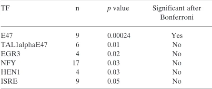
Finally, the Bonferroni-correction was subsequently applied to compensate for the effect of multiple testing (Table I).
Results
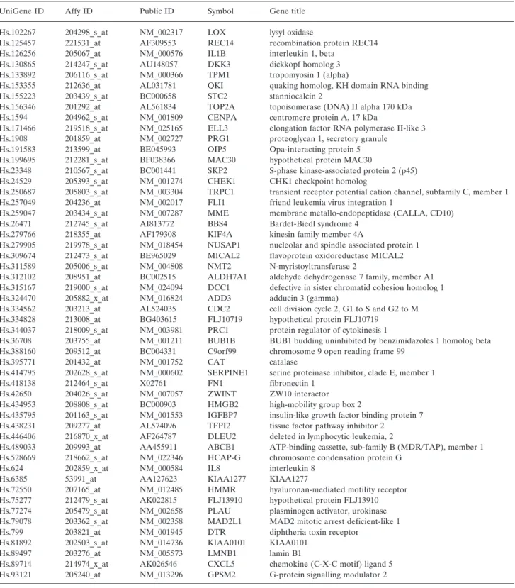
For the comparative promoter analysis the three different gene lists were combined. Out of the total of 312 individual genes, 52 were repeatedly associated with doxorubicin resistance (see Table II). To increase the accuracy of our model, other genes were dismissed and our attention focused solely on the repeated genes. Then, out of the 52 repeated genes, promoter sequences for 45 genes were retrieved (see Figure 1).
Table I.Over-represented transcription factor (TF)-binding sites before and after Bonferroni-correction.
TF n pvalue Significant after
Bonferroni
E47 9 0.00024 Yes
TAL1alphaE47 6 0.01 No
EGR3 4 0.02 No
NFY 17 0.03 No
HEN1 4 0.03 No
ISRE 9 0.05 No
The comparative promoter analysis revealed the E47 transcription-factor as being over-represented in our gene set (p=0.00024, n=9, prior frequency: 8.19E-6). These TFBSs
were distributed on eight different genes, of which the SKP2 gene had two E47 TFBSs. The consensus sequence for E47 TFBS is: RCAGNTG (TRANSFAC accession no: R02139).
Table II.Genes associated with doxorubicin resistance in at least two different published gene lists.
UniGene ID Affy ID Public ID Symbol Gene title
Hs.102267 204298_s_at NM_002317 LOX lysyl oxidase
Hs.125457 221531_at AF309553 REC14 recombination protein REC14 Hs.126256 205067_at NM_000576 IL1B interleukin 1, beta
Hs.130865 214247_s_at AU148057 DKK3 dickkopf homolog 3
Hs.133892 206116_s_at NM_000366 TPM1 tropomyosin 1 (alpha)
Hs.153355 212636_at AL031781 QKI quaking homolog, KH domain RNA binding
Hs.155223 203439_s_at BC000658 STC2 stanniocalcin 2
Hs.156346 201292_at AL561834 TOP2A topoisomerase (DNA) II alpha 170 kDa Hs.1594 204962_s_at NM_001809 CENPA centromere protein A, 17 kDa
Hs.171466 219518_s_at NM_025165 ELL3 elongation factor RNA polymerase II-like 3 Hs.1908 201859_at NM_002727 PRG1 proteoglycan 1, secretory granule
Hs.191583 213599_at BE045993 OIP5 Opa-interacting protein 5 Hs.199695 212281_s_at BF038366 MAC30 hypothetical protein MAC30
Hs.23348 210567_s_at BC001441 SKP2 S-phase kinase-associated protein 2 (p45) Hs.24529 205393_s_at NM_001274 CHEK1 CHK1 checkpoint homolog
Hs.250687 205803_s_at NM_003304 TRPC1 transient receptor potential cation channel, subfamily C, member 1 Hs.257049 204236_at NM_002017 FLI1 friend leukemia virus integration 1
Hs.259047 203434_s_at NM_007287 MME membrane metallo-endopeptidase (CALLA, CD10) Hs.26471 212745_s_at AI813772 BBS4 Bardet-Biedl syndrome 4
Hs.279766 218355_at AF179308 KIF4A kinesin family member 4A
Hs.279905 219978_s_at NM_018454 NUSAP1 nucleolar and spindle associated protein 1 Hs.309674 212473_s_at BE965029 MICAL2 flavoprotein oxidoreductase MICAL2 Hs.311589 205006_s_at NM_004808 NMT2 N-myristoyltransferase 2
Hs.312102 208951_at BC002515 ALDH7A1 aldehyde dehydrogenase 7 family, member A1 Hs.315167 219000_s_at NM_024094 DCC1 defective in sister chromatid cohesion homolog 1 Hs.324470 205882_x_at NM_016824 ADD3 adducin 3 (gamma)
Hs.334562 203213_at AL524035 CDC2 cell division cycle 2, G1 to S and G2 to M Hs.334828 213008_at BG403615 FLJ10719 hypothetical protein FLJ10719
Hs.344037 218009_s_at NM_003981 PRC1 protein regulator of cytokinesis 1
Hs.36708 203755_at NM_001211 BUB1B BUB1 budding uninhibited by benzimidazoles 1 homolog beta Hs.388160 209512_at BC004331 C9orf99 chromosome 9 open reading frame 99
Hs.395771 201432_at NM_001752 CAT catalase
Hs.414795 202628_s_at NM_000602 SERPINE1 serine proteinase inhibitor, clade E, member 1
Hs.418138 212464_s_at X02761 FN1 fibronectin 1
Hs.42650 204026_s_at NM_007057 ZWINT ZW10 interactor
Hs.434953 208808_s_at BC000903 HMGB2 high-mobility group box 2
Hs.435795 201163_s_at NM_001553 IGFBP7 insulin-like growth factor binding protein 7 Hs.438231 209277_at AL574096 TFPI2 tissue factor pathway inhibitor 2
Hs.446406 216870_x_at AF264787 DLEU2 deleted in lymphocytic leukemia, 2
Hs.489033 209993_at AA455911 ABCB1 ATP-binding cassette, sub-family B (MDR/TAP), member 1 Hs.528669 218662_s_at NM_022346 HCAP-G chromosome condensation protein G
Hs.624 202859_x_at NM_000584 IL8 interleukin 8
Hs.6385 53991_at AA127623 KIAA1277 KIAA1277
Hs.72550 207165_at NM_012485 HMMR hyaluronan-mediated motility receptor Hs.75277 212479_s_at AK022815 FLJ13910 hypothetical protein FLJ13910 Hs.77274 205479_s_at NM_002658 PLAU plasminogen activator, urokinase Hs.79078 203362_s_at NM_002358 MAD2L1 MAD2 mitotic arrest deficient-like 1
Hs.799 203821_at NM_001945 DTR diphtheria toxin receptor
Hs.81892 202503_s_at NM_014736 KIAA0101 KIAA0101
Hs.89497 203276_at NM_005573 LMNB1 lamin B1
Hs.89714 214974_x_at AK026546 CXCL5 chemokine (C-X-C motif) ligand 5 Hs.93121 205240_at NM_013296 GPSM2 G-protein signalling modulator 2
Discussion
In eukaryotes, protein synthesis is regulated at many levels during the transcription from DNA to the mature protein.
The first and most important step is the transcriptional initiation, since none of the other steps is possible if no primary RNA transcript is formed. Given a set of genes that share a similar expression profile, a logical step in the analysis is to search for the mechanism that explains their co- expression. The basic assumption is that genes that show a similar expression profile in an experiment might be regulated by the same transcriptional regulators. The activities of many TFs are context-dependent and can be modulated by other modulators nearby. Thus, a single TF can induce the transcription of specific genes while repressing others.
An important step in the identification of relevant TFs in this process is the ability to identify regulatory elements, notably the binding sites in DNA for TFs. First, a co- regulated set of genes is identified, then for each gene in the selected set the promoter or upstream region is identified.
Finally, statistically over-represented motifs are retrieved from this set of sequences as potential TFBSs. This approach targets the difficult problem of what to do when little information is available regarding the regulatory system and sequence recognition by the protein.
In our study, the over-representation of the binding site of the E47 TF was identified in the promoter regions of genes associated with doxorubicin resistance. The E47 (accession number: T00207, also called TCF3, BCF-1;
E2A.E47; E2A-E47; IEBP1 (rat); and E2A) belongs to the basic helix-loop-helix factors (HLH) class of TFs, to the
family of ubiquitous (class A) factors. The E47 contains two trans-activation domains (24), a leucine repeat, a basic region and the helix-loop-helix domain. The basic region contacts the DNA, and the HLH domain is the dimerization interface (25). The dimerization is a prerequisite for DNA- binding. Upon dimerization, the alpha-helical content may significantly increase supporting the four-helix bundle dimerization interface. Upon DNA-binding of the dimer, the basic regions adopt alpha-helical conformation as well.
Id helix-loop-helix proteins function as regulators of cell growth and differentiation and, when overexpressed, can induce malignant transformation. Enforced, ectopic expression of the E47 basic HLH (bHLH) protein in human adenocarcinoma cell lines efficiently sequestered endogenous Id proteins as Id-bHLH heterodimers, leading to growth arrest of the cells. E47 plays an active role in tumor cell growth by promoting angiogenesis (26). As the E47 proteins establish a direct transcriptional link between a cell cycle inhibitor, p21(Cip1) and a neutrophic receptor, TrkB, these proteins would play an important role in co- ordinating key events of cell cycle arrest (27). Enforced overexpression of a mutant E47 protein, deficient in transactivation and DNA-binding function, also partially inhibited cell growth. Deregulated expression of Id proteins contributed to the uncontrolled proliferation of tumor cells in colorectal cancer (28). Interestingly, these tumor suppressor properties (29) are more specific for tumor proliferation, not for resistance.
The E47 also interacts with other transcription factors, such as the E12 (Accession no: T00204), Id2 (T01212), Myf-4 (T00520), Myf-5 (T00521), Myf-6 (T00522), MyoD (T00525), Tal-1 (T00790), Tal-1beta (T01448) and Tal-2 (T01630). The E47 is part of the MEF-1 complex; it induces hyper- phosphorylation of MyoD upon association. Additionally, E47 induces transcription of IgH, but also of the IgH-stimulating Oct-2 gene and the recombination-activating genes RAG-1 and -2, thus stimulating IgH rearrangement as well (30). Although a promoter polymorphism in the MHC gene influences the binding of the E47 TF (31), no similar phenomenon has been described in association with drug resistance.
Other transcription factors, e.g., NF-κB and E2F, have already been associated with doxorubicin resistance. The use of agents that block NF-κB function has been highly beneficial in the treatment of tumors in combination with standard anticancer therapies (32). Treatment with BAY 11-7082, an irreversible inhibitor of NF-κB-phosphorylation, induced a higher percentage of apoptosis in vincristine- and doxorubicin-resistant cell lines. The suppression of constitutive NF-κB activity by BAY 11-7082 may be a useful treatment for MDR leukemias (33). The targets of doxorubicin, the topoisomerase II proteins, are direct targets of the E2F TF-mediated transcription and E2F has been proposed to be involved in drug resistance (34).
Figure 1. Overview of the data input and the performed statistical analysis.
However, no over-representation for these TFBSs was detected in our study. The question arises of whether a change in the level of transcription of a specific gene is caused by the TF acting directly at the promoter of the gene or through regulation of other transcription factors working at the promoter. It is apparent that these kinds of models are highly complex and difficult to set up. Therefore, the results of this in silico analysis will need to be coupled with biological experiments.
Gene regulatory sequences hold the key to understanding how genes are regulated by programmed and environmental signals. Technologies that permit global transcription profiling force the researcher to take a holistic view and consider biological pathways and processes that would otherwise be ignored. The hypothesis that we have proposed should not be considered to be a model that can explain all possible mechanisms of doxorubicin resistance, but must be considered as just one of several influences.
However, our computational approach does not provide information about the functional impact of the identified TFs on gene activation. For the investigation of the TF-DNA complex interaction, experiments should be carried out using functional assays, such as the electrophoretic mobility shift assay (EMSA) or chromatin immunoprecipitation (ChIP).
EMSA is based on the observation that protein:DNA complexes migrate more slowly than free DNA molecules when subjected to non-denaturing polyacrylamide or agarose gel electrophoresis; hence sequence-specific interactions may be investigated. The principle of the ChIP assay is that DNA- bound proteins (including TFs) in living cells can be cross- linked to chromatin by a gentle formaldehyde fixation. Once the proteins are immobilized on the chromatin, the whole protein-DNA complex can be immunoprecipitated using an antibody against the protein in question. The isolated protein/DNA fraction can then be purified for DNA. The identity of the DNA fragments isolated in connection with the protein can then be determined by PCR. Once we have experimental data confirming the binding of the E47 TF, further functional studies can be performed. These include RNA interference and stable transfection to modulate the gene expression and sequencing to identify mutations and SNPs in the E47 gene and in the E47 TFBS.
Conclusion
In summary, based on the results of our computer-simulated analysis of the promoter regions of genes with altered gene expression, we hypothesize that the E47 transcription factor may contribute to doxorubicin resistance in cancer. These results shed a new light on E47, as to date it was only considered to be involved in cell proliferation. Thus, E47 might present a target for effective intervention against doxorubicin resistance.
Acknowledgements
B.G. was supported by the National Office for Research and Technology, Hungary. This study was supported by the Berliner Krebsgesellschaft and by grants OTKA T046086 and NKFP 1A/002/2004 grant.
References
1 Miyashita T and Reed JC: Bcl-2 gene transfer increases relative resistance of S49.1 and WEHI7.2 lymphoid cells to cell death and DNA fragmentation induced by glucocorticoids and multiple chemotherapeutic drugs. Cancer Res 52: 5407-5411, 1992.
2 Fisher TC, Milner AE, Gregory CD, Jackman AL, Aherne GW, Hartley JA, Dive C and Hickman JA: Bcl-2 modulation of apoptosis induced by anticancer drugs: resistance to thymidylate stress is independent of classical resistance pathways. Cancer Res 53: 3321-3326, 1993.
3 Gerwitz DA: A critical evaluation of the mechanisms of action proposed for the antitumor effects of the anthracycline antibiotics adriamycin and doxorubicin. Biochemical Pharmacology 57: 727-741, 1999.
4 Scherf U, Ross DT, Waltham M, Smith LH, Lee JK, Tanabe L, Kohn KW, Reinhold WC, Myers TG, Andrews DT, Scudiero DA, Eisen MB, Sausville EA, Pommier Y, Botstein D, Brown PO and Weinstein JN: A gene expression database for the molecular pharmacology of cancer. Nat Genet 24: 236-244, 2000.
5 Szakacs G, Annereau JP, Lababidi S, Shankavaram U, Arciello A, Bussey KJ, Reinhold W, Guo Y, Kruh GD, Reimers M, Weinstein JN and Gottesman MM: Predicting drug sensitivity and resistance: profiling ABC transporter genes in cancer cells.
Cancer Cell 6: 129-137, 2004.
6 Hofmann WK, de Vos S, Elashoff D, Gschaidmeier H, Hoelzer D, Koeffler HP and Ottmann OG: Relation between resistance of Philadelphia-chromosome-positive acute lymphoblastic leukaemia to the tyrosine kinase inhibitor STI571 and gene- expression profiles: a gene-expression study. Lancet 359: 481- 486, 2002.
7 Holleman A, Cheok MH, den Boer ML, Yang W, Veerman AJ, Kazemier KM, Pei D, Cheng C, Pui CH, Relling MV, Janka- Schaub GE, Pieters R and Evans WE: Gene-expression patterns in drug-resistant acute lymphoblastic leukemia cells and response to treatment. N Engl J Med 351: 533-542, 2004.
8 van't Veer LJ, Dai H, van de Vijver MJ, He YD, Hart AA, Bernards R and Friend SH: Expression profiling predicts outcome in breast cancer. Breast Cancer Res 5: 57-58, 2003.
9 Chang JC, Wooten EC, Tsimelzon A, Hilsenbeck SG, Gutierrez MC, Elledge R, Mohsin S, Osborne CK, Chamness GC, Allred DC and O'Connell P: Gene expression profiling for the prediction of therapeutic response to docetaxel in patients with breast cancer. Lancet 362: 362-369, 2003.
10 Tupler R, Perini G and Green MR: Expressing the human genome. Nature 409: 832-833, 2001.
11 Orphanides G and Reinberg D: A unified theory of gene expression. Cell 108: 439-451, 2002.
12 McKenna NJ and O'Malley BW: Combinatorial control of gene expression by nuclear receptors and coregulators. Cell 108: 465- 474, 2002.
13 Ogata K, Sato K and Tahirov TH: Eukaryotic transcriptional regulatory complexes: cooperativity from near and afar. Curr Opin Struct Biol 13: 40-48, 2003.
14 Kudoh K, Ramanna M, Ravatn R, Elkahloun AG, Bittner ML, Meltzer PS, Trent JM, Dalton WS and Chin KV: Monitoring the expression profiles of doxorubicin-induced and doxorubicin- resistant cancer cells by cDNA microarray. Cancer Res 60:
4161-4166, 2000.
15 Troester MA, Hoadley KA, Sorlie T, Herbert BS, Borresen- Dale AL, Lonning PE, Shay JW, Kaufmann WK and Perou CM: Cell-type-specific responses to chemotherapeutics in breast cancer. Cancer Res 64: 4218-4226, 2004.
16 Györffy B, Serra V, Abdul-Ghani R, Jürchott K, Garber M, Stein U, Petersen I, Lage H, Dietel M and Schäfer R:
Prediction of doxorubicin sensitivity in breast tumors based on gene expression profiles of drug resistant cell lines correlates with patient survival, Oncogene 24: 7542-7551, 2005.
17 Kang HC, Kim IJ, Park JH, Shin Y, Ku JL, Jung MS, Yoo BC, Kim HK and Park JG: Identification of genes with differential expression in acquired drug-resistant gastric cancer cells using high-density oligonucleotide microarrays. Clin Cancer Res 10:
272-284, 2004.
18 Györffy B, Surowiak P, Kiesslich O, Denkert C, Schäfer R, Dietel M and Lage H: Gene expression profiling of 30 cancer cell lines predicts resistance towards 11 anticancer drugs at clinically achieved concentrations. Int J Cancer 118: 1699-1712, 2006.
19 Zhang H, Ramanathan Y, Soteropoulos P, Recce ML and Tolias PP: EZ-Retrieve: a web-server for batch retrieval of coordinate-specified human DNA sequences and underscoring putative transcription factor-binding sites. Nucleic Acids Res 30: e121, 2002.
20 Wheeler DL, Church DM, Federhen S, Lash AE, Madden TL, Pontius JU, Schuler GD, Schriml LM, Sequeira E, Tatusova TA and Wagner L: Database resources of the National Center for Biotechnology. Nucleic Acids Res 31: 28-33, 2003.
21 Heinemeyer T, Wingender E, Reuter I, Hermjakob H, Kel AE, Kel OV, Ignatieva EV, Ananko EA, Podkolodnaya OA, Kolpakov FA, Podkolodny NL and Kolchanov NA: Databases on transcriptional regulation: TRANSFAC, TRRD and COMPEL. Nucleic Acids Res 26: 362-367, 1998.
22 Aerts S, Thijs G, Coessens B, Staes M, Moreau Y and De Moor B: Toucan: deciphering the cis-regulatory logic of coregulated genes. Nucleic Acids Res 31: 1753-1764, 2003.
23 Wingender E, Chen X, Fricke E, Geffers R, Hehl R, Liebich I, Krull M, Matys V, Michael H, Ohnhauser R, Pruss M, Schacherer F, Thiele S and Urbach S: The TRANSFAC system on gene expression regulation. Nucleic Acids Res 29: 281-283, 2001.
24 Quong MW, Massari ME, Zwart R and Murre C: A new transcriptional-activation motif restricted to a class of helix- loop-helix proteins is functionally conserved in both yeast and mammalian cells. Mol Cell Biol 13: 792-800, 1993.
25 Voronova A and Baltimore D: Mutations that disrupt DNA binding and dimer formation in the E47 helix-loop-helix protein map to distinct domains. Proc Natl Acad Sci USA 87: 4722- 4726, 1990.
26 Peinado H, Marin F, Cubillo E, Stark HJ, Fusenig N, Nieto MA and Cano A: Snail and E47 repressors of E-cadherin induce distinct invasive and angiogenic properties in vivo. J Cell Sci 117: 2827-2839, 2004.
27 Liu Y, Encinas M, Comella JX, Aldea M and Gallego C: Basic helix-loop-helix proteins bind to TrkB and p21(Cip1) promoters linking differentiation and cell cycle arrest in neuroblastoma cells. Mol Cell Biol 24: 2662-2672, 2004.
28 Wilson JW, Deed RW, Inoue T, Balzi M, Becciolini A, Faraoni P, Potten CS and Norton JD: Expression of Id helix-loop-helix proteins in colorectal adenocarcinoma correlates with p53 expression and mitotic index. Cancer Res 61: 8803-8810, 2001.
29 Quong MW, Romanow WJ and Murre C: E protein function in lymphocyte development. Annu Rev Immunol 20: 301-322, 2002.
30 Schlissel M, Voronova A and Baltimore D: Helix-loop-helix transcription factor E47 activates germ-line immunoglobulin heavy-chain gene transcription and rearrangement in a pre-T- cell line. Genes Dev 5: 1367-1376, 1991.
31 Boodhoo A, Wong AM, Williamson D, Voon D, Lee S, Allcock RJ and Price P: A promoter polymorphism in the central MHC gene, IKBL, influences the binding of transcription factors USF1 and E47 on disease-associated haplotypes. Gene Expr 12:
1-11, 2004.
32 Wang CY, Cusack JC Jr, Liu R and Baldwin AS Jr: Control of inducible chemoresistance: enhanced anti-tumor therapy through increased apoptosis by inhibition of NF-kappaB. Nat Med5: 412-417, 1999.
33 Jiao W, Lin HM, Timmons J, Nagaich AK, Ng SW, Misteli T and Rane SG: E2F-dependent repression of topoisomerase II regulates heterochromatin formation and apoptosis in cells with melanoma-prone mutation. Cancer Res 65: 4067-4077, 2005.
34 Garcia MG, Alaniz L, Lopes EC, Blanco G, Hajos SE and Alvarez E: Inhibition of NF-kappaB activity by BAY 11-7082 increases apoptosis in multidrug resistant leukemic T-cell lines.
Leuk Res 29: 1425-1434, 2005.