ORIGINAL ARTICLE
Nicotinamide mononucleotide (NMN) supplementation promotes neurovascular rejuvenation in aged mice:
transcriptional footprint of SIRT1 activation, mitochondrial protection, anti-inflammatory, and anti-apoptotic effects
Tamas Kiss&Ádám Nyúl-Tóth&Priya Balasubramanian&Stefano Tarantini &
Chetan Ahire&Andriy Yabluchanskiy&Tamas Csipo&Eszter Farkas&
Jonathan D. Wren&Lori Garman&Anna Csiszar&Zoltan Ungvari
Received: 8 January 2020 / Accepted: 29 January 2020
#American Aging Association 2020
Abstract Aging-induced structural and functional al- terations of the neurovascular unit lead to impairment of neurovascular coupling responses, dysregulation of cerebral blood flow, and increased neuroinflammation, all of which contribute importantly to the pathogenesis of age-related vascular cognitive impairment (VCI).
There is increasing evidence showing that a decrease in NAD+ availability with age plays a critical role in age-related neurovascular and cerebromicrovascular dysfunction. Our recent studies demonstrate that restor- ing cellular NAD+ levels in aged mice rescues neurovascular function, increases cerebral blood flow, https://doi.org/10.1007/s11357-020-00165-5
Tamas Kiss, Ádám Nyúl-Tóth, Priya Balasubramanian and Stefano Tarantini contributed equally to this work.
T. Kiss
:
Á. Nyúl-Tóth:
P. Balasubramanian:
S. Tarantini:
C. Ahire
:
A. Yabluchanskiy:
T. Csipo:
J. D. Wren:
Z. Ungvari
Vascular Cognitive Impairment and Neurodegeneration Program, Reynolds Oklahoma Center on Aging/Center for Geroscience and Healthy Brain Aging, Department of Biochemistry and Molecular Biology, University of Oklahoma Health Sciences Center, 975 NE 10th Street, BRC 1311, Oklahoma City, OK 73104, USA T. Kiss
:
E. Farkas:
A. Csiszar:
Z. UngvariInternational Training Program in Geroscience, Theoretical Medicine Doctoral School/Department of Medical Physics and Informatics, University of Szeged, Szeged, Hungary
Á. Nyúl-Tóth
Institute of Biophysics, Biological Research Centre, Szeged, Hungary
S. Tarantini
:
T. Csipo:
A. Csiszar:
Z. UngvariInternational Training Program in Geroscience, Doctoral School of Basic and Translational Medicine/Department of Public Health, Semmelweis University, Budapest, Hungary
T. Csipo
International Training Program in Geroscience, Department of Cardiology, Division of Clinical Physiology, Faculty of Medicine, University of Debrecen, Debrecen, Hungary
J. D. Wren
:
L. GarmanGenes & Human Disease Research Program, Oklahoma Medical Research Foundation, Oklahoma City, OK, USA
A. Csiszar
:
Z. UngvariThe Peggy and Charles Stephenson Cancer Center, University of Oklahoma Health Sciences Center, Oklahoma City, OK 73104, USA
Z. Ungvari (*)
Department of Health Promotion Sciences, College of Public Health, University of Oklahoma Health Sciences Center, Oklahoma City, OK, USA
e-mail: zoltan-ungvari@ouhsc.edu / Published online: 13February2020
and improves performance on cognitive tasks. To deter- mine the effects of restoring cellular NAD+ levels on neurovascular gene expression profiles, 24-month-old C57BL/6 mice were treated with nicotinamide mononu- cleotide (NMN), a key NAD+intermediate, for 2 weeks.
Transcriptome analysis of preparations enriched for cells of the neurovascular unit was performed by RNA-seq. Neurovascular gene expression signatures in NMN-treated aged mice were compared with those in untreated young and aged control mice. We identified 590 genes differentially expressed in the aged neurovascular unit, 204 of which are restored toward youthful expression levels by NMN treatment. The tran- scriptional footprint of NMN treatment indicates that increased NAD+ levels promote SIRT1 activation in the neurovascular unit, as demonstrated by analysis of upstream regulators of differentially expressed genes as well as analysis of the expression of known SIRT1- dependent genes. Pathway analysis predicts that neurovascular protective effects of NMN are mediated by the induction of genes involved in mitochondrial rejuvenation, anti-inflammatory, and anti-apoptotic pathways. In conclusion, the recently demonstrated pro- tective effects of NMN treatment on neurovascular func- tion can be attributed to multifaceted sirtuin-mediated anti-aging changes in the neurovascular transcriptome.
Our present findings taken together with the results of recent studies using mitochondria-targeted interventions suggest that mitochondrial rejuvenation is a critical mechanism to restore neurovascular health and improve cerebral blood flow in aging.
Keywords Aging . Geroscience . Vascular cognitive impairment . Mitochondria dysfunction . Transcriptomics
Introduction
In recent years, there has been increasing appreciation that the health of the neurovascular unit (NVU) is crit- ical for brain health (Kisler et al.2017; Zlokovic2010, 2011; Iadecola2017; Stanimirovic and Friedman2012).
The extended NVU consists of cerebral microvessels that receive input from neurons via astrocytic endfeet, pericytes, and perivascular microglia (Iadecola 2017;
Stanimirovic and Friedman2012). The NVU is respon- sible for the tight coupling between neural activity and regional cerebral blood flow (“neurovascular cou- pling”), which ensures adequate oxygen and nutrient
delivery to the brain (Tarantini et al.2017a; Toth et al.
2017). Endothelial dysfunction and/or impaired astro- cytic function results in neurovascular uncoupling con- tributing to cognitive impairment (Toth et al. 2017;
Tarantini et al. 2015, 2018, 2017b). In addition, cells constituting the NVU form and maintain the blood-brain barrier (Zlokovic 2010, 2011; Montagne et al. 2017;
Sweeney et al.2018,2019a; Zlokovic2008), regulate transport processes and waste removal, deposit the ex- tracellular matrix, control the structural remodeling of the cerebral microcirculation (including angiogenesis, vessel regression, adaptation to hypertension (Csiszar et al.2017; Tarantini et al. 2016,2017c; Tucsek et al.
2014; Warrington et al. 2013; Ungvari et al. 2013, 2018a,2017; Toth et al.2015)), form and operate the glymphatic system (Iliff et al.2013; Jessen et al.2015;
Kress et al. 2014), maintain stem-cell niches (Solano Fonseca et al. 2016), synthesize the glycocalyx, and control the adhesion and extravasation of inflammatory circulating cells that participate in central nervous sys- tem immune surveillance (Stanimirovic and Friedman 2012). With age, the phenotype and function of the cells constituting the NVU are altered, which fundamentally affects all of the aforementioned physiological process- es (Kisler et al.2017; Zlokovic2010,2011; Tarantini et al.2017a). Age-related neurovascular dysfunction is now considered as a critical contributing factor to the pathogenesis of both vascular cognitive impairment (VCI) and neurodegenerative diseases, including Alzheimer’s disease (Sweeney et al.2019b). In order to develop novel methods for prevention and treatment of these diseases and to preserve cognitive function in older adults, it is important to identify therapeutic inter- ventions that can reverse age-related impairment of the NVU. Understanding the role of fundamental cellular and molecular mechanisms of aging in age-related neurovascular impairment is critical in that regard.
Nicotinamide adenine dinucleotide (NAD+) is a co- enzyme central to hundreds of redox reactions in eu- karyotic cells. NAD+ also has a critical role in the regulation of the activity of NAD+-consuming enzymes, including SIRT1 and other sirtuins (Gomes et al.2013;
Michan et al. 2010; Mitchell et al. 2014; Yang et al.
2007). Sirtuin enzymes are implicated in regulation of cellular processes of aging, mitochondrial function, stress resilience, apoptosis, and inflammation (Das et al. 2018; Csiszar et al. 2009a, b, 2008a). Aging is associated with cellular NAD+depletion (Gomes et al.
2013; Massudi et al.2012) (Yoshino, 2018 #10180), GeroScience (2020) 42:527–546 528
which has been proposed to be a critical driving force of aging processes (Imai and Guarente 2014), impairing nuclear and mitochondrial functions and contributing to the genesis of many age-associated pathologies. Ac- cordingly, restoration of cellular NAD+ biosynthesis extends lifespan in model organisms (Anderson et al.
2002) and improves health span and extends lifespan in murine models of aging (Zhang, 2016 #10167) (Mitchell et al.2018). There is emerging evidence that vascular aging is also characterized by cellular NAD+ depletion (Tarantini et al. 2019a; Csiszar et al. 2019;
Kiss et al. 2019a). Importantly, our recent studies showed (Tarantini et al.2019a) that in murine models of aging restoration of cellular NAD+levels by chronic treatment with the NAD+ precursor, nicotinamide mononucleotide (NMN) (Yoshino et al.2018) confers potent anti-aging neurovascular effects, rescuing cerebromicrovascular endothelial dysfunction and neurovascular coupling responses, increasing cerebral blood flow, and improving cognitive performance. In cultured cerebromicrovascular endothelial cells derived from aged rats, 5 days of treatment with NMN restored NAD+ levels and rescued mitochondrial function and attenuated mitochondrial oxidative stress in a sirtuin- dependent manner (Tarantini et al.2019a).
The present study was designed to test the hypothesis that age-related NAD+depletion in the NVU is causally linked to dysregulated expression of genes important for normal neurovascular function. A corollary hypothesis is that functional neurovascular rejuvenation in NMN- treated aged mice is associated with SIRT1-mediated restoration of a youthful neurovascular mRNA expres- sion profile. To test these hypotheses, aged mice were treated with NMN for 2 weeks and transcriptomic sig- natures in cells of the neurovascular unit were compared with those in cells obtained from untreated young and aged control mice. The transcriptomic footprint of SIRT1 activation was analyzed, and the predicted mul- tifaceted protective effects of NMN supplementation on diverse aspects of cerebromicrovascular and neurovascular biology were tested.
Methods
Animals, NMN supplementation
Young (3 months old) and aged (24 months old) male C57BL/6 mice were purchased from the aging colony
maintained by the National Institute on Aging at Charles River Laboratories (Wilmington, MA). The biological age of 24-month-old mice corresponds to that of ~ 60- year-old humans. Mice were housed under specific pathogen-free barrier conditions in the Rodent Barrier Facility at University of Oklahoma Health Sciences Center under a controlled photoperiod (12 h light; 12 h dark) with unlimited access to water and were fed a standard AIN-93G diet (ad libitum). Mice in the aged cohort were assigned to two groups. One group of the aged mice was injected daily with NMN (IP injections of 500 mg NMN/kg body weight per day) or the equiv- alent volume of PBS for 14 consecutive days at 6 PM and 8 AM on day 14 and were sacrificed 4 h after last injection. Similar dosages of NMN has been shown to exert potent anti-aging effects on mouse health span (Csiszar et al.2019; de Picciotto et al.2016), including rescue of neurovascular coupling responses, attenuation of vascular oxidative stress, and rescue of gene expres- sion changes in the aorta (Tarantini et al. 2019a; Kiss et al. 2019b). All procedures were approved by the Institutional Animal Use and Care Committees of the University of Oklahoma Health Sciences Center. All animal experiments complied with the ARRIVE guide- lines and were carried out in accordance with the Na- tional Institutes of Health guide for the care and use of Laboratory animals (NIH Publications No. 8023, re- vised 1978). The effects of NMN treatment on cognitive function, neurovascular coupling responses, and micro- vascular and aorta endothelial function in a similar cohort of mice have been recently reported (Tarantini et al.2019a).
Isolation of cells of the neurovascular unit
Animals were killed and transcardially perfused with PBS as previously described (Tarantini et al. 2019a;
Kiss et al. 2019b). The brains were quickly removed and rinsed in ice-cold PBS, and minced into ≈1 mm2 pieces. The tissue was washed twice in ice-cold 1× PBS by low-speed centrifugation (50g, 3 min). The diced tissue was digested in a buffer solution containing col- lagenase (800 U/g tissue), hyaluronidase (2.5 U/g tis- sue), and elastase (3 U/g tissue) in 1 mL PBS/100 mg tissue for 45 min at 37 °C in a rotating humid incubator.
The digested tissue was passed through a 100-μm cell strainer. The single-cell lysate was centrifuged for 2 min at 70g. After removing the supernatant, the pellet was washed twice in cold PBS supplemented with 2.5% fetal
calf serum (FCS), and the suspension was centrifuged at 300gfor 5 min at 4 °C. To create fraction enriched for cells of the neurovascular unit, the cell suspension was centrifuged using an OptiPrep gradient solution (Axi- Shield, PoC, Norway). Briefly, the cell pellet was resus- pended in Hanks’ balanced salt solution (HBSS) and mixed with 40% iodixanol thoroughly (final concentra- tion 17% (v/v) iodixanol solution; ρ= 1.096 g/mL).
Two milliliters of HBSS was layered on top and centri- fuged at 400gfor 15 min at 20 °C. Endothelial cells with attached astrocytes and pericytes, which banded at the interface between HBSS and the 17% iodixanol layer, were collected. The neurovascular-enriched fraction was incubated for 30 min at 4 °C in the dark with anti- CD31/PE (BD Biosciences, San Jose, CA, USA) and anti-MCAM/FITC (BD Biosciences, San Jose, CA, USA). After washing the cells twice with MACS buffer (Milltenyi Biotech, Cambridge, MA, USA), anti-FITC and anti-PE magnetic bead–labeled secondary antibod- ies were used for 15 min at room temperature. The endothelial/neurovascular enriched fraction was collect- ed by magnetic separation using the MACS LD mag- netic separation columns according to the manufac- turer’s guidelines (Milltenyi Biotech, Cambridge, MA, USA). Our pilot studies indicated that this method using gentle cell dissociation protocols results in enrichment for cerebromicorvascular endothelial cells with astro- cytes and pericytes.
RNA isolation, cDNA synthesis, library construction, and next generation sequencing
RNA was isolated from the samples using AllPrep DNA/RNA Mini Kit (Qiagen) as previously described (Imperio et al.2016; Valcarcel-Ares et al.2018). RNA quantity and quality (> 8 RNA integrity number) were measured using the RNA 6000 Nano Assay with an Agilent 2100 Bioanalyzer (Agilent, Palo Alto, CA).
Using 1μg RNA, cDNA was synthesized from purified RNA using ABI High-capacity cDNA Reverse Tran- scription Kit (Applied Biosystems, Foster City, CA) (Imperio et al.2016; Valcarcel-Ares et al.2018). Library construction was performed in a stranded manner to retain the directionality of the transcripts for as de- scribed (Valcarcel-Ares et al. 2018). In brief, prior to RNA-seq analysis, quality control measures were im- plemented. Concentration of RNA was ascertained via fluorometric analysis on a Thermo Fisher Qubit Fluo- rometer. Overall quality of RNA was verified using an
Agilent Tapestation instrument. Following initial QC steps, sequencing libraries were generated using the Illumina Truseq Stranded mRNA with library prep kit according to the manufacturers protocol. Briefly, mature mRNA was enriched for via pull down with beads coated with oligo-dT homopolymers. The mRNA mol- ecules were then chemically fragmented, and the first strand of cDNA was generated using random primers.
Following RNase digestion, the second strand of cDNA was generated replacing dTTP in the reaction mix with dUTP. Double stranded cDNA then underwent adenylation of 3′ ends following ligation of Illumina- specific adapter sequences. Subsequent PCR enrich- ment of ligated products was further selected for those strands not incorporating dUTP, leading to strand- specific sequencing libraries. Final libraries for each sample were assayed on the Agilent Tapestation for appropriate size and quantity. These libraries were then pooled in equimolar amounts as ascertained via fluo- rometric analyses. Final pools were absolutely quanti- fied using qPCR on a Roche LightCycler 480 instru- ment with Kapa Biosystems Illumina Library Quantifi- cation reagents. Sequencing was performed on an Illumina NovaSeq 6000 instrument with paired-end 50 bp reads.
RNA-seq data analysis and visualization
Raw sequencing reads were trimmed of their Illumina TruSeq adapter sequences using Trimmomatic v0.35 (Bolger et al.2014), then aligned to the mouse genome version GRCm38 using Kallisto v0.43.03(Bray et al.
2016). Samples were checked for outliers and separation by principle components analysis (PCA) with the R functionprcomp. Raw expression counts were summa- rized at the gene level to transcript-length adjusted, library-size scaled counts per million (CPM) with the R packagetximport(Soneson et al.2015). Differential expression analysis was performed using the empirical Bayes approach implemented in the R/Bioconductor packageDESeq2(Love et al.2014). Significantly dif- ferentially expressed (DE) genes had an absolute log2 fold-change≥0.585 (corresponding to a change of 50%
or more in expression) and the false discovery rate (FDR)-adjusted p value ≤0.05. Gene annotation was done using biomaRt (Durinck et al. 2009) in R/
Bioconductor package. Hierarchical clustering was per- formed via the R package ComplexHeatmap.
GeroScience (2020) 42:527–546 530
Functional annotation
Theorg.Mm.eg.db v3.8.2R/Bioconductor package was used to collect Gene Ontology terms associated with our differentially expressed genes. The hypergeometric test implemented inGOstats v2.51.0R/Bioconductor pack- age was used to calculate enrichment of the individual terms (Falcon and Gentleman2007).
We used upstream regulator analysis (URA) algo- rithm (Kramer et al.2014) implemented in the Ingenuity Pathway Analysis (QIAGEN) software find upstream regulators that potentially explains the observed gene expression changes in our samples. The IPA uses a manually curated database (Ingenuity Knowledge Base) to calculate “enrichment” score (Fisher’s exact test (FET)p value), measures the overlap of observed and predicted regulated gene sets, and a z-score assessing the match of observed and predicted up/downregulation patterns.
Results
NMN treatment reverses age-related changes in neurovascular mRNA expression profile in mice To isolate NVU-enriched mRNA, we employed an en- dothelial cell isolation-based strategy. Using RNA-seq to sequence the neurovascular transcriptome, we com- pared normalized mRNA expression values for each sample to that in individual cell types constituting the NVU (endothelial cells, astrocytes, and pericytes). To achieve that goal, we developed a list of cell-specific markers from published RNA-seq data of purified cor- tical cell types (GEO dataset GSE52564 (Zhang et al.
2014)). Comparison of mRNA levels in the NVU sam- ples with the input shows a significant enrichment for endothelial cell, astrocyte, and pericyte genes (Fig.1a).
We assessed transcriptomic changes in the NVU associated with aging and with NMN treatment (Fig.
1b). We performed unsupervised clustering of RNA-seq data from all samples using the topmost variably expressed genes across all samples. This showed that biological replicates from the same group cluster togeth- er, and that young samples segregate away from aged ones (Fig.1b). PCA (Fig.1c) of the transcriptomic data also showed a clear separation between the young and aged groups. Aged control mice and aged NMN-treated mice were also segregated in the PCA and hierarchical
clustering. This finding indicated a clear difference be- tween the transcriptome profiles of the two age groups.
In contrast, mRNA expression in young mice and NMN-treated aged mice were similar, and these groups did not separate well in the PCA and hierarchical clustering.
We then determined the number of genes that are significantly upregulated or downregulated (DE, fold- change ≥1.5 or < 0.67;p< 0.05 adjusted for multiple comparisons) in the NVU by aging or by NMN treat- ment. We then filtered for genes that are significantly altered (adjustedp< 0.05), expressed at an appreciable level (fragments per kilobase of transcript per million mapped reads > 1), and are expressed in cells of the NVU. We identified 590 differentially expressed genes in aged animals compared with young controls. We also identified 459 DE genes in the NMN-treated aged mice compared with the untreated aged controls. In Fig.1d, a volcano plot shows statistical significance (p value) versus magnitude of age-related change in gene expres- sion. Red symbols denote genes, whose expression levels differed in the aged phenotype, but have shifted back toward the young phenotype by NMN treatment (discordant DE genes). The Venn diagram in Fig. 1f shows that neurovascular expression of 204 genes, which are differentially expressed in aged mice, was shifted back toward youthful levels by NMN treatment of aged mice.
We realized that significance cutoffs to identify dif- ferentially expressed genes shared between the age- effect and NMN-effect datasets may be too stringent, and the analysis illustrated in Fig.1dmay miss discor- dant patterns (youthful shifts) of gene expression with important biological relevance for NMN-induced neurovascular rejuvenation. Thus, we also used an ap- proach to detect discordant transcriptional patterns (youthful shifts) by comparing the age-effect and NMN-effect gene expression datasets using combina- tion criteria that took into account the effect direction.
Genes were ranked by their effect size direction, and ranked lists were compared to identify overlapping genes across a continuous significance gradient. Our analysis required that discordant genes with youthful shifts (1) are“differentially expressed”based on both p value and fold-change criteria either in aging or the NMN treatment group, (2) satisfy a fold-change criteri- on with a cutoff of≥1.5 or < 0.67 in the group in which expression did not satisfy the statistical significance p< 0.05, and (3) satisfy the criterion that the effect
directions of the age-effect and NMN-effect are oppo- site. We found that these combination criteria found more biologically meaningful sets of genes than pvalues alone.
In Fig.1e, the magnitude of age-related changes in gene expression is plotted against the magnitude of
NMN-induced changes in gene expression. Red sym- bols denote discordant DE genes, whose expression levels shifted back toward the young phenotype by NMN treatment with statistical significance. Genes which are DE only in one group but otherwise satisfy the other criteria are denoted by blue (DE in aging) and GeroScience (2020) 42:527–546 532
green (DE in NMN-treated) symbols. Using this ap- proach, we have identified 466 discordant genes with youthful shifts, which changed in opposite directions between the two datasets (Fig.1f). These data suggest that NAD+ depletion has a critical role in age-related dysregulation of neurovascular gene expression.
Transcriptional footprint of neurovascular SIRT1 activation in NMN-treated aged mice
Previous studies suggested that restoration of NAD+ levels in aged cells by NMN treatment activates the
NAD+-dependent histone deacetylase enzyme SIRT1 (Gomes et al.2013; Tarantini et al.2019a). To provide additional evidence that SIRT1 activation contributes to the neurovascular protective effects of NMN, we exam- ined the transcriptional footprint of neurovascular SIRT1 activation in NMN-treated aged mice using three approaches. First, we analyzed age-related and NMN- induced changes in expression of SIRT1-regulated genes identified by IPA. We found that aging is associ- ated with changes in the expression of several known SIRT1-regulated genes and that majority of these tran- scriptional changes are reversed by NMN treatment (Fig.2a).
Ingenuity upstream regulator analysis
We have also performed IPA upstream regulator analy- sis (Kramer et al.2014) to identify upstream transcrip- tional regulators that may contribute to the observed transcriptomic changes in our dataset, which can help to identify the mechanism of action of NMN in the aged neurovascular unit. The upstream regulator analysis is based on information in the Ingenuity Knowledge Base (a curated relational database of the available biomedi- cal literature) on the expected effects between transcrip- tional regulators and their target genes. Using the IPA upstream regulator analysis, it was examined how many known targets of each transcriptional regulator were differentially expressed in our samples, and the direction of these gene expression changes were compared with what is expected from the literature. On the basis of the observed direction of change, a prediction of the activa- tion state of the predicted transcriptional regulators (“ac- tivated” or “inhibited”) were made (not shown). For each potential transcriptional regulator, two statistical measures, an overlappvalue and an activation z-score, were computed. The overlap p value calls likely up- stream regulators based on significant overlap between the differentially expressed genes and known targets regulated by that particular transcriptional regulator.
The activation z-score is used to infer the activation state of the predicted transcriptional regulators (“activated”or
“inhibited”) based on comparison with a model that assigns random regulation directions. The results of the IPA upstream regulator analysis are visualized in Fig.2b. We also determined the link between the pre- dicted upstream regulators activated by NMN and SIRT1 using IPA. As indicated in Fig.2b, we found that
~ 38% of the predicted upstream regulators activated by
Fig. 1 NMN treatment reverses age-related changes in neurovascular mRNA expression profile.aHeatmap displaying normalized mRNA expression values for each sample as com- pared with that in cells of the neurovascular unit (endothelial cells, astrocytes, and pericytes) from the reference datasets by Zhang et al. (2014). Note that neurovascular genes are enriched in the samples.bThe heat map is a graphic representation of normalized expression values of differentially expressed genes in neurovascular samples derived from young (3 months old), aged (24 months old), and NMN-treated aged mice. Hierarchical clus- tering analysis revealed the similarities on neurovascular mRNA expression profiles in young and NMN-treated aged mice.cPrin- cipal component analysis (PCA) plot of neurovascular mRNA expression profiles in young, aged control, and NMN-treated aged mice. The profiles from aged mice (red) cluster separately from clusters representing young mice (blue) and NMN-treated aged mice (green). PC1, PC2, and PC3 are principal components 1, 2, and 3, respectively. d Volcano plot depicting differentially expressed genes comparing neurovascular samples derived from young and aged mice. Stratifiedp values are plotted against expression fold-changes for results obtained in aged samples nor- malized to young samples. Colored points refer to genes whose expression is significantly altered by NMN treatment.eNMN- induced changes in gene expression plotted against age-related changes in the neurovascular transcriptome. Red symbols indicate discordant differentially expressed genes with youthful shifts, whose expression significantly changes with age and is restored by NMN treatment toward youthful levels. Blue and green sym- bols denote discordant genes with youthful shifts, whose expres- sion changes in aging and is restored by NMN treatment toward youthful levels, but only the aging (blue) or the NMN effect (green) reaches the cutoff for statistical significance.fVenn dia- grams sowing the numbers of differentially expressed mRNAs in each group. The blue circle represents neurovascular genes, which are significantly up or downregulated in aged mice as compared with young mice. The green circle represents neurovascular genes, which are significantly up or downregulated in aged mice by NMN treatment. The red area represents discordant differentially expressed genes. Gray areas represent discordant genes, whose expression is changed by NMN treatment toward youthful levels, but the effect does not reach the cutoff for statistical significanceNMN are known to be regulated by SIRT1-dependent pathways. In particular, the IPA upstream regulator analysis predicts that NMN-induced SIRT1 activation
upregulates PGC-1α (PPARGC1A), FOXO3- and FOXO4-mediated pathways, whereas it inhibits HIF- 1α-regulated pathways (Fig.2b). We also attempted to Fig. 2 NMN reverses age-related changes in neurovascular ex-
pression of SIRT1-regulated genes.aIPA results showing age- related (left) and NMN-induced (right) changes in the expression of SIRT1-regulated genes (classified as such by IPA). Green, downregulation; red, upregulation.bResults of the IPA upstream regulator analysis. Shown are predicted upstream transcriptional regulators that may contribute to the observed NMN-induced transcriptomic changes in our dataset. Known links between the predicted upstream regulators activated by NMN and SIRT1 ac- tivity are indicated.cLiterature-based relationships with positive mutual information among the predicted upstream regulators.
Node size correlates with the activation z-score from IPA (bigger
= higher z-score), edge width correlates with the mutual informa- tion of the genes within the literature, green marks which are predicted activators and red marks predicted repressors. dThe heat map is a graphic representation of normalized expression values of differentially expressed SIRT1-dependent genes in neurovascular samples derived from young (3 months old), aged (24 months old), and NMN-treated aged mice. Hierarchical clus- tering analysis revealed the similarities on neurovascular expres- sion profiles of SIRT1-dependent genes in young and NMN- treated aged mice. SIRT1-dependent genes were identified based on their differential expression in the brain of SIRT1−/−mice (Libert et al.2011)
GeroScience (2020) 42:527–546 534
predict NMN-activated, SIRT1-dependent regulatory networks by identifying relationships between SIRT1 and the predicted upstream regulators utilizing the IRI- DESCENT (Implicit Relationship IDEntification by in- Silico Construction of an Entity-based Network from Text) system (Wren and Garner2004). IRIDESCENT processes all available MEDLINE abstracts and uses a statistical model to determine whether each upstream regulator co-occurs with a term of interest more fre- quently than would be expected by chance, and quan- tifies this in terms of the mutual information measure.
The results of this analysis provide additional support to the view that the predicted NMN-induced SIRT1 acti- vation results in inhibition of HIF-1α29and activation of PGC-1α- and FOXO3-dependent pathways (Hubbard et al.2013). PGC-1αand FOXOs are known targets for SIRT1-mediated deacetylation (Fig.2c).
In addition, we also intersected the list of differen- tially expressed genes in our dataset with the list of genes differentially expressed in the brains of SIRT1−/−
mice (NCBI Gene Expression Omnibus: GSE28790) (Libert et al.2011). The heat map showing the expres- sion pattern of these SIRT1-sensitive genes is shown in Fig. 2d. Hierarchical clustering of the data showed a clear separation between the young and aged groups.
Aged control mice and aged NMN-treated mice were also clearly separated. In contrast, expression of SIRT1- sensitive genes in young mice and NMN-treated aged mice were similar, and these groups did not separate well in the hierarchical clustering, consistent with the idea that aging is associated with dysregulation of SIRT1-sensitive genes, which is rescued by NMN treatment.
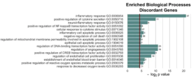
NMN-induced neurovascular transcriptomic changes in aged mice predict mitochondrial rejuvenation We performed GO enrichment analysis to explore po- tential biological functions of the NMN-regulated dis- cordant differentially expressed genes with youthful shifts. GO enrichment analysis of discordant differen- tially expressed genes with youthful shifts identified functions in mitochondrial regulation and oxidative stress, apoptosis, inflammation, endothelial activation, and transcriptional regulation (Fig.3).
Our recent studies, demonstrate that aging is associ- ated with mitochondrial dysfunction and oxidative stress in cerebromicrovascular endothelial cells, which play a critical role in dysregulation of cerebral blood
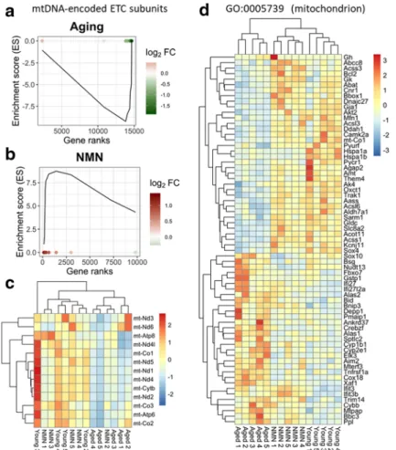
flow and impaired neurovascular coupling responses in aged mice (Tarantini et al. 2018, 2019a). To find out whether mitochondria-related gene expression is altered in the aging NVU, we analyzed expression of both nuclear-encoded and mtDNA-encoded mitochondria- related genes. We have used existing databases to com- pile a list of genes with mitochondrial targeting se- quences and known functions related to regulation of mitochondrial processes. We used Gene Set Enrichment Analysis (GSEA) for interpreting expression of mitochondria-related genes (Subramanian et al. 2005).
GSEA of mtDNA-encoded genes encoding components of the mitochondrial electron transport chain (ETC) was performed using a pre-ranked gene list based on the magnitude of the fold-change (largest upregulation to most downregulated; Fig. 4a, b). Figure4a, bdepict a running-sum statistic (enrichment score) based on Fig.
4, increasing when a gene is a member of the mtDNA- encoded ETC gene set and decreasing when it is not.
Note that in aged mice, GSEA scores increased predom- inantly on the right indicating downregulation of mtDNA-encoded ETC genes by aging. In contrast, in NMN-treated aged mice GSEA scores increased pre- dominantly on the left indicating upregulation of mtDNA-encoded ETC genes by NMN treatment in aged mice. The heat maps showing the expression pattern of nuclear-encoded and mtDNA-encoded mitochondria- related genes are shown in Fig.4c and d, respectively.
Hierarchical clustering of the data showed a clear sepa- ration between the young and aged groups. Aged con- trol mice and aged NMN-treated mice were also sepa- rated. In contrast, expression of mitochondria-related genes in young mice and NMN-treated aged mice were similar, and these groups did not separate well in the hierarchical clustering, consistent with the idea that age- related dysregulation of mitochondria-related genes in the NVU is reversed, at least in part, by NMN treatment.
Fig. 3 Most significantly enriched Gene Ontology (GO) terms for discordant genes. Note that NMN treatment is associated with transcriptional changes indicating multifaceted anti-inflammatory, anti-apoptotic, mitochondrial protective, and anti-oxidative effects
NMN-induced neurovascular transcriptomic changes in aged mice predict anti-apoptotic effects
Previous studies suggest that endothelial cell apoptosis plays a critical role in age-related structural remodeling of cerebromicrovascular network by contributing to mi- crovascular rarefaction (Ungvari et al. 2018a, b). To determine how NMN treatment alters apoptosis-related gene expression in the aging NVU, we analyzed
expression of genes known to be involved in regulation of programmed cell death. Apoptosis-related genes were identified based on GO classification. GSEA analysis suggested that aging is associated with upregulation of pro-apoptotic genes, which tends to be reversed by NMN treatment (Fig. 5a, b). KEGG pathway map depicting age- and NMN treatment-related changes in the expression of genes in the apoptosis pathways is shown in Fig.5c.
Fig. 4 NMN treatment reverses age-related changes in neurovascular expression of mitochondria-related genes. Gene Set Enrichment Analysis (GSEA) to test for enrichment of the set of mtDNA-endcoded subunits of the electron transport chain (ETC) by comparing NVU samples derived from aged (24 months old) mice with NVU samples derived from young (3 months old) mice (a) and NMN-treated aged NVU samples with untreated aged NVU samples (b). Aging-induced gene expression changes were ranked from most upregulated (left, red) to most downregu- lated (right, green). Dots represent identified mtDNA-encoded ETC genes. Panelsa,bdepict a running-sum statistic (enrichment score) based on panelc, increasing when a gene is a member of the mtDNA-encoded ETC gene set and decreasing when it is not.
Note that in aged mice, GSEA scores increased predominantly on
the right indicating downregulation of mtDNA encoded ETC genes by aging. In contrast, in NMN-treated aged mice GSEA scores increased predominantly on the left indicating upregulation of mtDNA-encoded ETC genes by NMN treatment in aged mice.
The heat maps are graphic representations of normalized expres- sion values of differentially expressed mtDNA-encoded ETC genes (c) and nuclear-encoded mitochondria-related genes (d).
Hierarchical clustering analysis revealed the similarities on neurovascular expression profiles of mitochondria-related genes in young and NMN-treated aged mice. Mitochondria-related genes were identified on the basis of GO classifications (GO:0005739).
Note that one young sample was a statistical outlier and was therefore excluded from the mtDNA-encoded gene expression analysis
GeroScience (2020) 42:527–546 536
NMN-induced neurovascular transcriptomic changes predict anti-inflammatory effects, including inhibition of endothelial activation in aged mice
Chronic low-grade inflammation, characterized by endo- thelial activation, is a hallmark of vascular aging (Ungvari et al.2018b,2007a; Csiszar et al.2007,2004, 2003). To elucidate the putative anti-inflammatory effects of NMN treatment, we assessed its effect on the expres- sion of endothelial activation–related genes. Endothelial activation–related genes were identified based on
published microarray data (GEO database; GSE45880), showing mRNA expression changes after activation of cultured cerebromicrovascular endothelial cells (CMVECs) by 10 ng/mL TNFα and IFNγ (Lopez- Ramirez et al.2013). GSEA analysis showed that aging is associated with upregulation of endothelial activation–
related genes in the NVU (Fig.6a). We found that NMN treatment exerts significant anti-inflammatory effects, downregulating endothelial activation–related genes in the NVU (Fig.6b). The heat map shown in Fig.6cis a graphic representation of normalized expression values Fig. 5 NMN treatment reverses age-related changes in
neurovascular expression of apoptosis-related genes. Gene Set Enrichment Analysis (GSEA) to test for enrichment of the set of pro-apoptotic genes by comparing NVU samples derived from aged (24 months old) mice with NVU samples derived from young (3 months old) mice (left,a) and NMN-treated aged NVU samples with untreated aged NVU samples (right,b). Aging-induced gene expression changes were ranked from most upregulated (left, red) to most downregulated (right, green). Dots represent identified pro-apoptotic genes. Panelsaandbdepict a running-sum statistic (enrichment score) based on panelb, increasing when a gene is a member of the apoptosis-related gene set and decreasing when it is not. Note that in aged mice, GSEA scores increased predominantly
on the left indicating upregulation of pro-apoptotic genes by aging.
In contrast, in NMN-treated aged mice, GSEA scores increased predominantly on the right indicating downregulation of pro- apoptotic genes by NMN treatment in aged mice.bKyoto Ency- clopedia of Genes and Genomes (KEGG) pathway map depicting age- and NMN treatment-related changes in the expression of genes in the apoptosis pathways. Each rectangle on the map represents a gene product in the apoptosis pathway. The rectangles are set to color by age-related (left side) and NMN treatment- induced (right side) changes in gene expression (fold-change).
Red color indicates upregulation, green color indicates downreg- ulation. Genes involved in positive regulation of apoptosis were identified based on GO classification
of differentially expressed endothelial activation–related genes in NVU samples derived from young, aged, and NMN-treated aged mice. We also found that 17 genes, which are important for blood-brain barrier integrity (Nyul-Toth et al. 2016) were significantly affected by NMN treatment (data not shown) (Fig.7).
Discussion
Our study demonstrates that protective effects of NMN treatment on cerebromicrovascular endothelial function and neurovascular coupling responses are associated
with anti-aging changes in the mRNA expression profile in the NVU in a mouse model of aging that recapitulates vascular alterations and deficits present in elderly humans at risk for vascular cognitive impairment.
To our knowledge, this is the first study to demon- strate that NMN treatment in aged mice reverses, at least in part, age-related, pro-inflammatory, pro-oxi- dative, pro-apoptotic, and endothelial dysfunction- promoting transcriptional alterations in the cerebral microcirculation. The results of the present study ex- tend the findings of earlier investigations showing that treatment with NMN confers potent anti-aging neurovascular effects in aged mice, rescuing Fig. 6 NMN treatment reverses age-related changes in
neurovascular expression of endothelial activation-related genes.
Gene Set Enrichment Analysis (GSEA) to test for enrichment of the set of endothelial activation-related genes by comparing NVU samples derived from aged (24 months old) mice with NVU samples derived from young (3 months old) mice (a) and NMN- treated aged NVU samples with untreated aged NVU samples (b).
Aging-induced gene expression changes were ranked from most upregulated (left, red) to most downregulated (right, green). Dots represent identified endothelial activation–related genes. Panelsa, bdepict a running-sum statistic (enrichment score) based on the upregulated endothelia activation–related genes in panelc, in- creasing when a gene is a member of the endothelial activation– related gene set and decreasing when it is not. Note that in aged mice, GSEA scores increased predominantly on the left indicating upregulation of endothelial activation–related genes by aging. In contrast, in NMN-treated aged mice, GSEA scores increased
predominantly on the right indicating downregulation of endothe- lial activation–related genes by NMN treatment in aged mice.c The heat map is a graphic representation of normalized expression values of differentially expressed endothelial activation–related genes in neurovascular samples derived from young, aged, and NMN-treated aged mice. Hierarchical clustering analysis revealed the similarities on neurovascular expression profiles of endothelial activation–related genes in young and NMN-treated aged mice.
Endothelial activation–related genes were identified based on published microarray data (GEO database; GSE45880), showing a distinct transcriptional signature of up and downregulated genes after activation of cultured cerebromicrovascular endothelial cells with 10 ng/mL TNFαand IFNγ(Lopez-Ramirez et al.2013).
Included in the figure are genes whose expression in aging chang- es similarly to the expressional changes observed in vitro upon cytokine stimulation. Discordant genes are shown in red font (bold, DE both in aging and NMN treated groups)
GeroScience (2020) 42:527–546 538
cerebromicrovascular endothelial dysfunction and neurovascular coupling responses, increasing cerebral blood flow, and improving cognitive performance (Yoshino et al. 2018). Demonstration of NMN- induced phenotypic and functional changes in the NVU is particularly important as neurovascular alter- ations (including impaired neurovascular coupling, blood-brain barrier disruption, and pro-inflammatory changes) associated with aging have been causally linked to the development of both neurodegenerative diseases and the entire spectrum of brain pathologies that contribute to vascular cognitive impairment (Iadecola 2017; Toth et al. 2017; Sweeney et al.
2019b). Endothelial protective effects of NMN have also been demonstrated in the peripheral circulation of aged mice (Das et al.2018; Yoshino et al.2018; de
Picciotto et al. 2016), suggesting that the effects of NMN on endothelial cells in the NVU likely play a key role in NMN-induced neurovascular rejuvenation.
Administration of NMN or other NAD+ precursors (e.g., nicotinamide riboside) to aged mice was report- ed to increase NAD+levels in homogenates of com- plex tissues derived from multiple organs (Yoshino et al. 2018; Mills et al. 2016; Zhang et al. 2016), including the aorta (Tarantini et al. 2019a). In vitro treatment with NMN was also demonstrated to restore NAD+levels in aged cerebromicrovascular endothe- lial cells (Tarantini et al.2019a). Future studies should determine how in vivo NMN treatment affects NAD+ levels in each cell type constituting the NVU and elucidate the cell type–specific functional and transcriptomic effects of NMN treatment in aging.
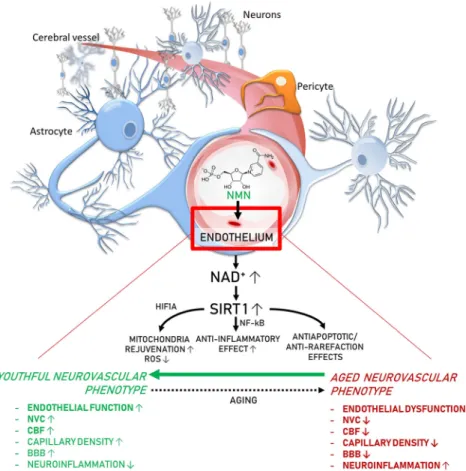
Fig. 7 Proposed scheme for the mechanisms by which restoration of NAD+levels in the aged neurovascular unit by NMN supple- mentation promotes neurovascular rejuvenation. The model, based on our present and previous findings and earlier data from the literature (Das et al.2018; Tarantini et al.2019a; Csiszar et al.
2019), predicts that increased NAD+ activates sirtuin-mediated pathways, which leads to anti-aging transcriptomic changes, re- stores cellular energetics, and attenuates mitochondrial ROS
production, rescuing a youthful neurovascular phenotype. These effects are predicted to act to improve endothelial function, in- crease neurovascular coupling responses, capillary density and cerebral blood flow (CBF), maintain blood-brain barrier (BBB) integrity, and inhibit neuroinflammation, protecting cognitive health (bold font, experimentally validated effects; regular fonts, predicted effects)
Our study demonstrates that NMN treatment, which augments the vascular NAD+metabolome (Tarantini et al.2019a), induces a neurovascular gene expression signature suggestive of SIRT1 activation. Our results expand the findings of previous studies showing that increases in NAD+levels induced by NMN treatment also activate SIRT1 in skeletal muscle (Gomes et al.
2013). As our recent studies demonstrate that shRNA knockdown of SIRT1 prevents the beneficial effects of NMN on aged cerebromicrovascular endothelial cells in vitro (Tarantini et al.2019a), we posit that NMN- induced SIRT1 activation plays a critical role also in neurovascular rejuvenation in vivo. Sirtuins are known to mediate beneficial anti-aging (Cohen et al.
2004; Moroz et al. 2014; Wood et al. 2004) and vasoprotective effects (Csiszar et al. 2009a, 2013;
2014a) of caloric restriction as well. Our bioinformat- ics analysis also revealed a role for Hif1αsignaling, confirming earlier findings (Gomes et al.2013). Fur- ther, our recent study demonstrate that NMN treatment reverses age-related changes in miRNA expression in the aged mouse aorta (Kiss et al.2019b). In that regard, it is significant that dysregulation of miRNA expres- sion has been shown to significantly contribute to age- related phenotypic and functional changes in the cerebromicrovascular endothelial cells as well (Ungvari et al.2013). These findings raise the possi- bility that complex changes in transcriptional and/or post-transcriptional control of expression of genes that encode critical factors determining neurovascular health contribute to the beneficial effects of treatment with NAD+boosters. GO enrichment analysis of dis- cordant differentially expressed neurovascular genes identified functions in mitochondrial regulation, apo- ptosis, inflammation, and endothelial activation.
Mitochondrial dysfunction and increased mito- chondrial oxidative stress have a critical role in the genesis of aging-induced cerebromicrovascular endo- thelial impairment and neurovascular dysfunction (Tarantini et al. 2018, 2019a). In support of this concept, attenuation of mitochondrial oxidative stress and restoration of mitochondrial energy metabolism in the cerebromicrovascular endothelial cells by treatment with the mitochondria-targeted antioxi- dants were shown to rescue neurovascular function in aged mice (Tarantini et al.2018). Here, we report that NMN treatment rescues aging-induced changes in mitochondria-related gene expression in the NVU.
Importantly, these NMN-induced changes in the
mitochondria-related transcriptome are associated with attenuated mitochondrial oxidative stress and restoration of mitochondrial energy metabolism in aging cerebromicrovascular endothelial cells (Tarantini et al. 2019a; Kiss et al. 2019a). On the basis of previous findings (Gomes et al. 2013), we posit that rescue of vascular mitochondrial function by restoring the expression of ETC subunits contrib- utes to the neurovascular protective effects of NMN.
Treatment with NMN was also shown to rescue ex- pression of mitochondrial-encoded ETC subunits in c e r e b r a l a r t e r i e s o f a g e d mi c e a n d i n a g e d cerebromicrovascular endothelial cells (Gomes et al.
2013). It is believed that rescue of electron flow through the electron transport chain, due to the re- stored expression of complex I and complex III (Kwong and Sohal 2000), likely attenuates electron leak, limiting mtROS production. Treatment with NAD+boosters was also demonstrated to upregulate mitochondrial gene expression in the mouse skeletal muscle (Canto et al. 2012). Our recent studies pro- vide evidence that NMN treatment exerts its mito- chondrial protective effects in cerebromicrovascular endothelial cells in a SIRT1-dependent manner (Gomes et al. 2013). Our observations accord with findings from earlier studies showing that many of the health benefits conferred by SIRT1 activation are linked to improved mitochondrial function (Baur et al. 2006). In addition to sirtuin-mediated transcriptomic effects, a mitochondrial ATP produc- tion requires NAD+as an essential cofactor, rescuing normal cellular NAD/NADH ratio per se may also promote efficient mitochondrial function in cells of the NVU.
Analysis of the transcriptomic signature of NMN treatment predicts potent anti-apoptotic effects in the NVU. This is significant, as endothelial cell apoptosis plays a critical role in age-related cerebromicrovascular rarefaction (Ungvari et al.2018a,b). Thus, future stud- ies should determine how NMN treatment affects the number of apoptotic endothelial cells in the NVU as well as capillary density in the aged brain. Recent stud- ies show that NMN also protects the integrity of the blood-brain barrier in a mouse model of brain ischemia (Wei et al.2017). On the basis of our findings that NMN upregulates factors controlling barrier integrity, it will be also of great interest to determine whether NMN treat- ment can also protect against age-related disruption of the blood-brain barrier.
GeroScience (2020) 42:527–546 540
Our studies demonstrate that NMN treatment in aged mice reverses, at least in part, age-related, pro- inflammatory alterations in mRNA expression pro- file in the NVU. Our findings expand the results of recent studies demonstrating that treatment of aged mice with NMN promotes anti-inflammatory pheno- typic changes in the peripheral vasculature as well (Kiss et al. 2019b). Previous studies attributed age- related endothelial activation and chronic sterile mi- crovascular inflammation to oxidative stress- mediated activation of NF-κB and upregulation of pro-inflammatory cytokines in the vascular wall (Csiszar et al. 2003, 2008b, 2014a; Ungvari et al.
2007a). SIRT1 activation is known to attenuate cel- lular and mitochondrial oxidative stress, inhibit NF-κB, and attenuate microvascular inflammation (Toth et al. 2015; Csiszar et al. 2008a, 2012; Baur et al. 2012; Ungvari et al. 2009; Zhang et al.2009;
Mattison et al. 2014). Thus, it is likely that SIRT1 activation and the previously documented anti- oxidative neurovascular effects contribute signifi- cantly to the observed anti-inflammatory effects as- sociated with NMN treatment.
Additional studies are warranted to determine the efficacy of combination treatments with NAD+ boosters (Mitchell et al. 2018; Yoshino et al. 2018;
Liu et al. 2018) and compounds that directly acti- vate SIRT1 and/or inhibit NAD+ overutilization for neurovascular protection. Similar to NAD+boosters, SIRT1-activating compounds (STACs; including resveratrol and SRT1720) were demonstrated to ex- ert important vasoprotective effects in models of aging and accelerated vascular aging (Csiszar et al.
2008a; Ungvari et al.2007a,b,2011; Pearson et al.
2008; Zarzuelo et al.2013; Chen et al.2015; Gano et al. 2014; Minor et al. 2011). These SIRT1- mediated effects include increased mitochondrial biogenesis (Csiszar et al. 2009b), attenuation of mitochondrial oxidative stress (Ungvari et al. 2009;
Csiszar et al. 2012), activation of anti-oxidative defense mechanisms (Csiszar et al. 2014b), and inhibition of apoptosis (Pearson et al. 2008). Treat- ment with the STAC resveratrol was shown to im- prove cerebromicrovascular endothelial function and rescue neurovascular coupling responses in aged mice (Toth et al. 2014; Wiedenhoeft et al. 2019).
Resveratrol was also shown to increase capillary density (Oomen et al. 2009) and prevent microvas- cular fragility (Toth et al. 2015) in the aged mouse
brain and to exert similar vasoprotective effects in non-human primate models as well (Mattison et al.
2014; Bernier et al. 2016). The molecular mecha- nisms contributing to age-related decline in NAD+ in cells of the NVU are likely multifaceted. In addition to downregulation of NAMPT (nicotin- amide phosphoribosyltransferase/NMN synthase;
which catalyzes the rate limiting step in the biosyn- thesis of NAD+) (Tarantini et al. 2019a), the in- c r e a s e d u t i l i z a t i o n o f N A D+ b y a c t i v a t e d poly(ADP-ribose) polymerase 1 (PARP-1) (Csiszar et al. 2019; Pacher et al. 2002) also likely plays an important role in age-related decline in NAD+in the NVU. Accordingly, treatment with PJ-34, a potent PARP inhibitor, restores neurovascular coupling re- sponses in aged mice, similar to the neurovascular protective effects of NMN treatment (Tarantini et al.
2019b). Thus, future studies should determine whether combination of NAD+ boosters with STACs, mitochondria-targeted agents, and/or PARP-1 inhibitors confers greater neurovascular and cognitive health benefits as compared with NAD+ booster treatment alone.
Conclusions
In conclusion, rescue of cerebromicrovascular endo- thelial function and neurovascular coupling re- sponses in NMN-treated aged mice are accompanied by marked anti-aging changes in the neurovascular transcriptome. We hope that our findings will facili- tate future endeavors to uncover the mechanistic role of neurovascular NAD+depletion in brain aging and the pathogenesis of VCI. The recently appreciated complex role of NVU dysfunction (ranging from impaired neurovascular coupling to blood-brain bar- rier disruption) in neurodegenerative diseases and VCI supports the concept that pharmacological treat- ments, which maintain neurovascular health, promote brain health (Kisler et al.2017; Zlokovic2010,2011;
Csipo et al. 2019a, b; de Montgolfier et al. 2019;
Farias Quipildor et al. 2019; Fulop et al. 2019;
Sorond et al. 2019; Sagare et al. 2013). Potentially, NAD+ booster treatments (e.g., in combination with STACs) could be harnessed for development of new pharmacological approaches for neurovascular pro- tection for the prevention and treatment of VCI and neurodegenerative diseases in older adults.
Funding information This work was supported by grants from the American Heart Association (ST), the Oklahoma Center for the Advancement of Science and Technology (to AC, AY, ZU), the National Institute on Aging (R01-AG047879; R01-AG038747;
R01-AG055395), the National Institute of Neurological Disorders and Stroke (NINDS; R01-NS056218 to AC, R01-NS100782 to ZU), the National Institute of General Medical Sciences Oklahoma Shared Clinical and Translational Resources (OSCTR) (GM104938, to AY and JW) and Molecular Mechanisms and Genetics of Autoimmunity COBRE (P30-GM110766, to LG), the Presbyterian Health Foundation (to ZU, AC, AY), the NIA- supported Geroscience Training Program in Oklahoma (T32AG052363), the Oklahoma Nathan Shock Center (P30AG050911), and the Cellular and Molecular GeroScience CoBRE (1P20GM125528, sub#5337). The funding sources had no role in the study design; in the collection, analysis, and inter- pretation of data; in the writing of the report; and in the decision to submit the article for publication.
Compliance with ethical standards
All procedures were approved by the Institutional Animal Use and Care Committees of the University of Oklahoma Health Sciences Center. All animal experiments complied with the ARRIVE guide- lines and were carried out in accordance with the National Insti- tutes of Health guide for the care and use of Laboratory animals (NIH Publications No. 8023, revised 1978).
References
Anderson RM, Bitterman KJ, Wood JG, Medvedik O, Cohen H, Lin SS, Manchester JK, Gordon JI, Sinclair DA (2002) Manipulation of a nuclear NAD+ salvage pathway delays aging without altering steady-state NAD+ levels. J Biol Chem 277:18881–18890
Baur JA, Pearson KJ, Price NL, Jamieson HA, Lerin C, Kalra A, Prabhu VV, Allard JS, Lopez-Lluch G, Lewis K, Pistell PJ, Poosala S, Becker KG, Boss O, Gwinn D, Wang M, Ramaswamy S, Fishbein KW, Spencer RG, Lakatta EG, Le Couteur D, Shaw RJ, Navas P, Puigserver P, Ingram DK, de Cabo R, Sinclair DA (2006) Resveratrol improves health and survival of mice on a high-calorie diet. Nature. 444:337–342 Baur JA, Ungvari Z, Minor RK, Le Couteur DG, de Cabo R (2012) Are sirtuins viable targets for improving healthspan and lifespan? Nat Rev Drug Discov 11:443–461
Bernier M, Wahl D, Ali A, Allard J, Faulkner S, Wnorowski A, Sanghvi M, Moaddel R, Alfaras I, Mattison JA, Tarantini S, Tucsek Z, Ungvari Z, Csiszar A, Pearson KJ, de Cabo R (2016) Resveratrol supplementation confers neuroprotection in cortical brain tissue of nonhuman primates fed a high-fat/
sucrose diet.Aging (Albany NY)8:899–916
Bolger AM, Lohse M, Usadel B (2014) Trimmomatic: a flexible trimmer for Illumina sequence data. Bioinformatics. 30:
2114–2120
Bray NL, Pimentel H, Melsted P, Pachter L (2016) Near-optimal probabilistic RNA-seq quantification. Nat Biotechnol 34:
525–527
Canto C, Houtkooper RH, Pirinen E, Youn DY, Oosterveer MH, Cen Y, Fernandez-Marcos PJ, Yamamoto H, Andreux PA, Cettour-Rose P, Gademann K, Rinsch C, Schoonjans K, Sauve AA, Auwerx J (2012) The NAD(+) precursor nicotin- amide riboside enhances oxidative metabolism and protects against high-fat diet-induced obesity. Cell Metab 15:838–847 Chen YX, Zhang M, Cai Y, Zhao Q, Dai W (2015) The Sirt1 activator SRT1720 attenuates angiotensin II-induced athero- sclerosis in apoE(−)/(−) mice through inhibiting vascular inflammatory response. Biochem Biophys Res Commun 465:732–738
Cohen HY, Miller C, Bitterman KJ, Wall NR, Hekking B, Kessler B, Howitz KT, Gorospe M, de Cabo R, Sinclair DA (2004) Calorie restriction promotes mammalian cell survival by inducing the SIRT1 deacetylase. Science. 305:390–392 Csipo T, Lipecz A, Fulop GA, Hand RA, Ngo BN, Dzialendzik M,
Tarantini S, Balasubramanian P, Kiss T, Yabluchanska V, Silva-Palacios F, Courtney DL, Dasari TW, Sorond F, Sonntag WE, Csiszar A, Ungvari Z, Yabluchanskiy A (2019a) Age-related decline in peripheral vascular health predicts cognitive impairment. Geroscience. 41:125–136 Csipo T, Mukli P, Lipecz A, Tarantini S, Bahadli D, Abdulhussein O,
Owens C, Kiss T, Balasubramanian P, Nyul-Toth A, Hand RA, Yabluchanska V, Sorond FA, Csiszar A, Ungvari Z, Yabluchanskiy A (2019b) Assessment of age-related decline of neurovascular coupling responses by functional near-infrared spectroscopy (fNIRS) in humans. Geroscience. 41:495–509 Csiszar A, Ungvari Z, Koller A, Edwards JG, Kaley G (2003)
Aging-induced proinflammatory shift in cytokine expression profile in rat coronary arteries. FASEB J 17:1183–1185 Csiszar A, Ungvari Z, Koller A, Edwards JG, Kaley G (2004)
Proinflammatory phenotype of coronary arteries promotes endothelial apoptosis in aging. Physiol Genomics 17:21–30 Csiszar A, Labinskyy N, Smith K, Rivera A, Orosz Z, Ungvari Z (2007) Vasculoprotective effects of anti-TNFalfa treatment in aging. Am J Pathol 170:388–698
Csiszar A, Labinskyy N, Podlutsky A, Kaminski PM, Wolin MS, Zhang C, Mukhopadhyay P, Pacher P, Hu F, de Cabo R, Ballabh P, Ungvari Z (2008a) Vasoprotective effects of res- veratrol and SIRT1: attenuation of cigarette smoke-induced oxidative stress and proinflammatory phenotypic alterations.
Am J Physiol Heart Circ Physiol 294:H2721–H2735 Csiszar A, Wang M, Lakatta EG, Ungvari ZI (2008b)
Inflammation and endothelial dysfunction during aging: role of NF-{kappa}B. J Appl Physiol 105:1333–1341
Csiszar A, Labinskyy N, Jimenez R, Pinto JT, Ballabh P, Losonczy G, Pearson KJ, de Cabo R, Ungvari Z (2009a) Anti-oxidative and anti-inflammatory vasoprotective effects of caloric re- striction in aging: role of circulating factors and SIRT1. Mech Ageing Dev 130:518–527
Csiszar A, Labinskyy N, Pinto JT, Ballabh P, Zhang H, Losonczy G, Pearson KJ, de Cabo R, Pacher P, Zhang C, Ungvari ZI (2009b) Resveratrol induces mitochondrial biogenesis in en- dothelial cells. Am J Physiol Heart Circ Physiol. 2009 J u l ; 2 9 7 ( 1 ) : H 1 3 - 2 0 . h t t p s : / / d o i . o r g / 1 0 . 1 1 5 2 /ajpheart.00368.2009
Csiszar A, Sosnowska D, Wang M, Lakatta EG, Sonntag WE, Ungvari Z (2012) Age-associated proinflammatory secretory phenotype in vascular smooth muscle cells from the non- human primate Macaca mulatta: reversal by resveratrol treat- ment. J Gerontol A Biol Sci Med Sci 67:811–820
GeroScience (2020) 42:527–546 542