ORIGINAL ARTICLE
Circulating anti-geronic factors from heterochonic
parabionts promote vascular rejuvenation in aged mice:
transcriptional footprint of mitochondrial protection, attenuation of oxidative stress, and rescue of endothelial function by young blood
Tamas Kiss&Stefano Tarantini&Tamas Csipo&Priya Balasubramanian&Ádám Nyúl-Tóth&Andriy Yabluchanskiy&Jonathan D. Wren&Lori Garman&Derek M.
Huffman&Anna Csiszar&Zoltan Ungvari
Received: 25 February 2020 / Accepted: 4 March 2020
#American Aging Association 2020
Abstract
Aging-induced functional and phenotypic al- terations of the vasculature (e.g., endothelial dysfunc- tion, oxidative stress) have a central role in morbidity and mortality of older adults. It has become apparent in recent years that cell autonomous mechanisms alone are inadequate to explain all aspects of vascular aging. The present study was designed to test the hypothesis that age-related changes in circulating anti-geronic factors contribute to the regulation of vascular aging processes in a non-cell autonomous manner. To test this hypothe- sis, through heterochronic parabiosis we determined the extent, if any, to which endothelial function, vascular
production of ROS, and shifts in the vascular tran- scriptome (RNA-seq) are modulated by the systemic environment. We found that in aortas isolated from isochronic parabiont aged (20-month-old) C57BL/6 mice [A-(A); parabiosis for 8 weeks] acetylcholine- induced endothelium-dependent relaxation was im- paired and ROS production (dihydroethidium fluores- cence) was increased as compared with those in aortas from young isochronic parabiont (6-month-old) mice [Y-(Y)]. The presence of young blood derived from young parabionts significantly improved endothelium- dependent vasorelaxation and attenuated ROS
Tamas Kiss, Stefano Tarantini, Tamas Csipo, PriyaBalasubramanian and Ádám Nyúl-Tóth contributed equally to this work.
T. Kiss
:
S. Tarantini:
T. Csipo:
P. Balasubramanian:
Á. Nyúl-Tóth
:
A. Yabluchanskiy:
J. D. Wren:
A. Csiszar:
Z. Ungvari (*)
Vascular Cognitive Impairment and Neurodegeneration Program, Reynolds Oklahoma Center on Aging/Center for Geroscience and Healthy Brain Aging, Department of Biochemistry and Molecular Biology, University of Oklahoma Health Sciences Center, 975 NE 10th Street, BRC 1311, Oklahoma City, OK 73104, USA e-mail: zoltan-ungvari@ouhsc.edu
T. Kiss
:
A. Csiszar:
Z. UngvariInternational Training Program in Geroscience, Theoretical Medicine Doctoral School/Departments of Medical Physics and Informatics, University of Szeged, Szeged, Hungary
S. Tarantini
:
T. Csipo:
Z. UngvariInternational Training Program in Geroscience, Doctoral School of Basic and Translational Medicine/Department of Public Health, Semmelweis University, Budapest, Hungary
S. Tarantini
:
Z. UngvariDepartment of Health Promotion Sciences, College of Public Health, University of Oklahoma Health Sciences Center, Oklahoma City, OK, USA
T. Csipo
International Training Program in Geroscience, Department of Cardiology, Division of Clinical Physiology, Faculty of Medicine, University of Debrecen, Debrecen, Hungary
/ Published online: 15March2020
production in vessels of heterochronic parabiont aged [A-(Y)] mice. In aortas derived from heterochronic parabiont young [Y-(A)] mice, acetylcholine-induced relaxation and ROS production were comparable with those in aortas derived from Y-(Y) mice. Using RNA- seq we assessed transcriptomic changes in the aortic arch associated with aging and heterochronic parabiosis.
We identified 347 differentially expressed genes in A-(A) animals compared with Y-(Y) controls. We have identified 212 discordant genes, whose expression levels differed in the aged phenotype, but have shifted back toward the young phenotype by the presence of young blood in aged A-(Y) animals. Pathway analysis shows that vascular protective effects mediated by young blood
–regulated genes include mitochondrial re- juvenation. In conclusion, a relatively short-term expo- sure to young blood can rescue vascular aging pheno- types, including attenuation of oxidative stress, mito- chondrial rejuvenation, and improved endothelial func- tion. Our findings provide additional evidence supporting the significant plasticity of vascular aging and evidence for the existence of anti-geronic factors capable of exerting rejuvenating effects on the aging vasculature.
Keywords
Aging . Senescence . Geronic factors
Introduction
Cardiovascular and cerebrovascular diseases are the most common causes of serious long-term disability and mortality among older adults in the developed world (Ungvari et al. 2018a), accounting for approximately one-third of all deaths in the USA at the age of 65 and nearly two-thirds of all deaths at an age of 85 (Health, United States 2016). Importantly, epidemiological stud- ies reveal that the effects of traditional risk factors (such as hypertension, dyslipidemia, cigarette smoking, obe- sity, diabetes mellitus, etc.) on the incidence of cardio- vascular and cerebrovascular diseases are dwarfed by the single most important risk factor for these diseases:
advanced aging (Ungvari et al. 2018a). In order to develop novel methods for prevention and treatment of vascular pathologies in older adults, it is important to understand the role of shared mechanisms of aging in the cellular and functional changes that occur in the vasculature during aging (Lakatta and Levy 2003).
In the past two decades, most studies on vascular aging have focused on cell autonomous mechanisms driving functional decline (Ungvari et al. 2018a). These studies have led to a comprehensive understanding of the critical roles of mitochondrial dysfunction (Tarantini et al. 2018; Gioscia-Ryan et al. 2014), increased pro- duction of reactive oxygen species(ROS) (Ungvari et al.
2018a; Csiszar et al. 2014; Csiszar et al. 2009; Toth et al.
2015a; Ungvari et al. 2007; Csiszar et al. 2002;
Labinskyy et al. 2006; Csiszar et al. 2007a; Csiszar et al. 2007b; Csiszar et al. 2012; Fulop et al. 2018;
Ungvari et al. 2011), impaired NO bioavailability (Csiszar et al. 2002; Tarantini et al. 2019a; Csiszar et al. 2015; Toth et al. 2014a), consequences of cellular NAD+ depletion (Tarantini et al. 2019b; Kiss et al.
2019a; Kiss et al. 2019b; Das et al. 2018), dysregulation of sirtuins(Csiszar et al. 2009; Mattison et al. 2014), and energetic dysfunction (Tarantini et al. 2018) in the eti- ology of vascular aging. However, it has also become apparent in recent years that these important cell auton- omous mechanisms alone are inadequate to explain all aspects of vascular aging (Ungvari et al. 2018a).
In the field of geroscience, compelling evidence has been accumulated that implicates non-cell autonomous mechanisms as also playing important roles in driving aging processes and pathogenesis of age-related dis- eases (Ashpole et al. 2016; Ashpole et al. 2017;
Lopez-Otin et al. 2013; Katsimpardi et al. 2014; Morri- son et al. 2019; Rebo et al. 2016). Recently, the
Á. Nyúl-TóthInternational Training Program in Geroscience, Institute of Biophysics, Biological Research Centre, Szeged, Hungary J. D. Wren
:
L. GarmanOklahoma Medical Research Foundation, Genes & Human Disease Research Program, Oklahoma City, OK, USA D. M. Huffman
Department of Molecular Pharmacology, Albert Einstein College of Medicine, Bronx, NY 10461, USA
D. M. Huffman
Institute for Aging Research, Albert Einstein College of Medicine, Bronx, NY, USA
D. M. Huffman
Department of Medicine, Albert Einstein College of Medicine, Bronx, NY, USA
A. Csiszar
:
Z. UngvariThe Peggy and Charles Stephenson Cancer Center, University of Oklahoma Health Sciences Center, Oklahoma City, OK 73104, USA
hypothesis was put forward that aging is associated with complex changes in inter-organ communication, which modulate cell autonomous aging processes in the vas- culature contributing to pathogenesis of age-related vas- cular diseases(Ungvari et al. 2018a). An important pre- diction based on this hypothesis is that circulating pro- geronic factors (characterized by their increased produc- tion with age and deleterious effects on vascular homeo- stasis, e.g., inflammatory cytokines) and/or the presence of anti-geronic factors (which reverse or prevent devel- opment of cellular aging phenotypes and presumably decline with age), which are derived from other organs (e.g., adipose tissue, the brain, endocrine system, im- mune system) orchestrate aging processes in cells within the vascular wall. Yet, additional experimental evidence is needed to better understand the relative contribution of circulating factors versus cell autonomous mecha- nisms to the genesis of vascular aging phenotypes.
Parabiosis is a surgical approach for joining the cir- culatory systems of two animals that has been widely used to study the role of circulating (humoral and cellu- lar) factors in the pathogenesis of various diseases, including endocrine and metabolic diseases (Harris 1997; Harris 1999; Coleman 1973; Coleman and Hummel 1969) as well as regulation of lifespan (Ludwig and Elashoff 1972; Horrington et al. 1960;
Lunsford et al. 1963; McCay et al. 1957). Parabiosis re-emerged over the past 15 years in geroscience re- search to investigate the complex interaction between cell autonomous and non-cell autonomous mechanisms of aging comparing heterochronic (young–old) and isochronic (young–young and old–old) parabiont pairs of animals (Katsimpardi et al. 2014; Rebo et al. 2016;
Lunsford et al. 1963; McCay et al. 1957; Bitto and Kaeberlein 2014; Cannata et al. 2017; Conboy et al.
2013; Fan et al. 2017; Flemming 2013; Ghosh et al.
2019; Gontier et al. 2018; Harrison and Astle 1982;
Hirayama et al. 1993; Katsimpardi et al. 2020; Sousa- Victor et al. 2019; Villeda et al. 2011; Villeda et al. 2014;
Zhang et al. 2019; DeCarolis et al. 2015; Middeldorp et al. 2016; Smith et al. 2015a). Using heterochronic parabiosis as an experimental tool, it was demonstrated that both pro-geronic and anti-geronic circulating fac- tors mediate non-cell autonomous effects driving aging in multiple organs, including the skeletal muscle, central nervous system, and the heart (Katsimpardi et al. 2014;
Fan et al. 2017; Villeda et al. 2011; Villeda et al. 2014;
DeCarolis et al. 2015; Smith et al. 2015a; Castellano et al. 2016).
The present study was designed to experimentally test the hypothesis that age-related changes in circulating pro- geronic factors and/or anti-geronic factors contribute to the regulation of vascular aging processes in a non-cell autonomous manner. To test this hypothesis we leveraged heterochronic parabiosis in combination with vascular functional assays and transcriptomic profiling, to deter- mine to what extent, if any, transposition of aging phe- notypes could be observed in the young and old aorta by exposure to an old and young environment, respectively.
Methods
Animals and parabiosis surgery
Young (4-month-old) and aged (18-month-old) male C57BL/6 mice were obtained from the aging rodent col- ony maintained by the National Institute on Aging at Charles River Laboratories (Wilmington, MA). Mice were housed under specific pathogen-free conditions at the rodent barrier facility at Albert Einstein College of Medicine under a controlled photoperiod (12 h light; 12 h dark) with unlimited access to water and were fed a standard AIN-93G diet (ad libitum). Parabiosis surgery in young and aged animals was carried out by the Einstein Chronobiosis and Energetics/Metabolism of Aging Core, according to published protocols (Harris 1997; Harris 1999) as reported previously (Morrison et al. 2019). Sur- gical unions were performed between young animals (isochronic; young Y–(Y);
n= 4 pairs), aged animals (isochronic old; A–(A);
n= 4 pairs), and young and aged mice (heterochronic Y–(A) and A-(Y);
n= 5 pairs) in the Einstein Health Span Core as described (Morrison et al.
2019). Following surgery, animals were kept on a partial heating pad overnight. Pairs were then intensively moni- tored and received subcutaneous (s.c) injections of Banamine (2 mg/kg each) immediately post-op and twice a day for t3 days and then once daily for 4 days. Animals also received 1 mL of Ringer
’s lactate (s.c.) immediately after, daily for 3 days post-op to prevent dehydration.
Animals remained joined for ~ 8 weeks prior to sacrifice.
All experimental procedures were approved by the Insti-
tutional Animal Care and Use Committee (IACUC) at the
Albert Einstein College of Medicine and the University of
Oklahoma Health Sciences Center. All animal experi-
ments were carried out in accordance with the National
Institutes of Health guide for the care and use of Labora-
tory animals (NIH Publications No. 8023, revised 1978).
Assessment of endothelial function in the aorta To assess the specific effect of circulating factors on endothelial function, endothelium-dependent vasorelax- ation was assessed in isolated aorta ring preparations as described previously (Pearson et al. 2008). In brief, animals were killed as reported (Morrison et al. 2019) and the thoracic aortas were cleaned from the connective tissue and perivascular fat, cut into ring segments 1.5 mm in length and mounted in myographs chambers (Danish Myo Technology A/S, Inc., Denmark) for mea- surement of isometric tension. The vessels were superfused with Krebs buffer solution (118 mM NaCl, 4.7 mM KCl, 1.5 mM CaCl
2, 25 mM NaHCO
3, 1.1 mM MgSO
4, 1.2 mM KH
2PO
4, and 5.6 mM glucose; at 37 °C; gassed with 95% air and 5% CO
2). After an equilibration period of 1 h during which an optimal passive tension was applied to the rings (as determined from the vascular length-tension relationship), they were pre-contracted with 10
−6M phenylephrine and relaxa- tion in response to acetylcholine was measured. The remaining aortic arch tissue was snap-frozen and stored at
−80 °C for transcriptomic analysis.
Assessment of vascular oxidative stress
To characterize vascular ROS production isolated seg- ments of the aorta were loaded with the redox-sensitive dye dihydroethidium (DHE, Invitrogen, Carlsbad CA;
3 × 10
−6mol/L; for 30 min) in oxygenated Krebs solu- tion (at 37 °C) as previously reported (Toth et al. 2015a;
Csiszar et al. 2007a; Pearson et al. 2008; Ungvari et al.
2003; Labinskyy et al. 2009). After loading the dye was washed out five times with warm Krebs buffer, and the vessels were allowed to equilibrate for another 20 min.
Then, the vessels were embedded in OCT medium and cryosectioned. Confocal images were captured using a Leica SP2 confocal laser scanning microscope (Leica Microsystems GmbH, Wetzlar, Germany). Average nu- clear DHE fluorescence intensities were assessed using the Metamorph software (Molecular Devices LLC, Sun- nyvale, CA) and values for each animal in each group were averaged.
RNA isolation, cDNA synthesis, library construction, and next generation sequencing
RNA was isolated from the aortic arch samples using AllPrep DNA/RNA Mini Kit (Qiagen) as previously
described (Imperio et al. 2016; Valcarcel-Ares et al.
2019). Prior to 3′-tag RNA-seq analysis quality control measures were implemented. Concentration of RNA was ascertained via fluorometric analysis on a Thermo Fisher Qubit fluorometer. Overall quality of RNA was verified using an Agilent Tapestation instrument. Fol- lowing initial QC steps sequencing libraries were gen- erated using the Lexogen Quantseq FWD library prep kit according to the manufacturer
’s protocol by the Quantitative Analysis Core of the Oklahoma Medical Research Foundation (Kiss et al. 2020). Briefly, the first strand of cDNA was generated using 5
′-tagged poly-T oligomer primers. Following RNase digestion, the sec- ond strand of cDNA was generated using 5′-tagged random primers. A subsequent PCR step with additional primers added the complete adapter sequence to the initial 5′ tags, added unique indices for demultiplexing of samples, and amplified the library. Final libraries for each sample were assayed on the Agilent Tapestation for appropriate size and quantity. These libraries were then pooled in equimolar amounts as ascertained via fluo- rometric analyses. Final pools were absolutely quanti- fied using qPCR on a Roche LightCycler 480 instru- ment with Kapa Biosystems Illumina Library Quantifi- cation reagents. Sequencing was performed by the Quantitative Analysis Core of the Oklahoma Medical Research Foundation (Kiss et al. 2020) using custom primers on an Illumina NextSeq 500 instrument with High Output chemistry and 75 bp single-ended reads.
RNA-Seq data analysis and visualization
Raw sequencing reads were trimmed of their Illumina TruSeq adapter sequences using
Trimmomatic v0.35(Bolger et al.2014), filtered for contaminants of ribosomal, mitochondrial, and hemoglobin transcripts, then aligned to the mouse genome version GRCm38 using
Kallisto v0.43.03(Bray et al. 2016). Samples were checked for outliers and separation by principle compo- nents analysis (PCA) with the R function
prcomp. Rawexpression counts were summarized at the gene level to transcript-length adjusted, library-size scaled counts per million (CPM) with the R/Bioconductor package
tximport(Soneson et al. 2015). Differential expression analysis was performed using the empirical Bayes ap- proach implemented in the R/Bioconductor package
DESeq2(Love et al. 2014). Significantly differentially expressed (DE) genes had an absolute log2 fold change
≥
0.585 (corresponding to a change of 50% or more in
expression) and the False Discovery Rate FDR-adjusted
pvalue
≤0.05. Gene annotation was done using
biomaRt(Durinck et al. 2009) in R/Bioconductor pack- age. The R package
pheatmap v1.0.12was used to perform hierarchical clustering and to draw the heatmaps. The
org.Mm.eg.db v3.8.2R/Bioconductor package was used to collect Gene Ontology, KEGG, and Reactome terms associated with the DE genes. The same package was used to translate Ensemble IDs to Entrez IDs when it was required by the statistical packages.
Functional annotation
The
org.Mm.eg.db v3.8.2R/Bioconductor package was used to collect Gene Ontology terms associated with our differentially expressed genes. The hypergeometric test implemented in
GOstats v2.51.0.R/Bioconductor pack- age was used to calculate enrichment of the individual terms (Falcon and Gentleman 2007).
We used Upstream Regulator Analysis (URA) algo- rithm (Kramer et al. 2014) implemented in the Ingenuity Pathway Analysis (QIAGEN) software to identify up- stream regulators that potentially explain the observed gene expression changes in our samples. The IPA uses a manually curated database (Ingenuity Knowledge Base) to calculate
“enrichment”score [Fisher’s exact test (FET)
pvalue] measures the overlap of observed and predicted regulated gene sets, and a
z-score assessing thematch of observed and predicted up/downregulation patterns.
Reactome pathway Knowledgebase (Fabregat et al.
2018)
Reactome is a free, open-source, curated, and peer- reviewed database of biological pathways. Enrichment of Reactome pathways was calculated and results were plotted by the
ReacomePAv1.29.0 R/Bioconductor package (Yu and He 2016).
org.Mm.eg.dbv3.8.2 R/Bioconductor package was used to convert Ensemble IDs to Entrez IDs since that is the required input format of the ReactomePA package.
KEGG pathway
org.Mm.eg.db
v3.8.2 R/Bioconductor package was used to convert Ensemble IDs to Entrez IDs. Pathview R/Bioconductor package was used to overlay the gene
expression data with the curated KEGG pathways. Heat maps were drawn by the
pheatmapv1.0.12 R package.
[https://CRAN.R-project.org/package=pheatmap].
Results
Young blood rescues endothelial function and attenuates oxidative stress in aged aorta
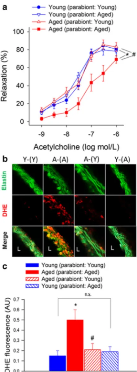
There is strong experimental and clinical evidence that endothelial dysfunction significantly contributes to the pathogenesis of age-related diseases of the cardiovascu- lar system (Ungvari et al. 2018b). To test the endothelial effects of circulating pro- and anti-geronic factors, endothelium-dependent vasodilator responses to acetyl- choline were tested. In aorta rings derived from Y-(Y) mice, administration of acetylcholine resulted in signif- icant relaxation, whereas these responses were signifi- cantly attenuated in aorta rings obtained from A-(A) mice (Fig. 1a). Acetylcholine-induced vasorelaxation was abolished by the pre-treatment of the aorta rings from each group with the NO synthase inhibitor L- NAME (data not shown). Exposure to young blood significantly improved NO-mediated, endothelium- dependent vasorelaxation in A-(Y) mice (Fig. 1a). In aorta rings derived from Y-(A) mice, acetylcholine- induced relaxation was comparable with that in aortas derived from Y-(Y) mice, suggesting that presence of old blood did not alter endothelial function considerably in young aorta.
Previous studies demonstrate that aging significantly increases vascular superoxide production and that treat- ment with antioxidants can restore endothelium- mediated vasodilation in aged rodents (Tarantini et al.
2018; Csiszar et al. 2002; Tarantini et al. 2019b;
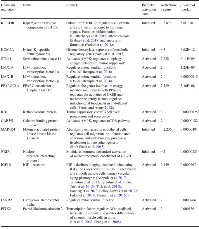
Ungvari et al. 2018b; Wiedenhoeft et al. 2019; Csiszar et al. 2009; Csiszar et al. 2007c). We found that DHE staining in the wall of aortas isolated from A-(A) mice was significantly stronger as compared with that in vessels isolated from Y-(Y) mice, indicating increased cellular ROS production (Fig. 1b). Aortas isolated from A-(Y) mice exhibited weaker DHE staining than vessels of A-(A) mice, indicating that the presence of young blood in old mice decreased vascular ROS production.
In agreement with the vasorelaxation responses, aortas
isolated from Y-(A) mice exhibited DHE staining that
was comparable with that in aortas isolated from Y-(Y)
mice (Fig. 1b), suggesting that the old environment per se, is not sufficient to provoke ROS production.
Young blood reverses age-related changes in vascular mRNA expression profile
Using RNA-seq we assessed transcriptomic changes in the aorta arch associated with aging and heterochronic parabiosis. In Fig. 2a, a heat map is shown as a graphic representation of normalized expression values of dif- ferentially expressed genes in aorta samples from each group. Hierarchical clustering analysis revealed groups of genes whose expression changes with age per se, and which is reversed by the presence of young blood in aged mice (A-(Y)). Principal component analysis (PCA;
Fig. 2c) of the transcriptomic data showed that biolog- ical replicates from the Y-(Y) and A-(A) groups, respec- tively, cluster together and that young samples segregate away from aged ones (Fig. 2b). The profiles from A-(A) mice also cluster separately from clusters representing A-(Y) mice. The Y-(Y) and A-(Y) expression profiles were more similar and clustered less discriminately in the PCA. These findings indicated a clear difference between the transcriptome profiles of young and old isochronic pairs, and a partial reversal of the aging effect by young blood. Meanwhile, the impact of old blood on expression in young aorta was less clear as the Y-(A) group did not exhibit good clustering in the space of the first three principal components due to the presence of an outlier (Y-(A) sample #3).
We then determined the number of genes that were significantly upregulated or downregulated (DE; fold change
≥1.5 or < 0.67;
p< 0.05 adjusted for multiple comparisons) in the aorta by aging or by heterochronic parabiosis. We then filtered for genes that are signifi- cantly altered (adjusted
p< 0.05), expressed at an appre- ciable level (fragments per kilobase of transcript per million mapped reads > 1). We identified 347 differen- tially expressed (DE) genes in A-(A) animals compared with Y-(Y) controls. We further identified 58 DE genes in A-(Y) mice compared with A-(A) controls. In Fig. 2c, a volcano plot shows statistical significance (p value) versus magnitude of age-related change in gene expres- sion. Red symbols denote genes, whose expression levels differed in the aged phenotype, but have shifted back toward the young phenotype by the presence of young blood in aged animals (discordant DE genes).
The Venn diagram in Fig. 2d shows that expression of 24 genes, which are differentially expressed in aged
Fig. 1 Young blood improves NO-mediated, endothelium-dependent vasorelaxation in aged mice.aAcetylcholine-induced relaxations in aortic ring preparations isolated from isochronic parabiont young mice [Y-(Y)], isochronic parabiont aged mice [A-(A)], heterochronic parabiont young mice [Y-(A)] and heterochronic parabiont aged mice [A-(Y)]. Age-related declines in endothelial function were reversed by the presence of circulat- ing factors derived from young parabionts. Data are mean ± S.E.M. (n= 4–5 mice for each data point).bRepresentative con- focal images showing stronger DHE staining (red fluorescence) indicating increased cellular ROS production in the wall of aortas isolated from A-(A) mice as compared with vessels isolated from Y-(Y) mice. Aortas isolated from A-(Y) mice exhibit weak DHE staining indicating decreased ROS production. Aortas isolated from Y-(A) mice exhibit DHE staining that is comparable to that in aortas isolated from Y-(Y) mice (original magnification: 10x).c Quantification of DHE fluorescence intensities. Data are means
±S.E.M. (n= 4–5 in each group) *p< 0.05 aged (parabiont: aged) vs young (parabiont: young);#p< 0.05 aged (parabiont: young) vs. aged (parabiont: aged)
mice, was shifted back toward youthful levels by the presence of young blood in aged mice.
We realized that significance cut-offs to identify DE genes shared between the age-effect and young blood
–effect datasets may be too stringent and the analysis illustrated in Fig. 2c may miss discordant patterns (youthful shifts) of gene expression with important bio- logical relevance for young blood–induced vascular rejuvenation. Thus, we also used an approach to detect discordant transcriptional patterns (youthful shifts) by comparing the age-effect and young blood–effect gene expression datasets using combination criteria that took into account the effect direction. Genes were ranked by their effect size direction, and ranked lists were com- pared to identify overlapping genes across a continuous significance gradient. Our analysis required that discor- dant genes with youthful shifts (1) are
“differentiallyexpressed
”based on both
pvalue and fold-change criteria either in aging or the young blood effect group, (2) satisfy a fold-change criterion with a cutoff of
≥1.5 or < 0.67 in the group in which expression did not satisfy the statistical significance
p< 0.05, and (3) satis- fy the criterion that the effect directions of the age-effect and young blood effect are opposite. We found that these combination criteria found more biologically meaningful sets of genes than
pvalues alone.
In Fig. 3a, the magnitude of age-related changes in gene expression is plotted against the magnitude of young blood–induced changes in gene expression in the aged aorta. Red symbols denote the 24 dis- cordant DE genes, whose expression levels shifted back toward the young phenotype by the presence of young blood in aged animals with statistical signif- icance. Genes which are DE only in one group but
Fig. 2 Young blood reverses age-related changes in vascularmRNA expression profile.aThe heat map is a graphic represen- tation of normalized expression values of differentially expressed genes in aorta samples derived from isochronic parabiont young mice [Y-(Y)], isochronic parabiont aged mice [A-(A)], heterochronic parabiont young mice [Y-(A)] and heterochronic parabiont aged mice [A-(Y)]. Hierarchical clustering analysis re- veals groups of genes whose expression is similar in young mice and young blood exposed aged mice. It was noted that the sample Y-(A)#3 is an outlier, yet the data were included in further analy- ses.bPrincipal component analysis (PCA) plot of aortic mRNA expression profiles in the four experimental groups. The profiles
from A-(A) mice (red) cluster separately from clusters representing Y-(Y) mice (blue) and A-(Y) mice (green). The profiles of Y-(A) mice (yellow) did not exhibit good clustering in the space of the first three principal components. PC1, PC2 and PC3: Principal components 1, 2, and 3, respectively. cVolcano plot depicting differentially expressed genes comparing aortic samples derived from Y-(Y) and A-(A) mice. Stratifiedpvalues are plotted against expression fold changes for results obtained in A-(A) samples normalized to Y-(Y) samples. Colored points refer to genes whose expression is significantly altered in A-(Y) mice.dVenn diagrams sowing the numbers of differentially expressed mRNAs in A-(A) mice (“aging effect”) and A-(Y) mice (“young blood effect”)
otherwise satisfy the other criteria are denoted by blue (DE in aging) and green (DE in A-(Y)) sym- bols. The Venn diagram in Fig. 3c shows that using this approach we have identified 188 discordant genes with youthful shifts, which changed in oppo- site directions between the two datasets. These data suggest that changes in circulating anti-geronic fac- tor(s) have a critical role in age-related dysregula- tion of vascular gene expression.
Using a similar approach, we also analyzed old blood-induced transcriptional changes in Y-(A) animals.
In Fig. 3b, the magnitude of age-related changes in gene expression is plotted against the magnitude of old blood- induced changes in gene expression. Due to the pres- ence of an outlier (Y-(A) sample #3) the analysis of the effects of the presence of old blood in young animals yielded fewer results. There was only one concordant DE gene (Prelid1; PRELI domain containing 1
Fig. 3 Young blood reverses age-related changes in vascularmRNA expression profile: identification of discordant genes.a Young blood–induced changes in gene expression (log2fold changes; heterochronic parabiont aged [A-(Y)] mice vs. isochronic parabiont aged [A-(A)] mice) plotted against age-related changes (log2fold changes; isochronic parabiont aged [A-(A)] mice vs.
isochronic parabiont young [Y-(Y)] mice) in the aortic tran- scriptome. Red symbols indicate discordant differentially expressed genes, whose expression significantly changes with age and is restored by young blood toward youthful expression levels. Blue and green symbols denote discordant genes, whose expression changes in aging and is restored by young blood toward youthful expression levels, but only the aging (blue) or the young blood effect (green) reaches the cutoff for statistical significance.bOld blood-induced changes in gene expression (log2fold changes; heterochronic parabiont young [Y-(A)] mice vs. isochronic parabiont young [Y-(Y)] mice) plotted against age- related changes (log2fold changes; isochronic parabiont aged [A- (A)] mice vs. isochronic parabiont young [Y-(Y)] mice) in the aortic transcriptome. Red symbols indicate concordant differen- tially expressed genes, whose expression significantly changes with age and the direction of this effect is mimicked by exposure to old blood. Blue and green symbols denote concordant genes,
whose expression similarly changes in aging and by old blood exposure, but only the aging (blue) or the old blood effect (green) reaches the cutoff for statistical significance.cVenn diagrams showing the numbers of differentially expressed mRNAs in each group. The blue circle represents genes, which are significantly up- or downregulated in aged mice as compared with young mice. The green circle represents genes, which are significantly up- or down- regulated by the presence of young blood in aged mice. The red area represents discordant differentially expressed genes. Gray areas represent discordant genes, whose expression is changed by the presence of young blood in aged mice toward youthful levels, but the effect does not reach the cutoff for statistical significance.dVenn diagrams showing the effect of old blood on vascular gene expression. The blue circle represents genes, which are significantly up- or downregulated in aged mice as compared with young mice. Gray areas represent concordant genes, whose expression is changed by the presence of old blood in young mice toward the aging phenotype, but the effect does not reach the cutoff for statistical significance. The red area represents discordant differentially expressed genes.eThe heat map is a graphic representation of normalized expression values of discor- dant genes in aorta samples derived from Y-(Y), A-(A) and A-(Y) mice
pseudogene) whose expression was significantly upreg- ulated by the presence of old blood in the young animals and whose expression was also significantly upregulat- ed in aortas of A-(A) animals (Fig. 3b). Next, we used an approach to detect concordant transcriptional patterns (pro-geronic shifts) by comparing the age-effect and old blood-effect gene expression datasets using combina- tion criteria that took into account the effect direction.
Our analysis required that concordant genes with pro- geronic shifts (1) are
“differentially expressed”based on both
pvalue and fold-change criteria either in aging or the old blood effect group, (2) satisfy a fold-change criterion with a cutoff of
≥1.5 or < 0.67 in the group inwhich expression did not satisfy the statistical signifi- cance
p< 0.05, and (3) satisfy the criterion that the effect directions of the age-effect and old blood effect are the same. Genes which are DE only in one group but otherwise satisfy the other criteria are denoted by blue in Fig. 3b. The Venn diagram in Fig. 3d shows that using this approach we have identified 114 concordant genes with pro-geronic shifts, which changed in the same directions between the two datasets. These data suggest that pro-geronic circulating factors may also have a role in age-related dysregulation of vascular gene expres- sion, though these effects do not appear to compromise function in our model.
Young blood–induced vascular transcriptomic changes in aged mice predict mitochondrial rejuvenation We performed GO enrichment analysis to explore po- tential biological functions of the young blood–induced discordant genes with youthful shifts. GO enrichment analysis of discordant genes with youthful shifts identi- fied functions in mitochondrial regulation and produc- tion of ROS (Fig. 4a). Overrepresentation analysis of GO terms revealed that extracellular matrix related genes (including the GO terms
“extracellular matrix”[GO:0031012] and
“collagen-containing extracellularmatrix
”[GO:0062023]) were also overrepresented among the aging-induced DE genes, but these biological functions were unaffected by young blood. Figure 4 b depicts Reactome pathways, which are overrepresented (enriched) in the discordant genes induced by young blood exposure. This analysis also showed that expo- sure to young blood is associated with transcriptional changes in the aorta consistent with multifaceted mito- chondrial protective effects as well as anti-inflammatory and anti-oxidative effects. Specifically, Reactome
pathways identified included the Krebs cycle and oxi- dative phosphorylation as well as nonsense-mediated mRNA decay (NMD), a homeostatic mRNA quality maintenance system. Recently, the concept has emerged that activity of NMD decreases with aging, which may contribute to a decline in mRNA quality in older animals (Nguyen et al. 2018; Son and Lee 2017; Son et al. 2017;
Tabrez et al. 2017).
Previous studies demonstrate that aging is associated with mitochondrial dysfunction and mitochondrial oxi- dative stress in endothelial and smooth muscle cells, which play a critical role in the genesis of endothelial dysfunction and vascular diseases (Tarantini et al. 2018;
Csiszar et al. 2014; Ungvari et al. 2007; Csiszar et al.
2012; Tarantini et al. 2019b; Csiszar et al. 2019a; Dai et al. 2012; Springo et al. 2015). To elucidate how mitochondrion-related gene expression is altered by cir- culating factors present in young blood we analyzed expression of mitochondrion-related genes. We have used the existing database of the Broad Institute (MitoCarta2.0, accessed at: https://www.broadinstitute.
org/files/shared/metabolism/mitocarta/mouse.
mitocarta.2.0.html on 2019/11/01) to compile a list of genes with mitochondrial targeting sequences (;
accessed on 2019/11/01) and known functions related to regulation of mitochondrial processes. We used Gene Set Enrichment Analysis (GSEA) for interpreting ex- pression of mitochondrion-related genes (Subramanian et al. 2005). GSEA of mitochondrion-related genes was performed using a pre-ranked gene list based on the magnitude of the fold change (largest upregulation to most downregulated; Fig. 4c, d). Figure 4 c and d depict a running-sum statistic (enrichment score) based on Fig.
4 , increasing when a gene is a member of the mitochondrion-related gene set and decreasing when it is not. Note that in aged mice enrichment scores in- creased predominantly on the right indicating downreg- ulation of mitochondrion-related genes by aging. In contrast, in young blood exposed aged mice (A-(Y)) enrichment scores increased predominantly on the left indicating upregulation of mitochondrion-related genes by the presence of young blood in aortas of aged mice.
The heatmap showing the expression pattern of mitochondrion-related genes is shown in Fig. 4e. Hier- archical clustering of the data showed that expression of
~ 40% of the mitochondrion-related genes tends to be
downregulated in aging and these changes also tend to
be returned toward youthful levels by the presence of
young blood in the aged A-(Y) animals. Oxidative
phosphorylation and the tricarboxylic acid (TCA;
Krebs) cycle emerged as the most significantly enriched
mitochondrial pathways for discordant genes induced by young blood exposure. KEGG pathway maps
Fig. 4 Young blood reverses age-related changes in vascular ex-pression of mitochondrion-related genes. a Most significantly enriched gene ontology (GO) terms for discordant genes induced by young blood exposure.bReactome pathways, which are over- represented (enriched) in the discordant genes induced by young blood exposure. Note that exposure to young blood is associated with transcriptional changes indicating multifaceted mitochondrial protective, anti-inflammatory and anti-oxidative effects.cGene Set Enrichment Analysis (GSEA) to test the effect of aging on enrich- ment of the set of mitochondrion-targeted genes by comparing aorta samples derived from young mice (isochronic parabiont: young) and aged mice (isochronic parabiont: aged). Aging-induced gene ex- pression changes were ranked from most upregulated (left, red) to most downregulated (right, green). Dots represent genes encoding mitochondrion-targeted proteins. Shown is a running-sum statistic (enrichment score) based on (e), increasing when a gene is a member of the mitochondrion-related gene set and decreasing when it is not.
dGSEA showing the effect of exposure to young blood on enrich- ment of mitochondrion-targeted genes. Aorta samples derived from isochronic parabiont aged mice (parabiont: aged) and heterochronic parabiont aged mice (parabiont: young) were compared. Note that in
aged mice enrichment scores increased predominantly on the right indicating downregulation of mitochondrion-related genes by aging.
In contrast, in response to the presence of young blood in aged mice enrichment scores increased predominantly on the left indicating upregulation of mitochondrion-related genes.eThe heat maps are graphic representations of normalized expression values of mitochondrion-related genes. Hierarchical clustering analysis re- vealed the similarities on aortic expression profiles of mitochondrion-related genes in young mice and young blood ex- posed aged mice. Note that expression of ~ 75% of mitochondrion- related genes are downregulated in aging, of which ~ 81% are restored toward youthful expression levels by the presence of young blood in aged animals.f,gKEGG pathway diagrams for oxidative phosphorylation (f) and TCA cycle (tricarboxylic acid cycle,g).
Oxidative phosphorylation and the TCA (Krebs) cycle emerged as one of the most significantly enriched pathways for discordant genes induced by young blood exposure. The upper part of the figure in (f) displays the respiratory chain complexes and the lower portion represents their subunits as rectangles. Aging- (left side) and young blood–induced (right side) expressional changes are shown
depicting age-related and young blood–induced chang- es in the expression of genes in the TCA cycle and oxidative phosphorylation pathways are shown in Fig.
4 f and g, respectively. These findings are consistent with the idea that young blood elicits mitochondrial rejuvenation in the aged vasculature.
Ingenuity upstream regulator analysis
We have performed IPA Upstream Regulator analysis (Kramer et al. 2014) to identify upstream transcriptional regulators that may contribute to the observed transcriptomic changes in our dataset, which can help to identify the mechanism of action of anti-geronic factors present in the young blood. The upstream regu- lator analysis is based on information in the Ingenuity Knowledge Base (a curated relational database of the available biomedical literature) on the expected effects between transcriptional regulators and their target genes.
Using the IPA Upstream Regulator analysis it was ex- amined how many known targets of each transcriptional regulator were differentially expressed in our samples and the direction of these gene expression changes were compared with what is expected from the literature. On the basis of the observed direction of change a predic- tion of the activation state of the predicted transcription- al regulators (“activated” or
“inhibited”) was made (notshown). For each potential transcriptional regulator two statistical measures, an overlap
pvalue and an activation
z-score were computed. The overlappvalue calls likely upstream regulators based on significant overlap be- tween the DE genes and known targets regulated by that particular transcriptional regulator. The activation
z-score is used to infer the activation state of the predicted transcriptional regulators (“activated” or
“inhibited”)based on comparison with a model that assigns random regulation directions. The results of the IPA Upstream Regulator analysis are shown in Table 1. In particular, the IPA Upstream Regulator analysis predicts that young blood
–induced vascular rejuvenation is associat- ed with activation of PGC-1α (PPARGC1A) and IGF1R-mediated pathways and inhibition of mTOR signaling (Table 1).
Effect of young blood on transcriptional footprint of shared mechanisms of vascular aging
Restoration of cellular NAD+ levels by treatment with the NAD+ precursor nicotinamide mononucleotide (NMN)
(Tarantini et al. 2019b; Kiss et al. 2019a; Kiss et al.
2019b; Das et al. 2018; Csiszar et al. 2019b), activation of SIRT-1(Gano et al. 2014) and inhibition of mTOR pathways (Lesniewski et al. 2017; Lin et al. 2013) were shown to reverse vascular aging phenotypes. To investi- gate the contribution of these mechanisms to the vascular rejuvenation induced by young blood we analyzed expres- sion of known NMN-regulated genes (Fig. 5a), SIRT-1- dependent and PGC1
α-regulated genes (Fig. 5b, c) and mTOR-regulated genes (Fig. 5d). NMN-regulated genes were identified based on their differential expression in response to restoration of cellular NAD+ levels by treat- ment with the NAD+ precursor nicotinamide mononucle- otide (NMN) in aged cerebromicrovascular endothelial cells (Kiss et al. 2020). SIRT1 dependent genes were identified based on their differential expression in the heart of mice with cardiac-specific overexpression of
Sirt1(Oka et al. 2011) and the brain of
Sirt1−/−mice (Libert et al.
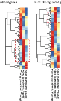
2011). The lists of PGC-1α- and mTOR-regulated genes were retrieved from Ingenuity Knowledge Base. Hierar- chical clustering analysis revealed only weak similarities on aortic expression profiles of NMN-regulated, SIRT1- and PGC-1α-dependent, and mTOR-regulated genes in young mice and aged mice exposed to young blood (red box). The ratio of discordant genes within the gene list was 9.5% (NMN-regulated genes), ~ 3.6% (SIRT-1-regulated genes), 8.5% (PGC-1α-regulated genes), and 3.4%
(mTOR-regulated genes).
Effect of young blood on the transcriptional footprint of signaling pathways activated by putative circulating anti-geronic factors in the vasculature
IGF-1 (insulin-like growth factor-1), GDF11 (growth dif- ferentiation factor 11; also known as bone morphogenetic protein 11 or BMP11), TGFβ (transforming growth factor-
β), and oxytocin (Elabd et al. 2014) were suggested to act as circulating factors regulating aging phenotypes in a context-dependent manner (Ungvari et al. 2018a). IGF-1 has clearly established anti-aging effects in many tissues, including the vasculature, myocardium, and brain, which are unrelated to the effects of developmental IGF-1 defi- ciency on lifespan (Ashpole et al. 2016; Ashpole et al.
2017; Tarantini et al. 2017; Tarantini et al. 2016a; Toth et al. 2015b; Toth et al. 2014b; Sonntag et al. 2013;
Bailey-Downs et al. 2012a; Mitschelen et al. 2011; Colon
et al. 2019). To determine the extent, if any, to which
changes in the aforementioned factors contribute to vas-
cular rejuvenation through heterochronic parabiosis, we
Table 1 Predicted upstream transcriptional regulators that may mediate the observed vasoprotective effects of circulating anti- geronic factors present in young blood. Results of the IPA Up- stream Regulator analysis. Shown are predicted upstream tran- scriptional regulators that may contribute to the observed young
blood–induced transcriptomic changes in our dataset. The overlap pvalue calls likely upstream regulators based on significant over- lap between dataset genes and known targets regulated by a transcriptional regulator
Upstream regulator
Name Remark Predicted
activation state
Activation z-score
pvalue of overlap
RICTOR Rapamycin-insensitive companion of mTOR
Subunit of mTORC2; regulates cell growth and survival in response to hormonal signals. Promotes inflammation, (Bhattacharya et al.2013) atherosclerosis, (Babaev et al.2018) and aneurysm formation (Parker et al.2018);
Inhibited −3.873 5.8E−18
KDM5A lysine [K]-specific demethylase 5A
Histone demetylase, repressor of metabolic regulatory genes (Varaljai et al.2015)
Inhibited −3 6.62E−12
STK11 Serine/threonine kinase 11 Activates AMPK; regulates autophagy, energy metabolism; tumor suppressor;
Activated 2.828 6.11E−09
LMX1A LIM homeobox
transcription factor 1α Regulates mitochondrial functions (Doucet-Beaupre et al.2016)
Activated 2 1.23E−08
LMX1B LIM homeobox
transcription factor 1β Regulates mitochondrial functions (Doucet-Beaupre et al.2016)
Activated 2 0.00000015
PPARGC1A PPARG coactivator
1 alpha; PGC-1α Regulates the genes involved in energy metabolism; interacts with PPARγ;
regulates the activities of CREB and nuclear respiratory factors; regulates mitochondrial biogenesis in endothelial cells (Patten and Arany2012);
Activated 2.789 3.36E−08
RB1 Retinoblastoma protein Tumor suppressor; controls cell cycle progression and senescence
Activated 3 0.000000256
CAB39L Calcium-binding protein 39-like
Activates AMPK, regulates mTOR pathway Activated 2 0.00000122 MAP4K4 Mitogen-activated protein
kinase kinase kinase kinase 4
Abundantly expressed in endothelial cells;
regulates cell migration, proliferation and adhesion, and inflammatory processes;
its ablation inhibits atherogenesis (Roth Flach et al.2015)
Inhibited −2.236 0.00000491
NRIP1 Nuclear
receptor-interacting protein 1
Modulates hormone-dependent activation of nuclear receptors; coactivatot of NF-kB
Inhibited −2 0.00000831
IGF1R IGF-1 receptor IGF-1 declines in aging; decline in circulating IGF-1 or knockdown of IGF1R in endothelial and smooth muscle cells mimics vascular aging phenotypes (Ashpole et al.2017;
Tarantini et al.2017; Tarantini et al.2016a;
Toth et al.2015b; Toth et al.2014b;
Sonntag et al.2013; Bailey-Downs et al.2012a;
Fulop et al.2019; Tarantini et al.2016b)
Activated 2.449 0.0000207
ESRRA Estrogen-related receptor alpha
Regulates mitochondrial function Activated 2 0.0000766
PITX2 Paired-like homeodomain 2 Transcription factor; regulates Wnt-mediated beta catenin signaling; regulates differentiation of smooth muscle cells in aorta
(Liu et al.2002; Shang et al.2008)
Activated 2 0.000136
analyzed changes in the expression of known downstream targets of IGF-1, GDF11, TGFβ, and oxytocin. Figure 6 shows heatmaps representing normalized expression values of genes known to be regulated by IGF-1 via IGF1R activation (Fig. 6a), GDF11 (Fig. 6b), TGFβ (Fig. 6c), activation of SMAD2 and SMAD3 signaling (Fig. 6d, e,), and oxytocin (Fig. 6f). Hierarchical clustering analysis revealed weak similarities on aortic expression profiles of specific gene clusters known to be regulated by IGF-1, GDF11, TGFβ, and oxytocin, respectively, in Y-(Y) mice and young blood exposed A-(Y) mice.
Discussion
The key finding of this study is that a relatively short- term exposure to a young humoral environment can rescue vascular aging phenotypes, including opposing increased oxidative stress, mitochondrial impairment, and consequential endothelial dysfunction as well as dysregulation of vascular gene expression in a mouse model of aging that recapitulates key aspects of age- related vascular dysfunction manifested in elderly patients.
Fig. 5 Effect of young blood on transcriptional footprint of shared mechanisms of vascular aging. Restoration of cellular NAD+
levels by treatment with the NAD+ precursor nicotinamide mono- nucleotide (NMN) (Tarantini et al.2019b; Kiss et al.2019a; Kiss et al.2019b; Das et al.2018; Csiszar et al.2019b), activation of SIRT-1 (Gano et al. 2014) and inhibition of mTOR pathways(Lesniewski et al.2017; Lin et al.2013) were shown to reverse vascular aging phenotypes. The heat maps are graphic representation of normalized expression values of NMN- regulated genes (a), SIRT-1-dependent genes (b), PGC-1α- regulated genes (c) and mTOR-regulated genes (d) in aorta sam- ples derived from isochronic parabiont young mice (parabiont:
young), isochronic parabiont aged mice (parabiont: aged) and heterochronic parabiont aged mice (parabiont: young). NMN- regulated genes were identified based on their differential
expression in response to restoration of cellular NAD+ levels by treatment with the NAD+ precursor nicotinamide mononucleotide (NMN) in aged cerebromicrovascular endothelial cells (Kiss et al.
2020). SIRT1 dependent genes were identified based on their differential expression in the heart of mice with cardiac-specific overexpression ofSirt1(b) (Oka et al. 2011) and the brain of Sirt1−/−mice (Libert et al.2011) (b, right). The lists of PGC-1α- and mTOR-regulated genes were retrieved from Ingenuity Knowl- edge Base (c,d). Hierarchical clustering analysis revealed the only weak similarities on aortic expression profiles of NMN-regulated, SIRT1- and PGC-1α-dependent and mTOR-regulated genes in young mice and aged mice exposed to young blood (red box).
The ratio of discordant genes within the gene list was 9.5% (NMN- regulated genes), ~3.6% (SIRT-1 regulated genes), 8.5% (PGC-1α regulated genes) and 3.4% (mTOR-regulated genes)
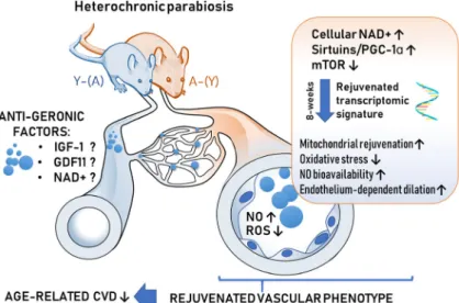
Elucidating the mechanisms by which aging impairs vascular responses and their potential reversal are criti- cal for the development of new targets and effective therapies for prevention and treatment of cardiovascular and cerebrovascular diseases in older adults. Here, we show for the first time that circulating factors present in the blood of young mice rescue NO-mediated endothe- lium-dependent vasodilation and attenuate vascular ox- idative stress in aged mice. Previous studies also dem- onstrated that presence of young blood in the circulation of aged heterochronic parabionts also reverses age- related cerebromicrovascular rarefaction (Katsimpardi et al. 2014). In that regard, it is also important that circulating non-cellular factors present in sera derived from caloric restricted non-human primates was also shown to improve endothelial angiogenic processes in cultured endothelial cells (Csiszar et al. 2013). The presence of young blood in the circulation of aged heterochronic parabionts (Fig. 7) also reversed select
transcriptional changes of aging in the aorta supporting the concept that non-cell autonomous mechanisms play important roles in driving vascular aging processes and pathogenesis of age-related cardiovascular diseases (Ungvari et al. 2018a). The remarkable level of mallea- bility of vascular aging phenotypes in response to anti- geronic circulating factors highlight the potential for therapeutic interventions to functionally reverse the del- eterious effects of aging in the circulatory system via targeting the systemic milieu or the endothelium directly to prevent cardiovascular morbidity and mortality.
Mitochondrial dysregulation plays a central role in the genesis of vascular aging phenotypes, including increases in oxidative stress and endothelial dysfunction (Tarantini et al. 2018; Csiszar et al. 2014; Ungvari et al.
2007; Springo et al. 2015; Ungvari et al. 2008). In the aged vasculature, the efficacy of the respiratory chain diminishes due to dysregulated expression of electron transport chain subunits, promoting electron leakage,
Fig. 6 Effect of young blood on transcriptional footprint of sig-naling pathways activated by putative circulating anti-geronic factors. IGF-1, GDF11 (Katsimpardi et al.2014; Katsimpardi et al.2020), TGFβand oxytocin (Elabd et al.2014) were sug- gested to act as circulating factors modulating cellular aging phenotypes. The heat maps are graphic representation of normal- ized expression values of genes regulated by IGF-1 via IGF1R activation (a), GDF11 (b), TGFβ(c), activation of SMAD2 and
SMAD3 signaling (d,e) and oxytocin in aorta samples derived from isochronic parabiont young mice (parabiont: young), isochronic parabiont aged mice (parabiont: aged) and heterochronic parabiont aged mice (parabiont: young). Gene lists were retrieved from Ingenuity Knowledge Base. Hierarchical clustering analysis revealed the similarities on aortic expression profiles of genes regulated by circulating anti-geronic factors in young mice and aged mice exposed to young blood (red box)
and increased mitochondrial ROS (mtROS) production and reducing cellular ATP generation (Tarantini et al.
2019b). Importantly, treatments that restore mitochon- drial gene expression and/or normalize mtROS produc- tion in the vasculature have been shown to improve endothelial function in rodent models of aging (Tarantini et al. 2018; Gioscia-Ryan et al. 2014;
Tarantini et al. 2019b; Pearson et al. 2008). Here, we report that circulating factors present in the blood of young mice rescue aging-induced changes in mitochondrion-related gene expression in the aorta of aged parabionts. On the basis of previous findings, (Gomes et al. 2013) we posit that rescue of vascular mitochondrial function by restoring the expression of ETC subunits contributes to the endothelial protective effects of young blood. In addition, upregulation of enzymes involved in the TCA cycle and antioxidant defenses may also contribute to young blood
–mediated vasoprotection.
Importantly, endothelial cells come in contact with each plasma constituent (including hormones, proteins, peptides, lipid mediators, micropeptides, metabolites, and circulating exosomes) as well as circulating cellular factors, all of which may exert important vasoprotective effects. Interestingly, circulating non-cellular factors
present in the young blood were also shown to confer rejuvenating effects on the central nervous system of aged mice after intravenous delivery, including an increased neurogenesis, improved learning and memory, and atten- uated neuro-inflammation (Wyss-Coray 2016), suggest- ing that key circulating anti-geronic factor(s) may pene- trate the blood-brain barrier and/or exert their neuropro- tective effects by rejuvenating the microcirculation.
The exact nature and the cellular origins of circulat- ing anti-geronic factors responsible for the vascular rejuvenating effects observed in our studies remain ob- scure. Recent evidence supports the concept that aging is associated with cellular NAD
+depletion (Gomes et al.
2013; Massudi et al. 2012; Yoshino et al. 2018) and consequential SIRT1 dysfunction, which are critical driving forces of vascular aging processes contributing to increased oxidative stress and the genesis of endothe- lial dysfunction (Tarantini et al. 2019b; Kiss et al.
2019a; Kiss et al. 2019b). Another evolutionarily con- served fundamental mechanism involved in vascular aging and the endothelial dysfunction is mTOR (mech- anistic/mammalian target of rapamycin) activation that governs cellular responses to nutrient availability and growth signals controlling biosynthesis of proteins, lipids, and nucleic acids (Lin et al. 2013; Parlar et al.
Fig. 7 Proposed scheme for the mechanisms contributing to young blood–mediated vascular rejuvenation in aged heterochonic parabionts. The model, based on our present and previous findings and earlier data from the literature (Ungvari et al.2018a; Tarantini et al.2018; Tarantini et al.2019b; Csiszar et al.2019b; Van Skike et al.2018), predicts that circulating anti-geronic factors present in young blood (derived from heterochronic parabiont young mice [Y-(A)]) restore cellular NAD+, activate sirtuins/PGC-1αand/or inhibit mTOR mediated pathways, promoting transcriptomic
changes, which rescue a youthful vascular phenotype in and heterochronic parabiont aged mice [A-(Y)]. Young blood– mediated vascular rejuvenation is associated with restored mito- chondrial function, attenuation of cellular and mitochondrial ROS production, increased bioavailability of NO and improved endothelium-mediated vasodilation. All of these effects are pre- dicted to act to improve vascular health, preventing the pathogen- esis of age-related vascular diseases. CVD: cardiovascular dis- eases; Y-(A)
2010; Cheng et al. 2008; Corbin et al. 1994; Milliard et al. 1998). Accordingly, restoration of cellular NAD+
levels by treatment with NMN (Tarantini et al. 2019b;
Kiss et al. 2019a; Kiss et al. 2019b; Das et al. 2018;
Csiszar et al. 2019b), activation of SIRT1,(Gano et al.
2014) and inhibition of mTOR pathways (Lesniewski et al. 2017; Lin et al. 2013; Van Skike et al. 2018) were shown to reverse vascular aging phenotypes and im- prove endothelial function. Heterochoronic parabiosis is expected to increase levels of NAD+ precursors in the circulation, and consequently restore NAD+ levels in the vasculature, of aged parabionts. There are known circulating anti-geronic factors (e.g., induced by caloric restriction) that activate SIRT1 (Csiszar et al. 2009;
Csiszar et al. 2013; de Cabo et al. 2003) and its down- stream target PGC-1α and/or regulate mTOR.
Transcriptomic analysis of the aortas suggests that these pathways may contribute to vascular rejuvenation in- duced by young blood. Further studies should investi- gate in detail the effects of heterochronic parabiosis or systemic administration of young plasma to aged mice on cellular NAD+ levels and SIRT1 and mTOR activi- ties in the vasculature. In that regard, it is interesting that a recent study demonstrates that age-related decline in NAD+ levels in red blood cells is not corrected by heterochronic parabiosis (Morrison et al. 2019). The effects of young blood on vascular aging phenotypes should be also investigated in mice with genetic deple- tion of SIRT1.
Previous studies identified age-related changes in cir- culating anti-geronic factors (e.g., IGF-1 (Ashpole et al.
2016; Ashpole et al. 2017; Tarantini et al. 2017; Toth et al. 2015b; Toth et al. 2014b; Bailey-Downs et al.
2012a; Mitschelen et al. 2011; Bailey-Downs et al.
2012b; Fulop et al. 2019; Tarantini et al. 2016b; Ungvari and Csiszar 2012), which can be causally linked to the development of aging phenotypes in multiple organ sys- tems. Importantly, decreases in circulating levels of IGF- 1 (Tarantini et al. 2016b) were shown to contribute to key aspects of vascular aging, including endothelial dysfunc- tion (Toth et al. 2015b), impaired autoregulation of cere- bral blood flow (Toth et al. 2014b), pathological vascular remodeling, atherogenesis (Higashi et al. 2012), impaired vascular stress resilience (Bailey-Downs et al. 2012a;
Bailey-Downs et al. 2012b), impaired angiogenic pro- cesses, and microvascular rarefaction (Lahteenvuo and Rosenzweig 2012). In the present study, IGF1R signaling was identified as a likely upstream regulator involved in young blood–mediated vascular rejuvenation. Future
studies should further interrogate experimentally the role of circulating IGF-1 in mediation of the vasoprotective effects of young blood transfer (e.g., using animal models with endothelium-specific knockdown of IGF1R as parabionts).
Other circulating factors, whose levels are rescued in aged mice by heterochronic parabiosis or systemic ad- ministration of young plasma and which may confer rejuvenating effects in multiple organs (e.g., brain, heart, and skeletal muscle (Katsimpardi et al. 2014;
Katsimpardi et al. 2020; Loffredo et al. 2013; Poggioli et al. 2016; Sinha et al. 2014)) include GDF-11, a mem- ber of the TGF-β superfamily of growth factors (Katoh and Katoh 2006). GDF11 is known to activate SMAD2/
SMAD3 signaling in endothelial cells (Poggioli et al.
2016; Finkenzeller et al. 2015). However, transcriptomic analysis of the aortas did not confirm that GDF11/
SMAD2/3 signaling plays a major role in young blood
–mediated vascular rejuvenation. Importantly, the possible rejuvenating effects of GDF11 were also questioned by several recent studies (Egerman et al. 2015; Rodgers and Eldridge 2015; Smith et al. 2015b). It is of note that the pleiotropic growth factor TGFβ also induces SMAD2/
SMAD3 (Nakao et al. 1997) and that increased TGF
βsignaling has been linked to the genesis of vascular aging phenotypes (Gaertner et al. 2005; Masliah et al. 2001;
Wyss-Coray et al. 1997a; Wyss-Coray et al. 1995; Wyss- Coray et al. 2000; Wyss-Coray et al. 1997b). Oxytocin was also proposed to mediate rejuvenating effects in the aged skeletal muscle (Elabd et al. 2014), yet it is unlikely that it plays an important role in young blood–induced changes in the vascular transcriptome.
Perspectives
Our findings provide additional evidence supporting the significant plasticity of vascular aging and its amenabil- ity to modulation by circulating anti-geronic factors.
Future studies will be necessary to mechanistically ex- pand on the present observations, including investiga- tions aimed at defining the exact nature of the non- autonomous factors that drive vascular aging. The role of age-related changes in circulating hormones, cyto- kines, and growth factors in development of vascular aging phenotypes should be methodologically elucidat- ed. Studies using mass spectrometry
–based approaches led to the identification of complex age-related changes in circulating metabolites, proteins (Lehallier et al.
2019), micropeptides, and lipid mediators. Subsequent
studies should identify changes that are not strain-, species-, or sex-dependent and elucidate their role in modulating vascular health. Further studies are also needed to identify the critical organ(s), tissues, and cell types that contribute to the regulation of vascular aging processes via circulating factors. It should be noted that in heterochronic parabionts all the organs are shared via the conjoined circulation. In addition to the brain (e.g., by releasing hypothalamic hormones and peptides and neuroendocrine factors) and endocrine organs, aged mice can benefit from access to the young bone marrow, liver, lungs, immune system, adipose tissue, skeletal muscle, heart, kidneys, and the gastrointestinal system and microbiome. In addition to the decline in anti- geronic factors, pro-geronic circulating factors, whose levels increase with age, may also contribute to the genesis of vascular aging phenotypes (Rebo et al.
2016). For example, there is strong evidence that TNFα(Csiszar et al. 2007c) and other cytokines (e.g., CCL11, TGFβ (Masliah et al. 2001) may serve as pro- geronic factors. Importantly, many of these factors are secreted by senescent cells in distant organs (e.g., the adipose tissue (Baker et al. 2011; Tchkonia et al. 2013).
Further analysis of our dataset may lead to the identifi- cation of important circulating pro-geronic factors that contribute to the induction of aging phenotypes in young parabionts. It will also be important to determine which of the non-autonomous changes observed in the vascular tissues are actually causative of age-related vascular pathologies. Targeting these mechanisms might offer potential new treatments for prevention of age-related vascular diseases.
Funding information This work was supported by grants from the American Heart Association (ST), the Oklahoma Center for the Advancement of Science and Technology (to AC, AY, ZU), the National Institute on Aging (R01-AG047879; R01-AG038747;
R01-AG055395), the National Institute of Neurological Disorders and Stroke (NINDS; R01-NS056218 to AC, R01-NS100782 to ZU), the Oklahoma Shared Clinical and Translational Resources (OSCTR) program funded by the National Institute of General Medical Sciences (GM104938, to AY and JW), the Presbyterian Health Foundation (to ZU, AC, AY), the NIA-supported Geroscience Training Program in Oklahoma (T32AG052363), the Oklahoma Nathan Shock Center (P30AG050911), and the Cellular and Molecular GeroScience CoBRE (1P20GM125528, sub#5337). DMH is supported by R21AG055026, the American Federation for Aging Research (AFAR) and the Einstein Nathan Shock Center (P30AG038072). The funding sources had no role in the study design; in the collection, analysis, and interpretation of data; in the writing of the report; and in the decision to submit the article for publication.
References
Ashpole NM, Herron JC, Mitschelen MC, Farley JA, Logan S, Yan H, et al. IGF-1 regulates vertebral bone aging through sex-specific and time-dependent mechanisms. J Bone Miner Res. 2016;31(2):443–54.https://doi.org/10.1002/jbmr.2689.
Ashpole NM, Logan S, Yabluchanskiy A, Mitschelen MC, Yan H, Farley JA, et al. IGF-1 has sexually dimorphic, pleiotropic, and time-dependent effects on healthspan, pathology, and lifespan. Geroscience. 2017;39:129–45.
Babaev VR, Huang J, Ding L, Zhang Y, May JM, Linton MF. Loss of Rictor in monocyte/macrophages suppresses their prolif- eration and viability reducing atherosclerosis in LDLR null mice. Front Immunol. 2018;9:215.
Bailey-Downs LC, Mitschelen M, Sosnowska D, Toth P, Pinto JT, Ballabh P, et al. Liver-specific knockdown of IGF-1 de- creases vascular oxidative stress resistance by impairing the Nrf2-dependent antioxidant response: a novel model of vas- cular aging. J Gerontol A Biol Sci Med Sci. 2012a;67:313–
29.
Bailey-Downs LC, Sosnowska D, Toth P, Mitschelen M, Gautam T, Henthorn JC, et al. Growth hormone and IGF-1 deficiency exacerbate high-fat diet-induced endothelial impairment in obese Lewis dwarf rats: implications for vascular aging. J Gerontol A Biol Sci Med Sci. 2012b;67:553–64.
Baker DJ, Wijshake T, Tchkonia T, LeBrasseur NK, Childs BG, van de Sluis B, et al. Clearance of p16Ink4a-positive senes- cent cells delays ageing-associated disorders. Nature.
2011;479:232–6.
Bhattacharya I, Dragert K, Albert V, Contassot E, Damjanovic M, Hagiwara A, et al. Rictor in perivascular adipose tissue controls vascular function by regulating inflammatory mole- cule expression. Arterioscler Thromb Vasc Biol. 2013;33:
2105–11.
Bitto A, Kaeberlein M. Rejuvenation: it's in our blood. Cell Metab.
2014;20:2–4.
Bolger AM, Lohse M, Usadel B. Trimmomatic: a flexible trimmer for Illumina sequence data. Bioinformatics. 2014;30:2114– 20.
Bray NL, Pimentel H, Melsted P, Pachter L. Near-optimal proba- bilistic RNA-seq quantification. Nat Biotechnol. 2016;34:
525–7.
Cannata A, Marcon G, Cimmino G, Camparini L, Ciucci G, Sinagra G, et al. Role of circulating factors in cardiac aging.
J Thorac Dis. 2017;9:S17–29.
Castellano JM, Palner M, Li SB, Freeman GM Jr, Nguyen A, Shen B, et al. In vivo assessment of behavioral recovery and circulatory exchange in the peritoneal parabiosis model. Sci Rep. 2016;6:29015.
Cheng C, Tempel D, Oostlander A, Helderman F, Gijsen F, Wentzel J, et al. Rapamycin modulates the eNOS vs. shear stress relationship. Cardiovasc Res. 2008;78:123–9.
Coleman DL. Effects of parabiosis of obese with diabetes and normal mice. Diabetologia. 1973;9:294–8.
Coleman DL, Hummel KP. Effects of parabiosis of normal with genetically diabetic mice. Am J Phys. 1969;217:1298–304.
Colon G, Saccon T, Schneider A, Cavalcante MB, Huffman DM, Berryman D, et al. The enigmatic role of growth hormone in age-related diseases, cognition, and longevity. Geroscience.
2019;41:759–74.