CHAPTER X I I Biochemistry of Androgens B Y R A L P H I. D O R F M A N
C O N T E N T S
Page
I. Sources of Androgens 469
A. Testis 469 B. Ovary 470 C. Adrenal Cortex 470
D . Placenta 471 II. Isolation of Androgens and Related Compounds 471
A. Urinary Androgens 471 B. Isolation of Androsterone and Dehydroisoandrosterone 472
C. Conversion of Dehydroisoandrosterone to Androsterone 474
D . Partial Synthesis of Androsterone and Isomers 475 E. Isolation of Testosterone from Testis Tissue 476
F. Partial Synthesis of Testosterone 477 G. Androgens from Adrenal Tissue 477 H. Isolation of 11-Hydroxyandrosterone 478
I. Isolation of A5-Androstenetriol-3(/3), 16,17 479
J. C2I Compounds Isolated from Testis Tissue 481 K . Isolation of A1 6-Androstenol-3(a) and A1 6-Androstenol-3(/3) 481
L. An Androstanol-3(/3)-one from Pregnant Mare Urine 481 M . Miscellaneous Substances Isolated from Swine Testes 482
N . Summary of Androgens and Related Substance from Natural Sources 483
I I I . Form in Which Androgens Occur in Urine 484
A. Isolation of Sulfate Esters 485 B. Artifacts in Urinary Extracts 486
1. Artifacts of Degradation 486 2. Artifacts of Substitution 487 3. Artifacts of Dehydration 487 IV. Assay of Androgens and Related Substances 488
A. Bioassay of Androgens 488 1. Capon's C o m b Assay b y Intramuscular Injection 489
2. Capon Assays b y Direct Application to the C o m b 490
3. Chick's C o m b M e t h o d of Assay 490 4. Mammalian Assay Methods 492 5. T h e International Androgen Standard 493
B. Polarographic Determination of Androgens and Related Compounds 493 C. Chemical Determination of Androgens and Related Compounds 494 V. Concentration of Androgens and 17-Ketosteroids in Urine and Blood 496
A. Concentration of Androgens and 17-Ketosteroids in Human Urine 497 467
Page
1. Children 497 2. Normal Adult Men and Women 500
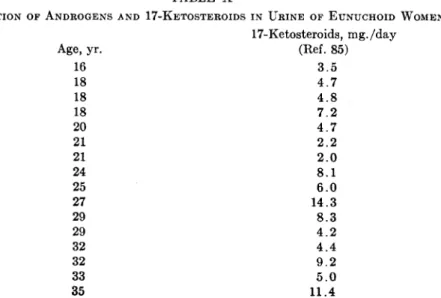
3. Senile Men and W o m e n 502 4. Eunuchoid and Castrate Men 506 5. Hypogonadal and Ovariectomized Women 506
6. Addison's Disease 508 7. Pitu tary Abnormalities 508 8. Thyroid Disease 509 9. Testicular and Embryonic Tumors 510
10. Precocious Puberty 511
11. Hirsutism 511 12. Ovarian Tumors 511 13. Adrenal Cortical Hyperactivity 512
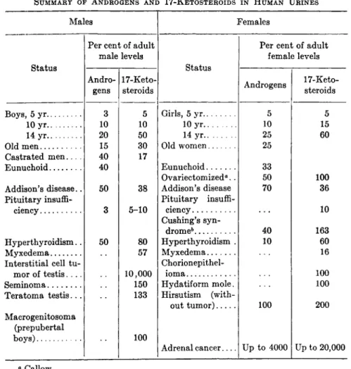
14. Urine 17-Ketosteroid Excretion in Miscellaneous Conditions 512 15. Summary of Changes in Androgens and 17-Ketosteroids in
Human Urines 515 B. Androgens and 17-Ketosteroids in Urines Other Than Human 516
1. Stallion, Ram, Bull, Cow, and Rat Urine 516
2. Chimpanzee Urine 516 3. Monkey Urine 516 C. Concentration of Androgens in Blood 517
VI. Metabolism of Androgens 517 A. Absorption of Androgens b y Various Routes 517
B. Anabolic Considerations 519 C. Catabolic Considerations 520
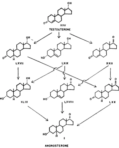
1. Metabolism of Testosterone 520
2. Significance of A6-Stenols 524
3. Possible Conversion of Androgens to Phenolic Estrogens 525 4. Possible Conversion of Adrenal Cortical Steroids to 17-Keto-
steroids 526 a. 5-Oxygen Compounds Having a 3(ß)-Hydroxyallo
Configuration 527
b. 5-Oxygen-A4-stenone Compounds 530
c. 5-Oxygen-3(a)-hydroxyallo Compounds 530 d. 4-Oxygen Compounds having a 3(/3)-Hydroxyallo Config-
uration 530
e. 4-Oxygen-A4-stenone Compound 530
/ . 17-Ketosteroids in Adrenal Cortex 532 g. 3-Oxygen Compounds with a 17-Hydroxy Croup 532
h. Concerning Metabolism of Desoxycorticosterone Acetate
and Adrenal Cortical Extracts to 17-Ketosteroids 533
5. Site of Inactivation of Androgens 534 D . Enzymic Changes Due to Bacteria and Yeasts 534
V I I . Mechanism of Action of Androgens 535 V I I I . Inhibitory Effects of Certain Compounds on Action of Androgens 538
I X . Ability of Androgens to Inhibit Action of Other Steroid Hormones 539 X . Possible Androgenic Activity of Other Hormones and Pregneninolone... . 539
References 540 Compound Numbers 546
XII. BIOCHEMISTRY ΟΈ* ANDROGENS 469 I. Sources of Androgens
As a result of detailed investigations involving sometimes delicate bioassay methods, sometimes careful clinical observations, and some- times histological and cytological studies, it has now been established that androgens are produced in at least three tissues and probably in a fourth. Well-defined pure substances possessing androgenic activity have been isolated and identified from testicular and adrenal tissue.
Although no pure androgen has been isolated from ovarian tissue, the physiological evidence of its production there seems convincing. The placenta may elaborate androgenic material, but the evidence at the pres- ent moment seems indecisive.
A. TESTIS
The earliest investigators were able to relate the physiological influ- ences of castration and the loss of a biologically active substance, the androgen. The first convincing demonstration of androgens in testis came from the laboratory of Professor Koch, where McGee in 1927 (148) demonstrated androgenic material in lipid extracts of bull testis. Sub- sequent studies, including isolations of pure androgens from testis tissue, are discussed elsewhere in this chapter (see page 464).
Sufficient evidence is at hand to prove that the androgenic hormone of the testis is produced by the interstitial tissue. Thus, it is well known that, if the seminal epithelium is caused to atrophy without damage to the interstitial tissue, androgenic material continues to be secreted by the modified testis. If an animal is made cryptorchid (158) or the testis treated with adequate dosage of X-rays (207), such a condition is realized. Experiments of this sort have been carried out on dogs, horses, pigs, guinea pigs, rabbits, and rats with similar results. In spite of the destruction of the seminal epithelium, the accessory genital organs are preserved in a normal functioning state. In one experiment extracts of the cryptorchid testis of swine were demonstrated to contain androgenic material when tested on the capon's comb.
A second line of proof that interstitial tissue rather than seminal epithelium is responsible for the elaboration of androgenic material is found in those experiments in which the interstitial tissue is caused to atrophy while the seminal epithelium remains intact. When pitch was administered to rodents, it was found that the interstitial tissue of the testis was damaged with a parallel atrophy of the secondary sex glands, in spite of the fact that the seminal epithelium remained normal (9).
In another report (159), interstitial damage was produced in rats by feeding the animals a vitamin-B-complex-deficient diet. Again the
seminal epithelium remained normal, but the prostate and seminal vesicles atrophied.
Finally, it has been shown that certain interstitial cell tumors of the testis in humans produce enormous quantities of androgenic material.
In one such case Venning et al. (212) have reported a titer of 1015 mg./day of 17-ketosteroids, which represents an increase of about 10,000%.
Evidence, therefore, indicates that the source of the androgens in the testis is the interstitial tissue.
B . OVARY
In experiments on fat-soluble extracts of sow ovarian tissue, it has been shown that such extracts do contain androgenically active material
(172). This work is reinforced by the striking experiments of Hill (106-108), who showed that, if ovaries are transplanted to the ears of castrated male mice, androgenic stimulation of the otherwise involuting accessories is observed. Here it is of interest to note that, although the ovary normally secretes androgenic material, exteriorizing the organ, as in transplantation to the ear, increases the rate of synthesis of the androgens. From the work of Hill it appears that the important factor is that of temperature, since ovaries grafted into the abdomen of cas- trated males were unable to maintain the accessories. In a series of experiments with rats Deanesley (48) confirmed the findings of Hill.
C. ADRENAL CORTEX
Androgens in the adrenal cortex have been demonstrated by the direct isolation of androgenic substances from adrenal cortical extracts and by the presence of high concentrations of androgenic material in body fluids in conditions of increased activity of the adrenal cortex, and indirectly by the presence of androgenic material in the complete absence of the gonads.
Adrenosterone ( X X I ) (188,189), androstanediol-3(0),ll(0)-one-17 ( X X I I I ) (166), A4-androstenedione-3,17 ( X X I I ) (215), and 17-"/3"- hydroxyprogesterone ( X X I V ) (177,178) have been isolated from adrenal cortical extracts (for formulas, see page 532). All four have been shown to possess androgenic activity. The possibility still exists that the C i9
steroids are not present in the gland as such, but rather that they are formed during the process of working up the extracts. On the other hand, other androgens may be present in the gland which as yet have escaped isolation. This may be said since no isolations of adrenal corti- cal extracts have been carried out with the object of isolating androgens, and therefore important glandular androgens may not yet have been isolated.
XII. BIOCHEMISTRY OF ANDROGENS 471 The administration of adrenotrophic extracts to castrated rats caused the development of the prostate and seminal vesicles (47). The adrenal cortices were greatly enlarged in these animals. The effect was absent in adrenalectomized rats but demonstrable in hypophysectomized animals. These experiments have been confirmed (165).
Female mice of the strain CE, when spayed at one to three days of age, develop tumors which appear to secrete androgenic material as evidenced by the growth and development of the accessory sex organs.
The same was true for castrated males of the same strain (221,222).
Masculinization as a result of hyperactivity of the adrenal cortex is well known in women. Increases in androgenic material in the urine of such patients are found. In 1936 Callow (30) reported the isolation of massive amounts of the androgen, dehydroisoandrosterone,from the urine of a six-year-old girl suffering from an adrenal cortical cancer. Numer- ous other similar cases in females have been reported and are discussed elsewhere (page 513).
Ovariectomized women excrete considerable quantities of androgenic material (31), and it has been possible to isolate two androgens, andro- sterone (I) and dehydroisoandrosterone (III), from the urine of such sub- jects in amounts only slightly less than that found in the urine of normal women (110).
The castration of week-old rats does not arrest the development of the seminal vesicles and prostate, but instead these organs continue to develop until the fifth week of life (185). If, however, young rats are adrenalectomized and castrated, complete atrophy of the prostate results (11,12).
D . PLACENTA
It has been reported that extracts of human placental tissue contain androgenic material (42). Experiments in the author's laboratory have not confirmed these findings.
II. Isolation of Androgens and Related Compounds A. URINARY ANDROGENS
Androgens have been studied in the urines of normal individuals as well as of those with various pathological conditions. Human urines contain relatively high concentrations of androgens as compared to other species. These biologically active substances have been demonstrated in the urine of men and women as well as children of both sexes. In addition to these substances, which possess androgenic activity, a number of steroids have been isolated which possess no biological activity, yet are chemically closely related to the androgens. In some cases these
472
biologically inert substances have been shown to be derived from andro- genically active material. The metabolic considerations are discussed elsewhere (page 517).
Within a period of two years three laboratories were able to show the presence of androgenic material in both normal men's and women's urine. Loewe et al. (128) were able to show that men's urine contained material capable of stimulating the seminal vesicles of castrated mice.
Ο Ο
m
D E H Y D R O I S O A N D R O S T E R O N E
FI G . 1.—3-Chloro-A5-androstenone-17 converted to dehydroisoandrosterone.
Funk and Harrow (86) and Funk, Harrow, and Lejwra (87) also reported urinary androgens which could be extracted with fat solvents. From the Chicago laboratories papers by Womach and Koch (220) and Gallag- her and Koch (89) demonstrated that androgens were present in female as well as male urine.
B. ISOLATION OF ANDROSTERONE AND DEHYDROISOANDROSTERONE
Butenandt (17,18) announced the first isolations of crystalline material from concentrates of men's urine which possessed androgenic activity. In a later group of papers (19,20,25), the presence of an andro- gen with the empirical formula C19H30O2 (I) was demonstrated, as well as two other substances, C19H27OCI (II) and C i9H28 02 (HI).
The compound C I 9 H3o 02 (I) was named androsterone. It proved to be saturated, since it did not decolorize bromine. One oxygen atom
XII. BIOCHEMISTRY OF ANDROGENS 473 was present as a ketone group, since an oxime, a semicarbazone, and a phenylhydrazone could be prepared. The second oxygen atom was present as a secondary alcohol group, since the compound formed a monoacetate, and oxidation with chromic acid yielded a diketone. A structure was assigned to the compound which was later shown to be correct by partial synthesis. Butenandt and co-workers were able to show the relationship among the three substances isolated.
The chloro compound C19H27OCI (II) was found to be physiologically inactive. That the compound contained an unsaturated grouping was
H V
3 - C H L 0 R 0- A5- A N D R 0 S T E W 0 N E- L 7
Κ A C E T A T E A C E T I C A C I D
I VI
A N D R O S T E R O N E
FI G. 2.—3-Chloro-A5-androstenone-17 converted to androsterone.
shown by the facts that it decolorized bromine, gave a yellow color with tetranitromethane, and took up one mole of hydrogen when subjected to catalytic hydrogénation to form a saturated ketone. On treatment of the chloro compound with potassium benzoate and benzoic acid the compound Ci9H280(OOCC6H5) (IV) was obtained, which on hydrolysis yielded an unsaturated hydroxy ketone C I9H28 02 (III) identical with the second androgen isolated (Fig. 1). Treatment of the unsaturated hydroxy-ketone C 1 9 H 2 8 O 2 (III), which was biologically active, with hydrochloric acid and heat produced the androgenically inactive chloro compound C 1 9 H 2 7 O C I (II). Thus it was concluded that the chloro compound was probably an artifact and had been formed during the course of extraction and isolation.
C . CONVERSION OF DEHYDROISOANDROSTERONE TO ANDROSTERONE
It still remained to show the structural relationship between andro- sterone and dehydroisoandrosterone (III). The chloro derivative was subjected to catalytic hydrogénation and the saturated chloro derivative was obtained (V). This was treated with potassium acetate and acetic acid to form androsterone acetate (VI), which yielded androsterone (I) on saponification (Fig. 2).
FI G . 3.—Partial synthesis of dehydroisoandrosterone from cholesterol.
Dehydroisoandrosterone (III) was shown later to have the same steric configuration at C 3 as cholesterol (VII). It was possible to prepare this androgen from cholesterol (VII). The cholesterol was acetylated and brominated. The side chain was oxidized with chromic acid to yield the C 1 7 ketone which, on removal of the bromine, and hydrolyzing, yielded dehydroisoandrosterone identical with that prepared from the urinary extracts (Fig. 3) (19-21,23,25).
The partial synthesis of dehydroisoandrosterone was carried out almost simultaneously by three other groups of workers. Thus Ruzicka and Wettstein (196) and Wallis and Fernholz (216a) accomplished the partial synthesis from cholesterol, while Oppenauer (170) was able to convert α-sitosterol to dehydroisandrosterone.
XII. BIOCHEMISTRY OF ANDROGENS 475
D . PARTIAL SYNTHESIS OF ANDROSTERONE AND ISOMERS
Clarification of the structure of androsterone was accomplished by Ruzicka's group in 1934 (193), when androsterone and the other three sterieoisomers, involving carbon atoms 3 and 5, were prepared by partial synthesis. For the synthesis of androsterone, cholesterol was reduced
IX
A N D R O S T E R O N E
FI G . 4.—Partial synthesis of androsterone from cholesterol.
to dihydrocholesterol ( V I I I ) which was oxidized in turn to cholestanone ( I X ) with chromic acid. On hydrogénation in acid solution and sub- sequent acetylation, the cholestanone ( I X ) was converted into epidihy- drocholesterol acetate ( X ) . Oxidation of the side chain with chromic acid yielded androsterone acetate ( V I ) , and on hydrolysis androsterone which was identical with that isolated by Butenandt from the urine was realized (Fig. 4).
Ruzicka et al (193) also prepared, by partial synthesis from choles- terol, the three additional isomers of androsterone at C-3 and C-5 as illustrated in Fig. 5.
The partial synthesis of androsterone (I) was also accomplished by Butenandt et al from cholesterol (21), by Dirscherl (54) from cinchol, by
CPI0IHY0ROCHOLESTEROL ANDROSTERONE
X V I I X V I I I E P I C O P R O S T E R O L E T I O C H O L A N O L ' 3 ( ° < ) ' 0 N E - I 7
FI G . 5.—Schematic representation of partial synthesis of androsterone and three isomers from cholesterol.
Dalmer et al (43) from sitosterol and stigmasterol, and by Marker (135) and Marker et al (137) from cholesterol.
E . ISOLATION OF TESTOSTERONE FROM TESTIS TISSUE
After the isolation of the two androgens, androsterone and dehydroiso- androsterone, from men's urine it became apparent that the androgenic material in bull testis must be due to still another substance. The work of Gallagher and Koch (90) had indicated that the androgenic material
XII. BIOCHEMISTRY OF ANDROGENS 477 in testis tissue was more labile to alkali than the androgens in urine.
Secondly, the highly purified fractions from bull testis were more active on a weight basis than the pure compounds isolated from urine. Finally, when testis extracts and urinary extracts were administered to castrated rats or mice at equivalent levels in terms of capon units, the testis material proved to be far more active than the urinary extract. David et al. (46) finally were able to isolate an androgen, testosterone, from bull testis which was shown to be approximately six times more active than androsterone (I) on the basis of the capon's comb, and which differed chemically from androsterone (I). Recently David's isolation was con- firmed in the laboratory of Ruzicka, where testosterone ( X X V ) was isolated from the testis of stallions (205).
F. PARTIAL SYNTHESIS OF TESTOSTERONE
The synthesis of testosterone and proof of structure was quickly accomplished by Ruzicka and Wettstein (196) and by Butenandt and Hanisch (21). The partial synthesis as accomplished by Ruzicka and Wettstein consisted of converting cholesterol (VII) by oxidizing its dibromo acetate derivative to dehydroisoandrosterone acetate (IV).
The ketone group of dehydroisoandrosterone acetate was reduced to the A5-androstenediol-3(/3),17(a)-3-monoacetate ( X I X ) , which wasbenzoylated to form the 3-acetoxy, 17-benzoxy derivative. On partial saponi- fication the acetate group was removed with the formation of A5-andro- stenediol-3(0),17(a)-17-benzoate ( X X ) . This compound on bromination followed by oxidation, debromination, and saponification yielded testo- sterone ( X X V ) , which was identical with the compound isolated from testis tissue (Fig. 6).
The synthesis of Butenandt and Hanisch is similar to that described by Ruzicka and Wettstein.
G . ANDROGENS FROM ADRENAL TISSUE
Studies on adrenal cortical extracts have yielded four crystalline androgens: adrenosterone ( X X I ) , A4-androstenedione-3,17 ( X X I I ) , androstanediol-3(ß),ll-one-17 ( X X I I I ) , and 17-"/3"-hydroxyprogesterone ( X X I V ) . Although the extracts were subjected to relatively mild chemical treatment, it has been pointed out by Reichstein that these compounds may represent artificial degradation products of C2i com- pounds. This does not apply to 17-"ß"-hydroxyprogesterone ( X X I V ) . Adrenosterone ( X X I ) was isolated from an adrenal cortical extract by Reichstein (188,189). From similar extracts androstanediol-3(/3),ll- one-17 ( X X I I I ) (215), and A4-androstenedione-3,17 ( X X I I ) (215) were realized. The structural considerations of the first two androgens are discussed elsewhere (Chapter X I I I ) in this volume, and Reichstein and
von Euw (215) as well as Pfiffner and North (178) have reported the isolation of 17"ß"-hydroxyprogesterone ( X X I V ) . The constitution of
17"0"-hydroxyprogesterone ( X X I V ) has been proved by degradation studies (177,178), and partial synthesis has been accomplished.
V I I I V C H O L E S T R O L I
X I X P A R T I A L
S A P O N I F I C A T I O N
T E S T O S T E R O N E
FI G . 6.—Partial synthesis of testosterone from cholesterol.
Note: For cholestrol, read cholesterol in VII.
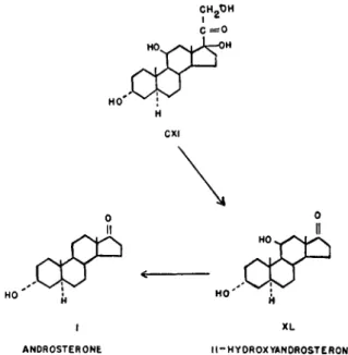
H. ISOLATION OF 11-HYDROXYANDROSTERONE
11-Hydroxyandrosterone (XL) was isolated first by Mason (141) and Mason and Kepler (144) from the urines of various patients showing adrenal cortical involvements, such as tumors and hyperplasia. The substance has also been isolated from the urine of a female pseudoherma- phrodite showing no apparent adrenal involvement by Miller, Dorfman, and Sevringhaus (154) and from normal male urine (142). The former workers found the compound to be androgenic by the chick comb test.
The formula C 1 9 H 3 0 O 3 was found by analysis, and on acetylation at 90°C. with acetic anhydride and pyridine, the compound yielded a monoacetate. The material was not precipitated with digitonin in 90%
methanol. It formed a yellow dinitrophenylhydrazone, and on oxida- tion with chromic acid gave a product identical with androstanetrione- 3,11,17 (XLI) (Fig. 7).
XII. BIOCHEMISTRY OF ANDROGENS 479 On treatment of the monoacetate with hydrochloric acid and acetic acid an androstenolone (XLII) was obtained, which appeared to be identical with one previously obtained from urine (153).
0
H X L I
A N O R O S T A N E T R I O N E - 3 , 1 1 , 1 7
G L A C I A L A C E T I C A C I O H Y 0 R 0 C H L O R I C A C I D
X L I I X L I I I A H D R 0 3 T A N E D I 0 L - 3 ( o < ) tl 7 ( < * >
FI G. 7.—Proof of structure of 11-hydroxyandrosterone.
I. ISOLATION OF A5-ANDROSTENETRIOL-3(/3), 16,17
A6-Androstenetriol-3(/3),16,17 (XLIV) was first isolated by Hirsch- mann (111) from the urine of a seven-year-old boy with an adenocar- cinoma of the left adrenal cortex. This steroid was also isolated from normal urine (139). The compound was inactive as an androgen at doses up to seventy times the amount necessary to produce a significant response in the chick's comb with androsterone (112). This steroid is of interest, however, particularly because of its relationship to dehy- droisoandrosterone (III). This relationship is similar to that found between estrone and estriol. Hirschmann was able to prove the struc- ture of this steroid by degrading the compound to a known ketodicar- boxylic acid (XLVI), and by the conversion of the monoacetate of the isolated compound into the known ß-3-hydroxy-A5-etiobilienic acid (XLVIII). Thus the isolated compound, which contained one nuclear double bond, was reduced with hydrogen and palladium to the saturated triol (XLV). The triol was oxidized with chromium trioxide at room temperature to form the ketodicarboxylic acid (XLVI) (Fig. 8).
In a second series of reactions, isoandrosterone (XIV) was converted to 16-benzylidinandrostanol-3(/3)-one-17 (XLVII), with sodium methyl- late and benzaldehyde. This product was acetylated with pyridine and acetic anhydride and oxidized with chromium trioxide, and after saponi-
o
X L
• l - H Y O R O X Y A N D R O S T E R O N E
H ' ο
ΧΙΝ ISOANOROSTERONR
F I G . 8.—Proof of structure of A6-androstenetriol-3(/3),16,17.
O H O H
β-3-HYDROXY-ZI-ETIOBILIENIC A C I O
. 9.—Conversion of AB-androstenetriol-3(ß),16,17 to £-3-hydroxy-A5-etiobilienie acid.
XII. BIOCHEMISTRY OF ANDROGENS 481 fication yielded the β-3-hydroxyetioallobilianic acid ( X L V I I I ) . The free ß-3-hydroxyetioallobilianic acid ( X L V I I I ) was oxidized with chromic acid to 3-ketoetioallobilianic acid ( X L V I ) ; this proved to be identical with that prepared from the isolated compound (Fig. 7).
From the above evidence it was apparent that the compound must be androstenetriol-3,16,17. The fact that the compound was precipitated with digitonin was indicative of a 3(/3)-hydroxy configuration. Com- parison of the optical rotations of androstenetriol triacetate and andro- stanetriol triacetate indicated that the double bond was between C-5 and C-6. Further evidence for the 3(0)-hydroxy and Δ5 was found by a third set of reactions. The 3-monoacetate of the isolated triol was brominated and oxidized with chromium trioxide. The reaction product was debrominated with sodium iodide, and j3-3-hydroxy-AB-etiobilienic acid ( L ) was isolated, which was identical with a known sample (Fig. 9).
J. C2I COMPOUNDS FROM TESTIS TISSUE
Marker et al. (136,138) have demonstrated that, although the bull excretes sizable amounts of such C21 steroids as pregnanediol-3(A),20(Û:)
( L X X V I I , page 432), allopregnanediol-3(OO,20(ÛO (CH) and allo- pregnanediol-3(/5),20(O!) ( L X X X I X , page 435), the steer does not. This would indicate that the testis is capable of producing some C21 steroids.
The work of Ruzicka and Prelog (194) has demonstrated the presence of two C21 compounds in swine testis, namely A5-pregnenol-3(/3)-one-20 ( L I I I , page 422) and allopregnanol-3(/3)-one-20 ( L I V , page 422).
K. ISOLATION OF A1 6-ANDROSTENOL-3(«) AND A1 6-ANDROSTENOL-3(/3) Prelog et al. (182) isolated from the lipid extracts of swine testis two isomeric androstenols, A1 6-androstenol-3(A) ( L V ) and A1 6-androstenol- 3(0) ( L V I ) . Both compounds had a musk-like odor. Their structures were established by partial synthesis (184). Androstanol-17(ß)-one-3 hexahydrobenzoate ( L V I I ) was heated to 300°C. in an atmosphere of nitrogen to form A16-androstenone-3 ( L V I I I ) . The latter compound was reduced with aluminum isopropylate (Meerwein-Pondorf) to form the two C-3 epimers, which in turn were separated with digitonin (Fig. 10).
L. A N ANDROSTANOL-3(£)-ONE FROM PREGNANT M A R E URINE Heard and McKay (102) isolated a digitonin-precipitable neutral steroid from the urine of pregnant mares which had the formula C I9H3o 02. The compound yielded androstane when reduced by the Clemmensen method, and a diketone on oxidation with chromic oxide which was not
identical with androstanedione-3,17 ( L X X , page 522). The authors feel that the most likely position for the carbonyl group is at C-6 or C-15
(103).
M . MISCELLANEOUS SUBSTANCES ISOLATED FROM SWINE TESTIS
Hirano (109) isolated a substance from swine testis which was phys- iologically inactive, and had the formula C21H32O3 and which he called testalolone. He suggested that the structure of this C21 compound was either allopregnanol-3(ß)-one-20-al-21 ( L I ) or pregnanol-3(ß)-one-20-al- 21 ( L I I ) . However, synthesis of both these compounds by Ruzicka
O-C,
3o<RC
H L V I I
A N D R O S T A N O L - I 7 Î / 3 ) - O N E - 3 — H E X A H Y 0 R O B E N Z O A T E
H O '
L V I I I
A L U M I N U M I S O P R O P Y L A T E
.05° jtf*
L V L V I
A, - A N 0 R 0 S T E N 0 L - 36 ( O ^ ) Δ 1 - A N D R O S T E N O L " 3 6
FI G . 10.—Partial synthesis of A1 6- a n d r o s t e n o l - 3 ( a ) and A1 6-androstenol-3(/3) from androstanol-17 (β) -one-3.
et al. (195) has shown this not to be the case. Ruzicka and Prelog (194) were able to isolate testalolone with melting point at 268°C, which analyzed for C21H32O2, as compared to Hirano's melting point of 258°-264°C. The compound gave a negative test with tetranitro- methane and reduced ammoniacal silver nitrate slowly. The compound also showed mutarotation.
A second compound was isolated from swine testis by Hirano (109) named testriol, which had the formula C19H40O3 and a melting point of 65-66°C. This compound has also been isolated from swine testis by
XII. BIOCHEMISTRY OF ANDROGENS 483
FI G . 11.—Androgens and related compounds isolated from natural sources.
ties represented are merely approximations on the basis of the capon's comb test. These would not necessarily be similar to relative activities derived from mammalian bioassays.
Prelog, Ruzicka, and Steinmann (183) and has been shown to be chimyl alcohol.
N . SUMMARY OF ANDROGENS AND RELATED SUBSTANCES
The various androgens and related compounds isolated from natural sources are represented in Fig. 11 and Table I. The androgenic activi-
T A B L E I
AN D R O G E N S A N D RE L A T E D SU B S T A N C E S IS O L A T E D F R O M NA T U R A L SO U R C E S C o m -
pound number
Systematic name C o m m o n
name Source
Approx.
am't equal to 1 L U . I Androstanol-3 (<*)- Androsterone Human, preg. cow, 100
one-17 and bull urine
II 3-Chloro-A5-andro- Human urine (prob- Inactive
sterone-17 ably artifact)
I I I A6-Androstenol-3(/3)- Dehydroisoan- Human, preg. c o w , 300 one-17 drosterone and bull urine
X I V Androstanol-3 (ß)- Isoandrosterone Human and preg. 700
one-17 mare urine
X V I I I Etiocholanol-3 (<*)- Human urine Inactive
one-17 at 1200
X X I A4-Androstenetrione- Adrenosterone Adrenal cortex 500 3,11,17
X X I I A4-Androstenedione- Adrenal cortex 100 3,17
X X I V Δ 4-Pregnenol-17 (β) - 1 7' V' - h y d r o x y Adrenal cortex 500 dione-3,20 progesterone
X X V A4- A n d r o s t e n o l - 1 7 ( a ) - Testosterone Bull and stallion 15
one-3 testis
X X V I Δ2 or 3-Androstenone- Human urine (prob- 1000
17 ably artifact)
X X V I I A3 6-Androstadienone- Human urine (prob- 400
17 ably artifact)
X X V I I I Δδ-Androstenediol- Path, human urine 3 ( / 3 ) , 1 7 ( a )
X L Androstanediol-3 (a) ,- 11-Hydroxy Human urine 300 ll-one-17 androsterone
X L I I Δ9 o r l l- A n d r o s t e n o l - Path, human urine 300
3(«)-one-17 (probably artifact)
X L I I I Androstanediol-3 (a) ,- Human urine? 20
1 7 ( a )
X L I V Δ5-Androstenetriol- Human urine Negative 3(0), 16,17
LV Δ1 6- Androstanol-3 (a) Boar testis L V I A1,5-Androstanol-3 (β) Boar testis
III. Form in Which Androgens Occur in Urine
Androgens in urine occur in a water-soluble, biologically inactive form.
On treatment with acid and heat, the water-soluble complex is split, yielding the fat-soluble, water-insoluble androgen, which now is biologi- cally active. However, the process of hydrolyzing the water-soluble complex, in addition to liberating the androgens, also causes structural
XII. BIOCHEMISTRY OF ANDROGENS 485 changes, to some extent, in the biologically active steroids, which tend to decrease the total androgenic activity.
As early as 1929, Funk et al. (87) were able to show that urine extracted after acidification gave larger amounts of active material than untreated urine. Adler (1) was able to show that butanol extracts of male urine which were inactive by the capon's comb test could be converted to biologically active material by heating with trichloroacetic acid. Other workers confirmed and extended these findings (90,175).
A. ISOLATION OF SULFATE ESTERS
The work of early investigators has been confirmed by the isolation of dehydroisoandrosterone (III) in the form of its sulfate ester from men's urine (162) and the isolation of androsterone sulfate (CIII, page 487) from the urine of a patient with an interstitial cell tumor of the testis (212).
In the procedure for the isolation of dehydroisoandrosterone sulfate, the urine was first extracted with n-butanol (162). The combined butanol extracts were treated with cold sodium bicarbonate and with sodium hydroxide. The butanol solution was subjected to repeated extraction with water. The aqueous extracts were treated with semi- carbazide and a semicarbazone was isolated which analyzed well for the semicarbazone of sodium dehydroisoandrosterone sulfate. Hydrolysis of the derivative with hydrochloric acid yielded a substance which, after sublimation and benzoylation, was found to be identical with dehydroiso- androsterone benzoate (CIV).
The isolation of androsterone sulfate by Venning et al. (212) consisted in a preliminary extraction of the urine with benzene to remove free steroids. This was followed by exhaustive extraction with n-butanol at pH 1, to remove the conjugates. The combined butanol extracts were neutralized and extracted with an aqueous solution of sodium hydroxide. The butanol extract was neutralized and evaporated to dryness. The residue was dissolved in ethanol and after removal of the ethanol-insoluble material, the solution was evaporated to dryness.
The residue was finally dissolved in water and precipitated with acetone.
After repeated precipitation followed by chromatographic separation, a crystalline conjugated 17-ketosteroid was obtained which analyzed for the sodium salt of androsterone sulfate. Proof for the structure of the conjugate was obtained by hydrolysis to the free steroid. The conjugate was refluxed for six hours in the presence of hydrochloric acid. After hydrolysis, both Δ2 and Δ3 androstenone-17 ( X X V I ) and androsterone (I) were obtained.
B . ARTIFACTS IN URINARY EXTRACTS
A certain number of isolated androgens and related steroids may be considered more as artifacts than as normal urinary constituents. The production of artifacts may distort the true picture of metabolites in the urine in three ways. First, when a true metabolite is modified, the concentration of this metabolite is decreased; second, the decrease in the concentration of the metabolite may be reflected in the formation of a substance or substances not originally present; and third, the change in the metabolite may result in the production of a second metabolite, thus causing the concentration of a metabolite to increase only as a result of the methods employed. These artifacts may be classified as artifacts of degradation, artifacts of substitution, and artifacts of dehydration.
1. Artifacts of Degradation
No clear-cut evidence has been presented to show that androgens or 17-ketosteroids may arise as a result of degradation. From the works
CH3 CH3 CH3
H - C - O H C = 0 C = 0
CVIII CVI CIX PREGNANETRI0L-3(°<>,L7T20 PREGNANEDI0L-3(*),L7-0NE-20 ^ ? - P R E G N E N E D I 0 L- 3 K ) ,
I 7 - 0 N E - 2 0
XVIII III ETI0CH0LAN0L-3 ( < Ό- 0 Ν Ε - Ι 7 0EHYDROISOAN0ROSTER0NE F I G . 12.—Possible artifacts of degradation.
of Talbot and Eitingon (208) as well as from the isolation studies of Butler and Marrian (26), Hirschmann and Hirschmann (113), and Lieberman and Dobriner (126), it is clear that human urines contain C21 compounds with 17-hydroxyl groups. The specific compounds isolated are pregnanetriol-3(a),17,20 (CVIII), pregnanediol-3 (a), 17-one-20 (CVI),
XII. BIOCHEMISTRY OF ANDROGENS 487
F I G . 13.—Artifacts of substitution.
2. Artifacts of Substitution
Artifacts of substitution are those in which hydroxy groups are replaced with chlorine to form the chloro derivative. Thus Butenandt and Dannenbaum (18) isolated 3-chloro-A5-androstenone-17 (II) from urinary extracts. The fact that this substance is an artifact was sug- gested by Butenandt. Venning et al. (212) were able to convert dehy- droisoandrosterone sulfate (CV, page 485) to 3-chloro-A5-androstenone-17 (II) by hydrolysis with hydrochloric acid (Fig. 13).
3. Artifacts of Dehydration
In the category of dehydrations, we have A2 or 3-androstenone-17 ( X X V I ) , A3'5-androstadienone-17 ( X X V I I ) , and A9 or n-androstenol- 3(a)-one-17 (XLII) (Fig. 14). Proof that A2 or 3-androstenone-17 ( X X V I ) is a hydrochloric acid artifact of androsterone has been produced by the conversion of a portion of androsterone sulfate (CIII, page 485) to this unsaturated steroid (212). Whether a portion of this sterone is present in urine as such is not known.
A3'5-Androstadienone-17 ( X X V I I ) appears to arise at least in part by the dehydration of dehydroisoandrosterone (III) (55,181). The latter workers used the characteristic spectrum as a means of identifying the dehydration product. They also claim from similar evidence that the compound is present in extracts of unhydrolyzed urines.
A third androgen of urine, isolated thus far only from the urine of a girl with an adrenal cancer (218) and from that of a female pseudoherma- phrodite (70), is A9 or n-androstenol-3(a)-one-17 (XLII), which appears to be an artifact. It has been shown that dehydration of 11-hydroxy- androsterone (XL) with a mixture of hydrochloric and acetic acids and A5-pregnenediol-3(ß),17-one-20 (CIX). If these compounds are degraded to 17-ketosteroids during the usual acid and heat treatment, we can expect the former two compounds to increase the titer of etiocholanol- 3(a)-one-17 (XVIII), while the latter compound would be converted to dehydroisoandrosterone (II) (Fig. 12).
yields an androstenolone which appears to be identical with that isolated from the urine (140).
II — HYDROXYANDROSTERONE Δ9 R 101 - ANDB0STEN0L-3(°0-0NE-L7
III XXVII DEHYDROISOANDROSTERONE ^ *& ANDROSTADIENONE — 1 7
F I G . 14.—Artifacts of dehydration.
IV. Assay of Androgens and Related Substances
The assay of androgens and related substances involves the considera- tion, first, of the bioassay methods which are dependent upon the biologi- cal activity of this class of compounds, and second, of the chemical methods which essentially involve the color produced by C-17 ketones under special conditions. Although the biological and chemical methods both measure some of the same compounds, certain obvious differences exist. Urinary steroids such as androsterone (I) and dehydroisoandro- sterone (III) both possess androgenic activity of varying degrees and give roughly, by the chemical methods usually employed, about the same intensity of color. On the other hand, a steroid such as etio- cholanol-3(a)-one-17 (XVIII) gives a positive test by the chemical methods but is inactive biologically.
A. BIOASSAY OF ANDROGENS
In studies of extracts containing androgenic material, the biological method is obviously indispensable. Biological methods are needed for
XII. BIOCHEMISTRY OF ANDROGENS 489 the characterization of a new androgen which must include its physiologi- cal action (qualitative) and its relative activity (quantitative).
In studies on urinary concentrates, it has been shown that a reasonable parallelism exists between the quantity of androgenic material (biological assay) and the amount of 17-ketosteroids (chemical assay) present.
Oesting (168) has demonstrated that relatively good correlation exists between the androgenic and 17-ketosteroid titer of normal children's urine. The work of Callow (31) indicates a close relationship between the two methods in a variety of urines. Holtorff and Koch (116) have studied this relationship, and, although their correlation appears to be poorer than that found by the earlier mentioned workers, it appears to be adequate. In human urines, for comparative studies, either the biological methods or chemical methods may be employed. For special studies, the choice of either the biological method, the chemical method, or both must be dependent upon the specific objectives.
1. Capon's Comb Assay by Intramuscular Injection
The capon's comb has served as a test object for the evaluation of androgenic activity since the time of Berthold's classic experiments.
It has served not only as a qualitative measure of androgenic activity, but has been used for quantitative studies. As a quantitative tool, many variables had to be discovered and controlled. Sueh factors as age at which cocks are caponized, breed of capon, weight of capon, influence of light, age of bird, and initial comb size have been subjected to critical analysis. Various methods of measuring comb size have been employed such as direct measurement of the comb with a millimeter rule, photograph of the comb with subsequent measurement of the areas, etc.
Detailed studies on the capon method have been reported particu- larly by Gallagher and Koch (91), Greenwood, Blythe, and Callow (96), and McCullagh and Cuyler (146). The method of Gallagher and Koch is representative of the capon method. This method consisted in the use of brown leghorn capons. Before administration of the test material, the length and height of the comb was obtained by direct measurement with a millimeter rule. The capons were injected intramuscularly once daily for five days and the combs again measured one day after the last injection. Each daily dose was contained in 1 ml. of oil. The increase in the length (L) plus the increase in height (H) was taken as the response.
The response (L plus H) was plotted against dosage and the charac- teristic curve determined. It was found that a response of 3 to 7 mm. in L plus H was the desirable range for assays. The capon unit was defined as the amount of material which, injected per day for five days, yields an average of 5-mm. increase in L plus H. This unit is approximately
equal to one international unit. The standard preparation employed was a highly purified bull testis preparation in which the activity \vas due principally to testosterone. These workers reported a mean error of 22.6% when the unknown was run in parallel with a standard and groups of 16 to 25 capons were used for both the unknown and standard.
Greenwood et al. (96) have also studied the dose-response relationship using androsterone and the brown leghorn capon. The five-day period was employed and the measurement of the comb done in a manner similar to that utilized by Gallagher and Koch. A log dose-response curve was constructed between the limits of 0.5 and 8 mg. of androsterone and was found to give a linear relationship within these limits. The authors found a slope of 12.6 when the comb response (L plus H) was expressed in millimeters and the dose expressed as logarithm of milli- grams. The authors claimed an accuracy of ± 1 8 % for the determina- tion of an unknown, using five capons, and claimed that if the number of capons employed was increased to ten, the error was decreased to ± 12%.
Although early studies indicated that the initial size of the comb was unimportant (7,89), more detailed studies seemed to indicate that the initial size of the comb must be taken into account for precise assays (91). The weight of the animals makes a slight difference in response.
No significant difference in response could be attributed to animals varying in age from four months to six years. Responses to subcuta- neous and intramuscular injections were similar but the amount and nature of the solvent employed was an important factor. Variations in intensity of light were reflected in changes in response to a standard dose.
A question of the strain of capons suitable for androgenic studies has been investigated. It has been found that, in addition to the white and brown leghorn, the English game bantam may be employed. How- ever, the heavier breeds such as the Rhode Island Red and the Plymouth Rock are not sensitive enough for the test (171).
2. Capon Assay by Direct Application to the Comb
A more sensitive method for the utilization of the capon's comb has been the direct inunction of the androgen on the comb. Studies of these methods (50,52,88,146) have indicated that this method is approxi- mately 100 to 200 times as sensitive as subcutaneous and intramuscular injection methods. The author is not aware of any statistical studies on the method using capon's comb b}' inunction, and the accuracy is difficult to evaluate.
3. Chick's Comb Method of Assay
The early observations of Ruzicka (192), Burrows, Byerly, and Evans (14), Danby (44,45), Dorfman and Greulich (59), and Frank
X I I . B I O C H E M I S T R Y OF A N D R O G E N S 491 et al. (82,83) indicated the advisability of using the chick comb as the test object for androgen assays. Ruzicka painted the chick's comb with a 0.5% solution of androsterone in oil each day for a period of several weeks and obtained large increases in comb area. He did not, however, study this reaction quantitatively. Frank and Klempner (83) applied the androgens in oil solutions directly to the base of the comb of white leghorn chicks. Applications were begun on the sixth day after hatching and were repeated on ten successive days. The animals were sacrificed and the comb weights were determined on the day following the last application. These workers were able to evoke a definite response with as little as 20 μg. of androsterone. Burrows and co-workers injected both androsterone and testosterone either into the base of the chick's comb or into the breast muscles and found that both these androgens stimulated comb growth. In all the studies mentioned, the end point consists of the weight of the comb, which perhaps represents an advan- tage over the less exact methods of measurement of size of the capon's comb. However, the capon's comb method has the advantage that each animal serves as its own control.
The method of Hollander et al. (115) and Frank et al. (84) is the most precise of the chick methods suggested and has been demonstrated to be an adequate method for the determination of androsterone and urinary androgen. This test was designed to utilize the two-to-three-day-old white leghorn chick. The total dose of material in 0.35 ml. of oil was administered in seven divided doses at 24-hour intervals. The material was administered by applying the test solution from a hypodermic needle moving lightly over the surface of the comb. Twenty-four hours after the last administration the animals were killed with chloroform and the combs removed and weighed. Mixed male and female chicks were employed. The calculations take into consideration the initial and final body weights as well as the sex of the animal and weight of the comb.
The following formulation was developed to calculate the andro- sterone equivalent in terms of milligrams:
A =
1.06(Sw) - 0.0043(2w2) - 0.397(Σ#0 - 0.267(EBt) + U.75Nm + 18MNf
Nm + Nf
where A = androsterone equivalent in mg., Σιν = sum of comb weights in mg., Σιυ2 = sum of squared comb weights, ΣΒχ = sum of initial body weights in g., 2Bt = sum of terminal body weights in g., Nm = number of males, and Nf = number of females.
Using this formulation, Klempner (120) has shown that in 24 deter- minations of androsterone using sixteen animals in a determination in
the dosage range of 20-40 Mg., the mean error was 13%, and in 39 deter- minations over the range of 10-50 Mg., the mean error was 24.6%. In another study, the results of Klempner were essentially confirmed (58).
In the latter study, with the range 20-40 μg. of androsterone, a mean error of 12% was found, and in the range 10-40 Mg., a mean error of 24%
was found.
The details of this method have been extended to the assay of testo- sterone propionate by Dorf man (58). Here the calculations were based on a simultaneous standard run according to the design formulated by Bliss (6). With the use of 32 chicks on the standard and 32 chicks on the unknown, errors in potency ratios of less than ± 3 8 % were realized.
Increase in light tends to increase the sensitivity of the comb to androgens, at least in the range of complete darkness to normal light (202). The body weights of animals in normal light were higher than those kept in darkness. The effect was still preserved, however, if the results were expressed as ratios of comb to body weight.
The sensitivity of the chick's comb to androgens varies with the breed employed. This is probably true for endogenous androgens as well as exogenous material since the comb ratios (comb weight per unit body weight) vary with the various breeds. Thus, when the comb ratios of the White Leghorn, Rhode Island Red, and Barred Rock untreated male chicks are compared, it is found that the White Leghorn is the largest followed in order by the Rhode Island Red and Barred Rock. The relative magnitudes of the ratios may be expressed as 8:6:4, respectively.
When relatively small doses of androgens were administered to male chicks of the three breeds, it was found that when the chick comb ratios of White Leghorns increased 300%, comb ratios of Rhode Island Reds increased 100% and ratios of Barred Rocks increased 70%, (58).
4. Mammalian Assay Methods
Various mammalian tests, usually on rodents, have been employed for the assay of androgens, such as the weight or histological change of the seminal vesicles or the prostate, the electrical ejaculation test (3,156), the ductus deferens test (210), the Cowper's gland test (104,105), and a pharmacological (pernoston and yohimbine) ejaculation test (127).
Among the various mammalian tests employed, the most important from the standpoint of sensitivity and accuracy has been the weight of the seminal vesicles, prostate, or both. The studies of Korenchevsky and Dennison (123), Deanesley and Parkes (49), Miescher, Wettstein, and Tschopp (149), Callow and Deanesley (32), Bulbring and Burns (10), and Greene and Burrill (93,95) are important in the development of
XII. BIOCHEMISTRY OF ANDROGENS 493 these methods. Recently Hays and Mathieson (100) and Mathieson and Hays (145) have reinvestigated the use of the seminal vesicles of the castrated rat for the assay of androgens. These workers specifically used testosterone propionate and the experimental design of Bliss (6).
The assay is so designed that a comparison could be made between standard and unknown solutions of testosterone propionate at two dose levels. By this method, an accuracy of ± 2 0 % could be achieved if each of the four groups of animals contained eight animals or a total of 32 animals on the standard plus the unknown.
In addition to the usual variables which influence biological assay methods—such as weight, strain, and age of animals, volume and nature of solvent (49), and diet of animals—the presence of contaminating estrogens may be considered in the seminal vesicle and prostate tests, since these substances have been shown to have an enhancing action.
A second factor is the question of such activators as palmitic acid, which apparently exert an enhancing action on the absorption of androgenic substances.
5. The International Androgen Standard
As a result of the League of Nations Committee meeting held in 1935, an international standard for androgens was established. The committee adopted 0.1 mg. of androsterone as equivalent to one international unit.
b. POLAROGRAPHIC DETERMINATION OF ANDROGENS AND RELATED COMPOUNDS
Studies on the applicability of the Polarographie method for the determination of androgens and related steroids have been reported by Wolfe, Hershberg, and Fieser (219). From the work of these investiga- tors, it appears that the 17-ketosteroids present in neutral urinary extracts can be determined accurately and rapidly by reacting these steroids with Girard's reagent Τ (trimethylacethydrazide ammonium chloride) and Polarographie analysis of a suitable aqueous solution of the reaction mixture. Under the conditions of analysis, 3-ketosteroids are indifferent, and the 20-ketosteroids give a distinctly different result than the 17-ketosteroids. The A4-3-ketosteroid may be easily distinguished from the 17-ketosteroids.
In a preliminary study of the relationship between 17-ketosteroid concentrations in urinary extracts, good agreement was found between the values obtained by the Polarographie and Zimmerman methods, although the range was from 1.7 mg. to 141 mg. of 17-ketosteroids per liter of urine.
C . CHEMICAL DETERMINATION OF ANDROGENS AND RELATED COMPOUNDS
For specific purposes such as the determination of urinary androgens and their related compounds, many of which are metabolites of body androgens, the chemical methods of detection have been applied as an alternative to biological assay. Zimmerman (224,225) demonstrated that pure ketonic steroids such as androsterone, testosterone, and estrone could be quantitatively determined by the use of the reaction of these substances with m-dinitrobenzene in alkaline solution to produce a characteristic color. This work was followed by that of Wu and Chou (223), who modified the test and studied concentrations of color-produc- ing material in urine, and expressed the results in terms of androsterone.
Following these initial efforts, an extensive literature has appeared deal- ing with modifications of the method as well as extensive applications to the study of urinary concentrations in normal and abnormal individuals.
Although numerous methods for the determination of 17-ketosteroids have been suggested, analysis of some of the factors operating in two of these methods (the details of which show differences) may suffice for our purposes. These two representative methods are those of Callow et al.
(28) and of Holtorff and Koch (116).
The method of Callow et at. (28) consists essentially in dissolving the material to be tested in absolute alcohol, adding a 2 % solution of m-dini- trobenzene in absolute alcohol, and finally a 2.5 Ν solution of potassium hydroxide in absolute alcohol. The solutions are mixed and incubated for one hour at 25 ± 0.1°C. and protected from strong light. A calibra- tion curve is constructed with known amounts of a crystalline standard such as androsterone. The "blank" consists of the solvent, absolute alcohol, plus the m-dinitrobenzene and potassium hydroxide solutions.
The spectroscopic studies of the reaction product between andro- sterone and m-dinitrobenzene showed a maximum at 5010 A, while the reagents alone gave a low general absorption with a maximum at 4650 A.
Callow suggested that any selective filter having maximum transmission somewhere between 5000 and 5400 A. would be suitable. It was noted that a broad absorption band with a maximum in the green was charac- teristic of carbonyl substitution at C-17. By this technique, distant substituents had little influence on the spectral characteristics of the color. Thus, dehydroisoandrosterone and estrone gave calibration curves similar to that of androsterone. Saturated 3-ketones show a very low general absorption after a one-hour development preceded by a rapid color development at five minutes. In the case of A4-stenones a longer time is required for the color development; the maximum is not
XII. BIOCHEMISTRY OF ANDROGENS 495 obtained at one hour. This group of compounds also shows a maximum in the yellow in addition to that found in the green. The 20-keto group has been shown to give only a low general absorption.
The studies on urinary extracts showed that both normal male and female urinary extracts had an absorption spectrum quite similar to that found for androsterone. However, in certain abnormal urines the read- ing in the green was partly due to substances other than 17-ketosteroids.
In such cases, it is found that relatively high absorptions were found in the region of the violet.
In the original work of Callow a good correlation was found between the chemical tests and the androgenic assay. In spite of the relatively high error of estimate in the biological assay, a correlation coefficient of 0.745 was found.
The question of nonspecific chromogen determined on the total neutral fraction has been studied by a number of workers. Essentially two methods have been employed, the first being to perform the deter- mination on ketonic fraction, the second, the use of a correction factor.
Talbot, Butler, and MacLachlan (206) have shown that higher accuracy can be attained with the Callow method when ketonic fractions are employed. Frazier et al. (85) have used a correction equation to compensate for thé overestimates inherent in measurements on the total neutral fraction. The interfering chromogens appear to absorb maxi- mally in the region of the violet at 4100 Α., as contrasted with the maximal absorption of the 17-ketosteroids at 5200 A. The validity of using a correction equation for the Callow procedure has been shown by the fact that net values so obtained agree well with the value derived from assays on the ketonic fractions (76,208).
Applying the formulations of Gibson and Evans (92) and making readings in the green and violet, the following correction equation may be used for the 17-ketosteroid determination by the Callow procedure:
Corrected reading in green = ^ _ ^ — For chromogens Ki = Ev/E0. For 17-ketosteroids Ks = EV/EG.
The Holtorff-Koch technique differs from the Callow method in a number of details. This method consists in the use of an aqueous 5 Ν potassium hydroxide solution and 95% ethanol solutions of the test material and a 2 % solution of ra-dinitrobenzene in 95% ethanol. The time of incubation was originally set at 45 minutes, but subsequent studies have indicated that the maximum color development is obtained at about 105 minutes (164). Unlike the Callow^ method, this method shows a difference in color produced by various 17-ketosteroids. This method shows a departure from linearity as the amount of total urinary
extract employed is increased. This is minimized if the measurements are made in the dilute range and completely removed if assays are done on the ketonic fraction even in an extended range. Since the curve departs from linearity, correction equations cannot be applied over an extended range of urinary concentrations (76).
Pincus (179) has described a colorimetric method for the determina- tion of urinary 17-ketosteroids which excludes a number of chromogens that react with m-dinitrobenzene. It involves reaction of neutral ketonic steroids with concentrated antimony chloride (SbCl3) in acid solution. Androsterone and its isomers produce an intense blue color, whereas the 20-ketosteroids and the 3-ketosteroids give yellowish or colorless reaction products. Androstenone-17 reacts as intensely as androsterone, and dehydroisoandrosterone with about one-seventh the intensity of androsterone. This reaction is applicable to human urine extracts and has also been used by Cohen and Salter (197) and Venning (211), who find it more spécifie than the m-dinitrobenzene reaction (see also Pincus, 179).
With the Holtorff-Koch technique such androgens or 17-ketosteroids as dehydroisoandrosterone and Δ2 θΓ 3-androstenone-17 tend to give higher color values than androsterone. Therefore, if urines are studied by this method after extraction procedures which cause extensive con- version of androsterone to the Δ2 θΓ 3-androstenone-17, the absolute values for 17-ketosteroids tend to be high when androsterone is used as the standard.
V. Concentration of Androgens and 17-Ketosteroids in Urine and Blood During the past fifteen years a rather large body of data has been accumulated with respect to the urinary levels of androgens and 17-keto- steroids in the urine of normal and diseased patients. Due to the diffi- culties in running androgen assays, only a relatively small amount of data on androgen concentrations has been presented, but with the advent of colorimetric methods for the determination of 17-ketosteroids many studies on these constituents of urine were presented, until at present a rather large literature has grown up.
Certain dynamic changes in urinary 17-ketosteroids have been found in various conditions of stress. The 17-ketosteroid concentrations in urines may be considered at two different levels. The first level, which is discussed under the adrenal cortical hormones, is concerned with the adaptation of adrenal cortex to stress, probably by way of pituitary stimulation and may involve changes from hour to hour (see Chapter X I I I ) . The second level deals with the average value of 17-ketosteroid excretion over a period of a day or many days. It is the latter level
XII. BIOCHEMISTRY OF ANDROGENS 497 which concerns us here and which may be correlated with urinary andro- gen excretion.
In discussing levels of 17-ketosteroid excretion in various urines, it is apparent from previous discussions that the magnitude is dependent
S - O TF>
(Ε 2 ÜJ UJ I— Ο
</> Ο Ο œ
Η Ο Ω Ζ
- τ 1 1 1 1 1 1 1 1 1 1 1 1 1 1 ι — τ - Ο I 2 3 4 5 6 7 8 9 1 0 I I 1 2 1 3 1 4 1 5 1 6 1 7
AGE IN YEARS
F I G . 15.—The excretion of androgens and 17-ketosteroids in the urine of boys.
Boys: X = #403 (ref. 169) Ο - #370 (ref. 155) 0 - #390 (ref. 163)
• = ref. 97, androgens
17-ketosteroids
upon such factors as nonspecific substances, the original method of urine extraction, and finally, the method of 17-ketosteroid determination.
A . CONCENTRATION OF ANDROGENS AND 17-KETOSTEROIDS IN HUMAN U R I N E
In Tables I I - X X I I an attempt has been made to compare the values of androgens and 17-ketosteroids reported by different investigators in normal and diseased subjects of varying ages.
1. Children
During the first six years of life, boys showed an extremely low level of androgenic material in the urine. A rather rapid increase in urinary excretion was observed beginning at about six to seven years of age. The rate of increase in urinary excretions continues up to about the seven- teenth to eighteenth year of life. Although levels up to 25 international