Lack of association of CD44‑rs353630
and CHI3L2‑rs684559 with pancreatic ductal adenocarcinoma survival
Manuel Gentiluomo
1, Chiara Corradi
1, Giuseppe Vanella
2,3, Astrid Z. Johansen
4, Oliver Strobel
5, Andrea Szentesi
6,7, Anna Caterina Milanetto
8, Péter Hegyi
6,7, Juozas Kupcinskas
9, Francesca Tavano
10, John P. Neoptolemos
5, Dania Bozzato
11, Thilo Hackert
5, Raffaele Pezzilli
12, Julia S. Johansen
4,13, Eithne Costello
14,
Beatrice Mohelnikova‑Duchonova
15, Casper H. J. van Eijck
16, Renata Talar‑Wojnarowska
17, Carsten Palnæs Hansen
18, Erika Darvasi
7, Inna M. Chen
4, Giulia Martina Cavestro
19, Pavel Soucek
20, Liliana Piredda
21, Pavel Vodicka
22, Maria Gazouli
23,
Paolo Giorgio Arcidiacono
2, Federico Canzian
24, Daniele Campa
1,25*& Gabriele Capurso
2,3,25 Although pancreatic ductal adenocarcinoma (PDAC) survival is poor, there are differences in patients’response to the treatments. Detection of predictive biomarkers explaining these differences is of the utmost importance. In a recent study two genetic markers (CD44‑rs353630 and CHI3L2‑rs684559) were reported to be associated with survival after PDAC resection. We attempted to replicate the associations in 1856 PDAC patients (685 resected with stage I/II) from the PANcreatic Disease ReseArch (PANDoRA) consortium. We also analysed the combined effect of the two genotypes in order to compare our results with what was previously reported. Additional stratified analyses
OPEN
1Department of Biology, University of Pisa, Via Derna 1, 56126 Pisa, Italy. 2Pancreato-Biliary Endoscopy and Endosonography Division, Pancreas Translational and Clinical Research Center, IRCCS San Raffaele Scientific Institute, Milan, Italy. 3Sant’Andrea Hospital, Faculty of Medicine and Psychology, Sapienza University of Rome, Rome, Italy. 4Department of Oncology, Herlev and Gentofte Hospital, Copenhagen University Hospital, Herlev, Denmark. 5Department of General, Visceral and Transplantation Surgery, University of Heidelberg, Heidelberg, Germany. 6Institute for Translational Medicine, Medical School, University of Pécs, Pecs, Hungary. 7First Department of Medicine, University of Szeged, Szeged, Hungary. 8Department of Surgery, Gastroenterology, Oncology-Clinica Chirurgica, University of Padua, Padua, Italy. 9Department of Gastroenterology and Institute for Digestive Research, Lithuanian University of Health Sciences, Kaunas, Lithuania. 10Division of Gastroenterology and Research Laboratory, Fondazione IRCCS Casa Sollievo Della Sofferenza, San Giovanni Rotondo, Foggia, Italy. 11Department of Medicine-Medicina Di Laboratorio, University of Padua, Padua, Italy. 12Gastroenterology Unit, San Carlo Hospital, Potenza, Italy. 13Department of Medicine, Herlev and Gentofte Hospital, Copenhagen University Hospital, Herlev, Denmark. 14National Institute for Health Research Liverpool Pancreas Biomedical Research Unit, University of Liverpool, Liverpool, UK. 15Department of Oncology, Faculty of Medicine and Dentristry, Palacky University Olomouc, Olomouc, Czech Republic. 16Department of Surgery, Erasmus Medical Center, Erasmus University, Rotterdam, The Netherlands. 17Department of Digestive Tract Diseases, Medical University of Lodz, Lodz, Poland. 18Department of Surgery, Rigshospitalet, Copenhagen University Hospital, Copenhagen, Denmark. 19Gastroenterology and Gastrointestinal Endoscopy Unit, Vita-Salute San Raffaele University San Raffaele Scientific Institute, Milan, Italy. 20Laboratory of Pharmacogenomics, Biomedical Center, Faculty of Medicine in Pilsen, Charles University, Pilsen, Czech Republic. 21ARC-Net Research Centre, University of Verona, Verona, Italy. 22Department of the Molecular Biology of Cancer, Institute of Experimental Medicine, Czech Academy of Sciences, Prague, Czech Republic. 23Laboratory of Biology, Medical School, National and Kapodistrian University of Athens, Athens, Greece. 24Genomic Epidemiology Group, German Cancer Research Center (DKFZ), Heidelberg, Germany. 25These authors contributed equally: Daniele Campa and Gabriele Capurso.*email:
daniele.campa@unipi.it
considering TNM stage of the disease and whether the patients received surgery were also performed.
We observed no statistically significant associations, except for the heterozygous carriers of CD44‑rs353630, who were associated with worse OS (HR = 5.01; 95% CI 1.58–15.88; p = 0.006) among patients with stage I disease. This association is in the opposite direction of those reported previously, suggesting that data obtained in such small subgroups are hardly replicable and should be considered cautiously. The two polymorphisms combined did not show any statistically significant association.
Our results suggest that the effect of CD44‑rs353630 and CHI3L2‑rs684559 cannot be generalized to all PDAC patients.
Pancreatic ductal adenocarcinoma (PDAC) is a lethal tumour type, with an increasing incidence over the past decades and a five year survival rate still as low as 9%1. PDAC is, therefore, projected to be the second cause of cancer-related deaths in the USA by 20302 with a similar figure in most Western countries. One of the reasons of this meager survival is the lack of specific symptoms and of biological markers for early diagnosis and risk stratification. Moreover, treatment options are poor, and surgery in the setting of multimodal treatments is the only possible cure. However, there are marked differences in patients’ responses to the treatments, which can- not be explained by the traditional prognostic factors such as tumour size, lymph node involvement and distal metastasis3. Thus, investigation of biomarkers able to predict the tumour behaviour is of the utmost importance.
Genetic variability accounts for a large part of the risk to develop PDAC4. Several germline mutations, associ- ated with well described hereditary syndromes, increase the risk of developing PDAC and subjects carrying such mutations are offered surveillance in the context of research protocols5. Besides these high penetrance germline mutations, there are overwhelming evidences on the role of germline genetic polymorphisms in the development of the disease, identified through genome-wide association studies (GWAS) or through large scale case–control studies6–21. However, the possible association between genetic variants and patients’ outcome in PDAC, both in terms of survival and response to treatments, has been poorly investigated with limited success22–29. The genetic contribution to PDAC survival has, however, been studied with GWAS. For example, a small-scale GWAS done on 252 PDAC cases, with subsequent validation done on 261 and 572 sets of patients, respectively, reported an association between a single nucleotide polymorphism (SNP) on chromosome 12 and overall survival (OS) of PDAC cases (p = 1.72 × 10⁻⁷ in the combined dataset)27. Innocenti et al. reported on a GWAS on OS of 351 pan- creatic cancer patients treated with gemcitabine in the context of a randomized clinical trial. This study showed an association of a SNP in IL17F with OS (p = 9.51 × 10⁻⁷)29. Wu et al. performed the largest GWAS on OS to date in 1005 PDAC cases (10), of whom 642 were of European descent (from prospective cohort studies), used as discovery phase, and 363 retrospectively collected cases of Chinese descent used for replication25. In the first stage 131 SNPs at 28 loci showed association with OS of the PDAC cases (p < 10−5). Combining the discovery and the replication phases, a locus on chromosome 11 near the SBF2 gene was the most significantly associated with OS of PDAC patients (p = 1.72 × 10⁻⁷). It is worth noticing that none of the findings of the above mentioned GWAS reached genome-wide significance level, conventionally set at p < 5 × 10⁻⁸, and that only a few loci were successfully replicated across multiple reports. In one of the few studies of this kind, we genotyped 44 SNPs pro- posed by the GWAS by Wu et al. in one of the largest study so far consisting in 1722 PDAC cases, in the context of the PANDoRA consortium25. We validated associations of one SNP in the CTNNA2 gene (rs1567532) and one SNP in the RUNX2 gene (rs12209785) with OS26. The lack of replicability of the PDAC survival loci is in striking contrast with the situation observed for GWAS-identified risk loci, which are generally confirmed in multiple independent studies, to some extent even in populations of diverse ethnic background.
Recently, Dimitrakopoulos and colleagues carried out a genome-wide screening of functional variants aimed at identifying novel markers associated with PDAC survival after pancreatic resection30. The authors used a dense SNP array and investigated more than 2 million SNPs in 331 PDAC patients in a two-phase approach. The main findings consisted in association of CD44-rs353630 and CHI3L2-rs684559 with PDAC survival. The diplotype combination of the two alleles of the two SNPs associated with better survival (C and G, respectively) reached genome-wide significance (HR = 0.38, 95%CI 0.27–0.53, p = 1.00 × 10⁻⁸) in the merged population consisting of the two phases.
One of the reasons behind the lack of success of studies investigating genetic variants as biomarkers of disease outcome is that most studies so far were underpowered and therefore prone to statistical fluctuation, and/or that a proper replication phase in an independent adequately sized population was lacking. Considering the capricious nature of association studies, we aimed at validating the findings by Dimitrakopoulos and colleagues in a large cohort of 1856 PDAC cases among which 685 resected patients with tumour stage I or II from the PANcreatic Disease ReseArch (PANDoRA) consortium.
Results
Population description and data filtering.
The population investigated in the present study consisted overall of 1856 PDAC patients: 105 with stage I, 717 with stage II, 348 with stage III and 686 with stage IV at the time of diagnosis. The median age was 66 and both sexes were represented with a slight majority of males (56%).685 patients with stage I or II were operated with curative intent. A description of the population is shown in Table 1.
The genotyping call rate was 95% for CD44-rs353630 and 96% for CHI3L2-rs684559, the concordance between the duplicated samples was higher than 99%. All the SNPs resulted to be in HWE equilibrium with non-significant p values (p > 0.05) when considering all the individuals together or dividing them by country of origin. The minor frequency (MAF) observed in PANDoRA is 25.62% for CD44-rs353630 and 36.07% for
CHI3L2-rs684559, in line with what observed in the European populations in the 1000 Genomes project: 25.35%
for CD44-rs353630 and 36.88% for CHI3L2-rs684559.
Survival analysis.
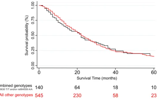
Analysing the whole PDAC case population and adjusting for age, sex and stage we observed no statistically significant association between the two SNPs under investigation and OS of PDAC patients, with the best finding consisting in a non-significant trend for the C/C genotype of the CD44-rs353630 of having a worse survival compared with the T/T homozygous (HR = 1.28, 95% CI 0.98–1.68, p = 0.074). We also performed stratified analysis by disease stage and when analysing the individuals that presented stage I disease at diagnosis we observed a statistically significant association for the heterozygous C/T carriers of CD44-rs353630 and worse OS compared with the reference (HR = 5.01; 95% CI 1.58–15.88; p = 0.006). The analysis considering only the 685 patients who received surgery did not show any statistically significant associations. We also per- formed an analysis considering the combination of the two markers and we observed no statistically significant result. The frequencies and distribution of the genotypes, the HR and 95% CI for the association with PDAC survival are shown in Table 2. A Kaplan–Meier curve with the survival of resected PDAC patients diagnosed in stage I or II (p = 0.64), according to the combined genotypes of the two SNPs is showed in Fig. 1. We performed additional analyses stratifying by country of origin. We did not observe any statistically significant associations, with the exception of a longer OS for patients from Hungary diagnosed with stage I or II PDAC and heterozy- gous for the CHI3L2-rs684559 SNP, in comparison with the homozygotes for the common allele (HR = 0.19; 95%CI 0.05–0.71; p = 0.014).
In silico functional characterization.
The GTEx portal identified only one eQTL for CD44-rs353630:the weak (p = 0.00025) association between the A allele and a decreased expression of the CD44 gene in the tibial nerve. GTEx predicts that CHI3L2-rs684559 is an eQTLs for the CHI3L2, DENND2D, CHIAP2 and CHIA genes across seven tissues, although none of them in the pancreas (p-values ranging from 0.000026 to 3.7 × 10⁻²⁰). For CD44-rs353630 RegulomeDB showed a rank of 4 that is the third lowest and indicates that the polymorphism may be situated in a possible transcription factor binding site. On the other hand, for CHI3L2-rs684559 Regu- lomeDB showed a rank of 1b indicating a very high degree of supporting data in the database and high prob- ability of functional relevance. Haploreg showed no SNPs in LD with CD44-rs353630 and no eQTLs, while six SNPs (rs2764546, rs12048900, rs12024633, rs2494004, rs1325284, rs2494006) in LD with CHI3L2-rs68455 and 14 eQTLs in lymphoblastoid cell lines and in the whole blood.
Discussion
In the last decades genetic epidemiological approaches, especially those performed through genome-wide asso- ciation studies in the context of multicentric studies, have identified thousands of SNPs and have shed light on the biology of many cancer types. As far as regards PDAC, around 30 risk loci showed a clear association with the development of the disease. However, differently from other cancer types such as breast, lung, prostate, colorectal cancers, and multiple myeloma31–37, the involvement of genetic variability in PDAC survival has been investigated with limited success. The most reliable factors in determining PDAC patient survival are, therefore, clinical parameters such as TNM stage, tumour grade, margins of resection and pre- and postoperative levels of C-reactive protein (CRP) and CA-19-938. Unfortunately, these factors have limited accuracy in predicting the outcome of PDAC patients and their response to treatments. Therefore, the identification of additional biological markers would be of the utmost importance as a basis to elaborate novel decisional algorithms aimed at optimiz- ing treatment and improving patients’ outcomes. Additionally, discovering new PDAC survival genetic loci may give insight to the molecular mechanisms of survival and suggest, directly or indirectly, new therapeutic targets.
Several SNPs have been proposed to be possible prognostic markers in PDAC patients, given the association with survival. However, with only a few exceptions, none has been consistently replicated in following studies25–29. Recently two novel candidates, identified through a GWAS approach, have been proposed. A total of 331 PDAC patients were investigated by means of a dense SNP array, encompassing more than 2 million SNPs, finding an Table 1. Description of the study population.
Country Patients
Sex (%) Stage Age (years)
Females Males I II III IV Median (Q1–Q3)
Italy 483 211 272 18 188 86 191 67 (60–74)
Denmark 431 189 242 17 149 64 201 68 (62–73)
Germany 351 136 215 14 244 28 65 65 (58–70)
Hungary 254 130 124 18 16 120 100 65 (59–73)
Czech Republic 151 63 88 27 69 19 36 62 (58–69)
Lithuania 87 41 46 1 21 13 52 69 (60–73)
Poland 46 26 20 8 15 6 17 61 (54–66)
Romania 30 11 19 2 0 10 18 63 (54–67)
The Netherlands 15 6 9 0 10 2 3 68 (60–76)
United Kingdom 8 5 3 0 5 0 3 69 (65–73)
Total 1856 818 1038 105 717 348 686 69 (59–72)
SNP
All patientsa All patientsb Stage Ic
No. HR (95% CI) p value HR (95% CI) p value No. HR (95% CI) p value rs353630
T/T 134 1 [Reference] NA 1 [Reference] NA 13 1 [Reference] NA
C/T 683 1.16 (0.94–1.44) 0.165 1.15 (0.93–1.43) 0.206 34 5.01 (1.58–15.88) 0.006 C/C 1039 1.13 (0.92–1.39) 0.259 1.13 (0.91–1.39) 0.258 58 2.20 (0.73–6.61) 0.162 C/T + C/C 1722 1.14 (0.93–1.40) 0.202 1.14 (0.93–1.40) 0.220 92 2.77 (0.95–8.06) 0.062 Resected
T/T 85 1 [Reference] NA 1 [Reference] NA 9 1 [Reference] NA
C/T 372 1.21 (0.92–1.61) 0.178 1.23 (0.93–1.63) 0.152 25 4.43 (0.91–21.64) 0.066 C/C 630 1.26 (0.96–1.65) 0.098 1.28 (0.98–1.68) 0.074 51 2.34 (0.51–10.62) 0.272 C/T + C/C 1002 1.24 (0.95–1.62) 0.111 1.26 (0.97–1.65) 0.087 76 2.70 (0.61–11.91) 0.190 rs684559
A/A 256 1 [Reference] NA 1 [Reference] NA 11 1 [Reference] NA
G/A 827 1.01 (0.86–1.19) 0.918 0.97 (0.82–1.14) 0.719 44 0.50 (0.21–1.20) 0.119 G/G 773 0.96 (0.82–1.13) 0.640 0.94 (0.80–1.11) 0.456 50 0.54 (0.23–1.26) 0.155 G/A + G/G 1600 0.99 (0.85–1.15) 0.849 0.96 (0.82–1.11) 0.558 94 0.52 (0.24–1.16) 0.110 Resected
A/A 157 1 [Reference] NA 1 [Reference] NA 6 1 [Reference] NA
G/A 479 0.99 (0.81–1.24) 0.984 0.97 (0.78–1.20) 0.777 38 0.61 (0.19–1.94) 0.407 G/G 451 1.00 (0.81–1.24) 0.981 0.98 (0.79–1.22) 0.860 41 0.59 (0.19–1.89) 0.376 G/A + G/G 930 1.00 (0.82–1.22) 0.999 0.97 (0.80–1.19) 0.804 79 0.60 (0.20–1.81) 0.367 Combined SNPsd
rs353630 T/T and/or
rs684559 A/A 368 1 [Reference] NA 1 [Reference] NA 20 1 [Reference] NA
All other genotypes 1488 1.04 (0.91–1.18) 0.582 1.02 (0.89–1.16) 0.826 85 1.38 (0.65–2.91) 0.403 Resected
rs353630 T/T and/or
rs684559 A/A 229 1 [Reference] NA 1 [Reference] NA 12 1 [Reference] NA
All other genotypes 858 1.06 (0.90–1.27) 0.480 1.06 (0.89–1.26) 0.526 73 1.5 (0.55–4.12) 0.431 SNP
Stage IIc Stage I and IIc
No. HR (95% CI) p value No. HR (95% CI) p value
rs353630
T/T 47 1 [Reference] NA 60 1 [Reference] NA
C/T 265 1.08 (0.73–1.59) 0.709 299 1.27 (0.88–1.83) 0.203
C/C 405 1.07 (0.73–1.56) 0.730 463 1.17 (0.82–1.67) 0.388
C/T + C/C 670 1.07 (0.74–1.55) 0.714 762 1.20 (0.85–1.71) 0.295 Resected
T/T 44 1 [Reference] NA 53 1 [Reference] NA
C/T 209 1.08 (0.72–1.62) 0.721 234 1.23 (0.83–1.81) 0.311
C/C 347 1.15 (0.78–1.71) 0.476 398 1.23 (0.84–1.79) 0.291
C/T + C/C 556 1.12 (0.77–1.65) 0.550 685 1.23 (0.85–1.77) 0.282 rs684559
A/A 102 1 [Reference] NA 113
G/A 333 0.93 (0.71–1.22) 0.617 377 0.87 (0.67–1.12) 0.270
G/G 282 0.92 (0.70–1.22) 0.569 332 0.89 (0.68–1.15) 0.364
G/A + G/G 615 0.93 (0.72–1.20) 0.567 709 0.88 (0.69–1.11) 0.278 Resected
A/A 89 1 [Reference] NA 95 1 [Reference] NA
G/A 276 0.89 (0.67–1.19) 0.438 314 0.88 (0.66–1.16) 0.358
G/G 235 0.95 (0.71–1.28) 0.739 276 0.93 (0.70–1.23) 0.606
G/A + G/G 511 0.92 (0.70–1.20) 0.539 590 0.90 (0.69–1.17) 0.432 Combined SNPsd
rs353630 T/T and/or
rs684559 A/A 144 1 [Reference] NA 164 1 [Reference] NA
All other genotypes 573 0.92 (0.74–1.16) 0.489 658 0.95 (0.77–1.17) 0.623 Resected
rs353630 T/T and/or
rs684559 A/A 128 1 [Reference] NA 140 1 [Reference] NA
All other genotypes 472 0.93 (0.73–1.19) 0.571 545 0.97 (0.77–1.22) 0.780
association between CD44-rs353630 and CHI3L2-rs684559 and PDAC survival in resected patients of whom 90.9% with stage I or II. The diplotype combination of these two alleles was, indeed, associated with longer sur- vival after surgery30. We genotyped these two SNPs and analysed them, separately or in combination, in a large set of PDAC cases belonging to the PANDoRA consortium. Considering the paucity of evidences on markers related to PDAC survival this attempt to generalize the results of the two SNPs was necessary. We had more than 99% power to replicate the associations, but we observed none when analysing the whole population or when considering only the resected patients. The most clinically relevant result of the study by Dimitrakopoulos and colleagues is the association of the two combined polymorphisms with better survival in 331 patients resected with intent of radicality. Availability of such a biomarker might be extremely relevant as it would help selection of patients to consider for upfront surgery or neoadjuvant chemotherapy, thus contributing to a personalized medicine approach for PDAC. However, in our subgroup analysis on 685 patients resected with stage I or II disease (therefore with a much larger sample size and a power to detect the association greater than 99%) the same SNP combination did not show any statistically significant association (the best association we observed had a p = 0.48).
Further stratifying the population by stage, we observed a statistically significant association between CD44- rs353630 C/T individuals and worse OS among PDAC cases diagnosed with stage I. The estimate is very high (HR > 5) and it goes in the opposite direction of the ones reported by Dimitrakopoulos, which all showed asso- ciation of the effect alleles/genotypes with better OS. These findings arise from an analysis conducted in a very small subgroup of individuals and only comparing heterozygous with the reference homozygous (N = 34 vs. 13, respectively) and therefore must be taken with caution. The analysis conducted to assess the functional relevance of the SNPs clearly shows that for CD44-rs353630 there is little or no evidence of its involvement in gene expres- sion, while CHI3L2-rs684559 is possibly an eQTL with regulatory potential in several tissues, but not in the pancreas. It is therefore difficult to link the SNPs to a functional role in pancreatic cancer progression or outcome.
Analyses stratified by country of origin did not show anything remarkable, with the exception of a weak association with longer OS in Hungarian patients diagnosed in stage I or II for heterozygous for the CHI3L2- rs684559 SNP. We note that this association is not significant if we consider a significance threshold adjusted for multiple testing (the observed association had p = 0.014, but analyses stratified by 10 countries of origin have an adjusted threshold of significance of p = 0.05/10 = 0.005). Considering also the relatively small number of cases used for this analysis (18 patients diagnosed with stage I and 16 in stage II), and the difficulty of explaining why the polymorphism should be associated with OS only in Hungarian patients, we are inclined to think that this result is due to statistical fluctuation and does not reflect a real association. A possible limitation of the present study is the lack of information on chemotherapeutic regimens administered to the patients, either before or after surgery. This prevents us to explore association between the genetic variability and drug response that may help personalization of treatments. In addition, the included patients were not consecutively enrolled in each center, and that could add a potential bias to the findings. We believe, however that given the samples size this bias should be diluted and unlikely to be responsible for the lack of statistically significant association that we observed.
Material and methods
Study population.
The PANDoRA consortium consists of a multicentric study that includes 12 European countries (Italy, Germany, Poland, United Kingdom, Hungary, Czech Republic, Greece, Lithuania, the Nether- lands, Denmark, Ukraine, Romania) and Japan and has been described in detail elsewhere9.Briefly, all cases included in the consortium population are defined by a confirmed diagnosis of pancreatic cancer and were collected with a non-consecutive sampling approach in 21 different centers, including depart- ments of surgery, endoscopy and gastroenterology. In addition to pancreatic cancer cases, controls have been selected among the general population, blood donors and among hospitalized subjects with different diagnosis excluding cancer. In the present study, 1856 PDAC cases of European descent were included, for whom informa- tion on country of origin, sex, age at diagnosis, overall survival (OS) and disease stage assessed by TNM stage was available. Disease stage was defined according to American Joint Committee on Cancer TNM system 7th version39, and patients were categorized as stage I (T1-2, N0, M0), stage II (T3, N0, M0/T1-3, N1, M0), stage III (T4, any N, M0), stage IV (any T, any N, M1). The patients that underwent surgery were categorized by the stage at the time of operation.
All subjects gave their informed consent for inclusion before they participated in the study. The study was conducted in accordance with the Declaration of Helsinki, and the protocol was approved by the Ethics Com- mission of the Medical Faculty of the University of Heidelberg (project identification code: S-565/2015). A description of the population is shown in Table 1.
SNPs selection, sample preparation and genotyping.
We selected two germline variants, CD44- rs353630 and CHI3L2-rs684559 that were proposed recently by Dimitrakopoulos and colleagues as associated with survival of PDAC patients that received surgery. For each subject DNA was extracted from whole blood Table 2. Survival analysis of CD44-rs353630 and CHI3L2-rs684559. NA, not applicable; HR, Hazard Ratio, CI, confidence interval. Statistically significant results (p < 0.05) are in bold. Numbers may not add up to the total number of subjects in the respective stratum due to missing data. a The analyses were adjusted by tumour stage (stages I through IV). b The analyses were adjusted by tumour stage, country of origin, sex and age. c The analyses were adjusted by, country of origin, sex and age. d The combined analysys was performed using the rs353630 T/T and rs684559 A/A genotypes as reference.using a QIAamp 96 DNA QIAcube HT Kit on QIAcube 96 instrument and kept frozen at − 20° till use. Geno- typing was conducted using TaqMan (ABI, Applied Biosystems, Foster City, CA, USA) allelic discrimination technology following the manufacturer’s recommendation (assay id C____779802_10 for CD44-rs353630 and assay id C___3138574_20 for CHI3L2-rs684559). For each reaction 10 ng of dried DNA were used. The geno- typing was conducted in 384-well plates and duplicated samples (8%) were included in each plate for quality control purpose. The identity of the samples was not known to the personnel performing the lab work. Genotype attribution was made using QuantStudioTM 5 Real-Time PCR system (Thermofisher, USA) and QuantStudio software.
Statistical analysis.
Deviation from Hardy–Weinberg equilibrium (HWE) was tested for both polymor- phisms in the overall population and dividing for country of origin using the Pearson corrected chi square test.Survival analysis was conducted by Cox proportional hazard models with OS as endpoint, computing hazard ratios (HR) and 95% confidence intervals (CI), using an additive and a codominant inheritance models and set- ting the same alleles used by Dimitrakopoulos and colleagues as the reference category (T for CD44-rs353630 and A for CHI3L2-rs684559). In addition, considering that the most statistically significant association observed by Dimitrakopoulos was the one combining the two genotypes, we also performed this analysis defining two groups: the individuals with the CD44-rs353630 T/T and/or CHI3L2-rs684559 A/A as the reference category and all the other genotype combinations as the other group. For this analysis we considered only individuals with 100% call rate. All analyses were adjusted for sex, age and TNM stage. We also performed an analysis adjusting only for disease stage, since Dimitrakopoulos and colleagues did not adjust for sex and age.
As the vast majority (90.9%) of the patients in the study by Dimitrakopoulos and colleagues were resected with stage I or II, stratified analysis was conducted considering patients showing TNM stage I, TNM stage II, and TNM stage I and II together. We also performed a stratified analysis considering TNM stage I and II for patients who received surgery, as well as by country of origin. The probability of survival was calculated by means of a Kaplan–Meier curve.
Functional analysis using bioinformatic tools.
To test the possible functional relevance of CD44- rs353630 and CHI3L2-rs684559 we used several bioinformatic tools. In particular, the Genotype-Tissue Expres- sion (GTEx) project portal (https:// gtexp ortal. org/ home/) was used to identify potential cis-acting expression quantitative trait loci (eQTLs) in order to establish whether the two SNPs could be involved in the expression of nearby genes (accession date 8th May 2020)40. We analysed all the tissues present in the database. We have used Regulome DB 2.0 (https:// www. regul omedb. org/ regul ome- search/) to evaluate the effect of the SNPs with regu- latory regions of the human non-coding genome41. Regulome DB 2.0 assigns to each SNP a rank consisting of 14 steps, that reflect the volume of the supporting data on the functional relevance of the SNP, going from 6 (lowest amount of evidence) to 1a (highest amount of evidence). Finally, we used Haploreg v4.1 (https:// pubs. broad insti tute. org/ mamma ls/ haplo reg/ haplo reg. php) to link the SNPs to functional annotations of the genome42. Hap- loreg is designed to explore the possible mechanistic interaction of the candidate SNPs (and all SNPs in LD with them) with regulatory motifs.Figure 1. Kaplan–Meier curve showing the survival of resected PDAC patients diagnosed in stage I or II, according to the combined genotypes of CD44-rs353630 and CHI3L2-rs684559.
Ethics declarations.
The study was conducted in accordance with the Declaration of Helsinki, and the protocol was approved by the Ethics Commission of the Medical Faculty of the University of Heidelberg (project identification code: S-565/2015).Conclusions
In conclusion, we attempted to replicate the results of a genome-wide investigation on genetic polymorphisms and survival of PDAC patients, with a well-powered study. Our findings suggest no involvement in the PANDoRA population of the two variants reported by Dimitrakopoulos and colleagues and highlight a lack of functional or regulatory role of CD44-rs353630 and CHI3L2-rs684559 in the pancreatic tissue. Our results strongly support the need to consider with caution findings on genetic variables as prognostic markers in PDAC, in the absence of replication in large and independent datasets or of compelling in vitro mechanisms to support the associations.
Moreover, it would be desirable for future studies to have information on the therapy and to include this information in the analyses. This could make possible to evaluate the different genetic predisposition in drug response, and further our knowledge of this deadly disease.
Data availability
Owing to ethical and legal reasons, raw data of this work are not publicly deposited. The PANDoRA primary data for this work will be made available to researchers who submit a reasonable request to the corresponding author, conditional to approval by the PANDoRA Steering Committee and Ethics Commission of the Medical Faculty of the University of Heidelberg. Data will be stripped from all information allowing identification of study participants.
Received: 26 August 2020; Accepted: 23 March 2021
References
1. Siegel, R. L., Miller, K. D. & Jemal, A. Cancer statistics, 2020. CA Cancer J. Clin. 70, 7–30 (2020).
2. Rahib, L. et al. Projecting Cancer Incidence and Deaths to 2030: The Unexpected Burden of Thyroid, Liver, and Pancreas Cancers in the United States. Cancer Research vol. 74 2913–2921 (American Association for Cancer Research, 2014).
3. Hidalgo, M. Pancreatic cancer. N. Engl. J. Med. 50, 545–555 (2010).
4. Gentiluomo, M. et al. Germline genetic variability in pancreatic cancer risk and prognosis. Semin. Cancer Biol. Online ahead of print (2020). https:// doi. org/ 10. 1016/j. semca ncer. 2020. 08. 003.
5. Signoretti, M. et al. Results of surveillance in individuals at high-risk of pancreatic cancer: A systematic review and meta-analysis.
Unit. Eur. Gastroenterol. J. 6, 489–499 (2018).
6. Petersen, G. M. et al. A genome-wide association study identifies pancreatic cancer susceptibility loci on chromosomes 13q22.1, 1q32.1 and 5p15.33. Nat. Genet. 42, 224–8 (2010).
7. Low, S.-K. et al. Genome-wide association study of pancreatic cancer in Japanese population. PLoS ONE 5, e11824 (2010).
8. Campa, D. et al. Functional single nucleotide polymorphisms within the cyclin-dependent kinase inhibitor 2A/2B region affect pancreatic cancer risk. Oncotarget 7, 57011–57020 (2016).
9. Campa, D. et al. Genetic susceptibility to pancreatic cancer and its functional characterisation: The PANcreatic Disease ReseArch (PANDoRA) consortium. Dig. Liver Dis. 45, 95–99 (2013).
10. Campa, D. et al. TERT gene harbors multiple variants associated with pancreatic cancer susceptibility. Int. J. Cancer 137, 2175–2183 (2015).
11. Obazee, O. et al. Germline BRCA2 K3326X and CHEK2 I157T mutations increase risk for sporadic pancreatic ductal adenocar- cinoma. Int. J. Cancer 145, 686–693 (2019).
12. Campa, D. et al. Genome-wide association study identifies an early onset pancreatic cancer risk locus. Int. J. Cancer 147, 2065–2074 (2020).
13. Corradi, C. et al. Genome-wide scan of long noncoding RNA single nucleotide polymorphisms and pancreatic cancer susceptibility.
Int. J. cancer https:// doi. org/ 10. 1002/ ijc. 33475 (2021).
14. Childs, E. J. et al. Common variation at 2p13.3, 3q29, 7p13 and 17q25.1 associated with susceptibility to pancreatic cancer. Nat.
Genet. 47, 911–916 (2015).
15. Wolpin, B. M. et al. Genome-wide association study identifies multiple susceptibility loci for pancreatic cancer. Nat. Genet. 46, 994–1000 (2014).
16. Klein, A. P. et al. Genome-wide meta-analysis identifies five new susceptibility loci for pancreatic cancer. Nat. Commun. 9, 556 (2018).
17. Wu, C. et al. Genome-wide association study identifies five loci associated with susceptibility to pancreatic cancer in Chinese populations. Nat. Genet. 44, 62–66 (2011).
18. Amundadottir, L. et al. Genome-wide association study identifies variants in the ABO locus associated with susceptibility to pancreatic cancer. Nat. Genet. 41, 986–990 (2009).
19. Zhang, M. et al. Three new pancreatic cancer susceptibility signals identified on chromosomes 1q32.1, 5p15.33 and 8q24.21.
Oncotarget. 7, 66328–66343 (2016).
20. Amundadottir, L. T. Pancreatic cancer genetics. Int. J. Biol. Sci. 12, 314–325 (2016).
21. Galeotti, A. A. et al. Polygenic and multifactorial scores for pancreatic ductal adenocarcinoma risk prediction. J. Med. Genet.
jmedgenet-2020–106961 (2020). https:// doi. org/ 10. 1136/ jmedg enet- 2020- 106961.
22. Mohelnikova-Duchonova, B. et al. SLC22A3 polymorphisms do not modify pancreatic cancer risk, but may influence overall patient survival. Sci. Rep. 7, 43812 (2017).
23. Tang, H. et al. Genetic polymorphisms associated with pancreatic cancer survival: A genome-wide association study. Int. J. Cancer 141, 678–686 (2017).
24. Rizzato, C. et al. Pancreatic cancer susceptibility loci and their role in survival. PLoS ONE 6, e27921 (2011).
25. Wu, C. et al. Genome-wide association study of survival in patients with pancreatic adenocarcinoma. Gut 63, 152–160 (2014).
26. Rizzato, C. et al. Association of genetic polymorphisms with survival of pancreatic ductal adenocarcinoma patients. Carcinogenesis 37, 957–964 (2016).
27. Willis, J. A. et al. A replication study and genome-wide scan of single-nucleotide polymorphisms associated with pancreatic cancer risk and overall survival. Clin. Cancer Res. 18, 3942–3951 (2012).
28. Gentiluomo, M. et al. Genetic variability of the ABCC2 gene and clinical outcomes in pancreatic cancer patients. Carcinogenesis 40, 544–550 (2019).
29. Innocenti, F. et al. A genome-wide association study of overall survival in pancreatic cancer patients treated with gemcitabine in CALGB 80303. Clin. Cancer Res. 18, 577–584 (2012).
30. Dimitrakopoulos, C. et al. Identification and validation of a biomarker signature in patients with resectable pancreatic cancer via genome-wide screening for functional genetic variants. JAMA Surg. 154, e190484 (2019).
31. Guo, Q. et al. Identification of novel genetic markers of breast cancer survival. J. Natl. Cancer Inst. 107, 81 (2015).
32. Khan, S. et al. Meta-analysis of three genome-wide association studies identifies two loci that predict survival and treatment outcome in breast cancer. Oncotarget 9, 4249–4257 (2018).
33. Escala-Garcia, M. et al. Genome-wide association study of germline variants and breast cancer-specific mortality. Br. J. Cancer 120, 647–657 (2019).
34. Ziv, E. et al. Genome-wide association study identifies variants at 16p13 associated with survival in multiple myeloma patients.
Nat. Commun. 6, 7539 (2015).
35. Pirie, A. et al. Common germline polymorphisms associated with breast cancer-specific survival. Breast Cancer Res. 17, 58 (2015).
36. Li, W. et al. Genome-wide scan identifies role for AOX1 in prostate cancer survival. Eur. Urol. 74, 710–719 (2018).
37. Phipps, A. I. et al. Common genetic variation and survival after colorectal cancer diagnosis: A genome-wide analysis. Carcinogenesis 37, 87–95 (2016).
38. Mizrahi, J. D., Surana, R., Valle, J. W. & Shroff, R. T. Pancreatic cancer. The Lancet 395, 2008–2020 (2020).
39. Edge, S. B. & Compton, C. C. The American Joint Committee on Cancer: The 7th Edition of the AJCC Cancer Staging Manual and the Future of TNM. Ann. Surg. Oncol. 17, 1471–1474 (2010).
40. GTEx Consortium, T. Gte. The genotype-tissue expression (GTEx) project. Nat. Genet. 45, 580–5 (2013).
41. Boyle, A. P. et al. Annotation of functional variation in personal genomes using RegulomeDB. Genome Res. 22, 1790–1797 (2012).
42. Ward, L. D. & Kellis, M. HaploReg: A resource for exploring chromatin states, conservation, and regulatory motif alterations within sets of genetically linked variants. Nucleic Acids Res. 40, (2012).
Author contributions
Conceptualization, D.C. and G.C.; Data curation, M.G. and C.C.; Formal analysis, M.G. and C.C.; Writing – original draft, M.G., F.C., D.C. and G.C.; All authors reviewed the manuscript.
Funding
This work was partially supported by: [D.C.; F.C.] intramural funds of Univerity of Pisa and DKFZ; [D.C.] Fon- dazione Tizzi (www. fonda zione tizzi. it/); [D.C.] Fondazione Arpa (www. fonda zione arpa. it); [B.M.D.] Ministry of Health of Czech Republic, NV 19–03-00097; [L.P.] Associazione Italiana Ricerca sul Cancro (AIRC 5 × 1000 n 12182); [P.S.] Czech Ministry of Health, project no. NV19-08–00113; [G.C.] Associazione Italiana Ricerca sul Cancro (AIRC IG IG 17177); [Division of Gastroenterology, Fondazione “Casa Sollievo della Sofferenza” IRCCS Hospital] Italian Ministry of Health (RC1803GA32) and the “5 × 1000” voluntary contribution.
Competing interests
The authors declare no competing interests.
Additional information
Correspondence and requests for materials should be addressed to D.C.
Reprints and permissions information is available at www.nature.com/reprints.
Publisher’s note Springer Nature remains neutral with regard to jurisdictional claims in published maps and institutional affiliations.
Open Access This article is licensed under a Creative Commons Attribution 4.0 International License, which permits use, sharing, adaptation, distribution and reproduction in any medium or format, as long as you give appropriate credit to the original author(s) and the source, provide a link to the Creative Commons licence, and indicate if changes were made. The images or other third party material in this article are included in the article’s Creative Commons licence, unless indicated otherwise in a credit line to the material. If material is not included in the article’s Creative Commons licence and your intended use is not permitted by statutory regulation or exceeds the permitted use, you will need to obtain permission directly from the copyright holder. To view a copy of this licence, visit http:// creat iveco mmons. org/ licen ses/ by/4. 0/.
© The Author(s) 2021