The association between sympatho-vagal balance and central blood pressures
P. L. LATCHMAN
1p, G. GATES
2, J. PEREIRA
3, R. AXTELL R
1, K. GARDNER
4, J. SCHLIE
5, Q. YANG
1, T. YUE
1, A. MORIN-VIALL
1and
R. DEMEERSMAN
61Department of Health and Movement Sciences, Southern Connecticut State University, New Haven, CT, USA
2Department of Pediatrics, Albert Einstein College of Medicine, Yeshiva University, Bronx, NY, USA
3Department of Heart and Vascular Echocardiography, Yale New Haven Hospital, New Haven, CT, USA
4Gerald Claude Eugene Foster College of Physical Education & Sports, Spanish Town, Jamaica
5Institute of Exercise Science, Human Performance and Training, University of M€unster, M€unster, Germany
6Department of Biobehavioral Sciences, Teachers College, Columbia University, New York, NY, USA
Received: April 6, 2019 • Accepted: December 13, 2019 Published online: April 23, 2020
© 2020 Akademiai Kiado, Budapest
ABSTRACT
Purpose:High central blood pressure is more predictive of cardiovascular disease (CVD) versus high pe- ripheral blood pressure. Measures of central pressures (CPs) include, central systolic blood pressure (CSBP) and central diastolic blood pressure. Measures of central pressures augmentation (CPsA) include augmentation pressure (AP) and the augmentation index @ 75 beats$min1(AIx@75). Increased sym- pathetic tone (ST) is also associated with CVD. The low to high frequency ratio (LF/HF) is often used to determine sympatho-vagal balance. Given the association between ST, CPs, CPsA and CVD there is a need to understand the association between these predictors of CVD. The aims of this study were to examine the association between the LF/HF ratio, CPs, and CPsA in men and women collectively and based on gender.
Methods:We measured the LF/HF ratio, CSBP, AP, and AIx@75 in 102 participants (41F/61M). The LF/
HF ratio was determined via power spectral density analysis. CSBP, AP, and AIx@75 were determined via applanation tonometry.Results:The LF/HF ratio was inversely associated with AP (r5–0.26) and AIx @75
pCorresponding author. Southern Connecticut State University, 501 Crescent Street, New Haven, CT 06515, USA. Tel.:þ1 (203) 392 6081; Fax:þ1 (203) 392 6093, E-mail: Latchmanp1@southernct.edu
(r5–0.29) in the combined group of men and women. The LF/HF ratio was inversely associated with CSBP (r5–0.27), AP (r5–0.28), and AIx@75 (r5–0.32) in men, but not in women.Conclusion:There is an inverse association between the LF/HF ratio, AP, and AIx@75 in men and women combined. The association between the LF/HF ratio, CSBP, AP, and AIx@75, differs based on gender.
KEYWORDS
sympatho-vagal balance, central systolic blood pressure, central diastolic blood pressure, augmentation pressure, augmentation index
INTRODUCTION
Cardiovascular disease (CVD) is still a main cause of health loss worldwide [32]. Although high peripheral blood pressure (PBP) increases the risk for CVD [30], research suggests that irre- spective of patients having similar PBP, those with higher central blood pressure (CBP) were at increased risk for CVD versus those with low CBP [29, 31, 38], suggesting CBP to be an in- dependent predictor of CVD [2, 29, 31, 36, 38]. In addition to elevated CBP, increased activity from the sympathetic nervous system is also associated with many negative cardiovascular outcomes [21].
In this paper, central pressures (CPs) will be indicative of central systolic blood pressure (CSBP) and central diastolic blood pressure (CDBP). Central pressures augmentation (CPsA) will be indicative of augmentation pressure (AP) and the augmentation index (AIx). As related to CPs, past works have found CSBP to be highly predictive of cardiovascular events [29, 35];
this relationship could be due in part to the concomitant increase in work by the myocardium imposed by an increase in CSBP. From a CDBP perspective, an attenuation in CDBP caused by the early return of reflective waves can promote reduced coronary perfusion leading to reduced bloodflow to the myocardium [12].
CPsA is expressed in absolute terms as the AP or as a percentage of pulse pressure expressed as the AIx. AP has been demonstrated to be predictive of negative cardiovascular health and the AIx has been shown to be predictive for the presence of coronary artery disease (CAD) [13, 16, 36], adverse cardiovascular events in patients with established CAD [8], and negative outcomes associated with end-stage renal disease [20].
The sympathetic branch of the autonomic nervous system is involved in many aspects of cardiovascular physiology, including its direct role in myocardial activity, vasomotor tone and its role in the renin-angiotensin-aldosterone system. Increased sympathetic tone (ST) is implicated in impaired cardiovascular functioning and is associated with heart failure [39], arrhythmias [21] and increased PBP [11]. Both the sympathetic and parasympathetic branch of the autonomic nervous system promotes changes in the variability of the R-R intervals between heart beats which is referred to as heart rate variability (HRV) [27]. HRV is therefore repre- sentative of the variation in time between R-R intervals. In the frequency domain the high frequency (HF) band is believed to be representative of parasympathetic modulation while the low frequency (LF) band is considered to be representative of both sympathetic and para- sympathetic modulation with a dominance of sympathetic modulation [14]. The LF–HF ratio (LF/HF) is considered to be indicative of sympatho-vagal balance [25, 26]. Thus, an increase in
the LF/HF ratio is considered to denote a shift towards sympathetic dominance over the parasympathetic. Although the use of the LF/HF ratio in assessing sympatho-vagal balance has been questioned [3], the LF/HF ratio has been used in numerous studies [15, 23, 34] and represents an easy, non-invasive, inexpensive way of measuring sympatho-vagal balance in young individuals, the elderly [23], pediatric [34] and certain clinical populations [15]. Due to the non-invasive nature of the LF/HF ratio measurement it is ideally suited for determining sympatho-vagal balance in a wider population range versus other more invasive methods of assessing ST.
The association between ST and PBP has been extensively studied, however, despite the understanding that CPs and CPsA are robust predictors of cardiovascular health and given the role of elevated ST in CVD, there is a surprising paucity of research examining the association between ST, CPs, and CPsA. One prior study examined the association between muscle sym- pathetic nerve activity (MSNA) and aortic wave reflections and suggested that in a healthy group of young men and women, MSNA was not associated with CPs, AP or AIx. However, when this group was divided by gender, MSNA was positively associated with AP and AIx in men but was inversely associated in women [4]. Although MSNA is a robust indicator of ST, this measure- ment requires great technical skill and involves the insertion of a tungsten microelectrode into the peroneal nerve. These characteristics reduce the suitability of MSNA as the method of assessing ST in many populations, including: children, the elderly, and certain clinical pop- ulations. Therefore, there is a need to be able to examine the association between ST and measures of CPs and CPsA using methods that are more suitable for a wider population range.
Given the associations between ST, CPs, CPsA, and CVD, there is a great need to not only understand the association between these predictors of CVD and CVD itself, but also for an understanding of the association between these predictors of CVD using a non-invasive method of ST assessment that is suited to a wider population range. An understanding of the associa- tions between ST, CPs, and CPsA could allow for an advancement in the physiological knowledge regarding the etiology and treatment of CVD.
Therefore, the primary aim of the current study was to examine the association between the LF/HF ratio, CPs, and CPsA in a group of young men and women. Additionally, based on our previous findings that the association between the Alx and autonomic modulation differ based on gender [19], and on otherfindings demonstrating differences in the regulation of PBP based on gender [11] the secondary aim was to examine the association between the LF/HF ratio, CPs, and CPsA based on gender. We hypothesized that there will be no association between the LF/
HF ratio, CPs and CPsA in young men and women combined but that these association will differ based on gender.
METHODS
Participants included 102 young men and women (M 5 61; W 5 41) recruited from the Southern Connecticut State University student population and the New Haven area. Inclusion criteria included no clinical indication of cardiovascular or metabolic disease, non-smoker, medication-free and possessing normal electrocardiogram (ECG) and blood pressure (BP) patterns. This research complied with the Helsinki Declaration and was approved by the Institutional Review Board (IRB) at Southern Connecticut State University. Each participant
reviewed and signed an IRB approved informed consent form. Participants arrived for testing between 7:00 and 10:00 a.m., prior to arrival they were advised not to exercise or consume food for 48 and 12 hours, respectively, prior to testing but were allowed to drink water. Anthropo- metric measurements were taken followed by two to three BP measurements after 5 min of seated rest. After anthropometric measurements were taken CPs and CPsA measurements were taken followed by measurement of the LF/HF ratio.
Measurement of the CPs, CPsA and LF/HF ratio
CSBP, CDBP, AP and AIx. The SphygmoCor system (AtCor Medical Pty Ltd, West Ryde, Australia) used pulse wave analysis to determine CSBP, CDBP, AP, and AIx [24]. A high-fidelity transducer applanation tonometer was placed over the left radial pulse. The system then analyzed the radial waveform via a generalized transfer function that was validated intra-arte- rially [7, 28] to determine CSBP, CDBP, AP, and AIx.
AP was established as the amplitude of the reflected wave and was defined as the difference between the initial and second systolic shoulders of the aortic systolic blood pressure [38]. AIx was established as AP divided by pulse pressure expressed as a percent of the central pulse pressure [38]. Since heart rate has an effect on AIx [24, 37], AIx values were adjusted to be a standard heart rate of 75 beats$min1, denoted as AIx@75. Measurements were taken from the left radial pulse after 6 min of rest in a seated position. The SphygmoCor system has built in quality control parameters. These parameters are used to determine validity and reproducibility of a waveform. An operator index of ≥85 represents the highest level of validity and repro- ducibility of a waveform. All pressure data collected in this study conformed to an operator’s index of≥85.
The LF/HF ratio. For the LF/HF ratio measurement, participants were instrumented with a Nexfin monitor (BMEYE, Netherlands), which used ECG to determine continuous R-R interval measurements. The R-R interval measurement was taken for 8 min in a seated position but only the final 5 min of data were used in the analysis. In the current study only the frequency–
domain method was used to assess HRV. Here, power spectral density analysis of HRV was used to determine autonomic modulation; with a priori power spectra of the R-R interval within the 0.15–0.4 Hz bandwidth indicating primarily parasympathetic modulation; defining the HF segment of HRV, referred to as high frequency of R-R interval denoted HFR-R. The LF segment of HRV was located within the 0.04–0.15 Hz bandwidth denoted as LFR-Rand is considered to represent a mixture of sympathetic and parasympathetic modulation [14]. Sympatho-vagal balance was determined as the ratio between the LF and HF spectra of HRV and is denoted as LF/HF [25]. Participants were instructed to breathe at 12 breaths$min1(0.2 Hz), guided by a light moving up and down on a computer screen. This breathing protocol avoided the effect of a varied respiratory rate on spectral distributions [9]. The absolute value of the LF and HF components were log transformed to remove skewness and minimize the large SD customarily present in these data and were indicated as lnLF and lnHF respectively. Women were tested within 5 days of the cessation of menstruation.
Statistical analysis. Means and standard errors are presented for the demographic variables of age (years), height (centimeters), body mass (kilograms) and body mass index (kg$m2). Means and standard errors are also presented for the LF/HF ratio, CSBP (mmHg), CDBP (mmHg), AP
(mmHg) and AIx@ 75 (%). Independent samplet-tests were performed to determine whether differences in the aforementioned variables exist by gender. To determine the associations be- tween LF/HF, CPs, and CPsA values, Pearson's correlation coefficients and linear regression analyses were performed. Significance was set at P < 0.05 for all statistics and analyses were obtained using SPSS for Windows, Version 23 (IBM Corporation 2015, Armonk, NY).
RESULTS
Participant demographics are presented in Table 1. ST, and measures of CPs and CPsA are presented in Table 2. Independent t-test revealed no significant difference in age based on gender, however men demonstrated greater height, body mass and BMI versus women (Table 1, P< 0.05). No significant differences were found in lnLF and lnHF between men and women (Table 2). There was no significant difference in the LF/HF ratio between men and women (Table 2). Men demonstrated greater CSBP and CDBP versus women (Table 2, P < 0.05).
Women demonstrated greater AP and AIx@75 versus men (Table 2,P< 0.05).
Table 1.Demographic variables for men (n561) and women (n541)
Men Women Pvalue
Age (y) 21.5±0.4 22.1±0.4 0.39
Height (cm) 175.7±0.8 164.8±1.0 <0.001**
Body mass (kg) 77.3±1.3 64.2±1.7 <0.001**
BMI (kg$m2) 25.0±0.3 23.6±0.5 0.035*
BMI: body mass index. Data are in means±SEM.
*P5<0.05.
**P5<0.001.
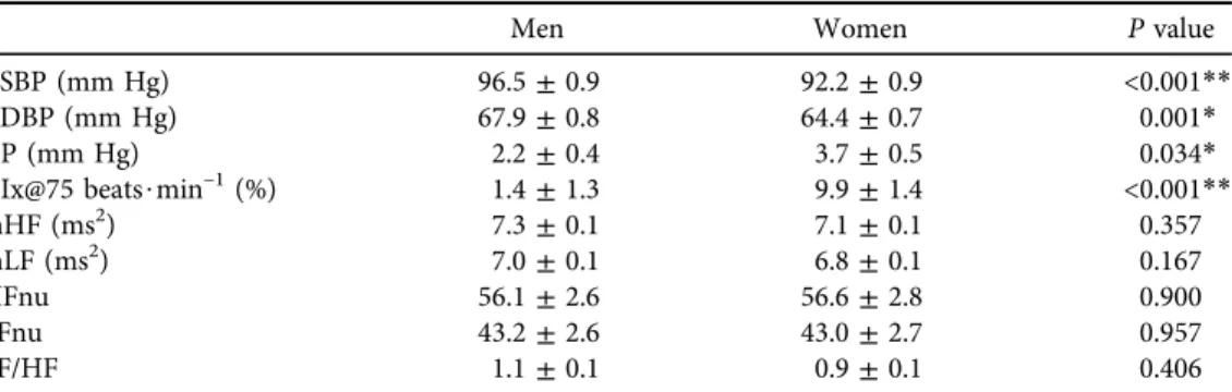
Table 2.Sympathetic tone and blood pressure parameters in men (n561) and women (n541)
Men Women Pvalue
CSBP (mm Hg) 96.5±0.9 92.2±0.9 <0.001**
CDBP (mm Hg) 67.9±0.8 64.4±0.7 0.001*
AP (mm Hg) 2.2±0.4 3.7±0.5 0.034*
AIx@75 beats$min–1(%) 1.4±1.3 9.9±1.4 <0.001**
lnHF (ms2) 7.3±0.1 7.1±0.1 0.357
lnLF (ms2) 7.0±0.1 6.8±0.1 0.167
HFnu 56.1±2.6 56.6±2.8 0.900
LFnu 43.2±2.6 43.0±2.7 0.957
LF/HF 1.1±0.1 0.9±0.1 0.406
CSBP: central systolic blood pressure; CDBP: central diastolic blood pressure; AP: augmentation pressure;
AIx@75 beatsmin1, augmentation index at 75 beats$min–1; lnHF: log transform high frequency; lnLF: log transform low frequency; HFnu: normalized high frequency; LFnu: normalized low frequency; LF/HF: low- high frequency ratio. Data are in means±SEM.
*P5<0.05.
**P5<0.001.
Table 3.Correlation between the LF/HF ratio, central pressures, and central pressures augmentation
ST Gender
CSBP (mm Hg)
P value
CDBP (mm
Hg) P value
AP (mm
Hg) P value
AIx@75 beats$min1
(%)
P value LF/
HF
Men –0.27* 0.032 –0.14 0.265 –0.28* 0.027 –0.32* 0.012
Women –0.05 0.744 0.08 0.604 –0.21 0.180 –0.21 0.169
All –0.18 0.068 –0.06 0.544 –0.26** 0.007 –0.29** 0.003 ST: sympathetic tone; CSBP: central systolic blood pressure; CDBP: central diastolic blood pressure; AP:
augmentation pressure; AIx@75 beats$min1: augmentation index at 75 beats$min–1.
*P5<0.05.
**P5<0.01.
Figure 1.Association between the LF/HF ratio and CSBP in men and women
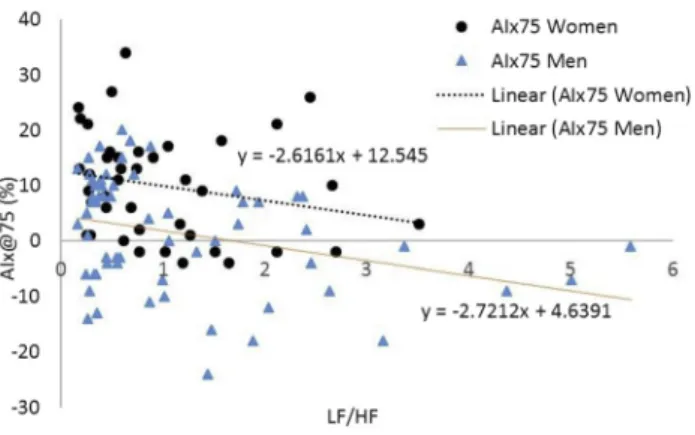
Figure 2.Association between the LF/HF ratio and AIx@75 in men and women
In the combined group, Pearson's correlation coefficients revealed that the LF/HF ratio was inversely associated with AP (r5–0.26;P5< 0.01) and AIx @75 (r5–0.29;P5< 0.01) but not with CSBP or CDBP (Table 3).
In men the LF/HF ratio was inversely associated with CSBP (r5–0.27;P50.03), (Fig. 1), AP (r5–0.28;P50.02), (Table 3) and AIx@75 (r5–0.32;P50.01), (Fig. 2) but not CDBP (Table 3). In women there were no associations between the LF/HF ratio, CSBP (r5–0.05;P5 0.74), (Fig. 1), CDBP (r50.08;P50.60), AP (r5–0.21;P50.18), (Table 3), or AIx@75 (r5 –0.21;P5 0.16), (Fig. 2).
DISCUSSION
The main finding of this study was that the association between the LF/HF ratio, CSBP, AP, and AIx@75, differed based on gender. Here, the new finding was that the LF/HF ratio was inversely associated with, CSBP, AP, and AIx@75, in young men but not in young women. This finding was in keeping with our hypothesis. Additionally, the LF/HF ratio was found to be inversely associated with AP and AIx@75 in a group of young men and women. However, this finding differed from our hypothesis that there would be no association between the LF/HF ratio, CPs and CPA in a group of young men and women.
We did not expect an inverse association between the LF/HF ratio, CSBP, AP and AIx@75 in men. Here, these associations suggest that as the LF/HF ratio increases, there is a concomitant attenuation in CSBP and CPA. This observation suggests that as the sympatho-vagal balance moved towards sympathetic dominance there was a concomitant attenuation in wave reflection.
While this finding was not expected, the concept of increased ST associated with an attenuation in measures of CPsA is not completely new, as one previous study demonstrated this association in women using MSNA [4]. The reason for the inverse association between the LF/HF ratio, CSBP, AP, and AIx@75 in young men cannot be explained by this study; however, one could speculate that a shift towards sympathetic dominance over the parasympathetic modulation could result in an increased heart rate. Decreased heart rate is associated with an increase in the ventricular ejection period which gives rise to summation of the incident and reflective wave [24], which could cause an increase in CPsA. Conversely, an increase in heart rate, which could result from an increase in the LF/HF ratio, could result in a decrease of the ventricularfilling period and decrease summation of the incident and reflective wave, which could in part help to promote the inverse association between the LF/HF ratio, CSBP, AP, and AIx@75. While the above would seem like a plausible speculation, the inverse association was observed even when the heart rate was held constant at 75 beats$min1, suggesting increased heart rate was unlikely responsible for the inverse association between the LF/HF ratio, CSBP, AP, and AIx@ 75.
One prior study using MSNA found a positive association between MSNA, AP, and AIx in young men [4]. The differences in the nature of the association between MSNA, CPs, and CPsA observed in that prior study and the association between the LF/HF ratio, CPs, and CPsA observed in the current study could perhaps be associated in some way to the understanding that MSNA is derived from direct vasoconstrictor ST to the vasculature of skeletal muscle [18], while the LF/HF ratio is obtained from sympatho-vagal balance derived from the heart, along with the understanding that ST directed towards different components of the BP equation could have different impacts on BP. Research suggests that MSNA was inversely associated with the cardiac
output (CO) component of the BP equation but was positively associated with total peripheral resistance (TPR) in young men [6]. Additionally, the association between MSNA and the different components of the BP equation has been demonstrated not to befixed in all people.
Since ST as determined by MSNA has been found to be associated in a different manner with various components of the BP equation, and since ST influence on the BP equation differs between individuals, if the LF/HF ratio for any reason was to be more associated with the reduced CO component versus the TPR component, then perhaps it could be speculated that the association between the LF/HF ratio, CSBP, AP and AIx@75 could be inverse in nature.
While we observed an inverse association between the LF/HF ratio, CSBP, AP, and AIx@75 in young men, we found no association in young women. Explaining the lack of association between the above variables is beyond the scope of this study. The finding of differences between the association of the LF/HF ratio, CSBP, AP, and AIx@75 based on gender was not surprising, since gender related differences in the association between MSNA and measures of CPs and CPsA have been previously reported [4]. One probable reason for the lack of association be- tween the LF/HF ratio, CSBP, AP, and AIx@75 in women could be that estrogen stimulates endothelial nitric oxide (NO) synthase (eNOS), which would result in an increase in endothelial NO availability [22]. Briefly, NO will stimulate soluble guanylyl cyclase, which will promote the formation of cyclic guanosine monophosphate (cGMP). cGMP would then activate protein kinase G (PKG), which would result in the movement of intracellular calcium (Ca) into the sarcoplasmic reticulum, the movement of Ca out of the cell and the opening of potassium channels leading to hyperpolarization. With a decreased intracellular Ca level, there would be a concomitant inactivation of calmodulin. Here, myosin light chain kinase (MLCK) would not be activated and would therefore not be able to phosphorylate myosin, which would lead to smooth muscle relaxation [40]. Here, plasma estrogen in women could serve to restrain ST to the vasculature resulting in vasodilation. This vasodilation could lead to an attenuation in wave reflection and could provide a basis for the observation of no association between the LF/HF ratio, CSBP, AP, and AIx@75 seen in young women in the current study.
Women were found to have significantly higher AIx@75 versus men, which corroborated with prior work [4]. This greater AIx@75 can perhaps be explained in part by women being significantly shorter than men. Being shorter is suggestive of smaller aortas, and here the reflective sites would be closer to the heart and therefore result in earlier wave reflections [33].
We found no association between the LF/HF ratio and CDBP in men and women combined or based on gender. The mean central systolic and diastolic blood pressure values in the current study pointed toward the lower end of the central blood pressure spectrums. However, the operator index for the systolic and diastolic pressure values collected during this study con- formed to an operator index of ≥85, which represents the highest values for validity and reproducibility of the data collected. Additionally, we observed other studies with similar central pressure values [1, 5]. Although inter- and intra-individual variability of HRV parameters is known, our HRV parameters were corroborated by a prior study [17].
To the best of our knowledge, this is the first study to examine the association between the LF/HF ratio, CPs, and CPsA. Our finding that these associations differ based on gender is corroborated by a prior study which used MSNA as an indicator of ST, however, the nature of the associations was different between these studies. The findings from the current study are not suggesting that ST is not associated with CVD, as increased ST has been found to be associated with hypertension, congestive heart failure, and myocardial infarction [10]. Importantly,
findings from the current study suggest that an increase in the LF/HF holds different cardio- vascular information for men versus women. Additionally, thesefindings suggest that the as- sociation between ST, CPs and CPsA differs based on the method used to assess ST. Findings from the current study, could serve to underscore the complex nature of the association between ST and measures of CPs and CPsA.
LIMITATION OF THE STUDY
This study was limited since body weight was not accounted for when determining the fre- quency domain parameters.
To summarize, in young men and women, the LF/HF ratio was inversely associated with AP and AIx@75; however, these associations were gender specific, with the LF/HF ratio in men being inversely associated with CSBP, AP, and AIx@75 with no association found in women.
These findings represent the first observations of the association between the LF/HF ratio and measures of CPs and CPsA. The authors hope that the nature of these findings will underscore the need for further research examining these associations.
Conflict of interest:The authors declare no conflict of interest.
REFERENCES
[1] Bauer P, Kraushaar L, H€olscher S, Tajmiri-Gondai S, D€orr O, Nef H, et al. Most A: Elite athletes as research model: vitamin D insufficiency associates with elevated central blood pressure in professional handball athletes. Eur J Appl Physiol 2019; 119: 2265–74.
[2] Ben-Shlomo Y, Spears M, Boustred C, May M, Anderson SG, Benjamin EJ, et al. Aortic pulse wave velocity improves cardiovascular event prediction: an individual participant meta-analysis of prospective observa- tional data from 17,635 subjects. J Am Coll Cardiol 2014; 63: 636–46.
[3] Billman GE. The LF/HF ratio does not accurately measure cardiac sympatho-vagal balance. Front Physiol 2013; 4: 26.
[4] Casey DP, Curry TP, Joyner MJ, Charkoudian N, Hart EC. Relationship between muscle sympathetic nerve activity and aortic wave reflection characteristics in young men and women. Hypertension 2011; 57: 421–7.
[5] Celik G, Yilmaz S, Kebapcilar L, Gundogdu A. Central arterial characteristics of gout patients with chronic kidney diseases. Int J Rheum Dis 2017; 20: 628–38.
[6] Charkoudian N, Joyner MJ, Johnson CP, Eisenach JH, Dietz NM, Wallin BG. Balance between cardiac output and sympathetic nerve activity in resting humans: role in arterial pressure regulation. J Physiol 2005;
568: 315–21.
[7] Chen CH, Nevo E, Fetics B, Pak PH, Yin FC, Maughan WL, et al. Estimation of central aortic pressure waveform by mathematical transformation of radial tonometry pressure. Validation of generalized transfer function. Circulation 1997; 95: 1827–36.
[8] Chirinos JA, Zambrando JP, Chakko S, Veerani A, Schob A, Willens HJ, et al. Aortic pressure augmentation predicts adverse cardiovascular events in patients with established coronary artery disease. Hypertension 2005; 45: 980–5.
[9] Demeersman R, Reisman S, Daum M, Zorowitz R, Findley T. Influence of respiration on metabolic, he- modynamic, psychometric, and R-R interval spectral parameters. Am J Physiol Heart Circ Physiol 1995; 269:
1437–544.
[10] Grassi G, Seravalle G, Mancia G. Sympathetic activation in cardiovascular disease: evidence, clinical impact and therapeutic implications. Eur J Clin Invest 2015; 45: 1367–75.
[11] Hart EC, Charkoudian N, Wallin BG, Curry TB, Eisenach JH, Joyner MJ. Sex differences in sympathetic neural- hemodynamic balance: implication for human blood pressure regulation. Hypertension 2009; 53: 571–6.
[12] Hashimoto J, Ito S. Central blood pressure and predictors of cardiovascular events. Curr Hypertens Rev 2012; 8: 108–13.
[13] Hayashi T, Nakayama Y, Tsumura K, Yoshimaru K, Ueda H. Reflection in the arterial system and the risk of coronary heart disease. Am J Hypertens 2002; 15: 405–9.
[14] Task Force of The European Society of Cardiology and The North American Society of Pacing and Electrophysiology. Heart rate variability. Standards of measurement, physiological interpretation, and clinical use. Circulation 1996; 93: 1043–65.
[15] Hoogerwaard AF, de Jong MR, Adiyaman A, Smit JJJ, Delnoy PPHM, Heeg JE, et al. Renal sympathetic denervation induces changes in heart rate variability and is associated with a lower sympathetic tone. Clin Res Cardiol 2019; 108: 22–30.
[16] Imanishi R, Seto S, Toda G, Yoshida M, Ohtsuru A, Koide Y, et al. High Brachial-Ankle pulse wave velocity is an independent predictor of the presence of coronary artery disease in men. Hypertens Res 2004; 27: 71–8.
[17] Phoemsapthawee J, Prasertsri P, Leelayuwat N. Heart rate variability responses to a combined exercise training program: correlation with adiposity and cardiorespiratoryfitness changes in obese young men. J Exerc Rehabil 2019; 15(1): 114–22.
[18] Joyner MJ, Charkoudian N, Wallin BG. Sympathetic nervous system and blood pressure in humans: indi- vidualized patterns of regulation and their implications. Hypertension 2010; 56(1): 10–6.
[19] Latchman PL, Thiel R, Gates G, Zhu W, Axtell R, Gardner K, et al. A predictor of heart rate adjusted augmentation index in men but not in women. Open J Mol Integr Physiol 2017; 7: 41–51.
[20] London GM, Blacher J, Pannier B, Guerin AP, Marchias SJ, Safar ME. Arterial wave reflections and survival in end-stage renal failure. Hypertension 2001; 38: 434–8.
[21] Manolis AJ, Poulimenos LE, Kallistratos MS, Gavras I, Gavras H. Sympathetic overactivity in hypertension and cardiovascular disease. Curr Vasc Pharmacol 2014; 12: 4–15.
[22] Miller VM, Duckles SP. Vascular actions of estrogens: functional implications. Pharmacol Rev 2008; 60(2):
210–41.
[23] Ohta M, Ueda T, Sakurai K. Changes in autonomic nervous activity during ingestion of soft food in older nursing home residents. Bull Tokyo Dent Coll 2018; 59: 257–63.
[24] O'Rourke MF, Pauca AL, Jiang X. Pulse wave analysis. Br J Clin Pharmacol 2001; 51: 507–22.
[25] Pagani M, Lambardi F, Guzzetti S, Rimoldi O, Furlan R, Pinzinelli P, et al. Power spectral analysis of heart rate and arterial pressure variabilities as markers of sympatho-vagal interaction in man and conscious dog.
Circ Res 1986; 59: 178–93.
[26] Pagani M, Lambardi F, Guzetti S, Sandrone G, Rimoldi O, Malfatto G, et al. Power spectral density of heart rate variability as an index of sympatho-vagal interaction in normal and hypertensive subjects. J Hypertens Suppl 1984; 2: 383–5.
[27] Panday KR, Panday DP. Heart rate variability. J Clin Exp Cardiol 2018; 9: 1–12.
[28] Pauca AL, O'Rourke MF, Kon ND. Prospective evaluation of a method for estimating ascending aortic pressure from the radial artery pressure waveform. Hypertension 2001; 38: 932–7.
[29] Pini R, Cavallini MC, Palmieri V, Marchionni N, Di Bari M, Devereux RB, et al. Central but not brachial blood pressure predicts cardiovascular events in an unselected geriatric population: the icare dicomano study. J Am Coll Cardiol 2008; 51: 2432–9.
[30] Rapsomaniki E, Timmis A, George J, Pujades-Rodriguez M, Shad AD, Denaxas S, et al. Blood pressure and incidence of twelve cardiovascular diseases: lifetime risks, healthy life-years lost, and age-specific associations in 1.25 million people. Lancet 2014; 383: 1899–911.
[31] Roman MJ, Devereux RB, Kizer JR, Lee ET, Gallowway JM, Ali T, et al. Central pressure more strongly relates to vascular disease and outcome than does brachial pressure: the strong heart study. Hypertension 2007; 50: 197–203.
[32] Roth GA, Johnson C, Abajobir A, Abd-Allah F, Abera SF, Abyu G, et al. Global, regional, and national burden of cardiovascular disease for 10 Causes, 1990 to 2015. J Am Coll Cardiol 2017; 70: 1–25.
[33] Smulyan H, Marchais SJ, Pannier B, Guerin AP, Safar ME, London GM. Influence of body height on pulsatile arterial hemodynamic data. J Am Coll Cardiol 1998; 31(5): 1103–9.
[34] Tessier MP, Pennestri MH, Godbout R. Heart rate variability of typically developing and autistic children and adults before, during and after sleep. Int J Phsycophysiol 2018; 134: 15–21.
[35] Wang KL, Cheng HM, Chuang SY, Spurgeon HA, Ting CT, Lakatta EG, et al. Central or peripheral systolic or pulse pressure: which best to target organs and future mortality? J Hypertens 2009; 27: 461–7.
[36] Weber T, Auer J, O'Rourke MF, Kvas E, Lassnig E, Berent R, et al. Arterial stiffness, wave reflections, and the risk of coronary artery disease. Circulation 2004; 109: 184–9.
[37] Wilkinson I, Maccallum H, Flint L, Cockcroft J, Newby D, Webb D. The influence of heart rate on augmentation index and central pressure in humans. J Physiol 2000; 525: 263–70.
[38] Williams B, Lacy PS, Thom SM, Cruickshank K, Stanton A, Collier D, et al. CAFE investigators; Anglo- Scandinavian cardiac outcomes trial investigators; CAFE steering committee and writing committee: dif- ferential impact of blood pressure-lowering drugs on central aortic pressure and clinical outcomes: principal results of the conduit artery function evaluation (CAFE) study. Circulation 2006; 113: 1213–25.
[39] Zhang DY, Anderson AS. The sympathetic nervous system and heart failure. Cardiol Clin 2014; 32: 33–45.
[40] Zhao Y, Vanhoutte PM, Leung SWS. Vascular nitric oxide: Beyond eNOS. J Pharmacol Sci 2015; 129: 83–94.