RESEARCH
COVID-19 infection in adult patients
with hematological malignancies: a European Hematology Association Survey (EPICOVIDEHA)
Livio Pagano
1,2*†, Jon Salmanton‑García
3,4†, Francesco Marchesi
5, Alessandro Busca
6,
Paolo Corradini
7, Martin Hoenigl
8,9,10, Nikolai Klimko
11, Philipp Koehler
3,4, Antonio Pagliuca
12, Francesco Passamonti
13, Luisa Verga
14,15, Benjamin Víšek
16, Osman Ilhan
17, Gianpaolo Nadali
18, Barbora Weinbergerová
19, Raúl Córdoba‑Mascuñano
20, Monia Marchetti
21, Graham P. Collins
22, Francesca Farina
23, Chiara Cattaneo
24, Alba Cabirta
25,26, Maria Gomes‑Silva
27, Federico Itri
28, Jaap van Doesum
29, Marie‑Pierre Ledoux
30, Martin Čerňan
31, Ozren Jakšić
32, Rafael F. Duarte
33, Gabriele Magliano
34, Ali S. Omrani
35, Nicola S. Fracchiolla
36, Austin Kulasekararaj
37,38,
Toni Valković
39,40,41, Christian Bjørn Poulsen
42, Marina Machado
43, Andreas Glenthøj
44,
Igor Stoma
45, Zdeněk Ráčil
46, Klára Piukovics
47, Milan Navrátil
48, Ziad Emarah
49, Uluhan Sili
50, Johan Maertens
51, Ola Blennow
52, Rui Bergantim
53,54,55,56, Carolina García‑Vidal
57, Lucia Prezioso
58, Anna Guidetti
59, Maria Ilaria del Principe
60, Marina Popova
61, Nick de Jonge
62, Irati Ormazabal‑Vélez
63, Noemí Fernández
64, Iker Falces‑Romero
65, Annarosa Cuccaro
66, Stef Meers
67,
Caterina Buquicchio
68, Darko Antić
69,70, Murtadha Al‑Khabori
71, Ramón García‑Sanz
72,73, Monika M. Biernat
74, Maria Chiara Tisi
75, Ertan Sal
3,4, Laman Rahimli
3,4, Natasa Čolović
69,70,
Martin Schönlein
76, Maria Calbacho
77, Carlo Tascini
78, Carolina Miranda‑Castillo
79, Nina Khanna
80, Gustavo‑Adolfo Méndez
81, Verena Petzer
82, Jan Novák
83, Caroline Besson
84, Rémy Duléry
85,
Sylvain Lamure
86, Marcio Nucci
87, Giovanni Zambrotta
14,15, Pavel Žák
16, Guldane Cengiz Seval
17, Valentina Bonuomo
18, Jiří Mayer
19, Alberto López‑García
88, Maria Vittoria Sacchi
21, Stephen Booth
22, Fabio Ciceri
23, Margherita Oberti
24, Marco Salvini
13, Macarena Izuzquiza
25,26, Raquel Nunes‑Rodrigues
27, Emanuele Ammatuna
29, Aleš Obr
31, Raoul Herbrecht
30, Lucía Núñez‑Martín‑Buitrago
33,
Valentina Mancini
34, Hawraa Shwaylia
89, Mariarita Sciumè
36, Jenna Essame
37, Marietta Nygaard
42, Josip Batinić
40,90,91, Yung Gonzaga
92, Isabel Regalado‑Artamendi
93, Linda Katharina Karlsson
44, Maryia Shapetska
94, Michaela Hanakova
46, Shaimaa El‑Ashwah
49, Zita Borbényi
47, Gökçe Melis Çolak
50, Anna Nordlander
52,95, Giulia Dragonetti
1,2, Alessio Maria Edoardo Maraglino
1,2, Amelia Rinaldi
58,
Cristina De Ramón‑Sánchez
96and Oliver A. Cornely
3,97,98,99,100on behalf of EPICOVIDEHA working group
© The Author(s) 2021. Open Access This article is licensed under a Creative Commons Attribution 4.0 International License, which permits use, sharing, adaptation, distribution and reproduction in any medium or format, as long as you give appropriate credit to the original author(s) and the source, provide a link to the Creative Commons licence, and indicate if changes were made. The images or other third party material in this article are included in the article’s Creative Commons licence, unless indicated otherwise in a credit line to the material. If material is not included in the article’s Creative Commons licence and your intended use is not permitted by statutory regulation or exceeds the permitted use, you will need to obtain permission directly from the copyright holder. To view a copy of this licence, visit http:// creat iveco mmons. org/ licen ses/ by/4. 0/. The Creative Commons Public Domain Dedication waiver (http:// creat iveco mmons. org/ publi cdoma in/ zero/1. 0/) applies to the data made available in this article, unless otherwise stated in a credit line to the data.
Open Access
*Correspondence: Livio.Pagano@unicatt.it
†Livio Pagano and Jon Salmanton‑García have contributed equally to this work1 Hematology, Fondazione Policlinico Universitario Agostino Gemelli ‑ IRCCS – Università Cattolica del Sacro Cuore, Rome, Italy
Full list of author information is available at the end of the article
Background
Coronavirus disease 19 (COVID-19), caused by severe acute respiratory syndrome coronavirus 2 (SARS-CoV-2) was declared a pandemic by the World Health Organi- zation (WHO) in March 2020 [1]. During that year, COVID-19 spread worldwide, causing over 1.5 million deaths. Patients with hematological malignancies (HM) are considered at high risk of developing severe and life- threatening infections, because of immune deficiency and immunosuppressive treatments. Severe infections in HM patients can determine a worsening of the clini- cal outcome, potentially affecting life expectancy. SARS- CoV-2 affects HM patients disproportionally, leading often to severe COVID-19 with a high mortality rate [2].
So far, various reports have been published on COVID- 19 HM patients, but in most cases on small patient cohorts [3–8], specific HM [9–12], or larger reports from single countries [13–16]. In June 2021, an ongoing world- wide registry of the American Society of Hematology (ASH) reported a total of 1013 cases of COVID-19 infec- tions in HM [17]. Altogether, these data show a signifi- cant mortality rate, ranging between 13.8 and 39%, and highlighting the major relevance of COVID-19 manage- ment in this frail patient population [3–17]. Advanced disease, one or more co-morbidities, older age, type of
malignancy, in particular acute myeloid leukemia (AML), and several laboratory parameters, for example high C-reactive protein, lymphopenia, and neutropenia, were found to be risk factors for COVID-19 in HM patients [14–16]. A possible role of some antineoplastic drugs has been reported to be protective in patients with myelopro- liferative disorders [18, 19]. Despite the current spread of vaccination programs among HM patients in several countries, the future trajectory of this pandemic seems still to be uncertain. Collecting further data and gaining a better knowledge about COVID-19 in HM is therefore relevant for hematologists around the world.
The EPICOVIDEHA, Epidemiology of COVID-19 Infec- tion in Patients with Hematological Malignancies: A Euro- pean Hematology Association Survey, multinational project aimed to collect COVID-19 cases occurring in HM patients in 2020, and was performed on behalf of the Scientific Working Group Infection in Hematology of the European Hematology Association (EHA). The objective was to assess epidemiology and outcomes of COVID-19 in HM patients.
Methods
Study design and patients
EPICOVIDEHA is an international open web-based reg- istry for patients with HM infected with SARS-CoV-2 Abstract
Background: Patients with hematological malignancies (HM) are at high risk of mortality from SARS‑CoV‑2 disease 2019 (COVID‑19). A better understanding of risk factors for adverse outcomes may improve clinical management in these patients. We therefore studied baseline characteristics of HM patients developing COVID‑19 and analyzed predictors of mortality.
Methods: The survey was supported by the Scientific Working Group Infection in Hematology of the European Hematology Association (EHA). Eligible for the analysis were adult patients with HM and laboratory‑confirmed COVID‑
19 observed between March and December 2020.
Results: The study sample includes 3801 cases, represented by lymphoproliferative (mainly non‑Hodgkin lymphoma n = 1084, myeloma n = 684 and chronic lymphoid leukemia n = 474) and myeloproliferative malignancies (mainly acute myeloid leukemia n = 497 and myelodysplastic syndromes n = 279). Severe/critical COVID‑19 was observed in 63.8% of patients (n = 2425). Overall, 2778 (73.1%) of the patients were hospitalized, 689 (18.1%) of whom were admitted to intensive care units (ICUs). Overall, 1185 patients (31.2%) died. The primary cause of death was COVID‑
19 in 688 patients (58.1%), HM in 173 patients (14.6%), and a combination of both COVID‑19 and progressing HM in 155 patients (13.1%). Highest mortality was observed in acute myeloid leukemia (199/497, 40%) and myelodysplastic syndromes (118/279, 42.3%). The mortality rate significantly decreased between the first COVID‑19 wave (March–May 2020) and the second wave (October–December 2020) (581/1427, 40.7% vs. 439/1773, 24.8%, p value < 0.0001). In the multivariable analysis, age, active malignancy, chronic cardiac disease, liver disease, renal impairment, smoking history, and ICU stay correlated with mortality. Acute myeloid leukemia was a higher mortality risk than lymphoproliferative diseases.
Conclusions: This survey confirms that COVID‑19 patients with HM are at high risk of lethal complications. However, improved COVID‑19 prevention has reduced mortality despite an increase in the number of reported cases.
Keywords: COVID‑19, Pandemic, Hematological malignancies, Epidemiology, EHA
[20]. The survey has been approved by the Institutional Review Board and Ethics Committee of the Fondazione Policlinico Universitario Agostino Gemelli—IRCCS, Uni- versità Cattolica del Sacro Cuore of Rome, Italy (Study ID: 3226). The corresponding local ethics committee of each participating institution has approved the EPI- COVIDEHA study when applicable. EPICOVIDEHA has been registered at www. clini caltr ials. gov with the identifier NCT04733729. Different medical hematol- ogy societies have joined this project (Additional file 1:
Table 1). Participating institutions documented episodes of COVID-19 in their patients with baseline HM between March 2020 and December 2020. Data were collected via the EPICOVIDEHA electronic case report form (eCRF), available at www. clini calsu rveys. net. This online sur- vey is provided by EFS Fall 2018 (Questback, Cologne, Germany).
Procedures
Experts at the University Hospital Cologne, Cologne, Germany, with previous experience in the research and study of HM and infectious diseases, reviewed each case included in the registry, for completeness and consist- ency. Each patient was reviewed for validity following the inclusion criteria: (a) HM (excluding non-malignant hematological disorders or solid tumors), (b) malig- nancy with activity during the 5 years before COVID-19 (either diagnosis or treatment), (c) patient over 18 years of age, (d) hematological diagnosis before COVID-19, and (e) laboratory diagnosis for COVID-19 (not clinical diagnosis). Data on patients’ demographic characteris- tics and baseline conditions before COVID-19 were col- lected. Additional variables, such as type of COVID-19 test, the reason for COVID-19 test, admission to ICU after COVID-19, day of death, and cause of death were collected.
The diagnosis of COVID-19 was made according to the international recommendations of the WHO [21].
At the time of the survey design, no well-defined crite- ria were yet available to establish a degree of infection severity. Therefore, the following definitions have been included: asymptomatic (no clinical signs or symp- toms); mild (non-pneumonia and mild pneumonia);
severe (dyspnea, respiratory frequency ≥ 30 breaths per min, SpO2 ≤ 93%, PaO2/FiO2 < 300, or lung infil- trates > 50%), and critical (patients admitted in intensive care for respiratory failure, septic shock, or multiple organ dysfunction or failure). However, our grading definition was very similar to the one suggested by the China Centers for Disease Control and Prevention defi- nitions [22]. Overall case-fatality rate (overall mortal- ity) was define as the proportion of deaths for any cause compared to the total number of patients registered
during the observation time. Attributable or contribut- able deaths were defined on the basis of subjective judg- ment of the local physician.
Study objectives
The primary objective of this study was to assess the epi- demiology and the outcome of HM affected by COVID- 19. Secondary objectives were: (1) to estimate the prevalence of disease severity (i.e., asymptomatic, mild, severe disease); (2) to evaluate the prevalence of ICU admission; (3) to estimate the frequency of pre-existing co-morbidities; (4) to evaluate the overall case-fatality rate; (5) to assess geographical patterns of the disease; (6) to stratify patients according to treatment of the under- lying HM (off/on) and according to type of therapy (i.e., chemotherapy, immunotherapy, targeted therapy, hemat- opoietic stem cell transplant [HSCT]).
Statistical analysis
The primary analysis describes the demographic and clinical characteristics of patients with COVID-19 after a previous HM diagnosis. Categorical variables are pre- sented with frequencies and percentages, and continu- ous variables with median, interquartile range (IQR) and absolute range. The secondary analysis studies independent predictors of overall mortality in hema- tological patients with COVID-19, by employing a Cox proportional hazard model. Univariable Cox regression model was performed with variables suspected to play a role in the mortality of HM patients with COVID-19 (i.e. sex [reference female], age, malignancy status [ref- erence controlled disease], hematological malignancy [reference Hodgkin lymphoma], COVID-19 infection [reference asymptomatic], ICU stay, chronic cardiopa- thy, liver disease, chronic pulmonary disease, diabetes mellitus, obesity, renal impairment, smoking history, neutrophils [reference ≤ 500 units/mm3], lymphocytes [reference ≤ 200 units/mm3], and last chemotherapy [reference > 3 months before COVID-19]). Variables with a p-value ≤ 0.1 were considered for multivariable analysis. A multivariable Cox regression model was calculated with the Wald backward method, and only those variables that were statistically significant dis- played. Mortality was analyzed using Kaplan–Meier survival plots. Log-rank test was used to compare the survival probability of the patients included in the dif- ferent models, based on COVID-19 severity, baseline malignancy, pandemic wave, and HSCT/non-HSCT. A p-value ≤ 0.05 was considered statistically significant.
No a priori sample size calculation was done for this exploratory study. SPSSv25.0 was employed for statis- tical analyses (SPSS, IBM Corp., Chicago, IL, United States).
Role of funding source
The funder of the study had no role in study design, data analysis, and interpretation, or writing of the report. All authors had full access to the data and had final responsi- bility for the decision to submit for publication.
Results
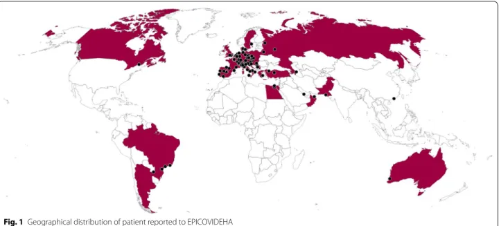
A total of 132 centers in 32 countries participated in this survey (Fig. 1, Additional file 2: Tables 2), and registered 4117 cases. Of these, 316 (7.7%) were excluded for the following reasons: age < 18 years old, clinical diagnosis of COVID-19, double-entry, non-malignant hematological diseases, incomplete information, more than 5 years off- therapy from the last chemotherapy, or solid cancer.
The demographic and clinical characteristics of 3801 valid cases are reported in Table 1. There was a higher prevalence of males (n = 2222, 58.5%) and Caucasian eth- nic background (n = 3289, 86.5%). The median age was 65 years (IQR: 54–74; range 18–95).
Patients with non-Hodgkin lymphoma (NHL) repre- sented the largest subgroup (n = 1084, 28.5%), followed by patients with multiple myeloma (MM) (n = 684, 18%) and those with AML (n = 497, 13.1%). Overall, 67.3% of the patients who developed COVID-19 had a baseline lymphoproliferative disease (n = 2557) (Table 1). More than 51% of the patients had active disease (n = 1963), and 2502 patients (65.8%) had received chemotherapy in the 3 months before the onset of COVID-19 (Table 2).
The most frequent treatments were chemotherapy with immunotherapy or immunotherapy alone administered to 983 patients (25.9%), compatible with the proportion of patients with NHL. In 271 patients (7.1%), the infec- tion occurred concomitantly with the diagnosis of HM,
and in 138 of those (50.9%) before treatment initiation for the baseline malignancy (Table 1). Five hundred fifty- seven patients (14.7%) had a transplant procedure per- formed in their clinical history (292 autologous HSCT [auto-HSCT] and 265 allogeneic HSCT [allo-HSCT]). In 247 patients, 173 allo-HSCT and 74 auto-HSCT recipi- ents, the transplant procedure was the last therapy before COVID-19 infection. In total 24 patients in the registry were treated with chimeric antigen receptor T (CAR- T) cells reinfusion, of which 3 patients had been treated with additional therapies after the CAR-T cell therapy (Table 2).
Overall, 2304 (60.6%) patients had at least one comor- bidity, with cardiovascular diseases being most frequent (n = 1146, 30.1%). In 447 patients (12.5%) smoking his- tory was reported (Table 1).
At the onset of COVID-19 infection, 280 patients (7.4%) had neutrophils below 0.5 × 10⁹/mm3, and 344 patients (9.1%) lymphocytes below 0.2 × 10⁹/mm3 (Table 3).
SARS-CoV-2 infection was diagnosed by naso- pharyngeal swab in almost all patients (n = 3700, 97.3%). COVID-19 tests were performed in 3027 patients (79.6%) because of pulmonary and/or extrapul- monary symptoms, and in 727 patients (19.1%) as part of asymptomatic screening. Reason for testing was unknown in 47 (1.2%). Presence of respiratory symp- toms, mainly cough and dyspnea, was the most fre- quent clinical presentation, reported in 2285 (60.1%), and in 831 of them (21.9%) it was combined with extra- pulmonary symptoms. In 742 patients (19.5%) extra- pulmonary symptoms, in particular anosmia, diarrhea,
Fig. 1 Geographical distribution of patient reported to EPICOVIDEHA
skin rash, were predominant in terms of clinical pres- entation (Table 3).
COVID-19 infection was determined to be critical in 689 patients (18.1%), severe in 1736 (45.7%), mild in 658 (17.3%), and asymptomatic in 675 (17.8%) (Table 3).
Overall, 2778 patients (73.1%) were hospitalized. The median duration of overall hospitalization was 15 days (IQR: 8–27, range 1–235), regardless of patient outcome.
Among the hospitalized patients, 689 (18.1%) required hospitalization in an ICU, 449 of these (65.2%) with inva- sive mechanical ventilation (MV) (Table 3).
Altogether, during the observation phase, 1185 patients (31.2%) died. The primary cause of death was COVID-19 in 688 patients (58.1%), HM in 173 patients (14.6%), and Table 1 Demographic and clinical characteristics of enrolled
patients at COVID‑19 diagnosis
Data can be super additive
a Onset patients had a contemporaneous diagnosis of the malignancy and the COVID-19, regardless of malignancy treatment initiation. Stable disease patients include patients at watch and wait
n %
Sex
Female 1579 41.5
Male 2222 58.5
Age, median (IQR) [range] 65 (54–74), [18–95]
Comorbidities
Chronic cardiopathy 1146 30.1
Chronic pulmonary disease 614 16.2
Diabetes mellitus 620 16.3
Liver disease 167 4.4
Obesity 345 9.1
Renal impairment 325 8.6
Smoking history 477 12.5
No risk factor identified 1463 38.5
Baseline hematological malignancies
Acute lymphoid leukemia 169 4.4
Chronic lymphoid leukemia 474 12.5
Acute myeloid leukemia 497 13.1
Chronic myeloid leukemia 161 4.2
Myelodysplastic syndrome 279 7.3
Low‑intermediate risk 138 3.6
High risk 48 1.3
Not stated 93 2.4
Hairy cell leukemia 23 0.6
Hodgkin lymphoma 135 3.6
Non‑Hodgkin lymphoma 1084 28.5
Indolent 497 13.1
Aggressive 516 13.6
Not stated 71 1.9
Essential thrombocythemia 69 1.8
Myelofibrosis 122 3.2
Polycythemia vera 70 1.8
Systemic mastocytosis 6 0.2
Multiple myeloma 684 18.0
Amyloidosis 8 0.2
Aplastic anemia 20 0.5
Statusa
Controlled disease 1760 46.3
Complete remission 1170 30.8
Partial remission 590 15.5
Activedisease 1963 51.6
Onset 888 23.4
Refractory/Resistant 473 12.4
Stable disease 524 13.8
Unknown 78 2.1
Unknown 78 2.1
Table 2 Summary of received treatments for Hematological Malignancies at the onset of COVID‑19
HSCT Hematopoietic stem cell transplantation, CAR-T chimeric antigen receptor T-cell therapies
a Bortezomib, ibrutinib, idelalisib, ruxolitinib, TKI (tyrosine kinase inhibitors), and venetoclax
b Data can be super-additive
n %
Last/ongoing treatment strategy before COVID-19
Immunochemotherapy 857 22.5
Targeted therapya 607 16.0
Conventional chemotherapy 597 15.7
No treatment 538 14.1
Palliative/supportive measures 226 6.0
Immunomodulators 218 5.7
Allogeneic HSCT 173 4.6
Anagrelide/Hydroxyurea 145 3.8
Hypomethylating agents 141 3.7
Immunotherapy only 125 3.3
Autologous HSCT 74 1.9
Unknown 41 1.1
Other 28 0.7
CAR‑T 21 0.6
Radiotherapy 10 0.3
Summary of received treatmentb
Chemotherapy 3178 83.6
In the last month 1979 52.1
In the last 3 months 523 13.8
Treatment ended > 3 months 631 16.6
Not stated 45 1.2
Radiotherapy 186 4.9
Allogeneic HSCT 265 7.0
Autologous HSCT 292 7.7
CAR‑T 24 0.6
Other strategies 150 3.9
No treatment 538 14.2
a combination of both COVID-19 and progressing HM in 155 patients (13.1%). In the remaining cases the cause was unknown or due to other reasons.
Patients over the age of 70 years had the highest mor- tality (661/1475, 44.8%). Considering the different HM, the higher number of fatalities was observed in AML (199/497, 40%) and in myelodysplastic syndromes (MDS) Table 3 Clinical features of COVID‑19 in our patient cohort
BAL Bronchoalveolar lavage, COVID-19coronavirus disease 19, HM hematological malignancy, ICU intensive care unit, MV mechanical ventilation, SARS-CoV-2 severe acute respiratory syndrome coronavirus 2
a Data can be super additive
b Data not available in all patients
n %
COVID-19 infection
Asymptomatic 675 17.8
Mild 658 17.3
Severe 1736 45.7
Critical 689 18.1
Unknown 43 1.1
COVID-19 test samplea
BAL 60 1.6
SARS‑CoV‑2 nasopharyngeal swab 3700 97.3
SARS‑CoV‑2 serology 86 2.3
Reason for COVID-19 testa
Pulmonary symptoms 1454 38.3
Pulmonary + extrapulmonary symptoms 831 21.9
Extrapulmonary symptoms 742 19.5
Screening 727 19.1
Unknown 47 1.2
Neutrophils level at COVID-19 diagnosisb
≤ 0.5 × 10⁹/mm3 280 7.4
0.501–0.999 × 10⁹/mm3 217 5.7
≥ 1 × 10⁹/mm3 2738 72.0
Lymphocytes level at COVID-19 diagnosisb
≤ 0.2 × 10⁹/mm3 344 9.1
0.201–0.499 × 10⁹/mm3 538 14.2
≥ 0.5 × 10⁹/mm3 2367 62.3
Stay during COVID-19
Admitted to hospital 2778 73.1
Length of hospital stay, median (IQR) [range] 15 (8–27), [1–235] –
ICU 689 18.1
Length of ICU stay, median (IQR) [range] 11 (5–20), [1–111] –
Invasive MV 449 11.8
Non‑invasive MV 221 5.8
Clinical outcome of COVID-19
Death 1185 31.2
Observation time, median (IQR) [range] 89 (21–172), [0–436] –
Reason for deatha
Not related to COVID‑19 125 3.3
Contributable by COVID‑19 155 4.1
Attributable to COVID‑19 843 22.2
Attributable to HM 328 8.6
Death due to other reasons 123 3.2
Death due to unknown reasons 78 2.1
(118/279, 42.3%) (Table 4). Mortality in AML/MDS was significantly higher when compared to mortality in other HM (p < 0.0001) (Fig. 2).
Regarding last underlying treatments for HM before COVID-19, the highest mortality rate was observed among
patients receiving demethylating agents (83/141, 58.9%
[95% confidence interval {CI} 50.6–66.7]) and in pallia- tive treatment settings (104/226, 46% [95%CI 39.4–52.5]).
Despite the small number of patients undergoing CAR-T reinfusion, mortality rate in these patients was high (47.6%
[95% CI 28.3–67.6]; 10/21 patients). Patients undergoing auto-HSCT or allo-HSCT had mortality rates of 27% ([95%
CI 18.2–38.1] 20/74 cases) and 24.8% ([95% CI 19.0–31.8]
43/173 cases), respectively (Table 4). The mortality rate of patients who received a transplant as most recent therapy was significantly lower when compared to non-transplant patients (p < 0.027) (Fig. 3).
Patients with critical COVID-19 (63.6% [95% CI 59.9–67.1, 438/689]) died in a higher proportion than those with severe (30.3% [95% CI 28.2–32.5, 526/1736]), p < 0.0001 or mild infection (16.7% [95%
CI 14.1–19.8, 110/658]), p < 0.0001. The mortality rate observed in patients with severe infection 30.3% ([95%
CI 28.2–32.5] 526/1736), was significantly higher than reported in patients with mild COVID-19 (16.7% [95%
CI 14.1–19.8] 110/658), p < 0.0001). The mortality in mildly symptomatic patients was not vastly different from that observed in initially asymptomatic patients:
15.4% ([95% CI 12.9–18.3] 104/675) p = 0.516 (Fig. 4, Additional file 3: Table 3). Clinical presentation with pulmonary symptoms was associated with a signifi- cantly higher mortality rate versus presentation with extrapulmonary symptomatology alone (mortality rate 876/2285, 38.3% [95% CI 36.4–40.4] vs. 163/742, 22.0% [95% CI 19.1–25.1], p < 0.0001).
A higher mortality rate was reported for patients admitted to ICU (438/689, 63.5% [95% CI 59.9–67.1]), compared to non-ICU patients (747/3112, 24% [95%
CI 22.5–25.5]) p < 0.0001. Furthermore, among the ICU patients, a significantly higher mortality rate was observed in patients with invasive MV versus those without (322/449, 71.7% [95% CI 67.4–75.7] vs.
116/240, 48.3% [95% CI 42.1–54.6] p < 0.0001).
Considering the two waves of COVID-19 (1st wave March–May 2020, 2nd wave October-December 2020), there was a significant decrease in the mortality rate in the second wave (581/1427, 40.7% [95% CI 38.2–43.3] vs.
439/1773, 24.8% [95% CI 22.8–26.8] p < 0.0001) (Fig. 5).
The reduction of mortality was consistent across different HM diagnoses (Fig. 6).
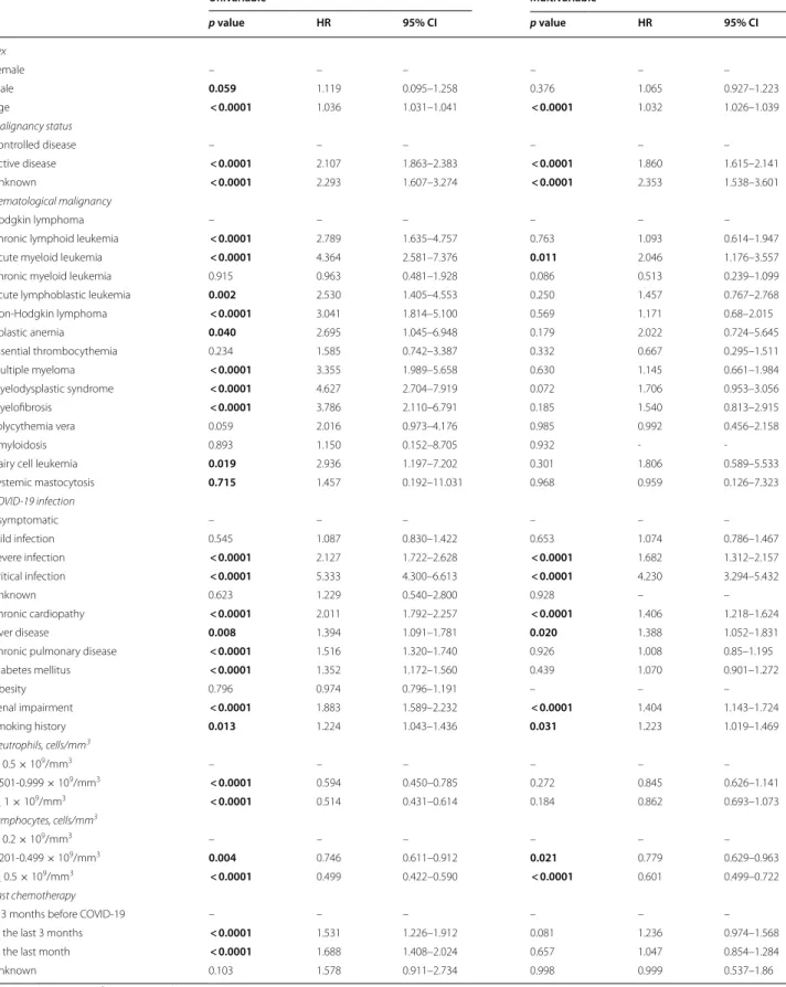
In the univariable Cox regression analysis, multiple fac- tors negatively influenced mortality (Table 5). Conversely, having a neutrophil count greater than 0.5 × 10⁹/mm3 or a lymphocyte count greater than 0.2 × 10⁹/mm3 were found to be protective.
In the multivariable analysis the following parame- ters were significantly associated with higher mortality:
age increase, active disease, chronic cardiopathy, liver Table 4 Overall mortality rate by disease and treatment received
HSCT Hematopoietic stem cell transplantation, CAR-T chimeric antigen receptor T-cell therapies
a Bortezomib, ibrutinib, idelalisib, ruxolitinib, TKI (tyrosine kinase inhibitors) and venetoclax
Overall mortality
Survived n (%) Died n (%) Baseline hematological malignancies
Acute lymphoid leukemia 125 (74) 44 (26)
Chronic lymphoid leukemia 340 (71.7) 134 (28.3)
Acute myeloid leukemia 298 (60) 199 (40)
Chronic myeloid leukemia 144 (89.5) 17 (10.5) Myelodysplastic syndrome 161 (57.7) 118 (42.3)
Low‑intermediate risk 77 (55.8) 61 (44.2)
High risk 26 (54.2) 22 (45.8)
Not stated 58 (62.4) 35 (37.6)
Hairy cell leukemia 15 (65.2) 8 (34.8)
Hodgkin lymphoma 120 (88.9) 15 (11.1)
Non‑Hodgkin lymphoma 739 (68.2) 345 (31.8)
Indolent 354 (71.3) 143 (28.7)
Aggressive 337 (65.3) 179 (34.7)
Not stated 48 (67.6) 23 (32.4)
Essential thrombocythemia 57 (82.6) 12 (17.4)
Myelofibrosis 77 (63.1) 45 (36.9)
Polycythemia vera 56 (80) 14 (20)
Systemic mastocytosis 5 (83.4) 1 (16.6)
Multiple Myeloma 458 (67) 226 (33)
Amyloidosis 7 (87.5) 1 (12.5)
Aplastic anemia 14 (70) 6 (30)
Last/ongoing treatment strategy before COVID-19
Anagrelide/Hydroxyurea 106 (73.1) 39 (26.9)
Conventional chemotherapy 423 (70.9) 174 (29.1)
Hypomethylating agents 58 (41.2) 83 (58.8)
Immunotherapy only 89 (71.2) 36 (28.8)
Immunochemotherapy 595 (69.4) 262 (30.6)
Immunomodulators 139 (63.8) 79 (36.2)
Targeted therapya 453 (74.6) 154 (25.4)
Allogeneic HSCT 130 (75.2) 43 (24.8)
Autologous HSCT 54 (73) 20 (27)
CAR‑T 11 (52.4) 10 (47.6)
Radiotherapy 9 (90) 1 (10)
Palliative/supportive measures 122(56) 104 (46)
Other 17 (60.7) 11 (39.3)
Unknown 28 (68.3) 13 (31.7)
No treatment 382 (71) 156 (29)
disease, renal impairment, smoking history, and ICU stay. Among HM, AML is the malignancy associated with a significantly high mortality (Table 5).
Discussion
The incidence of COVID-19 infection in HM ranges between 1 and 3.9% [23]. Mostly, patients get infected in the community, although in 1.1% to 15% of infections nosocomial transmissions are reported [24]. A clear cor- relation between the type of HM and the incidence of COVID-19 infection has not been described in the litera- ture, but current data indicate that lymphoproliferative disorders, in particular NHL, chronic lymphocytic leuke- mia, and MM are particularly associated with higher risk from COVID-19.
Here we presented a large survey on COVID-19 among HM patients, with almost 4000 patients reported from 132 hematology institutions mainly located in Europe. In addition, this survey has collected COVID-19 cases from March to December 2020, allowing us to analyze not only which patients were at risk, but also how the infectious process has evolved over time. Our data confirm that a larger number of COVID-19 cases was diagnosed among
patients with lymphoproliferative disorders, in particu- lar NHL and MM, as previously documented [9, 10].
However, we also observed a high number of COVID-19 among patients with AML (12.5%), which is considered a rare malignancy. As for comorbidities, our patient popu- lation reflects the overall population, with cardiovascular diseases being the most frequent comorbidity reported [16]. Most of the patients recorded in our survey had a severe/critical clinical presentation of COVID-19 (about 60%), over two-thirds were hospitalized and about 18%
required ICU admission. These data are not surprising and emphasize the frailty of HM patients, and are slightly higher compared with those reported in the literature, ranging between 15.5 to 52.4% and 6.9 to 14% for severe and critical clinical presentation, respectively [3–17].
The overall and the attributable mortality rates observed in our study (31.2% and 22.2%, respectively) are within the range of those reported in the literature among HM (published reports are summarized in Additional file 4: Table 4), confirming that COVID-19 mortality is significantly higher in HM patients than in the overall population, where current data show a mortality rate ranging between 0.1 and 9.4% across the different coun- tries around the world (www. coron avirus. jhu. edu/ data/
Fig. 2 Overall survival by the underlying disease
morta lity). Moreover, as expected, the overall mortality rate has been age-dependent, with higher mortality rates observed among patients aged over 70 years. In line with other studies [7, 14, 16], our data have shown that AML and MDS patients, especially those with high-risk MDS, have the worst clinical outcome and the highest mortal- ity rate (up to 45%). In fact, AML was the only that was independently associated with mortality in our multivari- able model. A recently published study focusing only on AML patients reported an overall mortality very similar to that described in our study [25]. There are several pos- sible explanations of this phenomenon. First, patients with AML/MDS are often aged over than 65 years old.
Second, they present a profound immunodeficiency as a consequence of both disease and treatments received.
Third, they are patients in which a treatment delay is often not possible due to the urgent need of starting an active therapy. This last aspect is quite relevant, espe- cially if we consider that a lower mortality in patients who delayed AML treatment was described compared to those with and without treatment modification [25]. In high-risk MDS patients, treatment with demethylating agents was associated with a particularly high mortal- ity rate. Our study highlights the role of these agents as
being potentially associated with high mortality in AML/
MDS patients with COVID-19. Our data also showed that patients undergoing HSCT (either autologous or allogeneic) presented a significantly lower mortality rate following COVID-19, compared to non-transplant patients. We report an overall mortality rate of 24.8% and 27% in allo-HSCT and auto-HSCT, respectively, almost identical to that very recently described in the study of the European Society for Blood and Marrow Transplan- tation [12]. This observation is coherent with previous published data, suggesting a significantly lower mor- tality rate among transplanted patients compared with non-transplanted HM patients [15]. Patients who receive HSCT, especially an allogeneic one, are by definition younger and healthier than the overall onco-hematolog- ical patients. In fact, we observed that most of condi- tions associated with higher overall mortality (i.e. older age, comorbidities, uncontrolled disease) were over- represented in the non-transplant cohort. These aspects may explain in part the lower mortality we observed in transplanted patients. Interestingly, patients undergoing CAR-T infusion have shown a worse clinical outcome in our survey, with 10 deaths among 21 COVID-19 patients registered in the database. Other significant predictors
25 125 44
247
278 594
3008 1363
0 9 70
154
535 267 0
0 0 0
200 300
Days from COVID-19 diagnosis
500 400
100 0
Survival probability (%)
100
80
60
40
20
0
Number of patients at risk /Autologous HSCT
No HSCT No treatment
Log-rank test p = 0.027
HSCT: Hematopoietic stem-cell transplantion Fig. 3 Overall survival by transplant vs no transplant
of mortality in the multivariable analysis included active disease, chronic cardiopathy, liver disease, renal impair- ment, smoking history, and ICU stay.
Moreover, we found a significantly lower mortality in COVID-19 HM patients in the second wave as com- pared to the first wave of COVID 19. Improved clinical outcome has been documented for many different dis- eases, including those with the highest mortality rates.
This improvement in the second wave of COVID-19 is of interest, and could be the result of several factors, including a better knowledge of the clinical course of the disease, more effective protective procedure for HM patients, a detection of a larger number of asymptomatic/
mild cases by screening swabs and/or an improvement of specific treatments against COVID-19, for example remdesivir, monoclonal antibodies, convalescent plasma.
Coherently with our hypothesis, in the second wave, we found a significantly higher rate of asymptomatic and mild infections and a significantly lower rate of severe infections. However, even though we did not observe significant differences in HM distribution, in the second wave we found more patients with controlled disease compared to the first one.
We strongly believe that our findings will impact the management of HM patients also in the near future.
Even if we are witnessing a huge worldwide vaccination program, preliminary data published so far suggest that anti-SARS-CoV-2 vaccines shows significantly less robust efficacy in eliciting an immune response in HM patients than observed in the general population [26, 27]. Moreo- ver, we are assisting to the wide diffusion of variants of concerns under the vaccine selective pressure. Indeed, sev- eral cases of breakthrough infections have been reported in the general population, with a significant mortality rate [28, 29]. We expect that, in the immediate future, we will assist to several cases of SARS-CoV-2 infections in fully vaccinated HM patients. From this point of view, the better understanding of epidemiologic features and risk factors for COVID-19 in HM patients, might surely help hema- tologists in the management of their patients and even in modifying the chemotherapeutic programs where possi- ble. HM patients still deserve special attention and protec- tive measures should continue.
Our large registry study comes with some limita- tions. First, at the time the study was designed, the role of thromboembolic phenomena of COVID-19 Fig. 4 Overall survival by COVID‑19 severity
Fig. 5 Overall survival by time distribution (first vs. the second wave)
Fig. 6 Overall survival in the different HMS by time distribution (first vs. the second wave)
Table 5 Overall mortality predictors in COVID‑19 HM patients
Univariable Multivariable
p value HR 95% CI p value HR 95% CI
Sex
Female – – – – – –
Male 0.059 1.119 0.095–1.258 0.376 1.065 0.927–1.223
Age < 0.0001 1.036 1.031–1.041 < 0.0001 1.032 1.026–1.039
Malignancy status
Controlled disease – – – – – –
Active disease < 0.0001 2.107 1.863–2.383 < 0.0001 1.860 1.615–2.141
Unknown < 0.0001 2.293 1.607–3.274 < 0.0001 2.353 1.538–3.601
Hematological malignancy
Hodgkin lymphoma – – – – – –
Chronic lymphoid leukemia < 0.0001 2.789 1.635–4.757 0.763 1.093 0.614–1.947
Acute myeloid leukemia < 0.0001 4.364 2.581–7.376 0.011 2.046 1.176–3.557
Chronic myeloid leukemia 0.915 0.963 0.481–1.928 0.086 0.513 0.239–1.099
Acute lymphoblastic leukemia 0.002 2.530 1.405–4.553 0.250 1.457 0.767–2.768
Non‑Hodgkin lymphoma < 0.0001 3.041 1.814–5.100 0.569 1.171 0.68–2.015
Aplastic anemia 0.040 2.695 1.045–6.948 0.179 2.022 0.724–5.645
Essential thrombocythemia 0.234 1.585 0.742–3.387 0.332 0.667 0.295–1.511
Multiple myeloma < 0.0001 3.355 1.989–5.658 0.630 1.145 0.661–1.984
Myelodysplastic syndrome < 0.0001 4.627 2.704–7.919 0.072 1.706 0.953–3.056
Myelofibrosis < 0.0001 3.786 2.110–6.791 0.185 1.540 0.813–2.915
Polycythemia vera 0.059 2.016 0.973–4.176 0.985 0.992 0.456–2.158
Amyloidosis 0.893 1.150 0.152–8.705 0.932 ‑ ‑
Hairy cell leukemia 0.019 2.936 1.197–7.202 0.301 1.806 0.589–5.533
Systemic mastocytosis 0.715 1.457 0.192–11.031 0.968 0.959 0.126–7.323
COVID-19 infection
Asymptomatic – – – – – –
Mild infection 0.545 1.087 0.830–1.422 0.653 1.074 0.786–1.467
Severe infection < 0.0001 2.127 1.722–2.628 < 0.0001 1.682 1.312–2.157
Critical infection < 0.0001 5.333 4.300–6.613 < 0.0001 4.230 3.294–5.432
Unknown 0.623 1.229 0.540–2.800 0.928 – –
Chronic cardiopathy < 0.0001 2.011 1.792–2.257 < 0.0001 1.406 1.218–1.624
Liver disease 0.008 1.394 1.091–1.781 0.020 1.388 1.052–1.831
Chronic pulmonary disease < 0.0001 1.516 1.320–1.740 0.926 1.008 0.85–1.195
Diabetes mellitus < 0.0001 1.352 1.172–1.560 0.439 1.070 0.901–1.272
Obesity 0.796 0.974 0.796–1.191 – – –
Renal impairment < 0.0001 1.883 1.589–2.232 < 0.0001 1.404 1.143–1.724
Smoking history 0.013 1.224 1.043–1.436 0.031 1.223 1.019–1.469
Neutrophils, cells/mm3
≤ 0.5 × 10⁹/mm3 – – – – – –
0.501‑0.999 × 10⁹/mm3 < 0.0001 0.594 0.450–0.785 0.272 0.845 0.626–1.141
≥ 1 × 10⁹/mm3 < 0.0001 0.514 0.431–0.614 0.184 0.862 0.693–1.073
Lymphocytes, cells/mm3
≤ 0.2 × 10⁹/mm3 – – – – – –
0.201‑0.499 × 10⁹/mm3 0.004 0.746 0.611–0.912 0.021 0.779 0.629–0.963
≥ 0.5 × 10⁹/mm3 < 0.0001 0.499 0.422–0.590 < 0.0001 0.601 0.499–0.722
Last chemotherapy
> 3 months before COVID‑19 – – – – – –
In the last 3 months < 0.0001 1.531 1.226–1.912 0.081 1.236 0.974–1.568
In the last month < 0.0001 1.688 1.408–2.024 0.657 1.047 0.854–1.284
Unknown 0.103 1.578 0.911–2.734 0.998 0.999 0.537–1.86
HR Hazard ratio, CI confidence intervals
infection was still unknown and therefore not included in the survey. Second, we have deliberately excluded the data relating to the various COVID-19 therapeutic approaches because they are extremely heterogeneous and treatment recommendations change rapidly. Third, due to our registry design we have not been able to calculate the incidence of COVID-19 in the various subclasses of HM. Last, due to the intrinsic limitations of the study, it is not pos- sible to provide cumulative incidences regarding rele- vant aspects, such as mortality, as there is no certainty about whether all participating sites documented all eligible cases.
These data need to be carefully interpreted consid- ering the incidence of individual HM in the general population and the patient performance status, which affects their social dimension and lifestyle in the community.
Conclusion
This study sheds light on the epidemiology, risk fac- tors and outcomes of COVID-19 among patients with HM. While the introduction of COVID-19 vaccina- tions will lead to a marked reduction of infections in HM patients, the possibility of a lower efficacy of vaccinations needs to be taken into account [30], pos- sibly resembling previous experiences with influenza vaccination. Future studies are needed to evaluate whether the use of vaccination will be able to prevent the development and above all mortality in the identi- fied risk categories of HM.
Abbreviations
allo‑HSCT: Allogeneic HSCT [hematopoietic stem cell transplantation]; AML:
Acute myeloid leukemia; ASH: American Society of Hematology; auto‑HSCT:
Autologous HSCT [hematopoietic stem cell transplantation]; BAL: Bronchoal‑
veolar lavage; CAR‑T: Chimeric antigen receptor T‑cell therapies; CI: Confidence intervals; COVID‑19: Coronavirus disease 19; eCRF: Electronic case report form;
EHA: European Hematology Association; EPICOVIDEHA: Epidemiology of COVID‑19 Infection in Patients with Hematological Malignancies: A European Hematology Association Survey; HM: Hematological malignancy; HR: Hazard ratio; HSCT: Hematopoietic stem cell transplantation; ICU: Intensive care unit;
IQR: Interquartile range; MDS: Myelodysplastic syndromes; MM: Multiple myeloma; MV: Mechanical ventilation; NHL: Non‑Hodgkin lymphoma; SARS‑
CoV‑2: Severe acute respiratory syndrome coronavirus 2; TKI: Tyrosine kinase inhibitors; WHO: World Health Organization.
Supplementary Information
The online version contains supplementary material available at https:// doi.
org/ 10. 1186/ s13045‑ 021‑ 01177‑0.
Additional file 1: Supplementary Table 1. Partnership from National and International Scientific Society.
Additional file 2: Supplementary Table 2. List of participating institutions.
Additional file 3: Supplementary Table 3. Demographic and clinical characteristics of enrolled patients depending on the COVID‑19 severity.
Additional file 4: Supplementary Table 4. Multicentre studies on COVID‑19 in patients with haematologic malignancies reported during 2020.
Acknowledgements
The authors thank all contributors for their utmost contributions and support to the project during a pandemic situation and to Susann Blossfeld and Corinna Kramer for their administrative and technical assistance.
Authors’ contributions
LP served as the principal investigator. LP and JSG contributed to study design, study supervision, and data interpretation and wrote the paper. LP, OC, FP, PC, NK, AP, MH, PK, PC, conceived the study idea. LP, JSG, and FM did the statistical plan, analysis and interpreted the data. All the authors recruited participants and collected and interpreted data. All authors contributed to manuscript writing and review of the manuscript. All authors agreed to be accountable for all aspects of the work in ensuring that questions related to the accuracy or integrity of any part of the work are appropriately investigated and resolved. All authors read and approved the final manuscript.
Funding
EPICOVIDEHA has received funds from Optics COMMITTM (COVID‑19 Unmet Medical Needs and Associated Research Extension) COVID‑19 RFP program by GILEAD Science, United States (Project 2020‑8223).
Availability of data and materials
Individual participant data that underlie the results reported in this Article, after de‑identification (text, tables, figures, and appendices), will be available together with the study protocol. This will be from 9 to 24 months follow‑
ing Article publication. Data will be available only for investigators whose proposed use of the data has been approved by an independent review com‑
mittee identified for this purpose.
Declarations
Ethics approval and consent to participate
The study was formally approved by the Ethical Committee of Fondazione Policlinico Universitario Agostino Gemelli—IRCCS, Università Cattolica del Sacro Cuore of Rome with the following registration number: 3226. The study was conducted in compliance wsith Helsinki declaration and Good Clinical Practice. The corresponding local ethics committee of each participating insti‑
tution has approved the EPICOVIDEHA study when applicable. EPICOVIDEHA has been registered at www. clini caltr ials. gov with the identifier NCT04733729.
The anonymized data that do not contain any personally identifiable informa‑
tion from any sources implies that the informed consent is not applicable.
Consent for publication Not applicable.
Competing interests
The authors declare that they have no competing interests.
Author details
1 Hematology, Fondazione Policlinico Universitario Agostino Gemelli ‑ IRCCS – Università Cattolica del Sacro Cuore, Rome, Italy. 2 Università Cattolica del Sacro Cuore, Rome, Italy. 3 Department I of Internal Medicine, Center for Integrated Oncology Aachen Bonn Cologne Duesseldorf, Excellence Center for Medical Mycology (ECMM), University of Cologne, Faculty of Medicine and University Hospital Cologne, Cologne, Germany. 4 Cologne Excellence Cluster On Cellular Stress Responses in Aging‑Associated Diseases (CECAD), University of Cologne, Faculty of Medicine and University Hospital Cologne, Cologne, Germany. 5 Hematology and Stem Cell Transplant Unit, IRCCS Regina Elena National Cancer Institute, Rome, Italy. 6 Stem Cell Transplant Center, AOU Citta’ Della Salute E Della Scienza, Turin, Italy.
7 University of Milan and Fondazione IRCCS Istituto Nazionale Dei Tumori, Milan, Italy. 8 Division of Infectious Diseases and Global Public Health,