Cognitive and psychiatric aspects of mitochondrial encephalomyopathies
Dissertation
Dr. Inczédy-Farkas Gabriella
Szentágothai János School of PhD Studies in Neurosciences Semmelweis University
Supervisor: Dr. Molnár Mária Judit DSc
Opponents: Dr. Nagy Ferenc PhD
Dr. Gonda Xénia PhD Chair of the oral exam: Dr. Túry Ferenc PhD
Members of the exam panel: Dr. Kelemen Anna PhD Dr. Baran Brigitta PhD
Budapest
2014
2 TABLE OF CONTENT
1. Abbreviations ... 4
2. Introduction ... 7
2.1. Mitochondria in health and disease... 7
2.1.1. General characteristics ... 7
2.1.2. The mitochondrial genome ... 8
2.1.3. Mitochondrial disorders ... 9
2.2. Epidemiology of mitochondrial disorders ... 11
2.3. Mitochondrial alterations in psychiatric disorders ... 12
2.3.1. Biochemical and morphologic data ... 12
2.3.2. Genetic data ... 13
2.4. Psychiatric symptoms in mitochondrial disorders ... 14
2.5. The role of mitochondria in neurodegenerative disorders ... 15
2.6. Cognitive symptoms in mitochondrial disorders ... 15
3. Objective ... 16
4. Patients and methods ... 17
4.1. Patients ... 17
4.1.1. Epidemiological study ... 17
4.1.2. Psychiatric and neuropsychologic assessment ... 18
4.2. Methods... 18
4.2.1. General clinical evaluation ... 18
4.2.2. Myopathology ... 19
4.2.3. Genetic studies ... 19
4.2.3.1. Analysis of the mtDNA ... 19
4.2.3.2. Analysis of the PMP22 gene ... 20
4.2.3.3. Determination of the 5HTTLPR genotype ... 20
4.2.4. Focused clinical evaluation ... 20
4.2.5. Psychiatric examination ... 21
4.2.6. Neuropsychological assessment... 21
4.2.7. Statistical analysis ... 22
5. Results ... 24
3
5.1. Epidemiology of mitochondrial disorders ... 24
5.1.1. Mutations in genes of the mitochondrial tRNA ... 24
5.1.1.1. The m.3243 A>G point mutation ... 24
5.1.1.2. The m.8344 A>G point mutation ... 24
5.1.1.3. Other mutations of the mitochondrial tRNA ... 25
5.1.2. Mutations in genes coding for mitochondrial proteins ... 25
5.1.2.1. The m.8993 T>C and m.8993 T>G point mutations ... 25
5.1.2.2. The m.3460 G>A, m.11778 G>A and m.14484 T>C point mutations... 26
5.1.2.3. Mutations detected with the Mitochip v.2.0. ... 26
5.1.3. Rearrangements of the mtDNA ... 26
5.2. Genetic background of the psychiatric study ... 27
5.2.1. MtDNA genotypes of the selected MTD patients ... 27
5.2.2. PMP22 genotypes of the control (HN) patients ... 28
5.2.3. Analysis of the 5HTTLPR gene ... 28
5.3. Clinical evaluation ... 29
5.3.1. Neurological examination ... 29
5.3.2. Medication ... 29
5.3.3. Neuroimaging ... 33
5.4. Psychiatric findings ... 35
5.5. Cognitive symptoms ... 40
5.5.1. Results of the neuropsychological assessment ... 40
5.5.2. Longitudinal follow-up of Patient 16 ... 44
5.6. La belle indifference – general characteristic or individual trait ... 46
5.7. NEPSYBANK... 48
6. Discussion... 49
6.1. Epidemiology of mitochondrial disorders ... 49
6.2. Mitochondrial psychiatry? ... 49
6.2.1. Affective spectrum disorders ... 49
6.2.2. Other psychiatric symptoms ... 50
6.2.3. The concept of mitochondrial psychiatry ... 51
6.3. Mitochondrial dementia? ... 51
4
6.3.1. Involvement of the prefrontal cortex/frontal lobe ... 51
6.3.2. La belle indifference ... 53
6.3.3. Involvement of non-frontal areas ... 53
6.3.4. Neurological symptoms as confounding factors ... 53
6.3.5. Intelligence ... 54
6.3.6. Correlations of cognitive performance and structural alterations ... 55
6.3.7. Patients with tRNA mutations are the most affected ... 55
6.3.8. The concept of mitochondrial dementia ... 56
6.3.9. The importance of the awareness to MTDs ... 57
7. Conclusion ... 58
8. Summary ... 59
9. Összefoglalás ... 60
10. References ... 61
11. List of publications ... 71
12. Acknowledgement ... 73
5
1. Abbreviations
ADP: adenosine-diphosphate AMP: adenosine-monophosphate AMPK: AMP-dependent kinase ANT: adenine nucleotide translocase ATP: adenosine-triphosphate
BCL-2: B-cell lymphoma-2
BDI-SF: Beck Depression Inventory – Short Form
CADASIL: cerebral autosomal dominant arteriopathy with subcortical infarcts and leukoencephalopathy
cAMP: cyclic AMP CI: confidence interval CK: creatine-kinase
CMT: Charcot – Marie – Tooth phenotype of the PMP22 mutation CNS: Central Nervous System
COX: cytochrome oxidase
CREB: cAMP response element-binding protein CT: computer tomography
Cyt-b: cytochrome b Cyt-c: cytochrome c
DCM: dilated cardiomyopathy DM: diabetes mellitus
DSM: Diagnostic and Statistical Manual of Mental Disorders EEG: electroencephalogram
EMG: electromyography ENG: electroneurography F: female
FSIQ: Full Scale Intelligence Quotient GERD: Gastro-Esophageal Reflux Disorder GSI: Global Severity Index of the SCL-90-R
HAQ-DI: Stanford Health Assessment Questionnaire 20-item Disability Index
6 HCM: hypertrophic cardiomyopathy
HDRS: Hamilton Depression Rating Scale HN: Hereditary Neuropathy
HNPP: Hereditary Neuropathy with liability to Pressure Palsy phenotype of the PMP22 mutation
HTN: hypertension
HTTLPR: 5HT (serotonin) – Transporter – Linked Polymorphic Region IgG: Immunoglobulin G
KSS: Kearns-Sayre Syndrome
LHON: Leber Hereditary Optic Neuropathy
L/L: long-long homozygous genotype of the 5HTTLPR L/S: long-short heterozygous genotype of the 5HTTLPR M: male
MAO: monoamine oxidase
MDD: Major Depressive Disorder
MELAS: Mitochondrial Encephalomyopathy, Lactic Acidosis and Stroke-like episodes MERRF: Myoclonus Epilepsy with Ragged Red Fibers
MIDD: Maternally Inherited Diabetes and Deafness MILS: Maternally Inherited Leigh’s Syndrome
MNGIE: Mitochondrial Neuro-Gastrointestinal Encephalopathy syndrome MOMP: Mitochondrial Outer Membrane Permeabilization
mPT: mitochondrial permeability transition pore MI: myocardial infaction
MR: mental retardation
MRI: magnetic resonance imaging MT-ATP6: mitochondrial ATP synthase MTD: mitochondrial disorders
mtDNA: mitochondrial DNA
NAD: nicotinamide adenine dinucleotide
NARP: Neuropathy, Ataxia, Retinitis Pigmentosa NCBI: National Center for Biotechnology Information ND5: NAD dehydrogenase subunit 5
7 NOS: not otherwise specified
OXPHOS: oxidative phosphorylation PCR: polymerase chain reaction
PDC: pyruvate dehydrogenase complex PEO: Progressive External Ophthalmoplegia PMP22: Peripheral Myelin Protein-22 POLG1: DNA polymerase gamma PQ: Performance Quotient
Pt/Pts: patient/ patients
PTSD: Post-Traumatic Stress Disorder RAVLT: Rey Auditory Verbal Learning Test ROS: reactive oxygen species
SCID-I and SCID-II: Structured Clinical Interviews for the DSM-IV SCL-90-R: Symptom CheckList – 90 – Revised
SDH: succinate dehydrogenase
SIDS: Sudden Infant Death Syndrome SNHL: Sensorineural Hearing Loss SNP: Single Nucleotide Polymorphism
S/S: short-short homozygous genotype of the 5HTTLPR StroopC: Stroop Color Condition
StroopCW: Stroop Color-Word Condition TCA: tricarboxylic acid cycle
TIA: Transient Ischemic Attack
TIM: Translocase of the Inner Membrane TMTA, TMTB: Trail Making Test part A and B TOM: Translocase of the Outer Membrane tRNA: transfer RNA
tRNALeu : Leucine of tRNA tRNALys: Lysine of tRNA
VDAC: Voltage-Dependent Anion Channel VQ: Verbal Quotient
WAIS: Wechsler Adult Intelligence Scale
8
2. Introduction
2.1. Mitochondria in health and disease
2.1.1. General characteristics
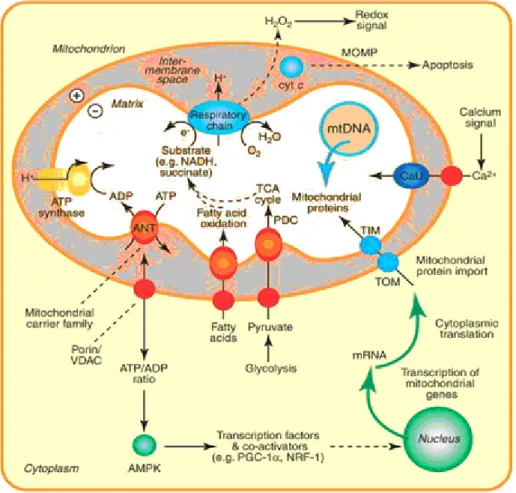
Mitochondria are intracellular organelles with possible common origin to bacteria (endosymbiotic theory). They are the sites ATP (adenosine triphosphate) production via oxidative phosphorylation (OXPHOS). Mitochondria are also involved in cell-specific functions such as steroid- (Miller, 2013) and amino acid synthesis (Araujo et al, 2012), neurotransmitter metabolism (Kann and Kovacs 2007), intracellular calcium homeostasis (Bygrave, 1978) and in the regulation of apoptosis (Mayer and Oberbauer 2003). These basic functions are summarized in Figure 1.
Figure 1. Mitochondrial function and biogenesis (www.sciencedirect.com, modified)
9 2.1.2. The mitochondrial genome
Mitochondria uniquely contain their own double-stranded 16.6 Kb circular genome (mtDNA) that is separate from the nuclear DNA but is in a complex interaction with it [nuclear-mitochondrial intergenomial communication]. MtDNA has only 37 genes; the majority of mitochondrial proteins are encoded by nuclear DNA. Thereby, mutations in either the mtDNA or the nuclear DNA can result in mitochondrial dysfunction. While clinically significant mutations can occur anywhere in the mitochondrial genome, common mutation sites, so called ‘hot spots’ has been identified. Probably the most frequent one is the m.3243 A>G mutation in the tRNALeu1(UUR) present in around 80%
of MELAS cases (Mitochondrial Encephalomyopathy, Lactic Acidosis and Stroke-like episodes, Goto et al, 1990). Another frequent tRNA mutation is the m.8344 A>G in the tRNALys, identified as the cause of MERRF syndrome (Myoclonus Epilepsy with Ragged Red Fibers, Schoffner et al, 1990).
MtDNA mutations may also occur in genes coding for mitochondrial proteins, such as the most commonly studied primary LHON (Leber Hereditary Optic Neuropathy) mutations (m.3460 G>A, m.11778 G>A and m.14484 T>C) that result in a very specific, easily recognizable clinical phenotype. Another important mutation site is at position mt8993 in the ATP synthase 6 gene (MT-ATP6), which results in NARP (Neuropathy, Ataxia and Retinis Pigmentosa) (Tatuch and Robinson 1993) or MILS (Maternally Inherited Leigh’s Syndrome), depending on the ratio of heteroplasmy (Degoul et al, 1995).
Another common cause of mitochondrial diseases is the rearrangement of mtDNA (Wallace et al, 1995). This can be single deletion, located between mt8470 and mt13447, or two or more deletions with different ranges, called ‘multiple’ deletions (Servidei et al, 1991) or mtDNA duplication (Poulton 1989). The 4977 base pair (bp)
‘common’ deletion, usually associated with Pearson syndrome in children and with PEO in adults, was detected in about 50% of the single deletion cases (Sadikovic et al, 2010). ‘Multiple’ mtDNA deletions are most frequently acquired (via aging, degenerative processes and autoimmune diseases) but they can also be due to intergenomial communication disturbances (Schröder and Molnar 1997).
10 2.1.3. Mitochondrial disorders
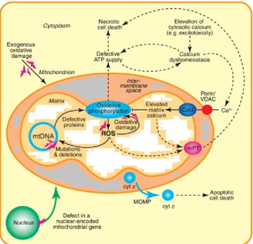
Mutations of the mtDNA cause primary mitochondrial dysfunction. Secondary dysfunction arises from extra-mitochondrial causes such as ischemia or neurodegeneration, among others. Energy depletion and chronic activation of AMP- dependent kinase (AMPK) shifts metabolism from anabolic to catabolic pathways (Bokko et al, 2007), shuts down or interferes with cellular functions (Bokko et al, 2007), results in further ROS production, disrupted calcium homeostasis and the induction of mitochondrial permeability via the mitochondrial permeability transition pore (mPT, Rasola and Bernardi, 2011) (Figure 2). These processes further damage mitochondrial function thereby establishing a vicious circle (Farrell et al, 2005).
Figure 2. Mitochondrial dysfunction (www.sciencedirect.com, modified)
11
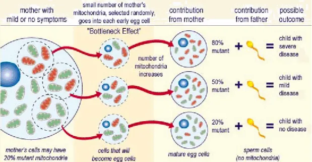
More than 200 genetic causes have been identified as potential causes of MTDs (Chinnery et al, 2012). The inheritance of the mtDNA and MTDs is maternal although paternal transmission has also been reported (Schwartz 2002). As we will demonstrate, the clinical picture can be very different in the offsprings and parents. This is due to heteroplasmy – the difference in mutation load on a tissue- and even cell level – and the genetic bottleneck that is a random shift and differences of mutated mtDNA load between generations (Taylor and Turnbull 2005) (Figure 3).
Figure 3. Inheritance of mtDNA mutations (http://medblog.medlink-uk.net/laurasmall/, modified)
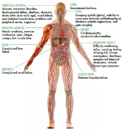
Threshold effect means that the percentage of mutant mtDNAs must be above a certain threshold to produce symptoms. This threshold, just as the number of mitochondria per cell, varies from tissue to tissue due to differences in oxidative requirements of the tissue. In cells highly dependent on OXPHOS, low amounts of mutant mtDNA cause dysfunction making the brain, heart and skeletal muscle the most heavily affected organs. MTDs frequently cause multisystemic, seemingly unrelated symptoms (Figure 4), making diagnosis difficult.
12
Figure 4. Symptoms of mitochondrial dysfunction in the human body (amdf.org.au, modified)
Manifestations of MTDs include classic syndromes, like the LHON, MELAS and MERRF as well as syndromes with high probability of mitochondrial involvement, such as PEO (Progressive External Ophthalmoplegia). Also, these mutations can be associated to common diseases or symptoms, like diabetes or hearing impairment (Taylor and Turnbull 2005). The inability to increase ATP production at times of higher energy demand explains why clinical symptoms typically worsen in association with physiological (Clay et al, 2010) or psychological (Gardner and Boles 2011) stress.
2.2. Epidemiology of mitochondrial disorders
Due to the multisystemic symptoms and multiple genetic causes, the diagnosis of MTDs is challenging and often requires referral to specialized research centers. Prevalence estimations thus vary widely and few systematic studies have been carried out so far. A prevalence of 6.57 - 9.2/100000 for symptomatic patients, 7.7 - 16.5/100000 including
‘at risk’ family members, a mean prevalence of 12.48 has been found in England
13
(Chinnery et al, 2000, Schaefer et al, 2008). The frequency of childhood mitochondrial encephalomyopathies has been estimated at 4.7/100000 in a Swedish study (Darin et al, 2001) while that of childhood respiratory chain diseases has been determined to be 6.2/100000 in Australia with a minimum birth prevalence estimated to be 13.1/100 000 for the total population (Skladal et al, 2003). Overall, mitochondrial dysfunction is probably the most prevalent metabolic abnormality considering the cardinal role mitochondria play in the metabolism.
2.3. Mitochondrial alterations in psychiatric disorders
With decades of research, the pathophysiology underlying psychiatric disorders seems more and more complex. Disease-associated genetic markers are being targeted for several psychiatric disorders by large-scale genome-wide association studies. Neurons critically depend on mitochondrial function for their metabolism, to establish membrane excitability and to execute the complex processes of neurotransmission and plasticity (Kann O and Kovacs R, 2007). In case of the chronic lack of ATP, reduced cellular resilience (Manji et al, 2012), synaptic plasticity (Manji et al, 2012) and abnormal neurotransmitter metabolism (Garcia-Cazorla et al. 2008) ensue. There is growing evidence that mitochondria are role players in the development of psychiatric conditions.
2.3.1. Biochemical and morphological data
Morphological changes (Inuwa et al, 2005), altered cellular location (Cataldo et al, 2010), decreased number (Kung and Roberts, 1999, Uranova et al, 2001) and function (Ben-Shachar and Karry, 2008, Andreazza et al, 2010) of mitochondria has been found in diverse psychiatric conditions. Downregulation of mtDNA genes (Konradi et al, 2004, Iwamoto et al, 2005, Sun et al, 2006, Shao et al, 2008, Washizuka et al, 2009), reduced expression profiles of electron transport chain subunits (Clay et al, 2010, Andreazza et al, 2010), impaired defense against oxidative stress (Erel, 2004, Naydenov et al 2007, Hovatta et al, 2010) and an overall dysfunction of brain metabolism at the mitochondrial level (Scaglia et al, 2010) has also been described in association with
14
psychiatric disorders. The most studied is bipolar disorder, where increased calcium signaling (Warsh et al, 2004), impaired molecular adaptation to energy stress (Naydenov et al, 2007), abnormal localization and size of mitochondria (Cataldo et al, 2010) as well as the global downregulation of mitochondrial genes in postmortem brain tissue has been described (Sun et al, 2006). Lactate levels were found to be significantly increased in subjects with bipolar disorder, primarily localized to the caudate and anterior cingulate cortices, suggesting that affective dysregulation may be related to metabolic abnormalities in the frontal-subcortical circuit (Chu WJ et al 2013). Post- mortem brain studies suggest that mitochondrial structure or function is altered in schizophrenia as well (Manji et al, 2012). In his interesting theory, Naviaux describes the “cell danger response” depicting the crucial role mitochondrial play in responding to harmful stimuli and how the resulting alteration in mitochondrial functioning can contribute to major psychiatric illnesses (Naviaux 2013).
2.3.2. Genetic data
Next generation sequencing of mtDNA did not yield significant mtDNA SNPs in association with bipolar disorder or schizophrenia. MtDNA common deletions have been the most consistent result in patients with bipolar disorder (Kato T et al 1997), schizophrenia (Sequeira et al 2010) and major depression (Gardner et al 2003). These deletions have been hypothesized to play a pathogenic role (Sequeira et al, 2010).
Point mutations and deletions (Kato M al, 2011), even polymorphisms of the mitochondrial DNA have been implied in the development of psychiatric conditions (Kato T et al 1997, Kato et al T 2000, 2001) and diverse personality traits (Kato C et al, 2004). Another group argued that it is a significantly higher cerebellar pH, associated with some mtDNA haplogroups (U, K and UK), that could be inherited through the mitochondrial genome and increase susceptibility to psychiatric diseases (Rollins et al, 2009).
15
2.4. Psychiatric symptoms in mitochondrial disorders
According to the above mentioned findings, patients with MTD frequently present with psychiatric symptoms. Before the publication of our work, only one systematic psychiatric assessment of mitochondrial patients has been published (Fattal et al, 2006).
In a cohort of 36 patients, lifetime diagnosis was found to be 54% for major depressive disorder, 17% for bipolar disorder, and 11% for panic disorder, all representing a higher rate compared to the general population and to cohorts with cancer and epilepsy.
Subjects with a comorbid psychiatric diagnosis were older (P=0.05), had more hospital admissions (P=0.02), more medical conditions (P=0.01), and lower quality of life (P=0.01) than subjects with MTD alone. Weakness of the study was that the diagnosis of mitochondrial disorder has not been confirmed by genetic investigation in all cases.
Since then, another study of 12 patients with depression, anorexia nervosa, bipolar disorder, and obsessive-compulsive disorder has been published (Anglin et al, 2012).
2.5. The role of mitochondria in neurodegenerative disorders
Mitochondria play a central role in apoptosis including the release of caspase activators, changes in electron transport, loss of mitochondrial transmembrane potential, alteration of cellular oxidation-reduction, and the release pro- and antiapoptotic Bcl-2 family proteins. Mutations in mitochondrial DNA and oxidative stress both contribute to aging, which is the greatest risk factor for neurodegenerative diseases (Perluigi et al 2013).
Mitochondrial alterations has been assumed to play a role in the pathogenesis of complex, multifactorial neurodegenerative diseases, most straightforward in Parkinson’s (Henchcliffe and Beal, 2008; Morais and De Strooper, 2010), but also clear in Alzheimer’s (Galindo et al, 2010) and Huntington’s disease (Turner and Schapira, 2010). In certain haplogroups, higher prevalence of dementia has been demonstrated (Tranah et al, 2012). On the other hand, neurodegeneration causes secondary mitochondrial dysfunction (Morais and De Strooper, 2010)
16
2.6. Cognitive symptoms in mitochondrial disorders
The chronic lack of ATP and CNS dysfunction results in neural dysfunction, manifesting not only in neurologic and psychiatric, but also in neuropsychologic symptoms (Finsterer 2006) ranging from mild cognitive impairment to full-blown dementia. Advanced neurodegeneration of mitochondrial origin is associated with structural abnormalities (Friedman et al, 2010), most commonly cerebral (Kim et al, 2009) or cerebellar (Scaglia et al, 2005) atrophy, also contributing to the development of cognitive symptoms.
Cognitive functions of adult patients with primary MTD have been evaluated so far in case reports of one single patient or family with MELAS (Sartor et al, 2002, Berg et al, 2011, Sharfstein et al, 1999, Inczedy-Farkas et al, 2011), NARP (Makela-Bengs et al, 1995, Gelfand et al, 2011) or MIDD (Maternally Inherited Diabetes and Deafness) (Lien et al 2001).
Studies assessing cohorts with distinct MTDs such as MELAS (Majamaa-Voltti et al, 2006), MERRF (Lorenzoni et al, 2011), PEO and KSS (Kearns-Sayre syndrome) (Bosbach et al, 2003) or MIDD (Fromont et al, 2009) also exist. Among further studies of more heterogeneous groups of MTD patients, some lack genetic diagnosis (Kartsounis et al, 1992), assessed a very limited number of patients (Lorenzoni et al, 2011), used only disease controls (Fromont et al, 2009, Kaufmann et al, 2004) or used no control group at all (Lang et al, 1995, Turconi et al, 1999). Some did not apply a comprehensive test battery (Lorenzoni et al, 2011) or did not specify it (Kaufmann et al, 2004), did not present the results in detail but reported ‘neuropsychological impairment’
(Kaufmann et al, 2004), or ‘dementia’ (Lorenzoni et al, 2011).
17
3. Objective
3.1. Complex clinical evaluation and assessment of psychiatric symptoms in a well- defined sub cohort of patients with genetically proven primary mtDNA mutations as compared to disease controls, harboring the CMT or HNPP type PMP22 mutation, living with similar level of disability.
3.2. The comprehensive neuropsychological assessment of the same cohort and compared the results to those of matched healthy controls.
3.3. Raising awareness to mitochondrial disorders in the international medical community.
3.4. Comprehensive assessment of the frequency rate of the most common mitochondrial mutations in Hungarian patients.
3.5. Biobank- and register-building activity (NEPSYBANK, SCHIZOBANK)
18 4. Patients and methods
4.1. Patients
4.1.1. Epidemiological study
A total of 1328 Hungarian patients were tested from the North-East, South-West and Central region of Hungary (Szabolcs-Szatmár-Bereg, Borsod- Abaúj-Zemplén, Hajdú- Bihar, Baranya, Pest counties and Budapest) between January 1999 and December 2012. A subcohort of 882 [485 female, 397 male; 740 adult and 142 children, mean age 38.8±19.1 years (female: 38.3±15.3 years, male 33.4±17.2 years) with a range of 1-75 years] patients were screened for m.3243 A>G, m.8344 A>G, m.8993 T>C, m.8993 T>G and mtDNA deletions. Patients were referred from county hospitals to the three genetic centers based on the symptoms and laboratory findings, for further evaluation and genetic analysis. All adult patients had a presentation consistent with mitochondrial disease i.e. the various combinations of symptoms such as short stature, epilepsy, ataxia, myopathy, lipomatosis, ophthalmoplegia externa, hypoacusis, exercise intolerance, myalgia, recurrent ischaemic stroke syndrome, cognitive dysfunction, psychiatric or endocrine disorder. In the case of young children, mitochondrial disease was suspected on the bases of muscle hypotonia, delayed psychomotor development, lactic acidosis and seizures.
We have screened 446 patients [217 male, 229 female, 366 adult and 80 children, 35.82±18.47 (male: 32.58±17.72 years, female: 38.98±18.64) with a range of 7-63 years] for the three primary LHON mutations (m.3460 G>A, m.17788 G>A, m.14484 T>C), based on the suggestive symptoms such as unilateral or bilateral decrease in visual acuity or sudden loss of vision. Written, informed consent was obtained from all participants. The study was carried out according to the Helsinki Declaration and was approved by the Research and Ethics committee of Semmelweis University.
19
4.1.2. Psychiatric and neuropsychologic assessment
Nineteen patients (Patient 1-19, 13 female, 6 male) with primary mutation of the mtDNA were selected for a detailed psychiatric and neuropsychologic assessment.
Selection was based on the ability and willingness of the patient to participate and the absence of severe ptosis, visual, hearing or mental disability that would have affected performance. Of the 19 patient, only 13 probands has been included in the statistical analysis in order to keep the variables independent. The results of the 6 family members are shown in the tables of the Results section.
Mean age for the 13 probands, included in the statistical analysis was 34±8.43 years (male: 27.25±6.8; female: 36.66±7.65), average years of education was 12.84±1.21 years (male: 13.25±1.5; female: 12.66±1.12).
As control for the psychiatric assessment, 10 patients (HN patients, Patient 20-29, 4 female, 6 male, mean age was: 40±10.99, average years of education was 14.2±3.85 years) with PMP22 mutation, causing CMT (Charcot-Marie Tooth) or HNPP type (Hereditary Neuropathy with liability to Pressure Palsy) sensorimotor neuropathy were examined. Diagnosis was based on clinical features supported by the presence of the PMP22 gene mutation in HN patients.
Thirteen healthy controls matched for age, sex and education (9 female, 4 male, mean age: 33.77±9.19, average years of education 14±0.9 years) were examined for the neuropsychological examination.
All participants of the study were Caucasian and visited the clinic within 1 year.
Written, informed consent was obtained from all participants. These studies were carried out according to the Helsinki Declaration and were approved by the Research and Ethics committee of Semmelweis University.
4.2. Methods
4.2.1. General clinical evaluation
Basic neurological evaluation has been carried out by the primary neurologist where patients were referred to by the primary care provider. If the suspicion of MTD was
20
present, the patient was referred to one of the three specialized genetic centers for further evaluation. The diagnosis of MTD has been confirmed by further clinical, neurological evaluation and myopathological and/or genetic studies. In some cases, segregation analysis of the proband’s family detected further patients with clinical symptoms.
4.2.2. Myopathology
Muscle biopsy have been performed in 312 cases and confirmed the mitochondrial disorder in 119 cases based on the presence of ragged blue/red and COX (cytochrome oxidase) negative fibers or on the presence of abnormal mitochondria on electron micrographs. Histopathological evaluation was carried out using standard methods.
Ragged blue fibers were detected with the modified intense succinate dehydrogenase histochemistry (SDH) reaction, while ragged red fibers were searched for by the modified Gomori Trichrome stain. COX negative fibers were discerned by large accumulations of morphologically abnormal mitochondria with significantly reduced or undetectable COX activity. Ultrastructural changes were investigated in the case of 152 patients by electron microscope.
4.2.3. Genetic studies
4.2.3.1. Analysis of the mtDNA
Molecular genetic analysis was performed for diagnostic purposes from the biological sample of all investigated patients. DNA was extracted from blood (in 570 cases) or if the blood sample yielded no result but the suspicion of MTD was high, skeletal muscle tissue (in 312 cases) by QIAamp DNA Blood or Tissue kit, according to the manufacturer’s (QIAgen, Hilden, Germany) instructions. The m.3243 A>G mutation in the tRNALeu (UUR), m.8344 A>G mutation in the tRNALys gene and the m.8993 T>C and m.8993 T>G mutations in the ATP synthase F0 subunit 6 (ATPase6) of mtDNA were screened based on a method described earlier using restriction fragment length polymorphism (RFLP) with HaeIII (m.3243 A>G), BanII (m.8344 A>G), HpaII
21
(m.8993 T>C), AvaI (m.8993 T>G) BSAHl (m.3460 G>A), SfaNl (m.11778 G>A) and Bccl (m.14484 T>C)restriction endonucleases after PCR amplification (Shoffner et al, 1990). The ratio of heteroplasmy was measured by Quantity One Software (Bio-Rad Corp. Hertfordshire, UK). The mtDNA deletion was screened for by long PCR methods using Phusion DNA Polymerase (Finnzymes). The entire mtDNA tRNA sequence has been investigated in 200 cases by bidirectional sequencing using the ABI Prism 3500 sequencing machine. The sequences were compared with the human mitochondrial complete reference genome (NC_012920.1) using the NCBI Blast program.
The entire mtDNA was resequenced with MitoChip v2.0 microarray (Affymetrix) in 17 patients, and the supposedly pathogenic alterations were validated by Sanger sequencing.
4.2.3.2. Analysis of the PMP22 gene
The PMP22 deletion and duplication in the HN group was detected with real time PCR according to earlier published methods (Aarskog et al, 2000).
4.2.3.3. Determination of the 5HTTLPR genotype
The 5HTTLPR (serotonin-transporter-linked polymorphic region) genotypes of both MTD and HN patients were analyzed according to earlier published methods (Heils et al, 1996).
4.2.4. Focused clinical evaluation
In the case of the 19 selected patients, functional ability was assessed using the Hungarian validated version of the Stanford Health Assessment Questionnaire 20-item Disability Index (HAQ-DI) (Ponyi et al, 2005). Patients’ charts were reviewed for duration of disease, medication and brain CT or MRI scans in case of the 19 patients selected for further evaluation.
22 4.2.5. Psychiatric examination
Detailed psychiatric assessment used the Symptom Checklist-90-Revised (SCL-90-R) (Derogatis et al, 1994) and the Beck Depression Inventory-Short Form (BDI-SF, Beck and Beck, 1972) self tests. The BDI-SF includes cognitive-affective but not somatic items to avoid spuriously high scores and overreporting of depression in somatic patients (Furlanetto et al, 2005). The clinician-administered 21-item Hamilton Depression Rating Scale (HDRS, Hamilton, 1960) and the clinical version of the Structured Clinical Interview for the DSM-IV axis-I (SCID-I) (First et al, 1996) and axis-II disorders (SCID-II) (First et al, 1997) were also used. The SCL-90-R helps evaluate a broad range of psychopathological symptoms. It yields 9 scores of primary symptom dimensions (somatization, obsession-compulsion, interpersonal sensitivity, depression, anxiety, hostility, phobic anxiety, paranoid ideation and psychoticism) and additional item subscale referring to sleep and memory problems. The mean of these subscales yields the global severity index (GSI). The BDI-SF is a 13-item questionnaire scored on a 4-point scale, from 0 to 3, with overall scores ranging from 0 to 39. The BDI-SF has been found to have a good correlation with the standard 21-item BDI (r = 0.96, p = 0.001) and relates the clinical depth-of-depression (r = 0.61) (Beck and Beck, 1972). The emphasis of the 21-item HDRS is on melancholic and physical symptoms of depression. In order to control for the confounding effect of cognitive impairment, we included patients with an FSIQ of 70 and above, as assessed by the Hungarian validated version of the Wechsler Adult Intelligence Scale (WAIS-III-R version, Kun and Szegedi 1996). Scales and interviews were administered by trained clinicians.
4.2.6. Neuropsychological assessment
Memory functions were evaluated by the Rey Auditory-Verbal Learning Test (RAVLT1 for immediate recall, RAVLT2 – RAVLT5 for learning ability over successive trials and RAVLT6 for delayed recall. The Stroop Color (Stroop C) and Color-Word (Stroop CW) tests assessed selective attention, cognitive flexibility and interference control, both the number of words read in 60 sec and the number of errors made. The Trail Making Tests (TMTA, TMTB) were used as a measure of visuo-spatial abilities,
23
attention and psychomotor speed and – the part B – of flexibility, working memory and executive functions in general (see for Maruta et al, 2011). With the category (aka semantic) and letter Fluency Tests (Rosen, 1980), we tested fluency of verbal associations under restricted conditions, executive functions and psychomotor speed.
Intelligence was assessed using the Hungarian version of the Wechsler Adult Intelligence Scale (WAIS, Kun and Szegedi, 1996).
In the case of Patient 16 we had the chance to do a longitudinal follow-up by retesting the patient 6 months apart. In her case, a more complex test battery was used comprising of the RAVLT, the Raven test (Raven Progressive Matrices, Raven 1936), the Toulouse-Pieron Attention Test (Toulouse et al, 1986), the Bells Test (Gauthier et al, 1989), the Benton Visual Retention Test (Benton Sivan A, 1992), the Rey-Osterrieth Complex Figure Test (Shin et al, 2006).
In all cases, neuropsychological assessment took place in one session of approximately two hours, with breaks if requested by the examinee. The tests were scored according to standard guidelines. Results have been correlated with the type of mtDNA mutation, age, duration of disease as well as scores on the Global Severity Index (GSI) of the Symptom Checklist-90-R (SCL-90-R) detecting the presence and severity of psychiatric symptoms.
4.2.7. Statistical analysis
In the epidemiological study, frequency of the mutations was calculated from the number of patients with pathogenic substitutions divided by the total number of investigated patients. The 95% confidence interval (CI) was calculated according to the standard method.
In the other studies, independent variables were obtained selecting only the proband (index case) from each family from the cohort of MTD patients. HN patients were all unrelated, statistics were thus performed using data from 13 MTD (Pt 1, 2, 3, 5, 8, 10, 11, 13, 14, 15, 16, 18, 19) and 10 HN patients. In the statistical analysis of the cognitive study, data of the same 13 MTD patients as well as 13 healthy controls, matched for age, sex and education was used. Correlation of total scores in GSI, BDI and HDRS with HAQ-DI in both groups was evaluated using Pearson's correlation. Differences
24
between the patient and control groups were assessed using Chi-Square test for categorical variables and parametrical (t-test) or non-parametrical tests (Wilcoxon Mann-Whitney test) for continuous variables (depending on the distribution of the variables). The normality of the data was checked by Shapiro-Wilk test (data not shown). All tests were two tailed and p values ≤ 0.05 were deemed significant.
SCL-90-R was analyzed with SAS System for Windows (Release 9.1 TS Level 1 M3, Statistical Analysis System, SAS-Institute USA).
25
5. Results
5.1. Epidemiology of mitochondrial disorders
In the epidemiology study, mutations of the mtDNA have been categorized into three groups; mutations in genes of the mitochondrial tRNA, mutations in genes coding for mitochondrial proteins and rearrangements of the mtDNA. The ratio of heteroplasmy was between 22% and 95% in the 570 blood sample, and between 25% and 79% in the 312 muscle sample (Remenyi et al, 2014).
5.1.1. Mutations in genes of the mitochondrial tRNA
5.1.1.1. The m.3243 A>G point mutation
The most commonly investigated m.3243 A>G substitution in the tRNALeu1(UUR) was found in a heteroplasmic form in 11 index patients and further 12 family members from the investigated 882 patients. The ratio of heteroplasmy varied between 22 and 80%.
From the positive cases, the mtDNA was isolated from blood in 20, and muscle in 3 cases. In one of the cases the mutation was found only in muscle tissue. All probands and family members carrying the mutation had clinical symptoms, mostly sensorineural hearing loss (SNHL) and diabetes mellitus (DM). Five cases had history of stroke. One family member had psychiatric symptoms whereas others were asymptomatic. The frequency of the m.3243 A>G mutation was 2.61 % (95% CI: 0.0207-0.0315).
5.1.1.2. The m.8344 A>G point mutation
The heteroplasmic m.8344 A>G mutation of the tRNALys gene was present in 13 patients (8 males, 5 females) from 3 families of the investigated 882 patients. The ratio of heteroplasmy varied between 25 and 82 %. The pathogenic mutation was detected from blood in 8 and from muscle in 5 cases. These patients also had diverse symptoms, mostly not the classic myoclonus epilepsy. The frequency of m.8344 A>G mutation was 1.47% in the investigated cohort (95% CI: 0.0106-0.0187).
26
5.1.1.3. Other mutations of the mitochondrial tRNA
With sequence analysis of the tRNA, we found 6 further pathogenic mutations in 17 (11 female, 6 male) patients out of the 200 investigated cases (1.93%). The m.3250 T>C substitution in the tRNALeu1 was present in 3 females of a family with myopathy and idiopathic pulmonary hypertension. The proband with PEO and myopathy had the homoplasmic, her mother and grandmother the heteroplasmic form. The heteroplasmic m.4298 G>A substitution in the tRNAIle in was present in a male patient with hypertension and exercise intolerance and a m.5698 G>A in the tRNAAsn in one female patient with familial PEO. In both cases, multiple deletions were also present. Two mutations were present in the tRNASer1(UCN), both presenting with bilateral SNHL; a heteroplasmic m.7445 A>G in 6 patients and the m.7510 T>C in a homoplasmic form in 3 patients with hearing impairment.
We found the new pathogenic m.8332 A>G mutation in the tRNALys, in a heteroplasmic form in two brothers and their mother with familial dystonia and juvenile stroke like syndrome (Gál et al). By sequencing the entirety of mtDNA tRNA genes 3-8 homoplasmic SNPs (single nucleotide polymorphisms) were detected per patient.
The overall frequency of tRNA mutations was found to be 6.35% (56/882 cases) in the Hungarian population.
5.1.2. Mutations in genes coding for mitochondrial proteins
5.1.2.1. m.8993 T>C and m.8993 T>G point mutations
We also screened for the m.8993 T>C and m.8993 T>G substitution that were present in 4 cases among 882 patients. With familial segregation analysis, the m.8993 T>C pathogenic mutation has been found in a heteroplasmic form in further 3 patients (2 male, 1 female). The heteroplasmic m.8993 T>G substitution was detected in further 1 female patient. Heteroplasmy varied between 58 and 95% in blood cells. The prevalent symptom of affected patients was cerebellar ataxia. The frequency of m.8993 T>C was 0.34% that of m.8993 T>G was 0.11% with an overall frequency of 0.45% for the nt8993 substitutions (95% CI: 0.00038-0.0086).
27
5.1.2.2. The m.3460 G>A, m.11778 G>A and m.14484 T>C point mutations
The three primary LHON mutations were detected altogether in 80 cases (41 male, 39 female) in homoplasmic form. The m.3460 G>A mutation was found in 9 cases (6 male, 3 female). The m.11778 G>A mutation was detected in most cases in 66 cases (34 male, 32 female) and the m.14484 T>C was found in 5 cases (2 male, 3 female) from the 446 patients. The frequency of each single mutation was as follows: 2.02% for the m.3460 G>A (95% CI: 0.0135-0.0269), 14.80% for the m.11778 G>A (95% CI: 0.1312-0.1648) and 1.12% for the m.14484 T>C (95% CI: 0.0062-0.0162), yielding an overall frequency of 17.94% (95% Cl: 0.1612-0.1976).
5.1.2.3. Mutations detected with the Mitochip v.2.0.
We resequenced the entire mtDNA, isolated from muscle tissue of 17 patients (with family history characteristic of MTD) using the Mitochip v.2.0. microarray (Affymetrix). This yielded the m.12770 A>G substitution in the NAD dehydrogenase subunit 5 (ND5) with a low rate of heteroplasmy, in a patient who was selected for further evaluation.
5.1.3. Rearrangements of the mtDNA
Single mtDNA deletions were detected in 132 out of 882 cases representing 14.97%
(95% CI: 0.1377-0.1617) of the investigated cohort. Forty percent of these cases harbored “common” deletion. Multiple deletions were present in 53 cases which is 6.01% (95% CI: 0.0521-0.0681). The majority (60.3%, 32 out of 53) of the cases had positive family history. Among the 132 cases with single deletion, 30 patients (22.7%) had PEO and 48 patients (36.3%) had myopathy. Out of the 132 single deletion cases, 74 were detected from muscle tissue and 58 from blood sample. Results of the epidemiological study are summarized in Table 1.
28
Table 1. Mutation frequency of the most common mtDNA mutations in Hungary
mtDNA mutation type
positive cases
number of investigated patients
frequency
tRNA mutations
m.3243 A>G 23 882 2.61%
m.8344 A>G 13 882 1.47%
m.3250 T>C 3 200 0.34%
m.4298 G>A 1 200 0.11%
m.5698 G>A 1 200 0.11%
m.7445 A>G 6 200 0.68%
m. 7510 T>C 3 200 0.34%
m. 8332 A>G 3 200 0.34%
Mutations in genes coding for mitochondrial proteins m.8993 T>C
m.8993 T>G
3 1
882 882
0.34 % 0.11%
m.3460 G>A 9 446 2.02%
m.11778 G>A 66 446 14.8%
m.14484 T>C 5 446 1.12%
Rearrangements of the mtDNA
single deletion 132 882 14.97%
multiple deletion 53 882 6.01%
5.2. Genetic background of the psychiatric study
5.2.1. MtDNA mutation types of patients selected for further evaluation
We selected patients for further psychiatric evaluation from the three groups identified in the epidemiological study and one further group consisting of patients with coexisting nonsynonymous mtDNA SNPs previously associated with symptoms suggestive of MTD (Table 2).
Mutations in genes of the mtDNA tRNA: Pts 1-8, 14. Nine MTD patients had common mutation of mtDNA. Four of them (Pt 1-4) harbored the m.3243 A>G substitution.
Interestingly, none of them had a history of stroke-like episodes. Four patients (Pt 5-8) had the m.8344 A>G substitution in tRNALys. Among them, only Pts 7 and 8 (twin brothers) had the classic myoclonus epilepsy. Pt 14 had the m.8332 A>G base substitution in the tRNALys. Pts 9, 10, 15 had mutations in protein-coding genes; Pts 9
29
and 10 carried the m.8993 T>G, but only the son had the classic NARP symptoms. Pt 15 had the m.12770 A>G substitution (detected with Mitochip sequencing as outlined above) resulting in a Glu - Gly substitution in the ND5 gene. Pts 11-13 and 18 had common deletion of the mtDNA; Pts 11 and 12 had PEO and Pt 13 had KSS. Pt 18 had cardiomyopathy and cognitive impairment.
The Mitochip sequencing yielded no pathogenic mutation but the combination of different nonsynonymous SNPs, previously associated to multisystemic symptoms characteristic of MTD (www.mitomap.org). Laboratory and myopathological results of these patients indicated mitochondrial dysfunction. In case of Pts 16 and 17, a C-T base substitution at nucleotide 14771 was present resulting in Pro-Ser substitution in the mitochondrial cytochrome b (Cyt-b) gene. In the case of Pt 19 - beside the A2706G polymorphism which is responsible for linezolid-induced severe lactic acidosis - 14 single nucleotide polymorphisms were present, previously associated with LHON, potentially exerting synergistic effect.
5.2.2. PMP22 gene analysis in control (HN) patients
As a disease control group patients with HN were used. In the HN group, 9 patients harbored a duplication (Charcot-Marie-Tooth phenotype, CMT), and one of them (Pt 22) had a deletion (Hereditary Neuropathy with Liability to Pressure Palsy phenotype, HNPP) in the PMP22 gene (Table 3).
5.2.3. Analysis of the 5HTTLPR gene
To rule out the confounding effect the 5HTTLPR gene might have, we obtained our patients’ 5HTTLPR genotype. In the MTD group, 5 patients harbored the long-long (L/L), 10 patients harbored the long/short (L/S) while 4 patient had the short-short (S/S) genotype (Pt 6, 7, 8, 19) (Table 5). In the HN group, 3 patients had the L/L, further 3 patients the L/S, and 4 patients the S/S polymorphism (Pt 22, 23, 27, 28) (Table 7).
30 5.3. Clinical evaluation
The MTD and HN groups did not differ significantly in gender (χ²=1.9652; p=0.1610), age (t= -1.42; p=0.1711) or education (t= -1.20; p=0.243). Mean HAQ-DI score was 0.82 in the MT (range: 0 - 1.625) and 0.71 in the HN group (range: 0 - 1.625) which did not differ significantly (p=0.6076) implying comparable level of disability in the two groups (Table 9).
5.3.1. Neurological symptoms
Hypoacusis, ataxia, myopathy, neuropathy and exercise intolerance were the most common neurological symptoms in the MTD group (Table 2). HN patients mostly had distal type paresis and muscle atrophy (Table 2).
5.3.2. Medication
Medication for the MTD comprised of Coenzyme Q10, Vitamin E and Vitamin C.
Some patients were taking psychiatric drugs at the time of the assessment.
Monotherapies were clonazepam (Pts 7, 10, 14), sertraline (Pt 12) and mirtazapine (Pt 13). Combination therapies were quetiapine and trazodone for Patient 16, clonazepam, sertraline and quetiapine for Pt 18 and aripiprazole with duloxetine and clonazepam for Pt 19. Anticonvulsant treatment was valproate for Pts 7 and 8, carbamazepine for Pt 14 and lamotrigine for Pt 16 (Table 2). In the HN group, prescribed CNS drugs were duloxetine and pregabalin for Pt 23, chlordiazepoxide for Pt 25 and gabapentin with duloxetine for Pt 28. (Table 3). Gender, age, duration of disease, mtDNA genotypes, clinical symptoms and medications of MTD patients (patient group) are outlined in Table 2. Gender, age, mtDNA genotypes, clinical symptoms and medications of of HN patients (control group) are outlined in Table 3.
31
Table 2. Gender, age, duration of disease, mtDNA genotypes, clinical symptoms and medication of MTD patients (patient group). Grey color fill indicates unrelated patients
(independent variables) included in the statistical analysis.
ID Gender, age, disease duration
(years)
mtDNA mutation
type
Clinical symptoms and findings
Medication (with total daily dose)
1 F 34 (20) m.3243 A>G MELAS
Hypoacusis, ataxia, myopathy, endocrine dysfunction
-
2 F 51 (30) m.3243 A>G MELAS
Ataxia -
3 F 34 (20) m.3243 A>G MELAS
PEO, myopathy (mother of Patient 4)
-
4 F 17 (10) [daughter of Pt 3]
m.3243 A>G MELAS
PEO -
5 M 32 (4) m.8344 A>G MERRF
Migraine -
6 F 61 (10) [mother
of Pt 8]
m.8344 A>G MERRF
Mild tremor in the upper limbs (mother of Pt 7 and 8)
-
7 M 34 (17) [twin brother of
Pt 8]
m.8344 A>G MERRF
Myoclonus epilepsy, thrombocytopenia (twin brother of Pt 8)
levetiracetam (1000 mg), clonazepam (2 mg), valproate (1500 mg), vinpocetine (20 mg) 8 M 34 (17) m.8344
A>G MERRF
Myoclonus epilepsy, ataxia, cognitive dysfunction, thrombocytopenia (twin brother of Pt 7)
levetiracetam (2500 mg),
valproate (2100 mg), vinpocetine (90 mg), betaxolol (10 mg), atorvastatine (10 mg), allopurinol (200 mg)
9 F 59 (30) [mother of Pt 10]
m.8993 T>G NARP
Developmental abnormality of the right arm,
hypothyreosis
-
10 M 23 (18) m.8993 T>G NARP
NARP clonazepam (1 mg)
vinpocetine (10 mg) 11 F 34 (20) mtDNA
del
PEO, iron-deficient anemia,
hypercholesterolemia -
32 12 F 40 (14)
[sister of Pt 11]
mtDNA del
Myalgia, exercise intolerance
sertraline (50 mg)
13 F 38 (20) mtDNA del
Kearns-Sayre syndrome
clonazepam (1mg) vinpocetine (20 mg) mirtazapine (30 mg) 14 M 20 (15) m.8332
A>G m.8270
C>T
Dystonia, early onset stroke-like
symptoms
levetiracetam (1000 mg), clonazepam (1.25mg) carbamazepine (1000 mg) 15 F 22 (9) m.12770
A>G
Ataxia, hypoacusis, spastic paraparesis
- 16 F 39 (20) m.15326
A>G m.750
A>G m.1438
A>G m.8860
A>G m.15326
A>G
Hypoacusis, limb tremor, myalgia, apraxia
(mother of Pt17)
trimetazidine (60 mg), metformin (850 mg),
bisoprolol (5 mg), quetiapine (400 mg), lamotrigine (50 mg),
trazodone (300 mg)
17 M 20 (7) [son of
Pt16]
see Pt 16.
- -
18 F 37 (23) m.3720 A>G m.3849
G>A m.13020
T>C m.13734
T>C m.12308
A>G m. 8473
T>C
Severe
cardiomyopathy, cognitive
dysfunction
trimetazidine (40 mg), enalapril (5 mg), molsidomine (4 mg),
quetiapine (400 mg),
lamotrigine (100 mg), sertraline (100 mg), clonazepam (1.5 mg)
19 F 41 (17) m.14766 C>T m.709
G>A m.73 A>G m.16126
T>C
Peripheral neuropathy
tolperisone (300 mg), aceclofenac (200 mg),
aripiprazole (15 mg), duloxetine (30 mg),
clonazepam (1 mg)
33
Table 3. Gender, age, mtDNA genotypes, clinical symptoms and medication of of HN patients (control group)
ID Gender and age (years)
PMP22 mutation type
Clinical symptoms and findings Medication (with total daily dose)
20 M, 41 CMT Atrophy of the hand and feet muscles - 21 M, 17 CMT Pes equinovarus, paresis in the distal
muscles of the legs
-
22 M, 49 HNPP Sensory disturbances in the lower limbs, neuropathy to pressure palsy
-
23 F, 33 CMT Generalized muscle weakness, walking difficulty, distal type hypesthesia, excavated feet
pregabalin (300 mg) duloxetine (30 mg)
24 M, 34 CMT Paresthesia, distal type hypesthesia in the limbs, muscle cramps, gait
instability
-
25 F, 46 CMT Distal type muscle weakness in the limbs, tremor in the hands
chlordiazepoxide (5 mg), tolperisone (150 mg)
, 26 F, 31 CMT Excavated feet, distal paresis of the
limbs, ataxia
-
27 M, 42 CMT Mild paresis in the hand and feet muscles
-
28 F, 55 CMT Diabetes mellitus, hyperlipidaemia, distal weakness of the limbs, distal type hypesthesia, head tremor, gait instability
duloxetine (60 mg) gabapentin (600 mg) rosuvastatin (20 mg)
ezetimibe (10 mg) 29 M, 47 CMT Bronchial asthma, atrophy and paresis
in the hand and feet muscles, gait instability
-
34 5.3.3. Neuroimaging
Various changes have been found on neuroimaging studies (Table 7). Multiple cerebral (Pt 2) or cerebellar (Pt 10) parenchymal lesions, focal contrast enhancing lesion at the left insula (Pt 5) and near the left occipital horn (Pt 18), paraventricular (Pt 18) and basal ganglia (Pt 2) calcifications were detected. Pt 3 had cortical, Pt 15 had cerebellar atrophy and Pt 1, 7 and 8 had both. Pt 2 and 18 had occipital lesions. MRI spectroscopy results showed low levels of choline, elevated glutamate for Pt 1, low levels of N- AcetylAspartic acid, creatine, choline for Pt 8 and elevated lactate peak for both of them. Pt 4 (the m.3243 A>G mutation with PEO symptoms), 6 (MERRF), 9 (NARP), 11-13 (mtDNA deletion), 14, 17, 19 (mt DNA polymorphism) had normal MRI findings. The mean HAQ-DI and GSI score of the probands in this subgroup did not differ significantly from those of the rest of the group (0.62 vs 0.93, p = 0.417 and 0.98 vs 1.65, p = 0.178, respectively). In this subgroup, only Pt 12 and 17 were free of physical symptoms. Pt 18 did have MRI alteration, but only exercise intolerance as physical symptoms and a high GSI score. Neurological symptoms, CT or MRI findings and WAIS FSIQ scores of MTD patients are presented in Table 4.
35
Table 4. Neurological symptoms, CT or MRI findings and FSIQ scores of MTD patients. Grey color fill indicates unrelated patients (independent variables) included in
the statistical analysis.
ID Neurological symptoms
CT/MRI finding VQ PQ FSIQ
1 Hypoacusis,ataxia,
lower limb paresis Cerebellar and cortical atrophy 97 53 74 2 Hypoacusis, myopathy,
neuropathy, exercise intolerance, ataxia
Multiple demyelinating lesion in the occipital lobe, at the sides of the lateral ventricles,
calcifications in basal ganglia 93 92 92 3 PEO, severe myopathy Slight cortical atrophy 81 72 74
4 PEO Normal findings 93 81 86
5 Migraine 9 mm contrast-enhancing lesion
at the left insula 118 119 120
6 Mild limb tremor Normal findings 119 119 119
7 Myoclonus epilepsy, ataxia (MERRF)
Slight cortical and cerebellar
atrophy 95 96 94
8 Myoclonus epilepsy,
severe ataxia (MERRF) Cerebellar and cortical atrophy 87 71 77
9 Limb deformation Normal findings 121 121 122
10 NARP, dysarthria, severe ataxia
Multiple parenchymal lesions in
both cerebellar hemispheres 68 47 55
11 PEO Normal findings 115 140 129
12 - Normal findings 126 132 131
13 KSS Normal findings 104 123 114
14 Dystonia, early onset
stroke-like symptoms Normal findings 77 76 75
15 Ataxia, hypoacusis, spastic paraparesis
Hypoplasia, cerebellar atrophy, Wider posterior scala and 4th
ventricle 108 101 105
16 Hypoacusis, limb tremor, apraxia
Left frontal cortical dysgenesis
with venous dilation 96 109 103
17 - Normal findings 120 135 129
18 Exercise intolerance contrast accumulating lesion at the left occipital horn,
paraventricular calcifications
temporally and frontally 100 96 98 19 Severe peripheral
neuropathy, distal type
weakness in the limbs Normal findings 117 124 122
36 5.4. Psychiatric findings
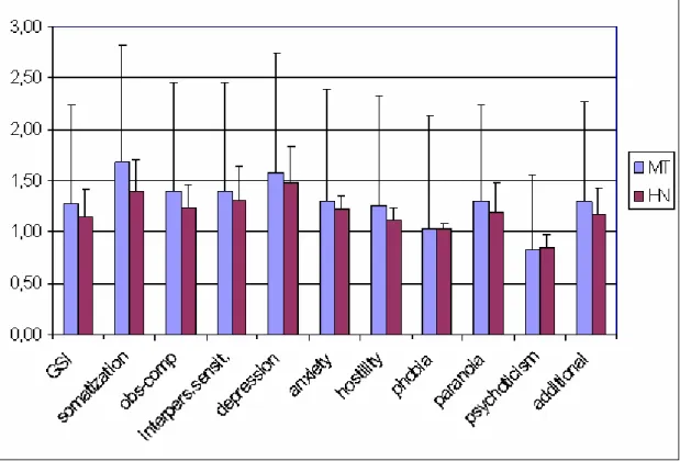
The MTD and HN groups’ BDI-SF and HDRS score differed significantly (12.85 vs 4.40, p<0.031, and 15.62 vs 7.30, p<0.043, respectively). Statistical difference was also found in the GSI score (1.44 vs 0.46, p<0.013) and the nine subscales of the SCL-90-R scale (Table 9). These subscales were obsession-compulsion (p<0.008), interpersonal sensitivity (p<0.008), depression (p<0.031), anxiety (p<0.031), hostility (p<0.143), phobic anxiety (p<0.031), paranoid ideation (p<0.01) psychoticism p<0.000) and additional items (p<0.013). No significant difference was found between the two groups’ somatization score (Figure 5).
Figure 5. Results of the SCL-90-R questionnaire with the GSI and subscales.
Patients harboring the S/S genotype had lower levels of depression than the rest of the group (BDI average score of 4.5 vs 11.8, see Table 5). BDI-SF, GSI, HAQ-DI scores and 5HTTLPR genotype of MTD patients are presented in Table 5.
37
Table 5. BDI-SF, GSI, HAQ-DI scores and 5HTTLPR genotype of MTD patients
ID BDI GSI HAQ-DI 5HTTLPR
genotype
1 22 1.92 1.375 L/S
2 8 1.09 0.875 L/S
3 8 1.09 1.25 L/S
4 14 1.24 0.125 L/S
5 21 2.46 0 L/L
6 0 0.33 0 S/S
7 2 0.31 0.375 S/S
8 3 0.87 1.625 S/S
9 8 0.59 0 L/S
10 7 0.44 1.375 L/L
11 10 0.86 0 L/L
12 2 0.22 0 L/S
13 16 1.19 0.375 L/S
14 4 0.78 0.125 L/S
15 5 0.74 1.625 L/S
16 30 3.02 0.625 L/L
17 2 0.25 0 L/L
18 20 3.2 0.625 L/S
19 13 1.08 0.75 S/S
A variety of psychiatric disorders; current diagnosis in 6 (31%), past diagnosis in 8 (42%), lifetime prevalence in 9 MTD cases (47%) were diagnosed with SCID-I (Table 6). Lifetime prevalence was 10% (2 patients) for major depressive disorder, for dysthymia, for bipolar II and for mood disorder due to general medical condition, 5% (1 patient) for major depression with psychotic features, for bipolar I, for mixed anxiety- depressive disorder, for postpartum depression and for PTSD. Three avoidant, 2 obsessive-compulsive personality were diagnosed in the MTD group. In 3 cases, personality disorder not otherwise specified (NOS) was detected referring to depressive
38
personality in case of Pt 5, and mixed personality disorder in case of Pts 16 and 18.
Personality disorder was found in 8 MTD cases (42%, Table 6).
Table 6: SCID-I and SCID-II results of MTD patients. Grey color fill indicates unrelated patients (independent variables) included in the statistical analysis.
ID Past diagnosis SCID I.
Current diagnosis SCID I.
Personality disorder SCID II.
1 Mixed anxiety- depressive disorder
Major depressive disorder
Avoidant
2 Dysthymia - -
3 - - -
4 - - -
5 - Dysthymia Personality
disorder NOS
6 - - -
7 Adjustment disorder with depressed mood
- -
8 Adjustment disorder with depressed mood
- Avoidant
9 - - Obsessive-
compulsive
10 - - -
11 - - -
12 - - -
13 Bipolar II disorder Bipolar II, current episode depressive
-
14 - - -
15 - - Obsessive-
compulsive 16 Major depression
with psychotic features
Major depressive disorder
Personality disorder NOS
17 - - Avoidant
18 PTSD Bipolar II, current episode depressive
Personality disorder NOS 19 Postpartum
depression
Bipolar I, latest episode depressive
-
39
In the HN group, depression was more present in those with the S/S (BDI score of 6.75 vs 2.83, Table 7.). Correlation analysis was not done due to the small number.
Table 7. BDI-SF, SCL-90-R GSI, HAQ-DI scores and 5HTTLPR genotype of HN pts
ID BDI GSI HAQ-DI 5HTTLPR
genotype
20 1 0.22 0 L/L
21 1 0.32 0.125 L/L
22 11 1.3 0.375 S/S
23 2 0.18 0.875 S/S
24 2 0.12 0.5 L/S
25 2 0.26 1.25 L/S
26 11 0.37 1.125 L/S
27 0 0 0 S/S
28 14 1.59 1.25 S/S
29 0 0.25 1.625 L/L
Three patients had past and current diagnosis. Lifetime prevalence was 20% (2 patients) for dysthymia, 10% (1 patient) for MDD, bipolar II, mood disorder due to general medical condition and alcohol abuse (Table 8). No personality disorder was found
Table 8. SCID-I and SCID-II results of HN patients.
ID Past diagnosis SCID I.
Current diagnosis SCID I.
20 - -
21 - -
22 Adjustment disorder with depressed mood
Dysthymia
23 - -
24 - -
25 - -
26 - -
27 - -
28 MDD Dysthymia
29 Alcohol abuse Bipolar II, latest episode hypomanic
40
Table 9. Results of GSI and the ten subscales of SCL-90-R, BDI-SF and HDRS in the group of MTD vs HN patients. Adjusted_p: p values with Bonferroni-Holm correction.
*Asterisks refer to statistically significant differences between the two groups.
MTD group
HN group
p-value (adjusted) Measured
items mean SD mean SD
GSI 1.44 0.91 0.46 0.53 0.0130*
Somatization 1.77 1.10 0.98 0.83 0.0817
Obsessive-
compulsive 1.65 1.04 0.47 0.68 0.0079*
Interpersonal
sensitivity 1.55 0.97 0.40 0.51 0.0079*
Depression 1.90 1.21 0.75 1.03 0.0309*
Anxiety 1.32 1.14 0.42 0.63 0.0309*
Hostility 1.26 1.00 0.48 0.50 0.0428*
Phobia 1.14 1.11 0.29 0.56 0.0309*
Paranoia 1.42 1.05 0.28 0.39 0.0101*
Psychoticism 0.92 0.60 0.13 0.24 0.0002*
Additional
items 1.48 0.96 0.43 0.66 0.0130*
BDI 12.85 8.33 4.40 5.36 0.0309*
HDRS 15.62 8.62 7.30 5.52 0.0428*
HAQ-DI 0.82 0.59 0.71 0.59 0.6076
41 5.5. Cognitive symptoms
5.5.1. Results of the neuropsychological assessment
Results of the RAVLT show that both groups learned new words with each repetition
from trial1 (RAVLT1) to trial 5 (RAVLT5) (Inczedy-
Farkas - Trampush et al, 2014). There is positive correlation between the number of words retained and the number of trials, which is slightly stronger in the control group (r=0.603, p<0.0001 for patients vs. r=0.748 p<0.0001 for controls). Patients performed below controls and also below normative data. We detected impaired short-term (RAVLT1, mean of patients = 5.46 ± 2.1062, mean of controls = 8.0 ± 1.354, p = 0.0015) and delayed recall (RAVLT6, mean of patients: 7.15, mean of controls = 12.15, p = 0.0001).
The number of read words differed significantly on both the Stroop C and the Stroop CW (Stroop C_60 Pt: 65.77±27.7, controls: 87.08±8.36, p = 0.018, Stroop CW_60 Pt:
42.77±22.9, Controls: 61±11.68, p = 0.021) No difference has been found in the number of errors on either test (Stroop C_error Pts: 0.77 ± 1.16, Controls: 0.75±1.35, p = 0.97, Stroop CW_error Pts: 3±3.96, Controls: 1. 08±1.44, p = 0.127). Controls performed close to normative data. No significant difference of the Stroop errors (StroopCW_error minus StroopC_error) was found in either group (Pts: 2.23, p = 0.064, controls: 0.33, p
= 0.601).
Results on TMT show that patients’ performance is over the cut-off values for abnormality (Lezak 2004) (TMTA: 96.39 vs. 86 (1st percentile), TMTB: 186.08 vs. 155 (10th percentile). Five patients could not complete the TMTB task within 300 seconds, which was discontinued and the TMTB time recorded as 300 ms. Error rate of these patients could not be recorded and were thus excluded from further calculations. Errors were supposed to be reflected in the overall completion time of the test.
Motor function was found to be impaired, although patients could perform the task without any mistakes – only in a much slower way (TMTA time Pts: 96.39 ± 62.5 msec (SD), controls: 34.15 ± 8.1 msec (SD), p = 0.0016). The difference was greater when motor and executive functions were assessed simultaneously a in a more complex task (TMTB time Pts: 186.08 ± 109.3 msec, controls: 64.39 ± 26.8 msec, p = 0.0007).
42
Patients with TMTA time exceeding 100 sec performed significantly worse on the WAIS Block Design subscale (mean of Pts with a TMTA_time>100 sec: 71.6, mean of Pts with TMTA_time<100 sec: 96.25, p = 0.0497).
In both groups, scores were higher for category than for letter fluency (Pts: 48.77 ± 21.8 vs 23 ± 11.9 (p = 0.0001), controls 67.77 ± 12 vs 38.8 ± 10.1 (p = 0.0001) showing better performance. Difference of means (mean of category fluency minus mean of letter fluency) was not found to be significant between the groups (mean of difference for Pts: 25.77. mean of difference for controls: 31.5, p = 0.4166) showing a similar pattern in both groups. Patients’ performance was 71.9% of the controls’ on the letter fluency, while it was 59.3% on the category fluency test (Figure 6).
Figure 6. Patients’ results on the fluency tests as compared to controls’ scores; semantic fluency (green): 71.9%, letter fluency (yellow): 59.3%.
General intelligence, assessed with the WAIS, was in a lower zone of the normal range for the Pt group (FSIQ Pts: 95.2 ± 22.8, controls: 123.7 ± 8.6, p = 0.0003, patients’
mean is 76.9% of the controls’ mean). Only one Pt exhibited mental retardation (FSIQ<70). The difference between the two groups was significant for both the VQ and the PQ, although a greater impairment was detected in the latter component showing primarily nonverbal impairment (VQ Pts: 97.00 ± 15.7, controls: 117.62 ± 9.9, p =
43
0.0007, patients’ mean is 82.5% of the controls), PQ Pts: 94.1 ± 28.8, controls: 127.23 ± 8.3, p = 0.0006, patients’ mean is 73.9% of controls). Results of the WAIS are shown in Table 10. Scores of the 19 patients as well as scores of the 13 probands as percentage of controls’ are shown in Figure 7.
Figure 7. Patients’ scores are shown as percentage of controls’ results. FSIQ: 76.9%, VQ: 82.5%, PQ: 73.9%.
Patients performed significantly weaker on most subscales, including the following VQ domains: Information (102.9 ± 15.54 vs 118.8 ± 12.1, p = 0.008), Digit Span (94.3 ± 13.4 vs 112.7 ± 12.7, p = 0.001), Arithmetic (86.2 ± 21.3 vs 104.4 ± 15.1, p = 0.021), Comprehension (92.9 ± 10.9 vs 114.8 ± 11.3, p = 0.000). PQ domains with significant difference were: Picture completion (109.6 ± 27.2 vs 130.3 ± 11.5, p = 0.018), Block design (86.7 ± 21.6 vs 117.1 ± 10.3, p = 0.00), Object assembly (84.2 ± 23.1 vs 107.9 ± 5.5, p = 0.001), Digit symbol (99.3 ± 22.9 vs 133.5 ± 8.6, p = 0.00). Subscales where no difference was found were Similarities (110.7 ± 13.4 vs 118.6 ± 8.5, p = 0.089) and Picture arrangement (101.5 ± 7.6 vs 112.6 ± 10.2, p = 0.136 Results of the RAVLT, Stroop, TMT, Fluency and FSIQ tests are summarized in Table 10.