ORIGINAL RESEARCH
Profiles of criteria and non-criteria
anti-phospholipid autoantibodies are associated with clinical phenotypes of the antiphospholipid syndrome
Ilan Volkov1,2, Luciana Seguro2,3, Elaine P. Leon3, László Kovács4, Dirk Roggenbuck5, Peter Schierack5, Boris Gilburd2, Andrea Doria6, Maria G. Tektonidou7 and Nancy Agmon‑Levin1,2,8*
Abstract
Background: Specific anti‑phospholipids antibodies (aPLs) are used as classification criteria of the antiphospholipid syndrome (APS). These aPLs, although essential for diagnosis, do not predict disease phenotypes, which may require specific therapies. Non‑criteria aPLs are rarely evaluated and their role is yet to be defined. In the current study, we aimed to examine the association between criteria and non‑criteria aPLs and APS phenotypes.
Methods: Serum samples from 188 subjects, 130 APS patients and 58 controls were analyzed for the presence of 20 aPLs (IgG and IgM isotypes to cardiolipin (CL), beta2‑glycoprotein1 (β2GP1), phosphatidic acid (P‑acid), phosphatidyl‑
choline (PC), phosphatidylethanolamine (PE), phosphatidylglycerol (PG), phosphatidylinositol (PI), phosphatidylserine (PS), annexin‑5 (AN) and prothrombin (PT) using a line immunoassay (GA Generic Assays, Germany). Sero‑positivity to the different aPLs/aPLs profiles was correlated to APS phenotypes (i.e. arterial thrombosis, CNS manifestations, venous thrombosis, relapsing disease, obstetric morbidity).
Results: In this cohort, arterial thrombosis was associated with accumulative number of ≥ 7/20 aPLs evaluated (OR 4.1; CI 95% 1.9–96, p = 0.001) as well as the sole presence of aPT (IgG) (OR 2.3;CI 95% 1.1–5.1, p = 0.03). CNS manifes‑
tations were linked with a profile of 4 aPLs (IgG): aPT, aPG, aPI and aAN (OR 2.6;CI 95% 1.1–6.3, p = 0.03). Symptom‑
free period of ≥ 3 years was linked with lower number of aPLs and the presence of aPI (IgG) (OR 3.0;CI 95% 1.08–8.1, p < 0.05) or aAN (IgG) (OR 3.4;CI 95% 1.08–10.9, p < 0.05). APS related pregnancy morbidity correlated with a profile of 2 aPLs (IgG): aCL and aPS (OR 2.9; CI 95% 1.3–6.5, p < 0.05) or the sole presence of aAN (IgG) (OR 2.8; CI 95% 1.02–8, p = 0.05).
Conclusion: In this study, we observed an association between specific criteria/non‑criteria aPLs or aPLs profiles and clinical phenotypes of APS. Our data suggest that examination of a wider variety of aPLs may allow better characteri‑
zation of APS.
Keywords: Anti‑phospholipid syndrome, Antiphospholipid antibody, Anti‑cardiolipin, Anti‑β2GP1, Anti‑prothrombin, Phosphatidic acid, Anti‑phosphatidylcholine, Anti‑phosphatidylethanolamine, Anti‑phosphatidylglycerol, Anti‑
phosphatidylinositol, Anti‑phosphatidylserine, Anti‑annexin 5, Phenotypes
© The Author(s) 2020. This article is licensed under a Creative Commons Attribution 4.0 International License, which permits use, sharing, adaptation, distribution and reproduction in any medium or format, as long as you give appropriate credit to the original author(s) and the source, provide a link to the Creative Commons licence, and indicate if changes were made. The images or other third party material in this article are included in the article’s Creative Commons licence, unless indicated otherwise in a credit line to the material. If material is not included in the article’s Creative Commons licence and your intended use is not permitted by statutory regulation or exceeds the permitted use, you will need to obtain permission directly from the copyright holder. To view a copy of this licence, visit http://creat iveco mmons .org/licen ses/by/4.0/.
Open Access
*Correspondence: nancy.agmon‑levin@sheba.health.gov.il
1 Clinical Immunology, Angioedema and Allergy Unit, Zabludowicz Center for Autoimmune Diseases, The Chaim Sheba Medical Center, Tel‑Hashomer 52621, Israel
Full list of author information is available at the end of the article
Background
The antiphospholipid syndrome (APS) is an acquired autoimmune disorder characterized by thrombotic events, obstetric morbidity and a myriad of systemic manifestations induced by the persistent presence of autoantibodies directed at phospholipids or phospho- lipid-binding proteins (aPLs) [1–4] APS classification cri- teria, defined in 2006, are currently in use, although their role for diagnosis or assessment of specific APS-related manifestations is less clear [6, 7]. Nevertheless, in the last decade, the significance of aPL persistency and accu- mulation (i.e. the co-presence of criteria aPLs: anti-car- diolipin (aCL), anti-beta2-glycoprotein1 (aβ2GP1) of the IgG or IgM subtypes and circulating lupus anticoagulant (LAC)) was established, particularly regarding the risk of APS evolvement. Moreover, sero-positivity of all three- classification criteria-aPLs termed the “triple positive”- variant is linked with a more aggressive disease [8–10].
The latter may require specific therapeutic interventions such as enhanced anti-coagulations or addition of other treatment modalities [8, 11–15]. In contrast transient appearance of aPLs during acute thrombosis or infections may not require intervention.
aPLs encompass also a spectrum of non-criteria aPLs which are typically not evaluated [4, 16]. Currently more than 30 different aPLs have been defined some of which bind directly to negatively charged phospholipids (e.g.
phosphatidylinositol, phosphatidylserine) while others react with phospholipid binding proteins (e.g. distinct domains of β2GP1, prothrombin, annexin-V) [17–19].
The pathogenic role of non-criteria aPLs as well as their importance in defining APS phenotypes is yet to be revealed. Nevertheless, such roles were described for some non-criteria aPLs, for instance anti-phosphatidy- lethanolamine (aPE) and anti-phosphatidylserine (aPS) with recurrent pregnancy losses [17–21] or anti-phos- phatidylserine/prothrombin (aPS/PT) with thrombosis [21]. Notably, inconsistencies regarding criteria and non- criteria aPLs have been reported [16] and most studies evaluated a single or a few non-criteria aPLs, frequently using different diagnostic platforms, which may be diffi- cult to compare. Lately, a new technique for aPLs testing was developed, using a line immunoassay (LIA), a multi- plex method that permits estimation of a relatively large profile of aPLs concomitantly [6, 22]. This novel assay technique appears to discriminate aPLs associated with APS from aPLs detected during infectious diseases and even asymptomatic carriers and may detect specific bind- ing of aβ2GP1 to domain1 (D1) of the β2GP1 [16, 23–25].
Hence, in the current study we evaluated the presence of 20 criteria and non-criteria aPLs amid a cohort of well- defined APS patients and the relationships between aPLs sero-positivity and clinical phenotypes of disease.
Patients and methods Patients
In this case–control multicenter study, we evaluated serum samples from 130 APS patients and 58 geographi- cally matched controls including 40 healthy individuals and 18 patients diagnosed with sepsis that may induce aPLs positivity transiently. Serum samples were stored at − 70 °C prior to their analysis. Diagnosis of APS was defined by the treating specialists, according to the APS classification criteria [5]. Data regarding prior aPL serol- ogy (e.g. lupus anti coagulants detected according to international guidelines, and anti-cardiolipin and anti B2GPI antibodies detected by different methods than line blot), APS clinical manifestations/phenotypes as arterial thrombosis, CNS manifestations, venous thrombosis, latency period from last thrombotic events, pregnancy morbidity as well as age, gender and other concomitant autoimmune diseases (i.e. primary or secondary APS) were collected from medical files prior to inclusion in this study and analyzed respectively. The study received approval by the ethics committee (Sheba medical center nu. 4784) and fulfilled the ethical guidelines of the decla- ration of Helsinki (Edinburgh 2000).
Methods
We analyzed all sera samples for the presence of 20 dif- ferent aPLs of the IgG and IgM isotypes directed at 10 antigens namely: cardiolipin (CL), beta-2 glycoprotein1 (β2GP1), phosphatidic acid (P-acid), phosphatidylcholine (PC), phosphatidylethanolamine (PE), phosphatidylglyc- erol (PG), phosphatidylinositol (PI), phosphatidylserine (PS), annexin 5 (AN) and prothrombin (PT) using a LIA (GA Generic Assays, Germany), as previously described [22, 25]. Briefly, the LIA strip is a hydrophobic membrane (polyvinylidenedifluoride, serving as a solid phase) that contains 11 lines: 10 with different APS-related autoan- tigenic targets and one for the positive control. Each strip was manually positioned in diluted sera according to the manufacturer’s instructions. Following aPL bind- ing to the target antigen on the solid-phase membrane, it underwent 30 min of washing in room temperature. In the second step, anti-human antibodies, conjugated with horseradish peroxidase were added for 15 min of incu- bation followed by an additional washing step. Once the horseradish peroxidase converted the colorless substrate into purple, the strips were densitometrically analyzed utilizing a scanner and data interpreted by a software supplied by the manufacturer (GA Generic Assays, Ger- many). The latter provides results on a scale of 0 to (+ 3) and consider positive reactivity for ≥ (+ 1) defined by the 99%. As this method was recently developed, and dif- ferences between populations have been reported, we assessed positivity in our cohort also according to the
analysis of healthy subjects in our population. Hence we considered positive only titers levels detected in less or equal to 5% of our healthy control group. Hence, 7 aPLs namely: aPG IgG, aPI IgG, aCL IgM, aPE IgM, aPI IgM, aPG IgM and aβ2GP1 IgM were considered positive if they were higher or equal to (+ 1) while the others were considered positive if the levels were higher or equal to (+ 2). LAC positivity was determined as recorded in the medical files, and was defined prior to initiation of treat- ment with warfarin.
Statistical methods
The collected data was transferred and processed using Microsoft excel version 2007 (Microsoft Corp, Seat- tle WA) and JMP version 7.0 (SAS institute, Cary, NC, USA). The statistical program SPSS 13.0 (SPSS, Chicago, IL, USA) was used for statistical analysis. For comparison between groups as well as correlations with clinical mani- festations, we used the Fisher’s exact test, student’s T-test and Pearson Chi Square as appropriate. Values of p less than 0.05 were regarded statistically significant.
Results
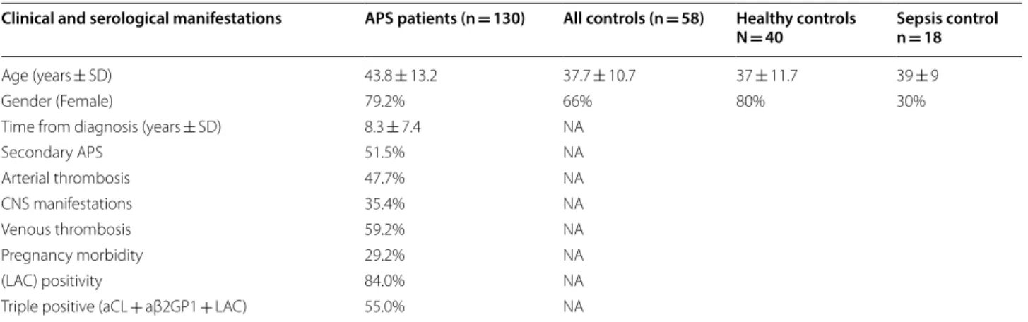
We studied 130 APS patients and 58 controls, of which 40 were healthy subjects and 18 disease controls diagnosed with sepsis. In our cohort of APS, patients’ prior serology was noticeable for LAC detected in 84% and “triple posi- tivity” (i.e. sero-positivity for aCL and a β2GP1 and LAC) in 55% of patients (Table 1). Clinical and serological data obtained from medical files prior to inclusion in this study were collected and analyzed, respectively (Table 1).
Prevalence of aPLs among APS patients and controls In this study, the sensitivity of the LIA including all aPLs of both IgM and IgG isotypes was 83% (49% for the IgM,
and 69% for the IgG isotype). Regarding non-criteria aPLs, the specificity of the test was 100% for the IgM isotype and 95% for the IgG antibodies compared to healthy controls. aPLs were more prevalent among APS patients compared to our entire control groups as well as the healthy and diseased control subjects with sepsis separately (Table 2). Among controls, some aPLs were numerically more prevalent in the subgroup of patients suffering from sepsis compared to healthy controls, whereas a statistical significance was reached only for aβ2GP1 IgG detected in 1/40 (2.5%) healthy controls vs.
4/18 (22.2%) septic patients (p = 0.03).
aPLs antibodies/profiles and phenotypes of APS
We analyzed the interactions of each aPL as well as dif- ferent combinations of aPLs (profiles) with the follow- ing clinical phenotypes of APS: arterial thrombosis, CNS manifestations, venous thrombosis, latency period from last thrombotic event and pregnancy morbidity (e.g.
recurrent early abortion, premature delivery etc.) as sum- marized in Table 3. In addition, we correlated serology with demographics (i.e. age, gender) and the presence of other autoimmune diseases, which in the vast majority of cases was systemic lupus erythematosus (i.e. primary or secondary APS).
Arterial thrombosis was associated with accumulation of any 7 or more of the 20 aPLs evaluated in this study compared to the presence of 6 or less (odds ratio [OR]
4.1; confidence interval [CI] 95% 1.9–96, p = 0.001) and the sole presence of aPT IgG (OR 2.3 (CI 95% 1.1–5.1, p = 0.03). Patients diagnosed with primary APS were more prone to suffer from arterial thrombotic events compared to those with secondary APS (OR 2.2; CI 95%
1.1–4.5, p = 0.03).
Table 1 Description of study cohort
Data retrieved from medical files
APS antiphospholipid antibody syndrome, CNS central nervous system, NA not applicable, aCL antibody to cardiolipin, aβ2GP1 antibody to beta2-glycoprotein1, LAC lupus anticoagulant
Clinical and serological manifestations APS patients (n = 130) All controls (n = 58) Healthy controls
N = 40 Sepsis control n = 18
Age (years ± SD) 43.8 ± 13.2 37.7 ± 10.7 37 ± 11.7 39 ± 9
Gender (Female) 79.2% 66% 80% 30%
Time from diagnosis (years ± SD) 8.3 ± 7.4 NA
Secondary APS 51.5% NA
Arterial thrombosis 47.7% NA
CNS manifestations 35.4% NA
Venous thrombosis 59.2% NA
Pregnancy morbidity 29.2% NA
(LAC) positivity 84.0% NA
Triple positive (aCL + aβ2GP1 + LAC) 55.0% NA
CNS manifestations correlated with the co-presence of a specific profile of four aPLs: aPT, aPG, aPI and aAN of the IgG isotype (OR 2.6; CI 95% 1.1–6.3, p = 0.03).
Venous thrombosis was present among our well- defined APS cohort in 77/130 patients of which 72(93%) were sero-positive for at least one aPL tested herein. Notably, the presence of aPLs was highly
associated with venous thrombosis in general, namely while comparing APS patients to our control groups, 72/77 patients with venous thrombosis were sero-pos- itive for at least 1 aPL (either IgG or IgM) whereas only 7/40 healthy control or 16/58 healthy and sepsis control were positive (p < 0.001 for both comparisons respec- tively). Among our APS patients, venous thrombosis Table 2 Prevalence of different anti-phospholipid antibodies (aPLs) among patients with the antiphospholipid syndrome (APS) and controls
HC healthy controls, SP sepsis patients, CL cardiolipin, β2GP1 beta2-glycoprotein1, P-acid phosphatidic acid, PC phosphatidylcholine, PE phosphatidylethanolamine, PG phosphatidylglycerol, PI phosphatidylinositol, AN annexin 5, PS phosphatidylserine, PT prothrombin, NS non-significant
APLs APS patients
N = 130 All controls
N = 58 p value
APS vs All controls Healthy controls (HC)
N = 40 p value
APS vs HC Sepsis patients (SP)
N = 18 p value
APS vs SP
CL IgM 34 (26%) 1 (2%) 0.001 0 < 0.001 1 (5%) 0.07
IgG 77 (59%) 1 (2%) 0.001 0 < 0.001 1 (5%) < 0.001
P‑acid IgM 17 (13%) 0 0.001 0 < 0.05 0 NS
IgG 73 (56%) 0 0.001 0 < 0.001 0 < 0.001
PE IgM 4 (3%) 0 NS 0 NS 0 NS
IgG 3 (2%) 0 NS 0 NS 0 NS
PG IgM 4 (3%) 0 NS 0 NS 0 NS
IgG 50 (38%) 0 0.001 0 < 0.001 0 < 0.001
PI IgM 11 (8%) 0 0.01 0 0.06 0 NS
IgG 59 (45%) 1 (2%) 0.001 1 (2%) < 0.001 0 < 0.001
PS IgM 16 (12%) 0 0.003 0 < 0.05 0 NS
IgG 73 (56%) 0 0.001 0 < 0.001 0 < 0.001
AN IgM 2 (1%) 1 (2%) NS 0 NS 1 (5%) NS
IgG 36 (27%) 5 (9%) 0.003 2 (5%) < 0.01 3 (16%) NS
β2GP1 IgM 42 (32%) 6 (10%) 0.003 4 (10%) < 0.001 2 (10%) NS
IgG 86 (66%) 5 (9%) 0.001 1 (2%) < 0.001 4 (22%) < 0.001
PT IgM 2 (1%) 0 NS 0 NS 0 NS
IgG 40 (30%) 1 (2%) 0.001 0 < 0.001 1 (5%) < 0.05
Table 3 APS phenotypes correlation with aPLs and aPLs profiles
Data is compared among APS patients with and without the defining phenotype
aCL cardiolipin, β2GP1 beta2-glycoprotein1, P-acid phosphatidic acid, PC phosphatidylcholine, PE phosphatidylethanolamine, PG phosphatidylglycerol, PI phosphatidylinositol, AN annexin 5, PS phosphatidylserine, PT prothrombin
APS phenotypes Associated aPLs and clinical manifestations O.R.
Arterial thrombosis > 7 any aPLsa 4.1 [CI 95% 1.9–96]
aPT IgG 2.3 [CI 95% 1.1–5.1]
Primary APS 2.2 [CI 95% 1.1–4.5]
CNS manifestations aPT + aPG + aPI and aAN (IgG) 2.6 [CI 95% 1.1–6.3]
Venous thrombosis aP‑acid IgM 0.3 [CI 95% 0.1–0.9]
Event free period > 3 years from last thrombotic events aPI IgG 3 [CI 95% 1.08–8.1]
aAN IgG 3.4 [CI 95%1.08–10.9]
Pregnancy morbidity aCL IgG and aPS IgG 2.9 [CI 95% 1.3–6.5]
anti‑AN IgG 2.8 [CI 95% 1.02–8]
Arterial thrombosis 3.3 [CI 95% 1.5–7.2]
CNS manifestations 3.9 [CI 95% 1.7–9]
Secondary APS 2.3 [CI 95%1.03–5.16]
as a phenotype was not linked to any specific aPL, but rather an inverse correlation was found between this phenotype and aP-acid IgM (OR 0.3; CI 95% 0.1–0.9, p = 0.02).
Latency from last thrombotic events The time range from last thrombotic event in our cohort was 0.5 to 16 years with an average of 4.8 years. Event-free period (i.e. with no recurrence of thrombotic events) of more than 3 years was significantly associated with sero- positivity to either aPI IgG or aAN IgG (OR 3; CI 95%
1.08–8.1, p < 0.05 and OR 3.4; CI 95% 1.08–10.9, p < 0.05, respectively), as well as with a lower cumulative number of aPLs (7.6 ± 6 vs. 11 ± 6, p < 0.05) compared to patients that experienced recurrent thrombosis within this time.
Pregnancy morbidity was observed in 38/103 (37%) of female APS patients. A profile of 2 aPLs: aCL IgG and aPS IgG was linked with this phenotype (OR 2.9; CI 95%
1.3–6.5, p < 0.05) as well as the sole positivity of anti-AN IgG (OR 2.8; CI 95% 1.02–8, p = 0.05). Notably, APS- related pregnancy morbidity was also linked to arterial thrombosis (OR 3.3; CI 95% 1.5–7.2, p = 0.004), CNS morbidity (OR 3.9; CI 95% 1.7–9, p = 0.001), and second- ary APS (OR 2.3; CI 95% 1.03–5.16, p = 0.04).
Older age allied with the presence of aP-acid IgM (51 y/o or older; p = 0.001) as well as with the profile of aβ2GP1 IgM and aCL IgM (46 y/o or older; p = 0.03) compared to sero-negative patients to these specific antibodies.
Gender was linked with specific aPLs: male with aCL IgG, aPG IgG, aPI IgG, aAN IgG and aPT IgG and female with aPE IgM (Table 4).
Concomitant autoimmune diseases 67/130 (51.5%) of our APS cohort had a concomitant autoimmune disease, which in the vast majority was systemic lupus erythema- tosus, defining secondary APS. In comparison to primary APS, secondary disease correlated with the presence of aβ2GP1 IgG (OR 3.9; CI 95%:1.8–8.2, p = 0.04).
Interactions between different aPLs
In the current study we observed interactions between most aPLs, as the presence of each aPL was statistically associated with sero-positivity of other aPLs of the same isotype (IgG or IgM; data not shown). An exception was noted for aPE of IgM isotype which was linked only with aβ2GP1 IgM (p < 0.05) and not with all other aPLs. Inter- estingly, the presence of LAC correlated with aPLs of the IgG subtype directed at CL, P-acid, PG, PI, PS, β2GP1, and PT (p < 0.05).
Discussion
In this study we evaluated 20 “criteria” and “non-criteria”
aPLs targeted at 10 different phospholipids or phospho- lipid-binding proteins in a cohort of well-defined “highly active serologically” APS patients (i.e. 84% were LAC pos- itive and 55% “triple positive”) aiming to correlate these aPLs with clinical phenotypes of APS.
APS is a unique acquired thrombotic condition, which causes both arterial and venous thrombosis that may reoccur despite anti-coagulation therapy. Arterial and recurrent thrombosis are both considered to have a worse outcome, thus are usually followed by a more forceful therapy [9, 26]. Herein, we found that certain aPLs and/or aPLs profiles are associated with three APS thrombotic phenotypes: arterial thrombosis, CNS mani- festations and recurrent thrombosis. Arterial thrombosis linked with the presence of any 7 or more aPLs compared to 6 or less. This stands in agreement with the notion that aPLs accumulation is an adverse marker of APS. The lat- ter was put forward in 2007 by Bizzaro. et al. [27] and in 2011 by Pengo. et al. [28] defining “triple positivity” as a risk factor for thrombosis. Later on Otomo. et al. [9]
evaluated criteria (aCL, aβ2GP1) and non-criteria (aPS/
PT complex) aPLs documented aPLs accumulation as a prognostic factor, so did Cervera R. et al. [29] that dem- onstrated “non-criteria” aPLs (aPT, aPE, anti-vimentin etc.,) relation to disease severity.
Additionally, we found a tie between arterial thrombo- sis and the existence of aPT. The thrombotic-predictive value of aPT was formerly suggested in a 15 year longitu- dinal study of SLE patients [27] as well as in a prospective study conducted by Forastiero et al. [30]. Lately Zhang et al. [31] found, similar to our data, that aPT relates to arterial thrombosis. In contrast, a review of 11 studies including 1440 patients concluded that aPT assessment was non-contributory for routine APS laboratory work- out [32]. These contradicting conclusions may result from different methods used for detection of aPT or dif- ferent cohorts of patients assessed. But perhaps the most striking difference is the role looked at as for routine APS workout aPT appears to render no benefit while a Table 4 Gender association with anti-phospholipids
antibodies (aPLs) among patients with the anti- phospholipids syndrome (APS)
PE phosphatidylethanolamine, CL cardiolipin, PG phosphatidylglycerol, PI phosphatidylinositol, AN annexin 5, PT prothrombin
APLs associated
with gender Odds ratio (CI 95%)
for female sex Odds ratio (CI 95%) for male sex
aPE IgM 2.5 (1.03–6) 0.39 (0.16–0.96)
aCL IgG 0.3 (0.1–0.6) 3.4 (1.6–7.4)
aPG IgG 0.2 (0.1–0.5) 2.2 (1–4.6)
aPI IgG 0.2 (0.1–0.5) 2.5 (1.1–5.4)
aAN IgG 0.2 (0.1–0.4) 3.0 (1.3–6.9)
aPT IgG 0.1 (0.4–0.3) 3.8 (1.7–8.5)
plausible role for risk stratification and phenotyping of
“well-defined” APS and SLE patients may be suggested.
APS commonly affects the central nervous system (CNS) manifesting as stroke, seizures, dementia, cogni- tive dysfunction, chorea, migraine, psychosis, demyelina- tion etc. [4, 33]. In this study, for the first time to the best of our knowledge, an association between APS-related CNS manifestations and a specific profile of four aPLs namely the co-presence of aPT aPG, aPI and aAN of the IgG isotype was observed, of which, like aPT also aAN and aPI were interrelated with thrombosis [34]. Annex- ins are a group of 12 regulatory proteins that are involved in vesicle trafficking, calcium signaling, cell growth, divi- sion, and apoptosis. Annexin 2 and 5 have an affinity to phospholipids, and antibodies directed at these proteins were found in patients with either arterial or venous thrombosis. Likewise, anti-phosphatidylinositol antibod- ies were significantly associated with thrombosis among APS and SLE patients [35].
One of the most difficult phenotypes of APS is the
‘recurrent thrombosis’ one. Approximately 20% of APS patients will experience recurrence within 3.4 to 16.3 years, depending on their treatments with antiplate- let, anticoagulant, or a combination of these therapies [36]. Currently, there are no established risk factors for
‘the thrombotic recurrence phenotype’ though the plau- sible role of aPLs has been suggested, and may eventually allude to enhanced therapy for patients at risk [37–39].
Herein, we found that a low recurrence rate, defined as thrombosis free period of more than 3 years, links with a lower accumulative number of aPLs, further support- ing the perception that more aPLs allied with a worst outcome.
APS is the most frequently acquired risk factor for recurrent pregnancy losses, ischemic placental dysfunc- tion, fetal growth restriction, preeclampsia, premature birth and intrauterine death [14, 40]. Apart from the thrombotic variants of APS, obstetric APS (OAPS) is probably the most common phenotype. Obstetric mor- bidity may be the only presentation of APS or may co- exist with thrombotic and non-thrombotic phenotypes [41]. In the current study, we found that obstetric mor- bidity was linked with CNS and arterial thrombotic phe- notypes, which stands in agreement with other reports such as a recent study by Gris et al. [42] documenting OAPS association with mental disorders. Besides, we found OAPS to be linked with a profile of two aPLs: aCL and aPS or the sole presence of aAN of the IgG isotype.
Recently, aCL was found to be the most common aPL present in a large cohort of 750 pregnancies [43]. Equally, aPS was linked with OAPS in several studies and in par- ticular, monoclonal aPS antibodies were found to reduce yolk sac growth in animal models as well as placental
trophoblastic cell growth and proliferation in humans [44]. In contrast to aCL and aPS, the role of aAN in OAPS is yet controversial. The latter was linked with recurrent pregnancy morbidity and losses in some studies [35, 45]
but this association was not ascertained in others [41, 46, 47]. Still, the prediction of aPL related pregnancy morbidity is an issue of great debate, especially among women defined as “only aPLs carriers” or those with less than three early miscarriages. Thus, although our results require further studies, the idea that certain aPLs or aPLs profile may be used as a marker of pregnancy morbidity is intriguing.
There is a strong link between aPLs and venous throm- bosis as was described in numerous studies as well as the current one while comparing APS patients to healthy subjects and patients diagnosed with sepsis. However, none of the aPLs studied herein was specifically linked with venous thrombosis among APS patients. In other words the vast majority of patients with venous thrombo- sis were aPL sero-positive and no differences were docu- mented compared to APS patients that did no exhibit venous thrombosis. In contrast, a striking observation in this study was the inverse association of anti P-acid anti- body of the IgM isotype with venous thrombosis. The anti P-acid antibody was rarely studied as a single anti- body and for the best of our knowledge this is the first report of such inverse association. Of note anti P-acid was linked with thrombosis in several studies which eval- uated mostly IgG antibodies and regarded thrombosis in general both arterial and venous. In a recent study of sero-negative APS patients, anti P-acid was linked with fetal losses and not with thrombosis [48]. Further studies are required to verify such an inverse correlation. Inter- estingly in this study the anti-P-acid IgM, as well as other IgM antibodies were linked with older age.
Last but not least, from the diagnostic perspective, the LIA was easy, efficient and with good sensitivity and specificity while employing cutoffs ascertained by healthy controls. This method enabled the discrimination of aPL found in APS patients from those in asymptomatic car- riers as reported previously [24]. Within our controls, some aPLs were numerically more prevalent among sep- tic control patients compared to healthy subjects, but a significant difference was observed only for anti-β2GP1 IgG. The latter transient appearance during infection was formerly reported [49, 50]. Furthermore, we observed interactions between different aPLs of the same iso- type. Similar observations have been reported and led to the hypothesis that broad aPLs positivity may be due to epitope spreading [51].
Our study has several limitations, as our cohort included “serologically active” APS patients (84% were LAC positive and 55% triple positive) that fulfill the
criteria for APS, which could cast doubt on the impli- cation of our results to patients with “lower serological activity” (e.g. single low titer aPL). A selection bias was inevitable as all patients had a least one clinical manifesta- tion of APS to fulfill its classification criteria. Noteworthy, this study aimed to define subtypes of APS rather than use aPL for diagnosis or classification of disease. Addi- tionally, aPLs were evaluated in this study using a single blood sample from each subject and a single method of detection. The former do not allow estimations of aPL profiles at the time of events, but rather a retrospective clinical correlation, as well as the lack of long term follow up, nor the role of other aPLs or aPL-complexes as anti- PS/PT (only PS and PT separately) or anti-domain-1 of β 2GPI. The latter two aPL may add value to the assess- ment of APS patients requiring further studies. Using multiple comparative methods for detection of aPLs is of significance, hence the use of the line dot blot (LIA) semi quantitative method as a single method is a limita- tion. However, all patients included in this study were originally criteria aPL positive and recently Thaler et al.
demonstrated similar results for detection of non-criteria aPL by the LIA method compared to ELISA [52]. Thus, we assume that there will be no significant differences by ELISA to our results by LIA. Moreover, Thaler et al [52]
reported a higher sensitivity of the LIA technique for aPL recognizing anionic phospholipids. Lastly, the aim of our study was to evaluate APS profiles, therefore correlation with a single criteria or non-criteria manifestation (e.g.
intra uterine fetal death of thrombocytopenia) was not evaluated.
Conclusions
Herein, we report that criteria and non-criteria aPLs and/
or aPLs profiles are statistically linked with APS pheno- types such as arterial thrombosis, CNS manifestations, recurrent thrombosis and obstetric APS. Additionally, we found that accumulation of these aPLs is associated with more severe variants of diseases namely arterial and recurrent thrombosis. Our data suggest that evaluating a broad spectrum of aPLs may enable defining APS pheno- types and, thus, may have a future role in precision choice of therapy for this autoimmune multifaceted disease.
Acknowledgements None.
Authors’ contributions
All authors have made substantial contributions to the conception and design of the work; IV, LS, EL, LK, BG, AD, MT, NAL contributed to acquisition and analysis of the samples, all authors contributed to interpretation of the data, creating, writing and revising the manuscript. All authors approved the sub‑
mitted version; including the list of authors as delineated above. All authors read and approved the final manuscript.
Funding No other funding.
Availability of data and materials
The clinical, serological data used to support the findings of this study are included within the article; further findings are available from the correspond‑
ing author upon request.
Ethics approval and consent to participate
The study received approval by the ethics committee of the Sheba medical center number 4784 and fulfilled the ethical guidelines of the declaration of Helsinki (Edinburgh 2000).
Consent for publication Not required.
Competing interests
This research was performed as part of the employment of the authors as well as supported by the arrow project for medical student (IV). The kits for serological evaluations were donated by (Medipan), DR is a shareholder of Medipan and GA Generic Assays and has a managerial position in both com‑
panies. The remaining authors do not have any conflict of interest.
Author details
1 Clinical Immunology, Angioedema and Allergy Unit, Zabludowicz Center for Autoimmune Diseases, The Chaim Sheba Medical Center, Tel‑Hashomer 52621, Israel. 2 Zabludowicz Center for Autoimmune Diseases, Sheba Medical Center, Tel‑Hashomer 52621, Israel. 3 Rheumatology Division, Hospital Das Clinicas HCFMUSP, Faculdade de Medicina, Universidade de Sao Paulo, Sao Paulo, SP, Brazil. 4 Department of Rheumatology and Immunol‑
ogy, Faculty of Medicine, University of Szeged, Szeged, Hungary. 5 Institute of Biotechnology, Faculty Environment and Natural Sciences, Brandenburg University of Technology Cottbus‑Senftenberg, Cottbus, Germany. 6 Rheu‑
matology Unit, Department of Medicine, University of Padova, Padova, Italy.
7 Rheumatology Unit, First Department of Propaedeutic Internal Medicine, School of Medicine, National and Kapodistrian University of Athens, Athens, Greece. 8 Sackler Faculty of Medicine, Tel‑Aviv University, Tel Aviv, Israel.
Received: 29 March 2020 Accepted: 6 May 2020
References
1. Schreiber K, Sciascia S, de Groot PG, et al. Antiphospholipid syndrome.
Nat Rev Dis Primers. 2018;4:18005.
2. Agmon‑Levin N, Shoenfeld Y. The spectrum between antiphospho‑
lipid syndrome and systemic lupus erythematosus. Clin Rheumatol.
2014;33(3):293–5.
3. Hughes GR, Shoenfeld Y. Antiphospholipid antibody testing—slow progress? Int J Clin Pract. 2012;66(6):533–5.
4. Gris JC, Nobile B, Bouvier S. Neuropsychiatric presentations of antiphos‑
pholipid antibodies. Thromb Res. 2015;135(Suppl 1):S56–9.
5. Miyakis S, Lockshin MD, Atsumi T, et al. International consensus statement on an update of the classification criteria for definite antiphospholipid syndrome (APS). J Thromb Haemost. 2006;4(2):295–306.
6. Roggenbuck D, Egerer K, von Landenberg P, et al. Antiphospholipid antibody profiling: time for a new technical approach? Autoimmun Rev.
2012;11(11):821–6.
7. Aggarwal R, Ringold S, Khanna D, et al. Distinctions between diagnostic and classification criteria? Arthritis Care Res (Hoboken). 2015;67(7):891–7.
8. Les I, Ruiz‑Irastorza G, Khamashta MA. Intensity and duration of antico‑
agulation therapy in antiphospholipid syndrome. Semin Thromb Hemost.
2012;38(4):339–47.
9. Otomo K, Atsumi T, Amengual O, et al. Efficacy of the antiphospholipid score for the diagnosis of antiphospholipid syndrome and its predictive value for thrombotic events. Arthritis Rheum. 2012;64(2):504–12.
10. Yelnik CM, Urbanski G, Drumez E, et al. Persistent triple antiphospho‑
lipid antibody positivity as a strong risk factor of first thrombosis, in a long‑term follow‑up study of patients without history of thrombosis or obstetrical morbidity. Lupus. 2017;26(2):163–9.
11. Pengo V, Ruffatti A, Legnani C, et al. Incidence of a first thromboembolic event in asymptomatic carriers of high‑risk antiphospholipid antibody profile: a multicenter prospective study. Blood. 2011;118(17):4714–8.
12. Agmon‑Levin N, Blank M, Zandman‑Goddard G, et al. Vitamin D: an instrumental factor in the anti‑phospholipid syndrome by inhibition of tissue factor expression. Ann Rheum Dis. 2011;70(1):145–50.
13. Sciascia S, Coloma‑Bazan E, Radin M, et al. Can we withdraw anticoagula‑
tion in patients with antiphospholipid syndrome after seroconvertion?
Autoimmun Rev. 2017;16(11):1109–14.
14. Mekinian A, Alijotas‑Reig J, Carrat F, et al. Refractory obstetrical antiphos‑
pholipid syndrome: features, treatment and outcome in a European multicenter retrospective study. Autoimmun Rev. 2017;16(7):730–4.
15. Leone A, Radin M, Almarzooqi AM, et al. Autologous hematopoietic stem cell transplantation in Systemic Lupus Erythematosus and antiphospho‑
lipid syndrome: a systematic review. Autoimmun Rev. 2017;16(5):469–77.
16. Abreu MM, Danowski A, Wahl DG, et al. The relevance of “non‑criteria”
clinical manifestations of antiphospholipid syndrome: 14th International Congress on Antiphospholipid Antibodies Technical Task Force Report on Antiphospholipid Syndrome Clinical Features. Autoimmun Rev.
2015;14(5):401–14.
17. Sciascia S, Bertolaccini ML. Antibodies to phosphatidylserine/pro‑
thrombin complex and the antiphospholipid syndrome. Lupus.
2014;23(12):1309–12.
18. Mekinian A, Bourrienne MC, Carbillon L, et al. Non‑conventional antiphos‑
pholipid antibodies in patients with clinical obstetrical APS: preva‑
lence and treatment efficacy in pregnancies. Semin Arthritis Rheum.
2016;46(2):232–7.
19. Shi H, Zheng H, Yin YF, et al. Antiphosphatidylserine/prothrombin antibodies (aPS/PT) as potential diagnostic markers and risk predictors of venous thrombosis and obstetric complications in antiphospholipid syndrome. Clin Chem Lab Med. 2018;56(4):614–24.
20. Wolgast LR, Arslan AA, Wu XX, Beyda JN, Pengo V, Rand JH. Reduction of annexin A5 anticoagulant ratio identifies antiphospholipid antibody‑
positive patients with adverse clinical outcomes. J Thromb Haemost.
2017;15(7):1412–21.
21. Sciascia S, Sanna G, Murru V, Roccatello D, Khamashta MA, Bertolaccini ML. Anti‑prothrombin (aPT) and anti‑phosphatidylserine/prothrombin (aPS/PT) antibodies and the risk of thrombosis in the antiphospholipid syndrome. A systematic review. Thromb Haemost. 2014;111(2):354–64.
22. Egerer K, Roggenbuck D, Buttner T, et al. Single‑step autoantibody profil‑
ing in antiphospholipid syndrome using a multi‑line dot assay. Arthritis Res Ther. 2011;13(4):R118.
23. Roggenbuck D, Somma V, Schierack P, Borghi MO, Meroni PL. Autoanti‑
body profiling in APS. Lupus. 2014;23(12):1262–4.
24. Roggenbuck D, Borghi MO, Somma V, et al. Antiphospholipid antibod‑
ies detected by line immunoassay differentiate among patients with antiphospholipid syndrome, with infections and asymptomatic carriers.
Arthritis Res Ther. 2016;18(1):111.
25. Nalli C, Somma V, Andreoli L, et al. Anti‑phospholipid IgG antibodies detected by line immunoassay differentiate patients with anti‑phospho‑
lipid syndrome and other autoimmune diseases. Auto Immun Highlights.
2018;9(1):6.
26. Pengo V, Ruiz‑Irastorza G, Denas G, Andreoli L, Khamashta M, Tincani A.
High intensity anticoagulation in the prevention of the recurrence of arterial thrombosis in antiphospholipid syndrome: ‘PROS’ and ‘CONS’.
Autoimmun Rev. 2012;11(8):577–80.
27. Bizzaro N, Ghirardello A, Zampieri S, et al. Anti‑prothrombin antibodies predict thrombosis in patients with systemic lupus erythematosus: a 15‑year longitudinal study. J Thromb Haemost. 2007;5(6):1158–64.
28. Pengo V. APS–controversies in diagnosis and management, critical over‑
view of current guidelines. Thromb Res. 2011;127(Suppl 3):S51–2.
29. Cervera R, Conti F, Doria A, Iaccarino L, Valesini G. Does seron‑
egative antiphospholipid syndrome really exist? Autoimmun Rev.
2012;11(8):581–4.
30. Forastiero R, Martinuzzo M, Pombo G, et al. A prospective study of anti‑
bodies to beta2‑glycoprotein I and prothrombin, and risk of thrombosis. J Thromb Haemost. 2005;3(6):1231–8.
31. Zhang S, Wu Z, Li J, et al. Clinical performance of antibodies to prothrom‑
bin and thrombin in Chinese patients with antiphospholipid syndrome:
potential interest in discriminating patients with thrombotic events and non‑thrombotic events. Rheumatol Int. 2017;37(4):579–84.
32. Galli M. Should we include anti‑prothrombin antibodies in the screening for the antiphospholipid syndrome? J Autoimmun. 2000;15(2):101–5.
33. Yelnik CM, Kozora E, Appenzeller S. Non‑stroke central neurologic manifestations in Antiphospholipid Syndrome. Curr Rheumatol Rep.
2016;18(2):11.
34. Iaccarino L, Ghirardello A, Canova M, et al. Anti‑annexins autoantibod‑
ies: their role as biomarkers of autoimmune diseases. Autoimmun Rev.
2011;10(9):553–8.
35. Amoroso A, Mitterhofer AP, Del Porto F, et al. Antibodies to anionic phospholipids and anti‑beta2‑GPI: association with thrombosis and thrombocytopenia in systemic lupus erythematosus. Hum Immunol.
2003;64(2):265–73.
36. Jackson WG, Oromendia C, Unlu O, et al. Recurrent thrombosis in patients with antiphospholipid antibodies and arterial thrombosis on antithrom‑
botic therapy. Blood Adv. 2017;1(25):2320–4.
37. Nalli C, Andreoli L, Casu C, Tincani A. Management of recurrent thrombo‑
sis in antiphospholipid syndrome. Curr Rheumatol Rep. 2014;16(3):405.
38. Legault KJ, Ugarte A, Crowther MA, Ruiz‑Irastorza G. Prevention of recurrent thrombosis in Antiphospholipid Syndrome: different from the general population? Curr Rheumatol Rep. 2016;18(5):26.
39. Amory CF, Levine SR, Brey RL, et al. Antiphospholipid Antibodies and Recurrent Thrombotic events: persistence and Portfolio. Cerebrovasc Dis.
2015;40(5–6):293–300.
40. Schreiber K, Radin M, Sciascia S. Current insights in obstetric antiphos‑
pholipid syndrome. Curr Opin Obstet Gynecol. 2017;29(6):397–403.
41. Alijotas‑Reig J, Esteve‑Valverde E, Ferrer‑Oliveras R, et al. The European registry on obstetric Antiphospholipid Syndrome (EUROAPS): a survey of 1000 consecutive cases. Autoimmun Rev. 2019;18(4):406–14.
42. Gris JC, Cyprien F, Bouvier S, et al. Antiphospholipid antibodies are associ‑
ated with positive screening for common mental disorders in women with previous pregnancy loss. The NOHA‑PSY observational study. World J Biol Psychiatry. 2017;20:1–13.
43. Saccone G, Berghella V, Maruotti GM, et al. Antiphospholipid antibody profile based obstetric outcomes of primary antiphospholipid syndrome:
the PREGNANTS study. Am J Obstet Gynecol. 2017;216(5):525 e521.
44. Ornoy A, Yacobi S, Matalon ST, et al. The effects of antiphospholipid antibodies obtained from women with SLE/APS and associated preg‑
nancy loss on rat embryos and placental explants in culture. Lupus.
2003;12(7):573–8.
45. Rand JH, Wu XX, Quinn AS, Taatjes DJ. The annexin A5‑mediated patho‑
genic mechanism in the antiphospholipid syndrome: role in pregnancy losses and thrombosis. Lupus. 2010;19(4):460–9.
46. Becarevic M. The IgG and IgM isotypes of anti‑annexin A5 antibodies:
relevance for primary antiphospholipid syndrome. J Thromb Thromboly‑
sis. 2016;42(4):552–7.
47. Zhang S, Wu Z, Li J, et al. Evaluation of the clinical relevance of anti‑
annexin‑A5 antibodies in Chinese patients with antiphospholipid syndrome. Clin Rheumatol. 2017;36(2):407–12.
48. Pignatelli P, Ettorre E, Menichelli D, Pani A, Violi F, Pastori D. Seronega‑
tive antiphospholipid syndrome: refining the value of non‑criteria antibodies for the diagnosis and clinical management. Haematologica.
2020;105(3):562–72.
49. Kivity S, Agmon‑Levin N, Blank M, Shoenfeld Y. Infections and autoim‑
munity–friends or foes? Trends Immunol. 2009;30(8):409–14.
50. Zinger H, Sherer Y, Goddard G, et al. Common infectious agents preva‑
lence in antiphospholipid syndrome. Lupus. 2009;18(13):1149–53.
51. Kasahara H, Matsuura E, Kaihara K, et al. Antigenic structures recog‑
nized by anti‑beta2‑glycoprotein I auto‑antibodies. Int Immunol.
2005;17(12):1533–42.
52. Thaler MA, Bietenbeck A, Steigerwald U, et al. Evaluation of the sensitivity and specificity of a novel line immunoassay for the detection of criteria and non‑criteria antiphospholipid antibodies in comparison to estab‑
lished ELISAs. PLoS ONE. 2019;14(7):e0220033.
Publisher’s Note
Springer Nature remains neutral with regard to jurisdictional claims in pub‑
lished maps and institutional affiliations.