Research Article
Physicochemical Characterization and Cyclodextrin Complexation of the Anticancer Drug Lapatinib
Gerg y Tóth,
1Ádám Jánoska,
1Gergely Völgyi,
1Zoltán-István Szabó,
2Gábor Orgován,
1Arash Mirzahosseini,
1and Béla Noszál
11Department of Pharmaceutical Chemistry, Semmelweis University, H˝ogyes Endre U. 9, Budapest 1092, Hungary
2Department of Drugs Industry and Pharmaceutical Management, University of Medicine and Pharmacy, Gh. Marinescu 38, 540139 Tˆargu Mures,, Romania
Correspondence should be addressed to Arash Mirzahosseini; mirzahosseini.arash@pharma.semmelweis-univ.hu Received 24 November 2016; Accepted 9 February 2017; Published 1 March 2017
Academic Editor: Jaime Villaverde
Copyright © 2017 Gerg˝o T´oth et al. This is an open access article distributed under the Creative Commons Attribution License, which permits unrestricted use, distribution, and reproduction in any medium, provided the original work is properly cited.
Lapatinib (LAP), the tyrosine kinase inhibitor drug with moderate bioavailability, was characterized in terms of physicochemical properties: acid-base characteristics, lipophilicity, and solubility. The highly lipophilic nature of the drug and its extremely low water solubility (𝑆0= 0.82nM) limit the development of a parenteral formulation. In order to enhance solubility and bioavailability, inclusion complex formation with cyclodextrins (CDs) is a promising method of choice. Therefore, LAP-CD interactions were also studied by a multianalytical approach. The stability constants of LAP with native cyclodextrins, determined by UV spectroscopy, identified the seven-membered𝛽-CD as the most suitable host. Continuous variation method (Job’s plot) by1H NMR showed a 1 : 1 stoichiometry for the complexes. The geometry of the complex was elucidated by 2D ROESY NMR measurements and molecular modeling, indicating that the partial molecular encapsulation includes the fluorophenyl ring of LAP. Phase-solubility studies with four CDs,𝛽-CD, (2-hydroxypropyl)-𝛽-cyclodextrin (HP-𝛽-CD), randomly methylated-𝛽- (RAMEB-) cyclodextrin, and sulfobutylether-𝛽-cyclodextrin (SBE-𝛽-CD), show an ALtype diagram and highly increased solubility via CD complexation.
The results are especially promising with SBE-𝛽-CD, exerting more than 600-fold gain in solubility. The equilibrium and structural information presented herein can offer the molecular basis for an improved drug formulation with enhanced bioavailability.
1. Introduction
Lapatinib (LAP) (Figure 1) is a potent and selective inhibitor of the epidermal growth factor receptor (EGFR) and human epidermal growth factor receptor 2 (HER2) [1].
The drug is marketed as its ditosylate salt, in form of a film-coated tablet (Tyverb/Tykerb). It is currently used in combination therapy with capecitabine or letrozole for the treatment of HER2-positive advanced or metastatic breast cancer [2, 3]. The pharmacokinetic properties of LAP are not optimal. Its oral bioavailability is moderate at best, it has considerable interpatient variability [4], and it is significantly affected by food intake. Both low-fat and high-fat diets before LAP administration increased its systematic exposure [4, 5]. Besides food intake, gastric pH also influences LAP bioavailability. Higher gastric pH reduces LAP absorption;
therefore, concomitant use of proton-pump inhibitors (PPIs)
can decrease its bioavailability [6]. The recommended dose of LAP is 1,250–1,500 mg once daily, which means taking 5-6 tablets at once, reducing patient adherence to therapy.
Increasing the bioavailability of LAP could reduce the dose.
Thus, a novel formulation with high solubility and bioavail- ability is currently an unmet medical need.
Enhancement of solubility and the concomitant bioavail- ability can be achieved by various means, such as solid disper- sion, size reduction, cosolvency, polymorphism, formation of water soluble prodrug, or complexes [7]. Cyclodextrin (CD) complexation is one of the most extensively investigated ways to improve drug solubility and bioavailability [8].
CDs are cyclic oligosaccharides comprising six, seven, or eight D-glucose units (𝛼-,𝛽-, and𝛾-CD, resp.) that have been recognized as pharmaceutical adjuvants for the past 20 years.
CDs have a hydrophilic surface responsible for their water solubility and a hydrophobic, inner cavity, where lipophilic
Volume 2017, Article ID 4537632, 9 pages https://doi.org/10.1155/2017/4537632
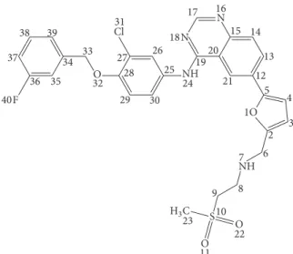
O F
O O NH Cl
N
1O 2 12
13 15 14 17 16 N
1920 24 21 25 27 26
28 29 30 31
32 34 33 35 36 40
37 38 39
3 5 4
7 6 9 8
10
11 22 23
NH
H3C S 18
Figure 1: Chemical structure and numbering of LAP.
molecules with adequate dimensions can be accommodated forming a noncovalently bonded host-guest complex, con- ferring an increased aqueous solubility for the encapsulated molecule. Besides native CDs, several modified derivatives are also available, differing in nature, position of substituents, and average degree of substitution, in order to enhance the inclusion capacity and improve the physicochemical prop- erties of the parent, native CDs [9, 10]. Many advantages of inclusion complexation have been reported, such as increased solubility, enhanced bioavailability, improved stability, taste and odor masking, reduced volatility, and modified drug release [11].
So far only a few attempts have been reported to improve the bioavailability of LAP [12–14]. To the best of our knowl- edge, LAP-CD inclusion complex characterization has yet to be described, despite the fact that CD complexation has been proven to increase the solubility, bioavailability, and anticancer activity of other tyrosine kinase inhibitors erloti- nib, gefitinib, imatinib, and so forth [15–19]; moreover, sulfobutylether-𝛽-CD was used for solubilizing LAP in some earlier studies [20, 21].
Here we report the quantified biorelevant physicochemi- cal properties of LAP. On the basis of the protonation con- stants, lipophilicity, and solubility parameters, we focus on the characterization and optimization of complex formation between various CDs and LAP. The information presented herein can be useful for the development of a new, improved LAP formulation. The methods used in this study include UV and NMR spectroscopy, electrospray ionization-mass spec- trometry (ESI-MS), phase-solubility, and molecular model- ing.
2. Materials and Methods
2.1. Materials. All CDs (𝛼-CD,𝛽-CD,𝛾-CD, (2-hydroxypro- pyl)-𝛽-CD (HP-𝛽-CD), random methylated𝛽- (RAMEB-) CD and sulfobutylether-𝛽-CD (SBE-CD)) were the products of Cyclolab R&D Ltd., Hungary. Degree of substitution (DS)
of HP-𝛽-CD, RAMEB, and SBE-CD was ∼4.5, ∼12, and 6.5, respectively. LAP was purchased from Eurasia’s Ltd., India. Tris(hydroxymethyl)aminomethane (Tris), H3PO4, KH2PO4, KOH, NaOH, HCl, and KCl were of analytical grade from commercial suppliers. D2O, DCl, dimethylsulfoxide (DMSO), hexadeuterated dimethylsulfoxide (DMSO-𝑑6), and n-octanol were obtained from Sigma-Aldrich, while MeOH was from Merck. Ultrapure, deionized water was prepared by a Milli-Q Direct 8 Millipore system.
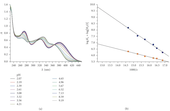
2.2. Determination of Protonation Constant by UV-pH Titra- tion. The log𝐾 value of LAP was determined by UV- pH titration, using the D-PAS ultraviolet spectropho- tometer attached to a GLpKa instrument (Sirius Analytical Instruments Ltd., Forest Row, UK). Multiwavelength UV absorbance of the sample solution was monitored throughout the titration using the fiber-optic probe in situ [22]. The UV- pH titrations were carried out under nitrogen atmosphere at constant ionic strength (𝐼 = 0.15M KCl) and temperature (𝑡 = 25.0 ± 0.5∘C). Due to the very low solubility of LAP, the cosolvent method was used to determine its log𝐾value.
The cosolvent protonation constants (log𝐾 values) were determined in six different MeOH-water mixtures within 23–
43% (w/w). A stock LAP solution of 7 mM concentration was prepared in DMSO. In each experiment, 75𝜇L of the stock solutions was transferred into sample vials containing 15 mL of 0.15 M KCl solution to achieve the required sample concen- tration. In each experiment, the acidity of the sample solution was adjusted using 0.5 M HCl to about pH∗ 2.5 and then titrated with 0.5 M KOH to about pH∗9. Spectral data were recorded in the 200–700 nm region after each pH measure- ment. About 20 pH readings and absorption spectra were collected from each titration. The log𝐾values were calcu- lated by RefinementProsoftware.
The measured log𝐾 values in MeOH-water solutions were then extrapolated to aqueous condition (zero MeOH concentration) according to the Yasuda-Shedlovsky proce- dure [23]. The Yasuda-Shedlovsky extrapolation method is based on the linear relation between log𝐾and the dielectric constant (𝜀) of the cosolvent mixture:
log𝐾+log[H2O] = 𝑎
𝜀 + 𝑏, (1)
where𝜀is the dielectric constant, and log[H2O] is the molar water concentration of the given MeOH-water mixture. Three parallel measurements were carried out.
2.3. Determination of the Intrinsic Solubility by Chasing Equi- librium Solubility Method (Cheqsol). The thermodynamic solubility of the nonionized form of the samples was deter- mined by the Chasing Equilibrium Solubility (CheqSol) method [24]. The apparatus used to perform the solubility determinations was a GLpKa titrator and a D-PAS spectrom- eter controlled by RefinementPro and CheqSol software (Sir- ius, Forest Row, UK). The titrations were performed under nitrogen atmosphere, at 0.15 M ionic strength and25.0±0.5∘C using standardized 0.5 M HCl and 0.5 M KOH solutions.
Three parallel measurements were carried out. The imple- mentation of the experiment is described in more detail in our previous articles [19, 25].
2.4. Partition Coefficient Measurement by the Stir-Flask Method. The distribution coefficients were measured from the absorbance of the molecules before and after partitioning using standard stir-flask method as we previously reported [26].
The partition coefficient (𝑃) cannot be measured directly due to the high log𝑃value of LAP. Therefore, the distribution coefficient (𝐷) was determined at acidic pH (pH= 2), where the molecule is in dicationic form, and at two other pH values at pH 5.83 and 6.11. Three parallel measurements were carried out at each pH. The pH was measured using a Metrohm 6.0204.100 combined pH glass electrode and a Metrohm 780 pH meter. The water/n-octanol phase ratio varied between 250 and 600. The log𝑃 values of each macrospecies were calculated based on the following equation:
𝐷pH= 𝜒𝑖𝑃𝑖= 𝜒LAP2+𝑃LAP2+∗ 𝜒LAP+𝑃LAP+∗ 𝜒LAP𝑃LAP, (2) where𝜒is the mole fraction of the corresponding macrospe- cies of the charge indicated.
2.5. Determination of Cyclodextrin Complex Stability by UV Spectroscopy. UV spectroscopic measurements were carried out on a Jasco V-550 diode-array spectrometer (Jasco, Japan) with 10 mm quartz cuvettes at 25∘C.
LAP was titrated with a constant amount of native CDs (𝛼-,𝛽-, and 𝛾-CD) at pH = 2 using 0.01 M HCl as solvent.
A stock solution of each CD was combined with different volumes of LAP solutions and filled to the same volume. The UV spectra were measured against a blind solution. Three parallel measurements were carried out for each CD. The complex formation equilibrium can be characterized by the following complex stability constant:
𝐾stab= [CD-LAP]
[CD] [LAP], (3)
where [CD], [LAP], and [CD-LAP] are the equilibrium concentrations of the CD, LAP, and the complex, respectively.
Since the total concentrations of CD ([CD]𝑇) and LAP ([LAP]𝑇)are known, assuming 1 : 1 stoichiometry, we can use the following relationships:
[CD]𝑇= [CD] + [CD-LAP]
[LAP]𝑇= [LAP] + [CD-LAP] . (4)
Since absorbance of any of the CDs is negligible and no association other than the CD-LAP formation takes place, it can be safely assumed that only LAP and the complex contribute significantly to the absorbance; therefore, the measured absorbance at any given wavelength is the weighed linear combination of the two absorbances (𝜀0and𝜀1being the molar extinction coefficients of LAP and the complex at a given wavelength, resp.):
𝐴 = 𝜀0[LAP] + 𝜀1[CD-LAP] . (5) Since the equilibrium concentration of LAP can be expressed in terms of the total LAP and the complex concentrations, the above equation takes on the following form:
𝐴 = 𝜀0[LAP]𝑇+ (𝜀1− 𝜀0) [CD-LAP] . (6)
The complex concentration can be expressed with the stability constant (𝐾stab) and the total concentrations:
𝐾stab= [CD-LAP]
([CD]𝑇− [CD-LAP]) ([LAP]𝑇− [CD-LAP]). (7)
Solving the above quadratic equation for[CD-LAP]one can derive an equation for absorbance (using (6)), which, if fitted onto the data, provides the complex stability constant:
𝐴 = 𝜀0[LAP]𝑇
+ (𝜀1− 𝜀0)(𝐾stab[CD]𝑇+ 𝐾stab[LAP]𝑇+ 1) − √(𝐾stab[CD]𝑇+ 𝐾stab[LAP]𝑇+ 1)2− 4𝐾stab2[CD]𝑇[LAP]𝑇
2𝐾stab .
(8)
Multiple fitting was performed with Opium program.
2.6. NMR Measurements. All NMR measurements were carried out on an Agilent/Varian VNMRS spectrometer (600 MHz for1H) with a 5 mm inverse-detection gradient probehead in D2O/DMSO-𝑑67/3 (v/v) mixtures at 25∘C. pD∗ was set to 2 using DCl solution. Addition of DMSO-𝑑6was necessary due to the low solubility of LAP in aqueous solvents, even at acidic pH. The chemical shifts were referenced to the
solvent signal of internal DMSO. Standard pulse sequences and processing routines available in VnmrJ 2.2C/Chempack 4.0 were used.
For determination of complex stoichiometry, Job’s con- tinuous variation method was used. Solutions were prepared from LAP and𝛽-CD in complementary amounts, to make up a 1.5 mM summary concentration. The solutions were mixed in different ratios and the1H NMR spectra were recorded after 24 h.
The average extent of penetration and the direction of inclusion were investigated by 2D ROESY NMR. Intermolec- ular NOEs between LAP and CD protons directly involved in the host-guest interaction were detected as cross-peaks.
In these experiments, the samples contained 1 mM LAP and 2 mM𝛽-CD. The ROESY spectrum was recorded applying a sweep width of 6 kHz and a spinlock of 3 kHz for a mixing time of 300 ms. 256 increments were collected with 32 repetitions and the measured data matrix was processed as a matrix of 4K (𝐹2) by 1K (𝐹1) data points.
2.7. ESI-MS Experiments. High resolution accurate mass of LAP-𝛽-CD inclusion complex was determined with an Agilent 6230 time of flight (TOF) mass spectrometer (Agilent, Germany). Samples were introduced by the Agilent 1260 Infinity LC system, and the mass spectrometer was operated in conjunction with a Jet Stream electrospray ion source in positive ion mode. Reference masses of m/z = 121.050873 and 922.009798 were used to calibrate the mass axis during analysis. The following ion source parameters were employed:
flow and temperature of the drying gas: 10 L/min and 320∘C;
pressure of the nebulizer gas (N2): 45 psi; capillary voltage:
3000 V; nozzle voltage: 500 V; sheath gas flow and tempera- ture: 10 L/min and 320∘C, respectively. TOF-MS parameters were as follows: fragmentor voltage: 300 V; skimmer poten- tial: 170 V; OCT 1 RF Vpp: 750 V. 1𝜇L of sample solution, containing 1 mM LAP and 1 mM𝛽-CD, was directly injected into the mass spectrometer. Data were acquired in a 100–3000 m/zmass range and peaks of the hypothesized complex were searched for in the 1000–3000m/zinterval. Mass spectra were processed by Agilent MassHunter B.04.00 software.
2.8. Phase-Solubility Studies. LAP phase-solubility studies were performed according to the method described by Higuchi and Connors [27]. Excess amount of LAP (5- 6 mg) was added to 5 mL stock solution with increasing concentrations of different CDs (𝛽-CD, HP-𝛽-CD, RAMEB, and SBE-CD) at pH 2.0 at 0.15 M ionic strength. The obtained suspensions were stirred for 24 hours. After sedimentation, LAP concentrations were measured from the supernatant spectrophotometrically on a Jasco V-550 instrument at 269 nm. Three parallel measurements were carried out.
Assuming 1 : 1 stoichiometry, the stability constant of the complex (𝐾stab) can be determined using the phase-solubility diagram according to the following equation:
𝐾stab= slope
𝑆0(1 −slope), (9) where𝑆0is the intrinsic solubility of LAP in the absence of CDs.
2.9. Molecular Modeling. Molecular modeling study was performed as previously reported [19, 28, 29] by using the semiempirical PM3 method of Hyperchem Professional software (version 8.0). The initial structures of neutral LAP and𝛽-CD were constructed with the help of Hyperchem 8.0 and optimized with PM3. A docking procedure was used to calculate the energy of the complex. LAP was moved into
the CD cavity from the wider rim, step-by-step, performing a geometry optimization after each step until the conformation with the lowest energy was achieved. The docking procedure was repeated using different starting positions, and the lowest achieved energy was considered as the optimal geometry of the complex.
The stability of the complex was estimated from the formation energy:
Δ𝐸 = 𝐸complex− (𝐸LAP+ 𝐸CD) . (10)
3. Results and Discussion
3.1. Physicochemical Characterization of LAP. Determination of the physicochemical properties of drugs is crucial to understand the pharmacokinetic behavior at the molecular level. Therefore, protonation constants, octanol/water parti- tion coefficient, and the intrinsic solubility of LAP were deter- mined. Due to the very low solubility of LAP, the protonation constants can only be determined by UV-pH titration with mixed-solvent procedure (Figure 2(a)). In our measurements, the MeOH-water cosolvent system was used, where the apparent protonation constants (log𝐾 values) were deter- mined in six different mixtures; then the aqueous log𝐾s were obtained by extrapolation using Yasuda-Shedlovsky method (see (1)) taking into account the dielectric constant of the different cosolvent mixtures (Figure 2(b)). This way log𝐾1= 7.72 ± 0.07and log𝐾2= 5.08 ± 0.05values were obtained.
Using the protonation constants, the distribution curves for protonation macrospecies of LAP can be depicted as a function of the pH (Figure 3(a)). The diagram shows that, in acidic media, at the pH of the stomach, the dicationic form is overwhelmingly dominant. The compound exists mainly in monocationic form at the pH of the blood (67.4%). Above pH 8, the neutral form dominates, which plays a crucial role in the transport mechanism in general, assuming that orally administrated LAP is absorbed mainly from the intestine.
Lipophilicity of LAP was characterized in terms of par- tition coefficient, which is the most widely used descriptor of lipophilicity in QSAR studies [30]. The working range of the used stir-flask method is−4 to +4 in log𝐷values, limited by the accuracy of the phase volume measurement. Based on this fact, the octanol/water partition coefficient at pH values where the nonionized form dominates cannot be measured directly due to the very high log𝑃value of LAP. Therefore, the log𝐷values were measured at lower pHs. At pH 2, where the molecule is practically 100% dicationic, the measured log𝐷pH2 = 2.71 ± 0.06characterizes the lipophilicity of the dicationic species. From log𝐷values at pH 5.83 (3.67 ± 0.07), pH 6.11 (3.90 ± 0.08), and pH 2 (2.71 ± 0.06), the log𝑃value of the monocationic and neutral species could be calculated using (2). The values are summarized in Table 1, while the pH- dependent lipophilicity profile of LAP is in Figure 3(b).
These values indicate that LAP is an extremely lipophilic molecule and even the dicationic form has the capability to penetrate membranes by passive diffusion. These high log𝑃 values also explain the ability of LAP to cross the blood-brain barrier, which could be a further therapeutic approach in
4.654.96 5.676.52 7.138.59 9.19 2.072.19
2.392.61 3.083.32 3.564.21 pH 0.0
0.2 0.4 0.6 0.8 1.0 1.2 1.4 1.6
A
260 280 300 320 340 360 380 400 440
240 420
(HG)
(a)
13.5 14.0 14.5 15.0
13.0 15.5 16.0 16.5 17.0
1000/
5.5 6.0 6.5 7.0 7.5 8.0 8.5 9.0 9.5 10.0
FIAKs+FIA[(2/]
(b)
Figure 2: Determination of protonation constants of LAP by (a) UV/pH titration of LAP in 43/57 (w/w%) methanol/water mixture and (b) Yasuda-Shedlovsky extrapolation.
0.0 0.2 0.4 0.6 0.8 1.0
X
14
10 12
8
4 6
2 0
pH
,!02+ ,!0
,!0+
(a)
14
10 12
8
4 6
2 0
pH
,!02+
,!0
,!0+
−6
−4
−2 0 2 4 6
logD
(b)
Figure 3: (a) Logarithmic distribution diagram for the LAP protonation macrospecies. (b) Contribution of the LAP protonation macrospecies to the lipophilicity profile (in black broad line).
brain metastasis [31]. As a part of our LAP profiling, its intrin- sic solubility (the equilibrium solubility of the nonionized form) was measured by the CheqSol method, which resulted in a value of only 0.48 ng/mL. LAP is a class II drug in the Biopharmaceutics Classification System, being of low water solubility and high permeability, which is in good agreement with our determined physicochemical values. These data
quantify that the very poor solubility is the Achilles heel of the LAP administration. It is the root cause for the low bioavailability of the drug the necessity of taking very high doses. These problems could be alleviated by enhancing LAP solubility through CD inclusion complexation. For further pharmaceutical formulation development, optimization of CD-complex formation is crucial; therefore, interactions
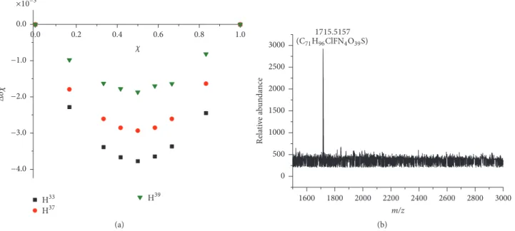
×10−3
(33
(37
(39
0.2 0.4 0.6 0.8 1.0
0.0
−4.0
−3.0
−2.0
−1.0 0.0
Δ
(a)
0 500 1000 1500 2000 2500 3000
Relative abundance
1800 2000 2200 2400 2600 2800 3000 1600
m/z 1715.5157
(#71(96ClF.4/393)
(b)
Figure 4: Determination of LAP-𝛽-CD stoichiometry. (a) Representative Job plot diagrams of selected LAP protons. (b) Representative mass spectrum of a LAP-𝛽-CD sample.
Table 1: Calculated log𝑃values of the different LAP macrospecies.
Macrospecies log𝑃
Dicationic 2.71 ± 0.07
Monocationic 3.15 ± 0.10
Neutral 5.49 ± 0.10
between LAP and CDs were characterized in solution with a wide variety of complementary analytical techniques and approaches, such as phase-solubility studies, 1H and 2D NMR techniques, UV spectroscopy, and electrospray ionization-time of flight-mass spectrometry (ESI-TOF-MS).
3.2. Determination of LAP-𝛽-CD Stoichiometry: NMR Job Plot and ESI-TOF-MS Studies. In order to draw adequate conclu- sions on the inclusion process, the knowledge of selector- selectand complexation stoichiometry is a prerequisite. The general method to determine complex stoichiometry is Job’s continuous variation method. In this experiment, the 1H NMR chemical shifts (𝛿) were measured at different LAP concentration/𝛽-CD concentration(𝑐LAP/𝑐CD) ratios, while the sum of 𝑐LAP + 𝑐CD was kept constant. The calculated factors (Δ𝛿𝜒LAP) were plotted as a function of LAP molar ratio (𝜒LAP). In all cases, the resulting Job plot diagrams showed maxima at 0.5, indicating the typical 1 : 1 binding stoichiometry. Representative Job plot curves of LAP and𝛽- CD are shown in Figure 4(a).
The complex stoichiometry was also studied by ESI-TOF- MS technique, which provides a powerful means to study noncovalent host-guest inclusion complexes between a CD and the guest molecule [32]. A large number of publications prove that a simple, fast ESI-MS analysis is suitable for the de- termination of inclusion complex stoichiometry [15, 19, 33];
however, ESI-MS studies imply the question whether the observed peaks represent actual inclusion complexes or rather electrostatic adducts formed during the electrospray process [34], if, however, additional adequate techniques prove that the complex formation ESI-MS is straightforward way to determine complex stoichiometry. Analyzing the MS spectra in the 100–1500m/zrange, the protonated molecular ion of the uncomplexed LAP (m/z 581.1419 [M+H]+), their source fragment ion (m/z458.1027 [M+H]+), and the charac- teristic m/z values of uncomplexed 𝛽-CD (m/z 1135.3732 [M+H]+, 1157.3531 [M+Na]+) can be observed. Figure 4(b) shows the renormalized upper part (1500–3000m/z) of the mass spectrum of a sample containing LAP and𝛽-CD in 1 : 2 molar ratio. The observed signal atm/z 1715.5157 with gen- erated C71H96ClFN4O39S molecular formula corresponds to the LAP-𝛽-CD 1 : 1 inclusion complex, which means that ESI- TOF-MS confirms the NMR Job result, verifying the 1 : 1 stoi- chiometry.
3.3. Determination of Stability Constant by UV. UV spec- troscopy can be a sensitive technique to characterize the CD inclusion complexes, provided that the guest molecule under- goes the significant absorption changes upon complexation.
This technique can be used for the determination of CD stability constant of molecules with low solubility as well.
Using (8), the stability constants between LAP and native CDs were determined to select the most suitable host. The stability constants determined at pH 2 are listed in Table 2.
Based on these data, we can conclude that, concerning the size, the seven-membered𝛽-CD is the most suitable host molecule for complexation. Thus, further experiments were carried out with𝛽-CD and its derivatives.
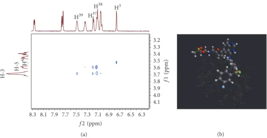
3.4. Structure of the LAP-𝛽-CD Complex. 2D ROESY NMR combined with molecular modeling is an appropriate way
4.1 4.0 3.9 3.8 3.7 3.6 3.5 3.4 3.3 3.2
H-3 H-5 (6
(39 (37
(38 (3
8.1 7.9 7.7 7.5 7.3 7.1 6.9 6.7 6.5 6.3 8.3
f2 (JJG)
f1(JJG)
(a) (b)
Figure 5: Determination of the LAP–𝛽-CD inclusion complex geometry. (a) Expansion of 2D ROESY NMR spectrum. (b) Energy-minimized structure of the complex based on the result of molecular modeling study.
Table 2: Determined𝐾stabvalues at pH 2 for different LAP-CD complexes with UV spectroscopy.
CD 𝐾stab(M−1)
𝛼-CD 46 ± 8
𝛽-CD 121 ± 12
𝛾-CD 89 ± 6
to characterize the geometry of inclusion complexes. ROESY experiment offers valuable information on spatial proximity between protons of the host and guest molecules by observing intermolecular dipolar cross-correlations, while molecular modeling seeks the energy-minimized structure, which can be visualized at the atomic level [19, 35, 36].
2D NMR ROESY spectra for LAP–𝛽-CD inclusion com- plex were recorded at pD∗2, where LAP exhibits an appro- priate solubility. The expansion of this 2D spectrum in Figure 5(a) contains an intramolecular cross-peak between H3 (6.76 ppm) and H6 (3.52 ppm) nuclei of LAP. From the viewpoint of encapsulation intermolecular cross-peaks of the fluorophenyl protons (H37,38,39) with the inner H-3 (3.68 ppm) and H-5 (3.58 ppm) protons of𝛽-CD are crucial.
Due to the fact that the H39 (7.49 ppm) proton of LAP gives a stronger cross-peak with the H-3 proton of the CD, we can assume that the fluorophenyl moiety is accommodated in the CD cavity from the wider rim. Based on ROESY data, molecular modeling at PM3 level was carried out. The energy- minimization structure is depicted in Figure 5(b). The energy of formation of the complex was found−10.7 kcal/mol, indi- cating that complex formation is thermodynamically favored.
3.5. Phase-Solubility Studies. Phase-solubility analysis accord- ing to Higuchi and Connors is an appropriate technique to demonstrate changes in drug solubility. Moreover, sta- bility constants and stoichiometry can also be determined [9]. Upon addition of increasing amounts of CDs (𝛽-CD,
Table 3:𝐾stabvalues calculated from phase-solubility studies for the CDs employed.
CD 𝐾stab(M−1)
𝛽-CD 131 ± 9
RAMEB 331 ± 15
HP-𝛽-CD 537 ± 20
SBE-CD 31622 ± 120
RAMEB, HP-𝛽-CD, and SBE-CD) at pH 2, linear solubility enhancement was observed for LAP, regardless of the CD employed (Figure 6).
The resulting AL-type linear positive isotherms reveal 1 : 1 binding stoichiometry, further supporting Job plot and ESI-TOF-MS results. The𝐾stab values calculated by (9) are summarized in Table 3.
The determined𝐾stabvalue for𝛽-CD by UV spectroscopy and phase-solubility is in agreement. Moreover similar stability constants were determined for other cyclodextrin complexes of tyrosine kinase inhibitors [15, 16, 19]. The values in Table 3 show that the substitution of the𝛽-CD enhances inclusion complex formation stability as observed also in the case of erlotinib [19]. However, in the case of imatinib, the methylation of the native𝛽-CD reduced the stability of the complex [15], which can be explained by the structural differ- ences of these anticancer drugs. The largest binding constant was observed for SBE-𝛽-CD, which is totally ionized under the applied conditions. Thus, the reason of the more stable complex is the additional electrostatic attractions between the multiply negatively charged hosts and the dicationic LAP.
Concerning the solubility enhancing capabilities, it can be stated that CD derivatives enhance the LAP solubility due pri- marily to the inclusion phenomenon, which can be extended by electrostatic forces. The results are especially promising with SBE-𝛽-CD where more than 600-fold solubility increase could be achieved.
×10−5
0.0 0.5 1.0 1.5 2.0 2.5
0.000 0.005 0.010 0.015 0.020 0.025 0.030
-CDHP--CD RAMEB
(GIF∗>G−3)cLAP
(GIF∗>G−3) c#$
(a)
×10−3
SBE-CD 0.0
0.2 0.4 0.6 0.8 1.0
0.005 0.015 0.020
0.000 0.010
(GIF∗>G−3 )
(GIF∗>G−3) c#$
c,!0
(b) Figure 6: Phase-solubility diagrams of LAP-CD complexes.
4. Conclusion
LAP, the anticancer drug, was characterized in terms of lipophilicity, solubility, acid-base, and CD-complex forma- tion properties, using complementary methods, such as UV, NMR, phase-solubility, and molecular modeling. An important practical result of this work is that CDs, especially the substituted derivatives, significantly increase the LAP solubility, which can be a starting point of a new LAP formulation of improved bioavailability.
Competing Interests
The authors declare no conflict of interests.
Authors’ Contributions
Gerg˝o T´oth and ´Ad´am J´anoska contributed equally to this work.
Acknowledgments
The research was supported through the New National Excellence Program of the Ministry of Human Capacities ( ´Ad´am J´anoska).
References
[1] G. M. Higa and J. Abraham, “Lapatinib in the treatment of breast cancer,”Expert Review of Anticancer Therapy, vol. 7, no.
9, pp. 1183–1192, 2007.
[2] C. E. Geyer, J. Forster, D. Lindquist et al., “Lapatinib plus capecitabine for HER2-positive advanced breast cancer,”New
England Journal of Medicine, vol. 355, no. 26, pp. 2733–2743, 2006.
[3] S. Johnston, J. Pippen Jr., X. Pivot et al., “Lapatinib combined with letrozole versus letrozole and placebo as first-line therapy for postmenopausal hormone receptor—positive metastatic breast cancer,”Journal of Clinical Oncology, vol. 27, no. 33, pp.
5538–5546, 2009.
[4] H. A. Burris III, C. W. Taylor, S. F. Jones et al., “A phase I and pharmacokinetic study of oral lapatinib administered once or twice daily in patients with solid malignancies,”Clinical Cancer Research, vol. 15, no. 21, pp. 6702–6708, 2009.
[5] L. A. Devriese, K. M. Koch, M. Mergui-Roelvink et al., “Effects of low-fat and high-fat meals on steady-state pharmacokinetics of lapatinib in patients with advanced solid tumours,”Investiga- tional New Drugs, vol. 32, no. 3, pp. 481–488, 2014.
[6] K. M. Koch, Y.-H. Im, S.-B. Kim et al., “Effects of esomeprazole on the pharmacokinetics of lapatinib in breast cancer patients,”
Clinical Pharmacology in Drug Development, vol. 2, no. 4, pp.
336–341, 2013.
[7] C. Leuner, “Improving drug solubility for oral delivery using solid dispersions,” European Journal of Pharmaceutics and Biopharmaceutics, vol. 50, no. 1, pp. 47–60, 2000.
[8] R. L. Carrier, L. A. Miller, and I. Ahmed, “The utility of cyclodextrins for enhancing oral bioavailability,” Journal of Controlled Release, vol. 123, no. 2, pp. 78–99, 2007.
[9] K. A. Connors, “The stability of cyclodextrin complexes in solution,”Chemical Reviews, vol. 97, no. 5, pp. 1325–1358, 1997.
[10] J. Szejtli, “Introduction and general overview of cyclodextrin chemistry,”Chemical Reviews, vol. 98, no. 5, pp. 1743–1753, 1998.
[11] T. Loftsson and D. Duchene, “Cyclodextrins and their pharma- ceutical applications,”International Journal of Pharmaceutics, vol. 329, no. 1-2, pp. 1–11, 2007.
[12] H. Gao, Y. Wang, C. Chen et al., “Incorporation of lapatinib into core-shell nanoparticles improves both the solubility and anti- glioma effects of the drug,”International Journal of Pharmaceu- tics, vol. 461, no. 1-2, pp. 478–488, 2014.
[13] L. Zhang, S. Zhang, S.-B. Ruan, Q.-Y. Zhang, Q. He, and H.- L. Gao, “Lapatinib-incorporated lipoprotein-like nanoparticles:
preparation and a proposed breast cancer-targeting mecha- nism,”Acta Pharmacologica Sinica, vol. 35, no. 6, pp. 846–852, 2014.
[14] Y. Song, X. Yang, X. Chen, H. Nie, S. Byrn, and J. W. Lubach,
“Investigation of drug-excipient interactions in lapatinib amor- phous solid dispersions using solid-state NMR spectroscopy,”
Molecular Pharmaceutics, vol. 12, no. 3, pp. 857–866, 2015.
[15] S. B´eni, Z. Szak´acs, O. Csern´ak, L. Barcza, and B. Nosz´al, “Cyclo- dextrin/imatinib complexation: binding mode and charge de- pendent stabilities,”European Journal of Pharmaceutical Sci- ences, vol. 30, no. 2, pp. 167–174, 2007.
[16] Y.-H. Phillip Lee, S. Sathigari, Y.-J. Jean Lin et al., “Gefitinib- cyclodextrin inclusion complexes: physico-chemical charac- terization and dissolution studies,” Drug Development and Industrial Pharmacy, vol. 35, no. 9, pp. 1113–1120, 2009.
[17] N. Devasari, C. P. Dora, C. Singh et al., “Inclusion complex of erlotinib with sulfobutyl ether-𝛽-cyclodextrin: preparation, characterization, in silico, in vitro and in vivo evaluation,”
Carbohydrate Polymers, vol. 134, pp. 547–556, 2015.
[18] S. M. L. Gontijo, P. P. G. Guimar˜aes, C. T. R. Viana et al.,
“Erlotinib/hydroxypropyl-𝛽-cyclodextrin inclusion complex:
characterization and in vitro and in vivo evaluation,”Journal of Inclusion Phenomena and Macrocyclic Chemistry, vol. 83, no. 3- 4, pp. 267–279, 2015.
[19] G. T´oth, ´A. J´anoska, Z.-I. Szab´o et al., “Physicochemical characterisation and cyclodextrin complexation of erlotinib,”
Supramolecular Chemistry, vol. 28, no. 7-8, pp. 656–664, 2016.
[20] D. W. Rusnak, K. Lackey, K. Affleck et al., “The effects of the novel, reversible epidermal growth factor receptor/ErbB-2 tyrosine kinase inhibitor, GW2016, on the growth of human normal and tumor-derived cell lines in vitro and in vivo,”
Molecular Cancer Therapeutics, vol. 1, no. 2, pp. 85–94, 2001.
[21] M. J. Sambade, R. J. Kimple, J. T. Camp et al., “Lapatinib in combination with radiation diminishes tumor regrowth in HER2+ and basal-like/EGFR+ breast tumor xenografts,”
International Journal of Radiation Oncology, Biology, Physics, vol. 77, no. 2, pp. 575–581, 2010.
[22] K. Y. Tam and K. Tak´acs-Nov´ak, “Multi-wavelength spec- trophotometric determination of acid dissociation constants: a validation study,”Analytica Chimica Acta, vol. 434, no. 1, pp.
157–167, 2001.
[23] G. V¨olgyi, R. Ruiz, K. Box, J. Comer, E. Bosch, and K.
Tak´acs-Nov´ak, “Potentiometric and spectrophotometric pKa determination of water-insoluble compounds: validation study in a new cosolvent system,”Analytica Chimica Acta, vol. 583, no.
2, pp. 418–428, 2007.
[24] M. Stuart and K. Box, “Chasing equilibrium: measuring the intrinsic solubility of weak acids and bases,”Analytical Chem- istry, vol. 77, no. 4, pp. 983–990, 2005.
[25] G. V¨olgyi, E. Baka, K. J. Box, J. E. A. Comer, and K.
Tak´acs-Nov´ak, “Study of pH-dependent solubility of organic bases. Revisit of Henderson-Hasselbalch relationship,”Analyt- ica Chimica Acta, vol. 673, no. 1, pp. 40–46, 2010.
[26] G. T´oth, K. Maz´ak, S. Hosztafi, J. K¨ok¨osi, and B. Nosz´al,
“Species-specific lipophilicity of thyroid hormones and their precursors in view of their membrane transport properties,”
Journal of Pharmaceutical and Biomedical Analysis, vol. 76, pp.
112–118, 2013.
[27] T. Higuchi and K. A. Connors, “Phase solubility studies,” in Advances in Analytical Chemistry and Instrumentation, C. N.
Reilly, Ed., pp. 117–212, Wiley-Interscience, New York, NY, USA, 1965.
[28] K. M. Al Azzam and E. Muhammad, “Host-guest inclusion complexes between mitiglinide and the naturally occurring cyclodextrins𝛼,𝛽, and𝛾: a theoretical approach,”Advanced Pharmaceutical Bulletin, vol. 5, no. 2, pp. 289–291, 2015.
[29] X. Wen, F. Tan, Z. Jing, and Z. Liu, “Preparation and study the 1:2 inclusion complex of carvedilol with𝛽-cyclodextrin,”Journal of Pharmaceutical and Biomedical Analysis, vol. 34, no. 3, pp. 517–
523, 2004.
[30] C. Hansch, “On the future of QSAR,” inMedicinal Chemistry for the 21st Century, C. G. Wermuth, N. Koga, and H. K¨onig, Eds., pp. 281–293, Metcalf Blackwell, Oxford, UK, 1994.
[31] A. Morikawa, D. M. Peereboom, Q. R. Smith et al., “Clinical evi- dence for drug penetration of capecitabine and lapatinib uptake in resected brain metastases from women with metastatic breast cancer,”Journal of Clinical Oncology, vol. 31, no. 15, supplement 1, abstract 514, 2013, Proceedings of the 2013 ASCO Annual Meeting.
[32] Y. Dotsikas and Y. L. Loukas, “Efficient determination and evaluation of model cyclodextrin complex binding constants by electrospray mass spectrometry,”Journal of the American Society for Mass Spectrometry, vol. 14, no. 10, pp. 1123–1129, 2003.
[33] N. Marangoci, M. Mares, M. Silion et al., “Inclusion complex of a new propiconazole derivative with𝛽-cyclodextrin: NMR, ESI-MS and preliminary pharmacological studies,”Results in Pharma Sciences, vol. 1, no. 1, pp. 27–37, 2011.
[34] J. B. Cunniff and P. Vouros, “False positives and the detection of cyclodextrin inclusion complexes by electrospray mass spec- trometry,”Journal of the American Society for Mass Spectrome- try, vol. 6, no. 5, pp. 437–447, 1995.
[35] D. Salvatierra, C. Jaime, A. Virgili, and F. S´anchez-Ferrando,
“Determination of the inclusion geometry for the𝛽-cyclodex- trin/benzoic acid complex by NMR and molecular modeling,”
Journal of Organic Chemistry, vol. 61, no. 26, pp. 9578–9581, 1996.
[36] Z.-I. Szab´o, F. Mohammadhassan, L. Sz˝ocs et al., “Stereoselec- tive interactions and liquid chromatographic enantioseparation of thalidomide on cyclodextrin-bonded stationary phases,”
Journal of Inclusion Phenomena and Macrocyclic Chemistry, vol.
85, no. 3-4, pp. 227–236, 2016.
Submit your manuscripts at https://www.hindawi.com
Hindawi Publishing Corporation
http://www.hindawi.com Volume 2014
Inorganic Chemistry
International Journal of
Hindawi Publishing Corporation
http://www.hindawi.com Volume 2014
Photoenergy
Hindawi Publishing Corporation
http://www.hindawi.com Volume 2014
Carbohydrate Chemistry
International Journal of
Hindawi Publishing Corporation
http://www.hindawi.com Volume 2014
Journal of
Chemistry
Hindawi Publishing Corporation
http://www.hindawi.com Volume 2014
Physical Chemistry
Hindawi Publishing Corporation http://www.hindawi.com
Analytical Methods in Chemistry
Journal of
Volume 2014
Bioinorganic Chemistry and Applications
Hindawi Publishing Corporation
http://www.hindawi.com Volume 2014
Spectroscopy
International Journal ofHindawi Publishing Corporation
http://www.hindawi.com Volume 2014
The Scientific World Journal
Hindawi Publishing Corporation
http://www.hindawi.com Volume 2014
Medicinal Chemistry
Hindawi Publishing Corporation
http://www.hindawi.com Volume 2014
Chromatography Research International
Hindawi Publishing Corporation
http://www.hindawi.com Volume 2014
Applied ChemistryJournal of
Hindawi Publishing Corporation
http://www.hindawi.com Volume 2014
Hindawi Publishing Corporation
http://www.hindawi.com Volume 2014
Theoretical Chemistry
Journal of
Hindawi Publishing Corporation
http://www.hindawi.com Volume 2014
Journal of
Spectroscopy
Analytical Chemistry
Hindawi Publishing Corporation
http://www.hindawi.com Volume 2014
Journal of
Hindawi Publishing Corporation
http://www.hindawi.com Volume 2014
Quantum Chemistry
Hindawi Publishing Corporation
http://www.hindawi.com Volume 2014
International
Electrochemistry
International Journal ofHindawi Publishing Corporation
http://www.hindawi.com Volume 2014
Hindawi Publishing Corporation
http://www.hindawi.com Volume 2014