UR JO
OFALCARNDIOVASCULAR EM ERG
ENSCIE
Journal of Cardiovascular Emergencies 2017;3(2):89-92
CASE REPORT
Symptomatic Coronary-Pulmonary Fistula Revealed with Coronary CT Angiography
Sára Papp, István Ferenc Édes, Béla Merkely, Pál Maurovich-Horvat, Mihály Károlyi
MTA-SE Cardiovascular Imaging Research Group, Heart and Vascular Center, Semmelweis University, Budapest, Hungary
Sára Papp: Városmajor u. 68, 1122 Budapest, Hungary. Tel: +36 1 458 6847 István Ferenc Édes: Városmajor u. 68, 1122 Budapest, Hungary. Tel: +36 1 458 6847 Béla Merkely: Városmajor u. 68, 1122 Budapest, Hungary. Tel: +36 1 458 6847 Mihály Károlyi: Városmajor u. 68, 1122 Budapest, Hungary. Tel: +36 1 458 6847 ABSTRACT
Introduction: Coronary artery fistulas are usually incidental findings and rarely cause any clinical symptoms. Case presentation: In this case a coronary pulmonary fistula was revealed by coronary CT angiography and as it was considered responsible for the patients’ symp- toms, its’ closure was performed during percutaneous coronary intervention. Conclusion:
The noninvasive coronary CT angiography is a valuable examination in the diagnosis of coro- nary anomalies.
Keywords:coronary CTA, invasive coronary angiography, coronary artery fistula, atypical chest pain
ARTICLE HISTORY Received: May 15, 2017 Accepted: June 26, 2017
CORRESPONDENCE Pál Maurovich-Horvat Határőr u. 18
H-1122 Budapest, Hungary Tel: +36 20 663 2485 Fax: +36 1 458 6842
E-mail: p.maurovich.horvat@mail.
harvard.edu
INTRODUCTION
Coronary artery fistulas (CAF) are abnormal connections between the coronary artery system and cardiac chambers or thoracic vascular structures. Most patients with CAF are asymptomatic, and fistulas are typically found incidentally during imaging studies. However, in some cases CAFs may cause serious conditions for the patient, such as myocardial ischemia, right heart failure, arrhythmias, or even sudden death, therefore is important to make the correct diagnosis.
CASE PRESENTATION
A 64-year-old man was scheduled for coronary CT angi- ography (CTA) due to atypical chest pain and intermedi- ate cardiovascular risk. Coronary CTA identified a partially
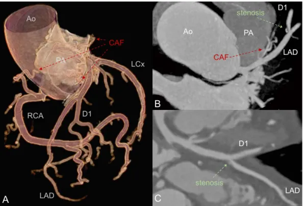
calcified plaque with high-risk features (low attenuation, positive remodeling, and napkin-ring sign) causing severe (70%) luminal stenosis in the mid segment of the left ante- rior descending (LAD) coronary artery. Additionally, several small tortuous branches were visualized connecting the sep- tal branch of the LAD with the pulmonary artery (Figure 1).
The patient underwent invasive coronary angiography (ICA), which confirmed a coronary fistula between the LAD-septal branch and the pulmonary trunk (Figure 2).
Fractional flow reserve (FFR) measurement was per- formed, which verified the significant stenosis in the mid LAD segment. Accordingly, percutaneous coronary inter- vention (PCI) was performed with a drug-eluting stent.
Post-PCI FFR value was normal; however, after nitro- glycerine administration, a significant decrease in the FFR DOI: 10.1515/jce-2017-0008
90 Journal of Cardiovascular Emergencies 2017;3(2):89-92
value was observed, presumably associated with the dila- tation of the fistula. Therefore, coil embolization of the fistula was performed (Figure 3). The FFR value normal- ized, and the patient became asymptomatic.
Twelve months later, due to the reoccurrence of his symptoms, the patient underwent control ICA, which ex-
cluded any significant restenosis in the mid LAD stent.
However, the complete reopening of the coronary fistula was detected. A second attempt for the coil embolization was made; however, due to embolization of the coil during deployment in the LAD-septal branch, it partially shifted into the LAD. Consequently, a coronary artery stent was FIGURE 1. Coronary CT angiography. A – 3D volume rendered image of the coronary tree. Red arrows
indicate the coronary artery fistula connecting the first septal branch of the LAD to the pulmonary trunk.
B, C – Coronary CTA images with multiplanar reconstruction. On Panel B small branches, susceptible for coronary fistula can be seen (red arrow). Panel C illustrates a high-grade stenotic, partially calcified plaque in the mid LAD segment (green arrow).
FIGURE 2. Invasive coronary angiography and percutaneous coronary intervention. A shows the coronary artery fistula (red arrow) originating from the first septal branch of the LAD. B illustrates the significant stenosis in the LAD-diagonal branch bifurcation (green arrow). C – Restored lumen of the LAD after stent implantation.
91 Journal of Cardiovascular Emergencies 2017;3(2):89-92
placed in the LAD to fix the coil position and prevent the occlusion of the LAD lumen. The stent deposition was successful, and no refill of the CAF was observed. Opti- cal coherence tomography (OCT) imaging was performed, which confirmed the good positioning of the LAD stent (Figure 4). The patient was discharged symptomless after the procedure.
The patient agreed to the publication of his data, and the institution where the patient had been admitted ap- proved the publication of the case.
DISCUSSION
Coronary artery fistulas (CAF) are direct precapillary communications between the coronary arteries, cardiac
chambers, or other vessels.1–4 CAFs can originate from the left and right coronary system, and the most common termination sites are the right ventricle, right atrium, and the pulmonary artery.2,3 CAFs usually have small calibers and do not have any hemodynamic impact.2,3 Rarely, large fistulas may cause symptoms and serious conditions for the patient.3 Invasive coronary angiography is the method of choice to identify coronary fistulas. However, due to the increasing number of coronary CT angiography used on patients with chest pain, CAFs and other coronary anomalies are more frequently diagnosed.2 Spontane- ous closure of these fistulas may happen, but surgical or transcatheter closure of the CAF may also be indicated in symptomatic patients with large, hemodynamically sig- nificant fistulas.4,5
FIGURE 3. Coil embolization of the coronary fistula. A – Microcatheter placement in the septal branch of the LAD; B – Coil placement in the coronary fistula; C – No flow is present in the coronary-pulmonary fistula after successful coil embolization.
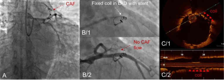
FIGURE 4. Second coronary angiography due to reoccurrence of patients’ symptoms. A – Reopened coronary fistula (red arrow). B/1 – Coil stabilization with a coronary stent in the LAD; B/2 – Complete coronary flow in the LAD with no refill of the fistula after coil embolization; C/1 –Cross-sectional OCT image of the coil in the LAD (red arrows); C/2 –Longitudinal OCT image of the LAD (red arrows indicate the coil).
92 Journal of Cardiovascular Emergencies 2017;3(2):89-92
CONCLUSION
Our case underlines the potential of noninvasive coronary CTA to detect not only significant coronary luminal steno- sis, but also extracoronary findings, such as fistulas as an underlying cause for patients’ chest pain.
CONFLICT OF INTEREST
None declared.ABBREVIATIONS
CAF coronary artery fistula
CTA computed tomography angiography FFR fractional flow reserve
ICA invasive coronary angiography
LAD left anterior descending coronary artery LCx left circumflex coronary artery
OCT optical coherence tomography
PCI percutaneous coronary intervention RCA right coronary artery
REFERENCES
1. Maurovich-Horvat P, Ferencik M, Voros S, Merkely B, Hoffmann U. Comprehensive plaque assessment by coronary CT angiography. Nat Rev Cardiol. 2014;11:390-402. doi:
10.1038/nrcardio.2014.60.
2. Zenooz NA, Habibi R, Mammen L, Finn JP, Gilkeson RC.
Coronary Artery Fistulas: CT Findings. RadioGraphics.
2009;29:781-789. https://doi.org/10.1148/rg.293085120.
3. Said SAM, Thiadens AAHJ, Fieren MJCH, Meijboom EJ,van der Werf T, Bennink GBWE. Coronary artery fistulas. Neth Heart J. 2002;10:65-78.
4. Natarajan A, Khokhar AA, Kirk P, Patel HH, Turner D.
Coronary-pulmonary artery fistula: value of 64-MDCT imaging. Q J Med. 2013;106:91-92. doi: 10.1093/qjmed/hcr254.
5. Armsby LR, Keane JF, Sherwood MC, Forbess JM, Perry SB, Lock JE. Management of coronary artery fistulae. Patient selection and results of transcatheter closure. J Am Coll Cardiol. 2002;39:1026-1032.