Research Article
Association between VEGF Gene Polymorphisms and
In-Stent Restenosis after Coronary Intervention Treated with Bare Metal Stent
Zsolt Bagyura,
1Loretta Kiss,
1Kristóf Hirschberg,
1,2Balázs Berta,
1Gábor Széplaki,
1Árpád Lux,
1Zsolt Szelid,
1Pál Soós,
1and Béla Merkely
11Heart and Vascular Center, Semmelweis University, V´arosmajor Utca 68, Budapest 1122, Hungary
2Department of Cardiology, University Hospital Heidelberg, Im Neuenheimer Feld 410, 69120 Heidelberg, Germany
Correspondence should be addressed to Zsolt Bagyura; bagyura@gmail.com
Received 28 November 2016; Revised 19 February 2017; Accepted 23 February 2017; Published 7 March 2017 Academic Editor: Giuseppe Murdaca
Copyright © 2017 Zsolt Bagyura et al. This is an open access article distributed under the Creative Commons Attribution License, which permits unrestricted use, distribution, and reproduction in any medium, provided the original work is properly cited.
Background. In-stent restenosis (ISR) is the gradual narrowing of the vessel lumen after coronary stent implantation due to the increase in vascular smooth muscle cell proliferation. Vascular endothelial growth factor (VEGF) protein plays an important role in this process. Our aim was to analyze the association of single nucleotide polymorphisms of the VEGF gene (rs2010963 and rs6999447) with the occurrence of ISR after coronary artery bare metal stent (BMS) implantation.Methods. 205 patients with a history of BMS implantation and a repeated coronarography were prospectively enrolled. Patients were assigned to diffuse restenosis group (𝑛 = 105) and control group (𝑛 = 100) and VEGF genotypes were determined.Results. Diffuse ISR was significantly more frequently observed in patients with homozygous normal genotype of rs2010963 polymorphism, and this polymorphism was independently associated with diffuse ISR.Conclusions. RS2010963 is associated with higher incidence of development of diffuse coronary ISR in patients treated with BMS implantation.
1. Introduction
In the era of balloon angioplasty without stent implantation in-stent restenosis (ISR) occurred in 40–50% [1]. Coronary stents were developed to lower the rate of early restenosis.
However, ISR still occurs in 10–30% of the interventions with deployment of bare metal stents (BMS) and therefore still forms a clinically important problem [2]. Although new generation drug eluting stents (DES) further reduced ISR rate and need for repeat revascularization, BMS are still widely used. Moreover, recently Bønaa et al. found no significant difference between receivers of BMS and DES in the composite outcome of death from any cause or nonfatal spontaneous myocardial infarction [3].
After coronary stent implantation mechanical injuries of vessel wall result in vascular smooth muscle cell (VSMC) activation and proliferation and a phenotype change from contractile to proliferative and secretory phenotype [4, 5].
Histological analyses have revealed that vascular smooth
muscle cells (VSMCs) represent the majority of neointima cells [6]. The neointima proliferation leads to narrowing of the coronary lumen [7].
Risk for restenosis is particularly high among patients with diabetes mellitus; this may be associated with metabolic alterations that promote endothelial dysfunction, accelerate intimal hyperplasia, and increase platelet aggregability and thrombogenicity [8]. There is evidence that gender itself (female) predisposes to restenosis [9] and some patients may have genetically higher risk [10, 11]. Genetic polymorphisms associated with high risk for restenosis include polymor- phisms in genes coding for angiotensin II receptor type 1 [12], CD18 [13], interleukin-1 receptor antagonist [14], glycopro- tein receptor IIIa [15], and mannose-binding lectin [16].
On invasive coronarography in-stent restenosis can be classified according to Mehran’s classification to focal (Mehran I) and diffuse (Mehran II–IV) groups [17]. Former type is determined by local and procedural factors, while the latter shows significant relation with general, patient-related
Volume 2017, Article ID 9548612, 7 pages https://doi.org/10.1155/2017/9548612
factors [18] such as diabetes or growth factors. Numerous studies of restenosis have indicated that transforming growth factor beta 1 (TGF-𝛽1), platelet-derived growth factor beta (PDGFB), epidermal growth factor (EGF) [19], basic fibrob- last growth factor (bFGF), and vascular endothelial growth factor (VEGF) play an important role in VSMC proliferation and migration to the tunica intima [20]. The rs699947 and rs2010963 polymorphisms are located in the 5UTR region of the VEGF gene affecting the transcription and expression of the protein. The rs699947 polymorphism of the VEGF gene is associated with a higher risk of developing certain neoplastic diseases [21] and with the development of coronary collaterals in patients with coronary artery disease [22] or with susceptibility to coronary heart disease [23].
According to these findings the involvement of the VEGF in pathological processes leading to restenosis is known, but only one study has investigated the role of rs699947 gene polymorphisms in relation to restenosis [24], whereas the roles of functional polymorphism rs2010963 in restenosis have not yet been studied. The aim of this study was to analyze the association of VEGF’s polymorphisms and the development of coronary in-stent restenosis after bare metal stent implantation.
2. Methods
2.1. Subjects, Interventions. 205 patients with a history of percutaneous coronary intervention (PCI) and BMS implan- tation, who presented with nonacute or acute cardiac symp- toms which warranted a repeat coronary angiogram, were prospectively enrolled between 2011 and 2013 in the Heart and Vascular Center, Semmelweis University. All patients received standard therapy according to the actual guidelines.
ISR has been evaluated by experienced clinicians according to Mehran’s classification and patients have been categorized to the diffuse restenosis group and control group (without diffuse restenosis).
2.2. Biological Samples and Genotyping. Genomic DNA was extracted from whole peripheral blood with a protease based technique (Flexigene DNA System, Qiagen, Hilden, Germany). Samples (1 mL) were added to a lysis buffer and were thoroughly mixed and centrifuged. After discarding the supernatant, samples were denaturized, and DNA was ethanol precipitated and reconstituted in the provided buffer.
Samples were stored at−80∘C. Estimation of the DNA yield and quality control was done by spectrophotometry and determination of the 260/280 absorption ratio (Nanodrop- 2000, Thermo Scientific, Wilmington, USA), and the average yield was 96.0𝜇g (range 25–370𝜇g).
Determination of the alleles of the VEGF gene C−2578A (rs699947) was performed with quantitative real-time PCR (RT-qPCR) (StepOne Plus, Applied Biosystems). Predesigned primers were provided by Applied Biosystems (kit number:
c___8311602_10). Reactions were performed according to the manufacturer’s protocol and for each run parallel samples with positive controls were used.
Genotyping of G+405C (rs2010963) was performed by RT-qPCR and melting curve analysis using LightCycler
(Roche GmbH, Penzberg, Germany). The following primers were used: 5-CCAGAAACCTGAAATGAAGG-3in the for- ward direction and 5-GGGCTCGGTGATTTAGC-3in the reverse direction, the probe sequences were 5-LC640-TGG AAT TGG ATT CGC CAT TTT ATT TTT CTT gC-3and 5- GAC CCA GCA CGG TCC CTC-FL. The PCR mix contained 1𝜇L of the genomic DNA, 5𝜇M of primers and probes, 1𝜇L of LightCycler FastStart DNA Master HybProbe kit (Roche), and 2.5 mM MgCl2. The initial 10 min denaturation at 95∘C was followed by 35 cycles, denaturation (95∘C; 10 s), annealing (52–56–60∘C; 15 s), and extension (72∘C; 10 s), on LightCycler.
Melting curve analysis was performed following the PCR and theTmof the products was determined. The melting points (Tm) were 59∘C for C and 67∘C for G alleles.
All assays were performed in 96-well arrays, and each plate contained controls. Genotyping of 10% of the samples was performed for quality control, with complete congru- ence. Genetic analysis was performed blinded to patient data, with the provided software. We established the genotypes for all patients. The genotype frequencies of two SNPs were all in agreement with the Hardy–Weinberg equilibrium test (𝑝 > 0.05).
2.3. Statistical Analyses. Data were collected in Microsoft Excel 2003 and were analyzed with SPSS 13.0 for Windows (SPSS, Chicago, USA) software. Data are presented as mean
± SD for continuous variables, or 𝑛 (%) for categorical variables. Comparisons between two groups were performed using Student’st-test for continuous variables, whereas for continuous nonparametric variables Mann–WhitneyUtests were performed. Categorical values were compared by using the chi-square test. Multivariate logistic regression has been performed with adjustment for clinical variables that reached a𝑝value of<0.3 when comparing patients with and without significant diffuse ISR. The genotype frequency was tested for deviation from Hardy–Weinberg equilibrium by using Pearson’s chi-square test. All analyses were performed two- tailed, and𝑝 < 0.05was considered as significant.
3. Results
3.1. Patient Characteristics. The total number of 205 patients was involved in our study and categorized into diffuse restenosis and control group. The diffuse restenosis group (𝑛 = 105) included patients with significant diffuse ISR (Mehran II–IV) at recoronarography (73.3% men). Control group (𝑛 = 100) included patients with no or only focal restenosis (Mehran I) at recoronarography in the target bare metal stent (74.0% men).
Clinical baseline characteristics of the 205 patients are described in Table 1. The mean age (control: 66.5 (±10.2) years versus restenosis: 65.7±9.8 years;𝑝 = 0.601) and distribution of genders (control: 74% male versus restenosis: 73.3% male;
𝑝 = 0.914) did not differ significantly. About one-third of patients had diabetes mellitus (71 patients, 35%), mostly type 2. Most patients had hypertension and hyperlipidaemia;
only 5.3% were not treated for hypertension and 9.1% were not on lipid-lowering therapy and had normal lipid levels.
Table 1: Patient characteristics. Continuous variables are presented as mean (±SD). Dichotomous variables are presented as number of patients (percentage).
Variable Control group(𝑛 = 100) Restenosis group(𝑛 = 105) 𝑝value
Age, years 66.5 (±10.2) 65.7 (±9.8) 0.601
Gender, male 74 (74%) 77 (73.3%) 0.914
Antihypertensive therapy 96 (97.0%) 98 (93.3%) 0.229
Lipid-lowering therapy 90 (91.8%) 95 (90.5%) 0.733
Diabetes mellitus 34 (35.1%) 37 (36.6%) 0.816
Obesity, BMI>25 79 (82.3%) 77 (74.8%) 0.197
BMI 28.0 (±3.8) 28.1 (±4.4) 0.817
Smoking 13 (13.0%) 8 (7.6%) 0.204
Peripheral vascular disease 13 (13%) 18 (17.1%) 0.396
Chronic renal failure 7 (7%) 3 (2.9%) 0.123
Maximal CKMB level 114.8 (±102.3) 95.3 (±140.3) 0.322
Acute coronary disease 56 (56.6%) 62 (59.0%) 0.720
Angina pectoris 40 (40%) 37 (35.2%) 0.482
Cardiogenic shock (Killip IV) 2 (2%) 4 (3.8%) 0.527
Multi-branch stented 8 (8.0%) 14 (13.3%) 0.004
Total number of stents 1.42 (±0.75) 1.65 (±0.97) 0.056
Total stent length in mm 30.7 (20.7) 38.9 (25.5) 0.119
Stent diameter in mm 3.1 (0.4) 3.2 (2.6) 0.614
Ejection fraction 52.5 (11.2) 52.8 (10) 0.864
Average time to repeated angiogram (years) 2.65 (±2.72) 1.12 (±1.43) <0.001
Obesity (body mass index (BMI)>25) was present in 76.1% of patients. 21 (10.2%) cases had history of stroke or TIA and 31 (15.1%) cases had peripheral artery disease in the anamnesis;
22 (10.5%) of the patients were current smokers at the time of the second angiography. 10 patients had chronic renal failure and 6 patients had cardiogenic shock. The presence of all above listed factors except chronic renal failure did not differ significantly in the groups. The average time to repeated angiogram was 2.6±2.7 years in the control group and 1.1 ± 1.4 years in the restenosis group (𝑝 < 0.001).
According to this, in-stent restenosis occurred and caused symptoms earlier while controls had a longer asymptomatic period before recoronarography was performed.
3.2. Interventions. There was no significant difference in the indication of stent implantation between the two groups:
acute coronary disease in 56.6% of the control group and 59.0% in the restenosis group. Total number of deployed stents was not significantly higher in restenosis group: 1.42 (0.75) versus 1.65 (0.97) (𝑝 = 0.056). Total stent length and stent diameter were not significantly different between the two groups (Table 1). One-tenth of the patients had multiple branches stented for the first intervention, 8 (8%) in the con- trol group and 14 (13.3%) in the restenosis group (𝑝 = 0.004).
3.3. Genetic Analysis Results. Allele frequencies were similar between genders (rs2010963: 50.1%/41.7%/9.3% in men and 40.7%/50%/9.3% in women, (G/G, G/C, C/C) 𝑝 = 0.454; rs699947: 25.5%/48.3%/26.2% in men and 15.4%/48.1%/36.5% in women, (A/A, A/C, C/C)𝑝 = 0.209).
The genotype distribution conformed to Hardy–Weinberg
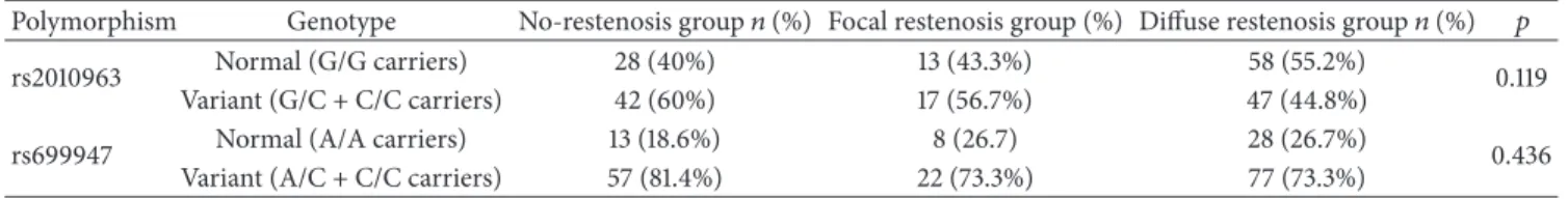
equilibrium (𝑝 = 0.45and𝑝 = 0.65, resp.). Genotype dis- tributions for the groups of patient with no restenosis, focal restenosis, and diffuse restenosis are shown in Table 2. There was no statistically significant difference among these groups.
For further analysis, we merged the focal restenosis group with the no-restenosis group as the focal in-stent restenosis is suspected to have different underlying pathomechanism compared to diffuse in-stent restenosis. Comparison of the genotype distribution of this control group to the diffuse in- stent restenosis group showed that diffuse in-stent restenosis was significantly less frequent in C/G and C/C genotypes (variant carrier) of rs2010963 polymorphism versus individ- uals with the G/G (homozygous normal) genotype (OR 0.56, 𝑝 = 0.04). Restenosis frequency did not differ between the two groups for rs699947 polymorphism (Table 3).
3.4. Multivariate Analysis. Multivariate analysis adjusted for clinical variables (BMI, hypertension, smoking, chronic renal failure, average time to repeated angiogram, multiple branch stent deployment, total stent length, and total number of implanted stents) revealed that the homozygous normal (A/A) genotype of rs2010963 is related to higher risk of diffuse ISR. The rs699947 polymorphisms of VEGF gene are not associated with a risk of diffuse ISR (Table 4).
4. Discussion
In our nonrandomized prospective study, we found a signif- icant association of rs2010963 VEGF polymorphism and the development of diffuse in-stent restenosis after coronary BMS implantation. This association was independent of certain
Table 2: Genotype distribution of no-restenosis group and focal and diffuse restenosis groups; G/G versus G/C + C/C and A/A versus A/C + C/C, chi-square.
Polymorphism Genotype No-restenosis group𝑛(%) Focal restenosis group (%) Diffuse restenosis group𝑛(%) 𝑝
rs2010963 Normal (G/G carriers) 28 (40%) 13 (43.3%) 58 (55.2%)
0.119
Variant (G/C + C/C carriers) 42 (60%) 17 (56.7%) 47 (44.8%)
rs699947 Normal (A/A carriers) 13 (18.6%) 8 (26.7) 28 (26.7%)
0.436
Variant (A/C + C/C carriers) 57 (81.4%) 22 (73.3%) 77 (73.3%)
Table 3: Genotype distribution of control group (no-restenosis + focal restenosis) versus diffuse restenosis group; G/G versus G/C + C/C and A/A versus A/C + C/C, chi-square.
Polymorphism Genotype Control group𝑛(%) Diffuse restenosis group𝑛(%) 𝑝
rs2010963 Normal (G/G carriers) 41 (41%) 58 (55.2%)
0.041
Variant (G/C + C/C carriers) 59 (59%) 47 (44.8%)
rs699947 Normal (A/A carriers) 21 (21%) 28 (26.7%) 0.342
Variant (A/C + C/C carriers) 79 (79%) 77 (73.7%)
Table 4: Results of the multivariate logistic regression analyses with adjustment for risk factors and VEGF rs2010963 variant genotype (G/C + C/C) with in-stent restenosis as a dependent variable. Hosmer–Lemeshow test𝑝 = 0.139.
Risk factor OR CI 𝑝
Lower Upper
Hypertension or antihypertensive therapy 0.45 0.043 4.692 0.503
BMI 1.05 0.906 1.235 0.478
Smoking 0.493 0.088 2.757 0.42
Chronic renal failure 0.754 0.034 16.807 0.859
Multi-branch stented 0.982 0.217 14.368 0.596
Total number of stents 1.441 0.421 4.939 0.561
Average time to repeated angiogram 0.226 0.756 0.413 0.004
Total stent length 1.002 0.958 1.049 0.992
VEGF rs2010963 0.754 0.034 0.535 0.003
clinical factors. VEGF is expressed by vascular endothelial cells, as well as additional cell types [25]. Association has been identified between VEGF polymorphisms and the risk of coronary artery disease [26], the development of collat- eral circulation in individuals with coronary artery disease [22]. VEGF contributes to mediating neovascularization of atherosclerosis plaques [27] and has been found to be asso- ciated with intimal thickening and thrombus development [28] and higher VEGF levels after PCI are related to restenosis after DES implantation [29].
Following PCI, a process similar to wound healing takes place in the affected coronary artery. VEGF has an important role directly in the progress of that endothelialization process and also it has an indirect effect on the inflammation cascade and VSMC proliferation and migration [30]. Accordingly, the endothelial stimulating effect of VEGF is essential for restor- ing the integrity of the vessel wall but it can also become the cause of restenosis through neointima hyperplasia [31]. Slight modifications of VEGF production or function due to gene polymorphisms can have remarkable consequences. Both rs699947 and rs2010963 are located in the promoter region (5UTR) and known to influence the transcription of VEGF gene resulting in a lower serum level of the protein [32].
Rs2010963 (+405C>G) is probably a functional polymor- phism, as serum VEGF levels in subjects with GG genotype have been found to be higher than in other genotypes [32].
Rs699947 has been previously associated with coronary artery disease [23]. It is noteworthy that Osadnik et al. [24]
investigated the role of this polymorphism in development of angiographically significant ISR, but, in their study, focal and diffuse ISR was not distinguished. However, we compared patients with diffuse ISR to patients with no or focal ISR because diffuse ISR has different underlying mechanism compared to focal one, as the former is related to general fac- tors, such as genotype, and the latter is rather related to pro- cedural factors. Although rs699947 has been previously asso- ciated with coronary artery disease, our findings are in con- cordance with Osadnik et al.’s [24] observation as we found no significant association between diffuse ISR and rs699947.
Polymorphism rs2010963 was demonstrated to be asso- ciated with several disorders, such as diabetic retinopa- thy [33], diabetic nephropathy [34], metabolic syndrome [35], myocardial infarction [36], and impaired prognosis in patients with chronic heart failure [37]. VEGF rs2010963 SNPs have been associated with development of collateral circulation [22] and CAD [38]. In contrast, no association has
been observed with HELLP syndrome (Hemolysis Elevated Liver enzymes Low Platelet count) according to Nagy et al.
[39], although rs699947 genotype carriers had an increased risk of HELLP syndrome. Merlo et al. found significant association between VEGF levels and rs2010963 polymor- phism but observed no association with carotid intima- media thickness [40]. Moreover, in a recent study [23] this SNP was not found to be associated with coronary artery disease. We found a significant association of rs2010963 VEGF polymorphism and the development of diffuse in- stent restenosis in our study population. The association remained present after adjusting for certain clinical factors in multivariate analysis. Particularly, we found that the association is independent of the presence of hypertension.
As an influence of the polymorphism on hypertension has been described before [41], it is relevant that our observation implies an effect of the polymorphism on ISR without regard to hypertension. It is notable that only the rs2010963 showed association with diffuse in-stent restenosis even if both SNPs are known to affect the transcription and expression of VEGF. The explanation for that could be due to the fact that in a highly polymorphic gene such as VEGF no single SNP is responsible for the VEGF production, but, rather, the influence of multiple SNPs (i.e., haplotypes) is more likely. Additional SNPs residing on the same haplotypes (i.e., in linkage disequilibrium) could represent the causal or functional variants. The genetic variations of the gene need further study in order to refine the “at-risk” haplotype, which can then be used in a larger prospective study of patients [42].
5. Conclusions
In our nonrandomized prospective study, we found a sig- nificant association of rs2010963 VEGF polymorphism and the development of diffuse in-stent restenosis after coronary BMS implantation. This association was independent of certain clinical factors. We found no significant association between diffuse ISR and rs699947. Preventing restenosis is an important issue of coronary interventions that should start even before the procedure. Early recognition of the diseases and quick reaction usually mean that we face a less complicated lesion, therefore a lower restenosis rate. But there are some factors associated with ISR that we cannot influence, such as genetic polymorphisms. With the rapid development of genotyping technologies, examination of several polymor- phisms may be accessible as routine diagnostics and therefore personalized risk assessment could be performed enabling better selection of patients for primary DES or bioabsorbable stent implantation.
Abbreviations
BMI: Body mass index BMS: Bare metal stent CAD: Coronary artery disease CI: Confidence interval
CKMB: Creatine kinase MB isoform DES: Drug eluting stent
DNA: Deoxyribonucleic acid
ISR: In-stent restenosis OR: Odds ratio
PCI: Percutaneous coronary intervention SD: Standard deviation
SNP: Single nucleotide polymorphism VEGF: Vascular endothelial growth factor.
Ethical Approval
All procedures followed were in accordance with the ethical standards of the responsible committee on human experi- mentation (Institutional and Scientific and Ethics Committee (TUKEB)) and with the Helsinki Declaration of 1975, as revised in 2000 (5).
Consent
Written informed consent was obtained from all patients for being included in the study.
Conflicts of Interest
All authors declare that they have no conflicts of interest.
Authors’ Contributions
Zsolt Bagyura, Loretta Kiss, Krist´of Hirschberg, and ´Arp´ad Lux carried out the molecular genetic studies and drafted the manuscript. Bal´azs Berta, Krist´of Hirschberg, Zsolt Szelid, and B´ela Merkely participated in the design of the study. Zsolt Bagyura, Loretta Kiss, and G´abor Sz´eplaki performed the statistical analysis. B´ela Merkely, Krist´of Hirschberg, and P´al So´os conceived of the study and participated in its design and coordination and helped to draft the manuscript. All authors read and approved the final manuscript.
Acknowledgments
This study was supported by the Hungarian Scientific Research Fund, OTKA K-105555.
References
[1] J. J. Ferguson and J. T. Willerson, “Lipids, atherosclerosis, and restenosis after percutaneous transluminal coronary angio- plasty,”Texas Heart Institute Journal, vol. 19, no. 1, pp. 54–61, 1992.
[2] A. K. Mitra and D. K. Agrawal, “In stent restenosis: bane of the stent era,”Journal of Clinical Pathology, vol. 59, no. 3, pp. 232–
239, 2006.
[3] K. H. Bønaa, J. Mannsverk, R. Wiseth et al., “Drug-eluting or bare-metal stents for coronary artery disease,”New England Journal of Medicine, vol. 375, no. 13, pp. 1242–1252, 2016.
[4] S. F. Louis and P. Zahradka, “Vascular smooth muscle cell motility: from migration to invasion,”Experimental and Clinical Cardiology, vol. 15, no. 4, pp. e75–e85, 2010.
[5] M. Simons, “VEGF and restenosis: the rest of the story,”
Arteriosclerosis, Thrombosis, and Vascular Biology, vol. 29, no.
4, pp. 439–440, 2009.
[6] B. P. Herring, A. M. Hoggatt, C. Burlak, and S. Offermanns,
“Previously differentiated medial vascular smooth muscle cells contribute to neointima formation following vascular injury,”
Vascular Cell, vol. 6, no. 1, article 21, 2014.
[7] R. Komatsu, M. Ueda, T. Naruko, A. Kojima, and A. E. Becker,
“Neointimal tissue response at sites of coronary stenting in humans: macroscopic, histological, and immunohistochemical analyses,”Circulation, vol. 98, no. 3, pp. 224–233, 1998.
[8] J. M. Ahmed, M. K. Hong, R. Mehran et al., “Influence of diabetes mellitus on early and late clinical outcomes in saphenous vein graft stenting,”Journal of the American College of Cardiology, vol. 36, no. 4, pp. 1186–1193, 2000.
[9] D. Trabattoni, F. Fabbiocchi, P. Montorsi et al., “Angiographic patterns of in-stent restenosis in men and women,”Italian Heart Journal, vol. 6, no. 2, pp. 138–142, 2005.
[10] X. Dai, S. Wiernek, J. P. Evans, and M. S. Runge, “Genetics of coronary artery disease and myocardial infarction,”World Journal of Cardiology, vol. 8, no. 1, pp. 1–23, 2016.
[11] Z. Szelid, ´A. Lux, M. Kolossv´ary et al., “Right ventricular adaptation is associated with the Glu298Asp variant of the NOS3 gene in elite athletes,”PLoS ONE, vol. 10, no. 10, Article ID e0141680, 2015.
[12] J. J. W. Verschuren, S. Trompet, I. Postmus et al., “Systematic testing of literature reported genetic variation associated with coronary restenosis: Results of The GENDER Study,” PLOS ONE, vol. 7, no. 8, Article ID e42401, 2012.
[13] W. Koch, C. B¨ottiger, J. Mehilli et al., “Association of a CD18 gene polymorphism with a reduced risk of restenosis after coronary stenting,”American Journal of Cardiology, vol. 88, no. 10, pp.
1120–1124, 2001.
[14] A. Kastrati, W. Koch, P. B. Berger et al., “Protective role against restenosis from an interleukin-1 receptor antagonist gene polymorphism in patients treated with coronary stenting,”
Journal of the American College of Cardiology, vol. 36, no. 7, pp.
2168–2173, 2000.
[15] B. Keavney, “Outcome following percutaneous coronary inter- vention: not, so far, in our genes,”Heart, vol. 89, no. 3, pp. 247–
248, 2003.
[16] Z. Bagyura, L. Kiss, B. Berta et al., “High rate of in-stent restenosis after coronary intervention in carriers of the mutant mannose-binding lectin allele,”BMC Cardiovascular Disorders, vol. 17, no. 1, p. 4, 2017.
[17] R. Mehran, G. Dangas, A. S. Abizaid et al., “Angiographic patterns of in-stent restenosis: classification and implications for long-term outcome,”Circulation, vol. 100, no. 18, pp. 1872–
1878, 1999.
[18] R. Hoffmann and G. S. Mintz, “Coronary in-stent restenosis—predictors, treatment and prevention,” European Heart Journal, vol. 21, no. 21, pp. 1739–1749, 2000.
[19] E. Sanchez-Guerrero, S. R. Jo, B. H. Chong, and L. M. Khachi- gian, “EGFR and the complexity of receptor crosstalk in the cardiovascular system,”Current Molecular Medicine, vol. 13, no.
1, pp. 3–12, 2013.
[20] S. M. Schwartz, “Perspectives series: cell adhesion in vascu- lar biology. Smooth muscle migration in atherosclerosis and restenosis,”The Journal of Clinical Investigation, vol. 99, no. 12, pp. 2814–2816, 1997.
[21] L. Wang, S. Ji, and Z. Cheng, “Vascular endothelial growth factor−2578C/A polymorphism and colorectal cancer risk: a meta-analysis,”Journal of Research in Medical Sciences, vol. 20, no. 8, pp. 811–817, 2015.
[22] T.-H. Lin, C.-L. Wang, H.-M. Su et al., “Functional vascular endothelial growth factor gene polymorphisms and diabetes:
effect on coronary collaterals in patients with significant coro- nary artery disease,”Clinica Chimica Acta, vol. 411, no. 21-22, pp.
1688–1693, 2010.
[23] L. Li, Y. Pan, L. Dai, B. Liu, and D. Zhang, “Association of genetic polymorphisms on vascular endothelial growth factor and its receptor genes with susceptibility to coronary heart disease,”
Medical Science Monitor, vol. 22, pp. 31–40, 2016.
[24] T. Osadnik, J. K. Strzelczyk, R. Reguła et al., “The relationships between polymorphisms in genes encoding the growth factors TGF-𝛽1, PDGFB, EGF, bFGF and VEGF-A and the restenosis process in patients with stable coronary artery disease treated with bare metal stent,”PLOS ONE, vol. 11, no. 3, Article ID e150500, 2016.
[25] L. F. Brown, M. Detmar, K. Claffey et al., “Vascular permeability factor/vascular endothelial growth factor: a multifunctional angiogenic cytokine,”EXS, vol. 79, pp. 233–269, 1997.
[26] Q. T. Cui, Y. Li, C. H. Duan, W. Zhang, and X. L. Guo, “Further evidence for the contribution of the vascular endothelial growth factor gene in coronary artery disease susceptibility,”Gene, vol.
521, no. 2, pp. 217–221, 2013.
[27] M. Inoue, H. Itoh, M. Ueda et al., “Vascular endothelial growth factor (VEGF) expression in human coronary atherosclerotic lesions: possible pathophysiological significance of VEGF in progression of atherosclerosis,”Circulation, vol. 98, no. 20, pp.
2108–2116, 1998.
[28] S. Bhardwaj, H. Roy, T. Heikura, and S. Yl¨a-Herttuala, “VEGF- A, VEGF-D and VEGF-DΔ𝑁Δ𝐶induced intimal hyperplasia in carotid arteries,”European Journal of Clinical Investigation, vol.
35, no. 11, pp. 669–676, 2005.
[29] K. M. Katsaros, S. P. Kastl, K. A. Krychtiuk et al., “An increase of VEGF plasma levels is associated with restenosis of drug-eluting stents,”EuroIntervention, vol. 10, no. 2, pp. 224–230, 2014.
[30] C. L. Grosskreutz, B. Anand-Apte, C. Dupl´aa et al., “Vascu- lar endothelial growth factor-induced migration of vascular smooth muscle cells in vitro,”Microvascular Research, vol. 58, no. 2, pp. 128–136, 1999.
[31] J. H. Br¨asen, A. Kivel¨a, K. R¨oser et al., “Angiogenesis, vascular endothelial growth factor and platelet-derived growth factor- BB expression, iron deposition, and oxidation-specific epitopes in stented human coronary arteries,”Arteriosclerosis, Thrombo- sis, and Vascular Biology, vol. 21, no. 11, pp. 1720–1726, 2001.
[32] C. J. Watson, N. J. A. Webb, M. J. Bottomley, and P. E.
C. Brenchley, “Identification of polymorphisms within the vascular endothelial growth factor (VEGF) gene: correlation with variation in VEGF protein production,”Cytokine, vol. 12, no. 8, pp. 1232–1235, 2000.
[33] M. Qiu, W. Xiong, H. Liao, and F. Li, “VEGF -634G>C polymorphism and diabetic retinopathy risk: a meta-analysis,”
Gene, vol. 518, no. 2, pp. 310–315, 2013.
[34] L. Sun, Q. Yuan, N. Cao et al., “VEGF genetic polymorphisms may contribute to the risk of diabetic nephropathy in patients with diabetes mellitus: a meta-analysis,”The Scientific World Journal, vol. 2014, Article ID 624573, 11 pages, 2014.
[35] Y. R. Kim and S.-H. Hong, “Association between the poly- morphisms of the vascular endothelial growth factor gene and metabolic syndrome,”Biomedical Reports, vol. 3, no. 3, pp. 319–
326, 2015.
[36] D. Petroviˇc, R. Verhovec, M. Globoˇcnik Petroviˇc, J. Osredkar, and B. Peterlin, “Association of vascular endothelial growth fac- tor gene polymorphism with myocardial infarction in patients
with type 2 diabetes,”Cardiology, vol. 107, no. 4, pp. 291–295, 2007.
[37] P. Van Der Meer, R. A. De Boer, H. L. White et al., “The VEGF +405 CC promoter polymorphism is associated with an impaired prognosis in patients with chronic heart failure: A MERIT-HF Substudy,”Journal of Cardiac Failure, vol. 11, no. 4, pp. 279–284, 2005.
[38] A. Moradzadegan, A. Vaisi-Raygani, A. Nikzamir, and Z.
Rahimi, “Angiotensin converting enzyme insertion/deletion (I/D) (rs4646994) and Vegf polymorphism (+405G/C;
Rs2010963) in type II diabetic patients: association with the risk of coronary artery disease,”JRAAS - Journal of the Renin- Angiotensin-Aldosterone System, vol. 16, no. 3, pp. 672–680, 2015.
[39] B. Nagy, H. Savli, A. Molvarec et al., “Vascular endothelial growth factor (VEGF) polymorphisms in HELLP syndrome patients determined by quantitative real-time PCR and melting curve analyses,”Clinica Chimica Acta, vol. 389, no. 1-2, pp. 126–
131, 2008.
[40] S. Merlo, J. N. Starˇcevi´c, S. Mankoˇc et al., “Vascular endothelial growth factor gene polymorphism (RS2010963) and its receptor, kinase insert domain-containing receptor gene polymorphism (RS2071559), and markers of carotid atherosclerosis in patients with type 2 diabetes mellitus,”Journal of Diabetes Research, vol.
2016, Article ID 1482194, 6 pages, 2016.
[41] M. Iaresko and E. Kolesnikova, “The role of polimorhyms—634 G/C (re 2010963) of VEGF-A gene in the development of hypertensionand obesity in premenopausal women,”Georgian Medical News, vol. 256-257, pp. 33–37, 2016.
[42] W. B. Breunis, M. H. Biezeveld, J. Geissler et al., “Vascular endothelial growth factor gene haplotypes in Kawasaki disease,”
Arthritis and Rheumatism, vol. 54, no. 5, pp. 1588–1594, 2006.
Submit your manuscripts at https://www.hindawi.com
Stem Cells International
Hindawi Publishing Corporation
http://www.hindawi.com Volume 2014
Hindawi Publishing Corporation
http://www.hindawi.com Volume 2014
INFLAMMATION
Hindawi Publishing Corporation
http://www.hindawi.com Volume 2014
Behavioural Neurology
Endocrinology
International Journal ofHindawi Publishing Corporation
http://www.hindawi.com Volume 2014
Hindawi Publishing Corporation
http://www.hindawi.com Volume 2014
Disease Markers
Hindawi Publishing Corporation
http://www.hindawi.com Volume 2014
BioMed
Research International
Oncology
Journal ofHindawi Publishing Corporation
http://www.hindawi.com Volume 2014
Hindawi Publishing Corporation
http://www.hindawi.com Volume 2014
Oxidative Medicine and Cellular Longevity
Hindawi Publishing Corporation
http://www.hindawi.com Volume 2014
PPAR Research The Scientific World Journal
Hindawi Publishing Corporation
http://www.hindawi.com Volume 2014
Immunology Research
Hindawi Publishing Corporation
http://www.hindawi.com Volume 2014
Journal of
Obesity
Journal ofHindawi Publishing Corporation
http://www.hindawi.com Volume 2014
Hindawi Publishing Corporation
http://www.hindawi.com Volume 2014
Computational and Mathematical Methods in Medicine
Ophthalmology
Journal ofHindawi Publishing Corporation
http://www.hindawi.com Volume 2014
Diabetes Research
Journal ofHindawi Publishing Corporation
http://www.hindawi.com Volume 2014
Hindawi Publishing Corporation
http://www.hindawi.com Volume 2014
Research and Treatment
AIDS
Hindawi Publishing Corporation
http://www.hindawi.com Volume 2014
Gastroenterology Research and Practice
Hindawi Publishing Corporation
http://www.hindawi.com Volume 2014
Parkinson’s Disease
Evidence-Based Complementary and Alternative Medicine
Volume 2014 Hindawi Publishing Corporation
http://www.hindawi.com