Studies on Cyclophilin-D and NAADP on Ca
2+-mediated events
PhD thesis
Miklós Mándi, MD
Semmelweis University
Neurosciences (‘János Szentágothai’) PhD School Functional Neurosciences Program
Supervisors: Veronika Ádám, MD, DSc
Official reviewers: Balázs Sümegi, MD, DSc Gábor Bánhegyi, MD, DSc Chairman of committee: Erzsébet Ligeti, MD, DSc Members of committee: Zsolt Liposits, MD, DSc Csaba Sőti, MD, PhD
Budapest, 2011
Table of contents
Table of contents
ABBREVIATIONS...3
1. INTRODUCTION...6
1 . 1 . M i t o c h o n d r i a a s C a2 + s t o r e s ... 7
1 . 1 . 1 . M e c h a n i s m s f o r C a2 + u p t a k e i n t o m i t o c h o n d r i a ... 8
1 . 1 . 2 . C a2 + e f f l u x p a t h w a y s f r o m m i t o c h o n d r i a ... 9
1 . 1 . 3 . C a2 + s t o r a g e i n t h e m i t o c h o n d r i a l m a t r i x ... 11
1 . 2 . M i t o c h o n d r i a l p e r m e a b i l i t y t r a n s i t i o n p o r e ( P T P ) ... 13
1 . 2 . 1 . F u n c t i o n s a n d c o n s e q u e n c e s o f t h e p e r m e a b i l i t y t r a n s i t i o n ... 13
1 . 2 . 2 . S t r u c t u r e o f t h e P T P ... 14
1 . 2 . 3 . M o d u l a t i o n o f m i t o c h o n d r i a l P T ... 15
1 . 2 . 4 . C o n t r i b u t i o n o f C y c l o p h i l i n - D t o P T ... 17
1 . 3 . N A A D P a n d C a2 + m o b i l i z a t i o n ... 20
1 . 3 . 1 . S t r u c t u r e o f N A A D P ... 21
1 . 3 . 2 . C a2 + r e l e a s e a c t i v i t y o f N A A D P i n m a m m a l i a n c e l l s y s t e m s ... 21
1 . 3 . 3 . T h e N A A D P r e c e p t o r ... 22
1 . 3 . 4 . T h e N A A D P / C a2 + s i g n a l l i n g p a t h w a y ... 24
2. AIMS AND OBJECTIVES...29
3. MATERIALS AND METHODS ...31
3 . 1 . M i t o c h o n d r i a l a n d m i c r o s o m a l p r e p a r a t i o n s ... 31
3 . 2 . M i t o c h o n d r i a l m e m b r a n e p o t e n t i a l (∆ Ψm) d e t e r m i n a t i o n ... 32
3 . 3 . C a2 + u p t a k e o f i s o l a t e d m i t o c h o n d r i a ... 32
3 . 4 . M e a s u r e m e n t o f m i t o c h o n d r i a l s w e l l i n g ... 33
3 . 5 . M a t r i x C a2 + i m a g i n g o f i s o l a t e d m i t o c h o n d r i a ... 33
3 . 6 . D e t e r m i n a t i o n o f A N T c o n t e n t o f m i t o c h o n d r i a ... 34
3 . 7 . A c t i v e l o a d i n g o f m i c r o s o m e s w i t h C a2 + a n d C a2 + r e l e a s e a s s a y ... 34
3 . 8 . P a s s i v e l o a d i n g o f m i c r o s o m e s a n d C a2 + r e l e a s e ... 34
3 . 9 . R e a g e n t s a n d s t a t i s t i c s ... 35
4. RESULTS AND DISCUSSION...36
4 . 1 . C o m p l e x c o n t r i b u t i o n o f C y c l o p h i l i n - D i n b r a i n - s p e c i f i c m i t o c h o n d r i a l p e r m e a b i l i t y t r a n s i t i o n i n d u c e d b y C a2 +... 36
4 . 1 . 1 . E f f e c t o f s u b s t r a t e a v a i l a b i l i t y o n C a2 +- i n d u c e d P T P a n d m o d u l a t i o n b y c y c l o s p o r i n A o r g e n e t i c d e l e t i o n o f c y c l o p h i l i n - D ... 36
4 . 1 . 2 . E f f e c t o f a n u n c o u p l e r a n d / o r i n h i b i t o r s o f t h e C a2 + u n i p o r t e r o n m i t o c h o n d r i a l C a2 + u p t a k e a n d l i g h t s c a t t e r ... 39
4 . 1 . 3 . E f f e c t o f C a2 + u n i p o r t e r i n h i b i t o r s o n m i t o c h o n d r i a l m a t r i x C a2 + a c c u m u l a t i o n o f i s o l a t e d m i t o c h o n d r i a i m a g e d u n d e r w i d e - f i e l d e p i f l u o r e s c e n c e . 40 4 . 1 . 4 . E f f e c t o f r e s p i r a t o r y c h a i n i n h i b i t i o n o n C a2 +- i n d u c e d P T P ... 42
4 . 1 . 5 E f f e c t o f c y p D a b l a t i o n o n C a2 +- i n d u c e d m i t o c h o n d r i a l s w e l l i n g w i t h i n n e u r o n s a n d a s t r o c y t e s ... 45 4 . 1 . 6 . E f f e c t o f C y p D a b l a t i o n o n i n s i t u m i t o c h o n d r i a l s w e l l i n g o f n e u r o n s
DOI:10.14753/SE.2012.1675
4 . 1 . 7 . D i s c u s s i o n ... 50
4 . 2 . C a2 + r e l e a s e t r i g g e r e d b y N A A D P i n h e p a t o c y t e m i c r o s o m e s ... 52
4 . 2 . 1 . N A A D P i n d u c e s C a2 + r e l e a s e f r o m h e p a t o c y t e m i c r o s o m e s ... 52
4 . 2 . 2 . D o s e - d e p e n d e n c e o f t h e N A A D P - m e d i a t e d C a2 + r e l e a s e ... 54
4 . 2 . 3 . U n i q u e h o m o l o g o u s d e s e n s i t i z a t i o n p a t t e r n o f t h e N A A D P r e c e p t o r s ... 56
4 . 2 . 4 . T h e e f f e c t o f t h a p s i g a r g i n a n d b a f i l o m y c i n A 1 o n t h e N A A D P - e v o k e d C a2 + r e l e a s e i n r a t l i v e r m i c r o s o m e s ... 57
4 . 2 . 5 . C a2 + a n d p H d e p e n d e n c e o f t h e N A A D P - i n d u c e d C a2 + r e l e a s e ... 59
4 . 2 . 6 . P h a r m a c o l o g i c a l p r o p e r t i e s o f t h e N A A D P - e l i c i t e d C a2 + e f f l u x ... 61
4 . 2 . 7 . D i s c u s s i o n ... 62
5. CONCLUSIONS...65
6. SUMMARY...68
7. ÖSSZEFOGLALÁS...69
LIST OF PUBLICATIONS ...70
R e l a t e d t o t h e p r e s e n t t h e s i s ... 70
N o t r e l a t e d t o t h e p r e s e n t t h e s i s ... 71
REFERENCE LIST ...72
ACKNOWLEDGEMENTS...101
Abbreviations
Abbreviations
∆Ψm: membrane potential 2-BO: 2-bromooctanoate ACh: acethylcholine
ADP: adenosine diphosphate
ADPRCs: ADP-ribosyl cyclase enzyme family AIF: apoptosis-inducing factor
ANT: adenine nucleotide translocase
AP5A: P1,P5-di-(adenosine-5′)-pentaphosphate AT-II: angiotensin II
ATP: adenosine triphosphate ATR: atraciloside
BCECF: 2’,7’-bis(carboxyethyl)-5,6-carboxyfluorescein BKA: bongkrekic acid
BSA: bovine serum albumin
cADPR(P): cyclic adenosine dinucleotide-ribose (phosphate) CaGreen: Calcium Green 5N
cATR: carboxyatractyloside CCK: cholecystokinine
CICR: Ca2 +-induced Ca2 + release CK: creatine kinase
CL: cardiolipin CypD: cyclophilin-D Cyt c: cytochrome c CysA: cyclosporine-A DAG: diacyl-glycerol DMSO: dimethyl-sulfoxide DTT: dithiothreitole
EC5 0: 50%-excitation concentration ER: endoplasmic reticulum
DOI:10.14753/SE.2012.1675
EDTA: ethylenediamine-tetraacetic acid
EGTA: ethylene glycol-bis(2-aminoethylether)-N,N,N′,N′-tetraacetic acid
Er e v_ANT: reversal potential of the ANT
Er e v_ATPase: reversal potential of the F0F1ATPase
FCCP: carbonyl cyanide 4-(trifluoromethoxy)-phenylhydrazone FFA: free fatty acid
GPN: glycyl-phenylalanyl-naphtylamide
Hepes: 4-(2-hydroxyethyl)-1-piperazineethanesulfonic acid HK: hexokinase
IC5 0: 50%-inhibition concentration IMM: inner mitochondrial membrane InsP3: D-myo-inositol-1,4,5-trisphosphate
mBR: peripheral mitochondrial benzodiazepine receptor MCF: mitochondrial carrier family
MgG: magnesium green
MOPS: 3-(N-morpholino)-propanesulfonic acid
NAD(P)+: nicotinamide-adenine dinucleotide (phosphate) NAADP: nicotinic acid-adenine dinucleotide phosphate NCE: Na+-Ca2 + exchanger
NICE: Na+-independent Ca2 + exchanger (H+-Ca2 + exchanger) OMM: outer mitochondrial membrane
pHi n: mitochondrial matrix pH pHo: extramitochondrial pH Pi: inorganic phosphate PPi: inorganic pyrophaosphate PiC: phosphate carrier
PLA2: phospholipase A2 pmf: protonmotive force
PMSF: phenylmethylsulfonyl fluoride PN: pyridine nucleotides
PPIase: peptidyl-prolyl cis-trans isomerase
Abbreviations
(m)PTP: (mitochondrial) permeability transition pore RaM: rapid (Ca2 +) uptake mode
ROS: reactive oxygen species RuRed: ruthenium-red
RyR: ryanodine receptor SCN-: thiocyanate
SERCA: sarco(endo)plasmic reticulum Ca2 +-ATPase SF 6847 (Tyrphostin 9, RG-50872, Malonaben):
2-[[3,5-bis(1,1-dimethylethyl)-4-hydroxyphenyl]methylene]- propanedinitrile
Sf-A: Sanglifehrin-A
SR: sarcoplasmic reticulum
TMPD: N,N,N′,N′-tetramethyl-p-phenylenediamine TPCN2: two-pore cation channel, type 2
Tris: tris-(hydroxymethyl)-aminomethane TRP: transient receptor potential channel VDAC: voltage-dependent anion channel WT: wild type
DOI:10.14753/SE.2012.1675
1. Introduction
Calcium is one of the most versatile and important intracellular messengers in living cells and organisms. Ca2 + signalling is involved in the control of numerous biological processes [1;2]. For example, Ca2 + is essential for oocyte fertilization, synaptic plasticity, muscle cell contraction, gene expression, enzyme / neurotransmitter secretion and cell proliferation, as well as cell death (both apoptosis and necrosis).
Ca2 + signals are intracellular relays of triggering events, such as depolarization, hormone or neurotransmitter stimulations, in order to control cell functions [1;2]. Ca2 + signals are not all-or-none events; they vary greatly in amplitude, duration and localization. Cytosolic Ca2 + signals can be generated by activation of Ca2 + entry and/or by mobilization of Ca2 + from intracellular stores. At the level of plasma membrane, Ca2 + influx occurs through several types of channels, such as voltage-gated channels, ligand-gated channels and transient receptor potential (TRP) ion channels [3-5]. Ca2 + is also stored in most organelles, including endoplasmic reticulum (ER), mitochondria, lysosomes, secretory granules, Golgi apparatus and nuclear envelope [6;7]. Thus, compartmentalization of Ca2 + to the extracellular matrix or to intracellular organelles is a crucial element of Ca2 + signalling [8].
Moreover, Ca2 + release from some of these stores can be triggered by intracellular second messengers. Ca2 + fluxes display complex spatial and temporal signatures, enabling more information to be encoded by Ca2 + signals. To meet the demands of this complexity, cells rely on precise regulation of Ca2 + channel activity [1].
The present thesis focuses on the role of cyclophilin-D in Ca2 +- triggered mitochondrial permeability transition and Ca2 + release from the cytosolic acidic Ca2 + stores.
Mitochondria as Ca2+ stores
1.1. Mitochondria as Ca
2 +stores
The importance of mitochondria in cellular Ca2 + homeostasis was questioned for a long period of time when the ER was identified as the main inositol-1,4,5-trisphosphate (InsP3)-dependent intracellular Ca2 + store [9;10]. Subsequently, the general consensus was that mitochondria would form a ‘Ca2 + sink’ [2;11;12], since it was hypothesized that the well-known low affinity but high capacity mitochondrial Ca2 + uptake mechanisms would only be significant under conditions of high- amplitude or prolonged [Ca2 +]c increases, i.e. in the Ca2 + overload.
However, changes of [Ca2 +]c in response to physiologically relevant stimuli were shown to coexist with rapid modulations of the [Ca2 +]m
[13;14], proving that mitochondria indeed play a more complex role in Ca2 + signalling [15]. Moreover, three mitochondrial enzymes participating in key steps of the intermediate metabolism (the pyruvate-, α-ketoglutarate- and isocitrate-dehydrogenases) are also regulated by Ca2 +, a notion that would imply that [Ca2 +]m should be suited to follow the physiological [Ca2 +]c events of the cell [16;17]. Uptake and efflux of Ca2 + in mitochondria occur by different processes (see Figure 1).
3 Na+
Na+ NCE
Ca2+
Ca2+
H+ n H+
NHE
[Ca2+]m
[Ca2+]c
H+
H+
Ca2+ Ca2+
NICE or mHCX
Ca2+uniporter RaM
Ca2+ Ca2+
Ca2+ Ca2+
- - - -
+ + + +
Ca2+ Ca2+
MPTP Ca2+
Ca2+
mRyR1 Ca2+
Ca2+
DAG-sensitive Ca2+channel
?
DOI:10.14753/SE.2012.1675
1.1.1. Mechanisms for Ca2 + uptake into mitochondria
Generally, the mitochondrial inner membrane potential (∆Ψm, that is usually between -150 and -180 mV) is the driving force of the two major mechanisms of Ca2 + entry to the mitochondria identified to date:
the Ca2 + uniporter and the rapid uptake mode (RaM) [18]. The Ca2 + uniporter ‘in vitro’ is a low-affinity and high capacity transporter that is non-competitively blocked by ruthenium compounds (e.g. Ruthenium Red [RuRed] [19;20], Ru360 [21]) or inhibited in a mainly competitive manner by divalent cations - that are themselves transported by the uniporter (e.g. Sr2 +, Mn2 +, Ba2 + and lanthanides), and activated by ATP, physiological concentrations of spermine and taurine [12;21-23]. In permeabilized cells however, the half-maximal activation (K0 . 5) for mitochondrial Ca2 + transport rate, presumably through the uniporter, was found to be 3-fold higher than ‘in vitro’ [24;25]. Hence it is possible that the affinity of the uniporter is higher in a cellular environment (possibly due to the presence of spermine or taurine or other intracellular factors). Moreover, regarding the factors that modulate Ca2 + influx through the uniporter, the most important is the extramitochondrial [Ca2 +]. Likewise, the rate of Ca2 + uptake through the uniporter shows a biphasic dependence on cytosolic Ca2 + in the presence of functional calmodulin [26;27] and physiological [Mg2 +] [28]. Thus, the uniporter may function indeed at physiological pCa. On the other hand, evidence has been presented for the formation of high Ca2 + microdomains [29-31], especially in the ER-mitochondrial junctions, in which the local Ca2 +-load can be enough to activate the Ca2 + uniporter even if its affinity for Ca2 + is as low as ‘in vitro’ [29-31].
In contrast, the other major pathway for mitochondrial Ca2 + uptake, RaM is a high affinity and high initial conductance Ca2 + transporter responding to small, but rapid Ca2 + transients of the cytoplasm [18;32]. RaM is inhibited by RuRed as well as Ca2 + itself, which exerts rapid inactivation of RaM following its activation [33].
Mitochondria as Ca2+ stores
Thus, RaM takes sufficient amounts of Ca2 + for the activation of the Ca2 +-sensitive dehydrogenases into the matrix [18] without causing Ca2 + overloading of mitochondria [34]. The molecular identity of both the Ca2 + uniporter and RaM has not yet been clarified and it is still uncertain whether RaM could be defined as a different ‘mode of transport’ through the uniporter or it may be a molecularly distinct entity [35].
On the other hand, a mitochondrial ryanodine receptor (mRyR1) has been identified in the inner membrane of rat heart mitochondria solely by Beutner et al. that shared pharmaco-kinetic properties with the skeletal type RyR (RyR1) [36-38]. It has been suggested that mRyR1 may play an important role in the excitation-metabolism coupling in rat heart mitochondria, thus behaving as a Ca2 + uptake mechanism [37;38].
1.1.2. Ca2 + efflux pathways from mitochondria
In the mitochondria of vertebrates, two different routes for Ca2 + efflux have been characterized: one is Na+-dependent (Na+/Ca2 + exchanger, mNCX or NCE) that is coupled to Na+/H+ exchange, while the other is Na+-independent (H+/Ca2 + exchanger, mHCX or NICE) [39]. The stoichiometry of NCE is 3Na+:1Ca2 + [23;34;39], while NICE shows a stiochiometry of nH+:1Ca2 +, where n is probably >2 [39], thus both are electrogenic. There are many established blockers of NCE including tetraphenyl phosphonium, trifluroperazine, diltiazem, verapamil, clonazepam, amiloride, and CGP-37157, while there are only few known inhibitors of NICE such as tetraphenyl phosphonium, cyanide and low levels of uncouplers [34]. The dominant Ca2 + efflux mechanism in heart, brain, skeletal muscle, parotid gland, adrenal cortex, brown fat and endothelial mitochondria is NCE [25;34;40], while NICE is mainly expressed on mitochondria in non-excitable tissues e.g. liver, lung, kidney and smooth muscle [23].
A diacylglycerol (DAG)-activated cation channel has also been described as the first second-messenger induced Ca2 + release mechanism
DOI:10.14753/SE.2012.1675
in the inner membrane of different mammalian mitochondria [41]. DAG analogs have been shown to release Ca2 + from loaded mitochondria in a biphasic manner. A rapid and transient initial Ca2 + release is observed, that is not accompanied by mitochondrial swelling and likewise, it is insensitive to permeability transition inhibitors. Following a relatively slow reuptake of Ca2 + into mitochondria, a secondary, progressive release of Ca2 + occurred, that was associated with swelling and attenuated by permeability transition inhibitors [41]. The initial peak of DAGs-induced Ca2 + efflux is abolished by La3 + (1mM) and potentiated by protein kinase C inhibitors. The putative DAG sensitive channel shows receptoric properties, since the 1,2-sn-DAG analogs induce mitochondrial Ca2 + efflux exclusively, while other DAG analogs or substitutes, such as phorbol esters, 1,3-diacylglycerols and 1- or 2- monoacylglycerols are inactive [41]. Moreover, the forementioned channel has been demonstrated to reside in the IMM since Ca2 +-loaded mitoplasts devoid of outer mitochondrial membrane also exhibit DAGs- induced Ca2 + release. Patch clamping of brain mitoplasts revealed that DAGs induced a slightly cation-selective channel activity which is insensitive to bongkrekic acid and abolished by La3 + [41].
Finally, the multiprotein complex of the permeability transition pore (PTP) could represent an alternative Ca2 + efflux pathway from mitochondria. PTP is believed to have both a small and a large conductance state [23]. Opening of the large-conductance PTP, induced by Ca2 + overloading of mitochondria or by other pathophysiological conditions, leads to the collapse of the inner membrane potential (∆Ψm) and the release of proapoptotic factors (e.g. cytochrome c, Smac/DIABLO and apoptosis-inducting factor, AIF) [42;43] and substantial amount of Ca2 + is also liberated. On the other hand, the small-conductance PTP might participate in physiological Ca2 + handling in the form of Ca2 +-induced Ca2 + release [44].
Mitochondria as Ca2+ stores
1.1.3. Ca2 + storage in the mitochondrial matrix
Mithochondria are capable of accumulating vast quantities of Ca2 + (up to 1M) while keeping the free Ca2 + concentration of the matrix in the low micromolar range (~1-5 µ M) [45]. This can be achieved by the formation of calcium- and phosphorus-rich precipitates in the matrix [46;47]. The relationship between the PO43 - concentration and pH shows a dependency on the third-power and the matrix pH is elevated due to pumping of H+ by the electron transfer chain during oxidative phosphorylation [48], enabling mitochondria to maintain the low free Ca2 + concentration in the matrix as several folds more Ca2 + is stored.
The precise composition of the precipitates remains uncertain, however recent studies indicate that they are composed mainly of tribasic calcium phosphate [Ca3(PO4)2] and/or dibasic calcium phosphate (CaHPO4) [47;49;50], as originally proposed nearly 40 years ago [46]. The maximal Ca2 + loading capacity of mitochondria is limited usually by the opening of the PTP and the Ca/P ratio of the precipitates may vary between ~1 (mainly CaHPO4) and ~1.5 (mainly [Ca3(PO4)2]) according to the Ca2 + load of mitochondria [50]. The acidic pH shift in the matrix upon the opening of the PTP and the dissipation of the mitochondrial membrane potential has been generally considered to play a crucial role in the mobilization of Ca2 + from the precipitates.
The author of the present thesis has contributed to the re- evaluation of the role of matrix acidification in uncoupler-induced Ca2 + release from mitochondria [51]. Our workgroup’s interest was raised by the fact that in the presence of abundant Pi, small ∆pH is observed across the inner mitochondrial membrane [52]. We also relied on the fact that the ∆pH remains relatively constant within a range of extramitochondrial pH (pHo) values [53]. We reasoned that, if matrix acidification does indeed underly the dissociation of the Ca2 +–phosphate complex and Ca2 + release by uncouplers, then at acidic pH (pHo 6.8), complete depolarization by combined inhibition of the respiratory chain and of the
DOI:10.14753/SE.2012.1675
reversal of the F0⁄F1-ATPase would produce the same effect. By the same token, at alkaline pH (pHo 7.8), complete depolarization by uncoupling would hinder the dissociation of this complex, and impair the release of sequestered Ca2 +. Furthermore, at alkaline pH (pHo 7.8), complete depolarization by combined inhibition of the respiratory chain and of the reversal of the F0F1-ATPase should not induce release of sequestered Ca2 +. The experimental findings presented by our workgroup (Vajda et al., [51]) do not support the above expectations, implying that matrix acidification by uncouplers cannot be the only explanation for the release of sequestered Ca2 +.
Mitochondrial permeability transition pore (PTP)
1.2. Mitochondrial permeability transition pore (PTP)
1.2.1. Functions and consequences of the permeability transition In normal aerobic conditions, the IMM always remains highly impermeable to water, ions and metabolites in order to maintain the pmf that is driving the ATP synthesis via the F0-F1 ATPase; only selective and controlled permeability via specific carriers and channels is preserved. The pmf is a large electrochemical driving force for protons (~200-220mV) across the IMM comprising of both ∆Ψm (~150-180 mV) and H+ gradient (∆pH), generated by the functional respiratory chain complexes pumping H+ into the intermembrane space [54]. However, in response to ischaemic, oxidative or any other type of stress, the permeability of the IMM may increase dramatically with the formation of a voltage-dependent, non-specific pore known as the mitochondrial permeability transition pore (PTP). In it’s open state, the PTP complex possesses a channel that is ~3nm vide in diameter, thus allowing the diffusion of all molecules with a molecular mass of less than 1500 Da [55;56]. As a consequence of the opening of the PTP megachannel, the
∆Ψm collapses and mitochondria start consuming ATP imported from the cytosol because of the reversal of the ANT and the F0-F1 ATPase.
Moreover, the high protein concentration of the matrix exert a high colloid osmotic pressure whilst non-protein solutes equilibrate with the cystosol, bringing vast quantities of water into the matrix, thus causing matrix swelling (expansion), de-folding of the cristae of the IMM and the rupture of the OMM. With the breaking of the OMM, pro-apoptotic proteins from the intermembrane space (including cytochrome-c, Smac/DIABLO and apoptosis-inducing factor [AIF]) are released [42;43]. While the prolonged permeability transition (PT) is an all-or- none event for an individual mitochondrion, transient PTP openings can be recorded electrophysiologically in the form of conductance
DOI:10.14753/SE.2012.1675
‘flickerings’ that do not lead to swelling and are unrelated to death signals. These physiological PT-transients (also known as small conductance PTP) might encompass matrix volume and pH regulation, redox equilibrium, protein import [57], bidirectional pyridine nucleotide funnelling [58] and a fast Ca2 + release mechanism (see also Section 1.1.2.). The small conductance PTP is most probably regulated by [Ca2 +]m, leading to a dynamic steady-state distribution of mitochondrial population with closed and open pores [57;59;60].
1.2.2. Structure of the PTP
The exact molecular composition of the PTP still remains uncertain and controversial, although numerous candidates have been investigated. Originally, based on biochemical and pharmacological studies, the PTP was proposed to consist of three major components:
pore-forming voltage-dependent anionic channel (VDAC or porin) and ANT (ANT1) in the OMM and IMM respectively, moreover the regulatory cyclophilin-D (CypD) on the matrix side of the IMM [61;62].
The primary structure of monomeric ANT, including altogether 6 transmembrane domains joined by hydrophilic regions grouped in 3 repeat domains [63], is well-suited for pore-forming. The native structure of the ANT homodimer, functioning as adenine nucleotide carrier with a stoichiometry of 1:1 [64;65], is stabilized by 6 tightly bound and non-detachable cardilopin (CL) molecules per dimer at the matrix side [66]. Since the determination of the ATP-ADP steady-state exchange rate mediated by ANT plays a central role in the understanding of how mitochondria can continually provide fresh ATP to the cytosol in different conditions, our workgroup has perfected and published a new method that is an on-line, high acquisition rate and quantitative measurement of changes in Magnesium Green (MgG) fluorescence based on the different affinity of ADP3 - and ATP4 - for Mg2 + [53].
Mitochondrial permeability transition pore (PTP)
However, mitochondrial permeability transition still occurred in ANT1/2 double knock-out experiments [67] and VDAC1,2,3 triple knock-out experiments [68] suggesting that ANT and VDAC are not essential or replaceable in forming the PTP. A new candidate, notably an other member of the MCF/SLC25 family: the mitochondrial phosphate carrier (PiC) has been considered to be part of the PTP [61] and in the revised model, VDAC does not appear as a compulsory element (although in this case cytochrome-c release from the intermembrane space is not sufficiently elucidated).
In addition, hexokinase (HK), creatine kinase (CK), members of the Bcl-2 family and the mitochondrial benzodiazepine receptor (mBR) have also been suggested to play additional regulatory role [56;69].
1.2.3. Modulation of mitochondrial PT
Various matrix and membrane factors modulate the PTP and it’s sensitivity to [Ca2 +]m, the increase of which is the most important promoter of pore- opening (see Table 1). In the case of Ca2 + overload of mitochondria, Ca2 + occupies critical amino acid residues
of the ANT
interacting with the surrounding bound cardilopin (CL) molecules and facilitating the conformational change of ANT from
Table 1. F a c t o r s i n d u c i n g a n d i n h i b i t i n g m i t o c h o n d r i a l p e r m e a b i l i t y t r a n s i t i o n ( m P T ) .
DOI:10.14753/SE.2012.1675
ADP/ATP antiporter c state to unspecific pore-forming, tensed-open uniporter state (c’) [70]. The peptidyl-prolyl cis-trans isomerase (PPIase) activity of the ANT-associated CypD may facilitate the above mentioned conformational changes of the ANT triggered by high [Ca2 +]m. Thus, inhibition of the association of CypD to ANT by cyclosporin-A (CysA) or inhibition of the PPIase activity of the ANT-associated CypD by sanglifehrin-A (Sf-A) hinders the aforementioned conformational changes of the ANT and reduces substantially the Ca2 + sensitivity of the PTP [71;72]; both CysA and Sf-A are established inhibitors of PTP.
Likewise, Mg2 + and other divalent cations (Mn2 +, Sr2 +) prevent pore- opening by competitively inhibiting Ca2 +-binding to ANT [73]. Protons (H+) also compete for the same binding sites and thus, PTP is inhibited by acidification of the matrix pH below 7.0 [74] and optimal matrix pH for pore-opening is 7.4 [75]. Oxidative stress, peroxide and increased ROS production promotes PTP by oxidizing critical residues on the ANT, now believed to be the Cys1 6 0 [76], strengthening the notion that ANT is indeed involved in PTP formation. Moreover, adenine nucleotide depletion ([ATP]↓, [ADP]↓) and high phosphate (Pi), pyrophosphate (PPi) concentrations (arsenate and vanadate as well) in the matrix enhance PTP opening by preventing adenine nucleotide binding to the ANT [56] and accordingly, the specificity and potency of different nucleotides as inhibitors of the PTP match their ability to be translocated by the ANT [77]. Amphipatic anions, such as free fatty acids (FFAs) produced by PLA2 promote pore opening by affecting the lipid structure of the IMM [78] around the ANT, on the other hand amphipathic cations (e.g. sphingozine, trifluoperazine or spermine) favour pore closure [79].
Also, PTP is sensitive to the redox state of the cell: NADH is protective and substrates that increase the reduction of NAD+ like pyruvate, α- ketoglutarate and glutamate are protective as well, while others that decrease the reduction like oxaloacetate or malonate are stimulators of pore opening [80]. Moreover, full-charged and resting membrane potential effectively prevents the pore-opening [81], parallel to the ∆Ψm-
Mitochondrial permeability transition pore (PTP)
dependency of the antiporter state of ANT and a vide variety of pathophysiological effectors alters the threshold voltage at which opening occurs either closer to the resting potential (PTP inducers), or away from the resting potential (PTP inhibitors) [82].
1.2.4. Contribution of Cyclophilin-D to PT
Cyclophilin-D (CypD) is an 18 kDa matrix protein exhibiting peptidyl-prolyl cis-trans isomerase activity (PPIase) that is encoded by the nuclear gene Ppif [83]. CypD appears in both structural models of the PT pore mentioned in Section 1.2.2, being the target for the inhibitory effect of the immunosuppressant cyclosporine-A (CysA) on MPTP. Likewise, the PPIase activity of CypD appears to facilitate the conformational changes of the ANT (or PiC) molecule when forming the non-selective megachannel of PT [71;84] triggered by high loads of Ca2 +. The importance of the Ca2 +- and ROS-dependent PPIase activity of CypD in the initiation of PTP has been further supported by the use of the non-immunosuppresive CypD antagonists Sanglifehrin A (Sf-A) and Debio-025 [71;85;86]. The contribution of CypD to PTP formation was confirmed by experiments in which the Ppif gene encoding CypD was knocked out [87-90]. These mice do not show a severe phenotype:
enchanced anxiety, avoidance behaviour, occurance of adult onset obesity [91] and a defect in platelet activation and predisposition for thrombosis [92]. CypD knockout mice exhibited lower sensitivity to focal cerebral and liver ischaemia, or in myocardial ischaemia/reperfusion and muscular dystrophy models [87-89;93-96].
Mitochondria from Ppif - / - mice were shown to be resistant to Ca2 + and oxidative stress-induced PTP and cell death, behaving identically to mitochondria from WT mice treated with CysA or Sf-A [87-90]. The loss of CypD did not prevent permeability transition, but increased substantially the load of stimuli necessary before pore opening occurred confirming the hypothesis, that MPTP opening involves a conformational
DOI:10.14753/SE.2012.1675
change in a membrane protein that is triggered by calcium and facilitated by CypD, but can occur in the absence of CypD if sufficient stimulus is given [97].
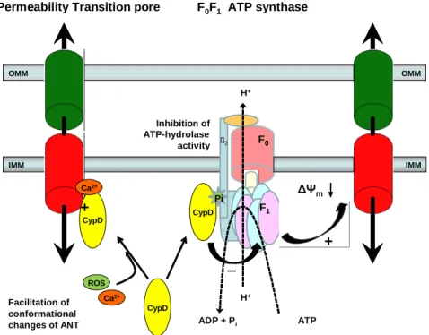
The physiological role of CypD remains unclear. However, apart from the high Ca2 +-induced binding to ANT, CypD has been shown recently to bind to the lateral stalk of the F0F1-ATP synthase as well (Figure 2) in a phosphate-dependent manner [98]; and it has been proposed that ablation of CypD or CysA treatment unmasks an inhibitory Pi binding site, rather than causing direct PTP inhibition [99] (reviewed in [100]). The absence of functional CypD leads to the disinhibition of the F0F1-ATP synthase resulting in accelerated ATP synthesis and hydrolysis rates [98;100]. Also, it has been postulated, that inorganic phosphate (Pi) may in fact be the true inhibitor of PTP acting on a binding site on F1 masked by functional CypD [98-100]. The different factors that favour Pi binding to this site (pmf or preserved membrane potential, high [Pi], high [Mg2 +], acidic pH) are strikingly similar to the inhibiting factors of PTP (see Section 1.2.3 and Table 1). Altogether,
IMM OMM OMM
IMM
Ca2+
ROS
ß2
CypD
ADP + Pi ATP
H+
F0
F1 Pi
Inhibition of ATP-hydrolase activity
Facilitation of conformational changes of ANT
H+
+
F0F1 ATP synthase Permeability Transition pore
CypD
CypD Ca2+
+
Figure 2. Direct and indirect promotion of PTP by cyclophilin-D (CypD).
Pi denotes the activating Pi-binding site
Mitochondrial permeability transition pore (PTP)
the activity of CypD leads to PTP in two converging ways: one is the direct binding of CypD to ANT triggered by Ca2 + overload, facilitating it’s conformational change to the pore-forming tensed open state and secondly, CypD inhibits the ATP hydrolase activity of the reversed ATP synthase by occupying a Pi binding site on the F1 subunit, thus preventing the salvage of membrane potential, favoring PTP indirectly [100] (Figure 2).
Furthermore, the studies with CypD KO mice also converged to the conclusion that CypD mediated MPTP regulates some forms of necrotic, but not apoptotic death, a notion originally suggested by the group of Crompton and colleagues [101]. The complex contribution of CypD to brain-specific mitochondrial permeability transition induced by Ca2 + is demonstrated in [102] and Section 4.1 of the present thesis.
DOI:10.14753/SE.2012.1675
1.3. NAADP and Ca
2 +mobilization
Mitochondrial functions are fundamentally affected by cytosolic signalling events. At the ER-mitochondrial junctions, the formation of high Ca2 + microdomains has been detected [6;14;29-31], which constitutes a tangible link between cytosolic Ca2 + signalization and mitochondrial Ca2 + homeostasis, as described in Section 1.1.1. This section is devoted to one of the major pathways of Ca2 + mobilization from intracellular organelles upstream from the Ca2 +-related modulation of mitochondrial functions (e.g. regulation of matrix enzymes and initialization of PTP).
Understanding of the regulation of intracellular Ca2 + release and its relationship to extracellular stimuli were greatly broadened by the discovery of the inositol-1,4,5-trisphosphate (InsP3) signalling pathway [1;2]. Although InsP3 appears to operate as a ubiquitous intracellular messenger for Ca2 + mobilization, numerous studies indicate that additional Ca2 + release mechanisms operate in many cells and are regulated by a family of pyridine nucleotide metabolites [103-107]. The first indication for this concept came from the pioneering work of Lee and colleagues who showed that β-NAD+ and β-NADP+ could trigger Ca2 + release from sea urchin egg microsomal fractions by a mechanism apparently independent of InsP3 [103]. Subsequently, the structure of the active metabolites has been determined, one of them being cyclic adenosine disphosphate ribose (cADPR) [108]. The Ca2 + mobilizing effect of β-NADP+ was shown to be due to a contaminant of commercially available β-NADP+ identified as nicotinic acid adenine dinucleotide phosphate (NAADP) [109]. Ca2 + release by both β-NAD+ and β-NADP+ appeared to operate via separate Ca2 + release mechanisms which were distinct from those gated by InsP3, since InsP3 showed homologous desensitization without affecting Ca2 + release by activators of the other two mechanism [108].
NAADP and Ca2+ mobilization
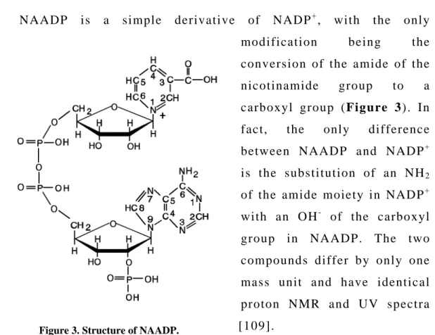
1.3.1. Structure of NAADP
NAADP is a simple derivative of NADP+, with the only modification being the conversion of the amide of the nicotinamide group to a carboxyl group (Figure 3). In fact, the only difference between NAADP and NADP+ is the substitution of an NH2
of the amide moiety in NADP+ with an OH- of the carboxyl group in NAADP. The two compounds differ by only one mass unit and have identical proton NMR and UV spectra [109].
1.3.2. Ca2 + release activity of NAADP in mammalian cell systems The first mammalian cell, in which NAADP was shown to be potent in mobilizing Ca2 + from internal stores, was the mouse pancreatic acinar cell in whole-cell patch configuration and whole-cell Ca2 +- activated current-detection [110]. In pancreatic acinar cells, what was immediately striking is the potency of NAADP compared to other Ca2 + mobilizing messengers: nanomolar concentrations of NAADP were effective, whereas micromolar concentrations of InsP3 or cADPR are needed to evoke such responses. Curiously, higher concentrations of NAADP produced no discernible response. Generally, application of supra-threshold concentrations of NAADP in mammalian cells desensitizes the pathway to any further NAADP stimulation [111-115]
Figure 3. Structure of NAADP.
DOI:10.14753/SE.2012.1675
and in contrast to the sea urchin egg model, the NAADP receptor desensitization by sub-threshold doses of NAADP has only been reported in rat liver microsomes [116]. Quite remarkably, the effects of NAADP in pancreatic acinar cells were blocked by heparin or 8-NH2-cADPR, specific antagonists of InsP3 and cADPR receptors, respectively [111].
An attractive hypothesis was put forward by Cancela, Charpentier &
Petersen to explain this finding. Namely, NAADP provides a trigger release of Ca2 + that is subsequently propagated by InsP3 and ryanodine receptors by Ca2 +-induced Ca2 + release [111]. Inactivation of NAADP receptors by high concentrations of NAADP has little effect on responses to InsP3 and cADPR, adding further support to the idea that NAADP receptors activate upstream of InsP3 and ryanodine receptors [111].
Similar Ca2 + release activity was seen with different techniques from a variety of mammalian cell types, such as brain- [117], heart- [118], kidney- [119], liver microsomes [116] (see also Section 4.2 of the present thesis), T-lymphocytes [120], skeletal muscle [121], microsomes derived from mammalian cell lines [122] and plant cells [114]. The pattern of NAADP-mediated Ca2 + release appeared to be biphasic, with an initial rapid release followed by a sustained but slower phase of release [116;118]. Similarly to the invertebrate systems, the NAADP- induced Ca2 + release was Ca2 +-independent in a wide range of pCa and pH-independent between pH 6.8 and 8.0 [116;118]. On the other hand, the activity of a lysosomal NAADP-reactive Ca2 + channel was highly potentiated by acidification of the medium in single-channel recordings [123].
1.3.3. The NAADP receptor
In sea urchin eggs, one big step towards the identification of the NAADP receptor has been made by solubilizing a protein that binds NAADP irreversibly and with high affinity [124]. The molecular weight
NAADP and Ca2+ mobilization
of the solubilized NAADP-binding protein (~471 kDa, presumably a tetramer of 120 ± 2 kDa subunits) was substantially lower than that of the InsP3R (~1000 kDa, tetramer of ~300 kDa subunits) and RyR (~2000 kDa, tetramer of ~560 kDa subunits) [125].
Biochemical, pharmacological and physiological evidences support the existence of a novel intracellular receptor [126]. First of all, [3 2P]NAADP binding studies showed that NAADP binds to specific binding sites in sea urchin egg homogenates [127;128], as well as in microsomes preparated from heart [118] and brain [129], and also in MIN6 insulinoma cells [113]. In brain and heart microsomes and MIN6 cells, [3 2P]NAADP binding was proven to be reversible [113;118;129], while in sea urchin eggs [3 2P]NAADP binding appears to be essentially irreversible [127]. The irreversible nature of NAADP receptor binding may contribute to the unusual, U-type self-desensitization of the NAADP-mediated pathway in sea urchin eggs [106;127;130] discussed previously. Neither Ca2 +, nor changes of the pH can displace the receptor binding of NAADP [131]. On the other hand, NAADP displays a high apparent affinity for the type 1 RyR in electrophysiological and biochemical experiments in some mammalian cells, e.g. Jurkat T- lymphocytes [120;132;133], skeletal muscle HSR vesicles [121] and the nuclear envelope of pancreatic acinar cells [134]. The EC5 0 (~30nM) of NAADP is comparable to that described for Ca2 + release in these systems [120;121;132;134], and inhibition of RyRs by RuRed concentration- dependently antagonized the Ca2 + mobilization by NAADP [120;121].
Meanwhile, the type 2 RyR from dogs, which represents the cardiac isoform, was also found to respond to NAADP in single-channel recordings [135]. However, it is important to underline that the interaction between NAADP and the RyRs (directly or via an accessory protein) does not rule out the existence of a genuine NAADP receptor but rather emphasizes the complexity of NAADP action [136].
Moreover, direct evidence has been provided with planar lipid bilayer technique that an NAADP-sensitive Ca2 + channel is present in rat
DOI:10.14753/SE.2012.1675
liver lysosomes [123]. The NAADP-induced activation of these lysosomal Ca2 + channels was markedly attenuated by blockade of TRP- ML1 (transient receptor potential-mucolipin 1) protein by anti-TRP-ML1 antibody, which are different from RyR and InsP3R Ca2 + release channels on the ER/SR [123].
The year 2009 has witnessed a major breakthrough in the search for the NAADP receptor: the two-pore cation channel 2 (TPCN) expressed on the acidic Ca2 + stores has fulfilled the basic criteria for being the long sought native lysosomal receptor for NAADP [137;138].
Nanomolar concentrations of NAADP trigger robust Ca2 + release from HEK293 cells transfected with TPCN2 which can be desensitized by the application of micromolar NAADP and abolished by pharmacologically blocking lysosomal Ca2 + storage capacity [137]. TPCN2 has been demonstrated to localize on lysosomes [137;138] and binding studies confirmed that TPCN2 enriched membranes have two affinities to NAADP with Kd~5nM and ~7µ M [138] comparable with other mammalian preparations [113;118;129].
1.3.4. The NAADP/Ca2 + signalling pathway
The Ca2 + stores activated by NAADP have been extensively studied and it appears that NAADP directly releases Ca2 + from a wider variety of intracellular organelles than InsP3 and cADPR. Alongside the ER/SR, which is the major target for InsP3 and cADPR, NAADP mobilizes Ca2 + from the nuclear envelope (alongside InsP3 and cADPR) [134] and from the acidic Ca2 + stores (e.g. lysosomes, reserve granules) in various cell types such as sea urchin eggs, smooth muscle cells, glial cells, mouse pancreatic acinar cells) [139-142] and insulin-containing vesicles in pancreatic beta cells [143]. The acidic Ca2 + stores sequester Ca2 + via a SERCA/thapsigargin-independent mechanism which is a combination of bafilomycin-sensitive V-type H+-ATPase and a Ca2 +/H+- exchanger (Figure 4 A) [139] and the acidic Ca2 + stores can be
NAADP and Ca2+ mobilization
selectively disrupted with GPN (glycyl-phenylalanyl-naphtylamide) [144]. Moreover, the intraluminal pH of rat liver lysosomes (pHL) has recently been shown to rise in accordance with Ca2 + release by NAADP [145] that may be due to Ca2 + re-uptake into lysosomes via the Ca2 +/H+- exchanger [145].
Likewise, bafilomycin A1 and GPN are able to abolish selectively the NAADP-induced Ca2 + signalling in the forementioned cell types [141;143;146;147]. In addition to acidic organelles, the NAADP- mediated Ca2 + response incorporates Ca2 + release via direct activation of RyRs on the ER/SR in heart, skeletal muscle and T-cells [120;121;135]
and/or Ca2 + entry described in both invertebrates (e.g. starfish oocytes) [148] and in mammalian T-lymphocytes [133]. Direct activation of RyRs by NAADP was confirmed on isolated RyRs reconstituted in lipid bilayers from rabbit skeletal muscle (RyR1) [121] and dog cardiac microsomes (RyR2) [135].
Cells thus possess multiple Ca2 + stores and interaction between these pools is crucial for the adequate Ca2 + response to different extracellular signals. In a wide range of cells, the two pool model or trigger hypothesis is best suited to depict the Ca2 + signalling by NAADP, cADPR and InsP3. In this model, NAADP activates the acidic Ca2 + stores and evokes localized Ca2 + signals that act as a trigger [149;150]. These
F i g u r e 4 . ( A ) I n t r a c e l l u l a r l o c a l i z a t i o n s o f A D P R C / C D 3 8 a c t i v i t y a n d N A A D P s y n t h e s i s a n d ( B ) m o d e l s f o r t h e s y n t h e s i s o f N A A D P e x t r a c e l l u l a r l y b y t h e e c t o e n z y m e , C D 3 8 o r i n t h e l y s o s o m a l c o m p a r t m e n t b y i n t e r n a l i z a t i o n o f C D 3 8 .
DOI:10.14753/SE.2012.1675
localized Ca2 + signals are then amplified and propagated by InsP3Rs and RyRs on the ER by CICR [110;149-151]. This model accounts for the finding that in many cells, the localized NAADP-induced signals persist in the presence of InsP3Rs and RyR antagonists or thapsigargin, but are abolished by bafilomycin A1 or GPN [139;141;143;146;147]. On the other hand InsP3Rs and RyR antagonists are able to abolish the NAADP- induced Ca2 + oscillations and global waves in these cells [141;146;147].
Alternatively, in those systems where NAADP was shown to interact directly with the RyRs expressed on the ER/SR (heart and skeletal muscle [121;135], T-lymphocytes [120] and nuclear envelope of pancreatic acinar cells [134]) a single-pool model can be applied.
Likewise, the effects of NAADP are completely abolished by inhibitors of RyR in these systems [120;121;134;135].
An important step towards the classification of NAADP as second messenger, i.e. the measurement of the changes in NAADP levels in response to extracellular stimuli has been solved by using a radioreceptor assay with the NAADP binding protein from sea urchin eggs [113;142;147;152-154]. NAADP levels have been shown to rise in sea urchin sperm during activation before fertilization [152;154] and in sea urchin eggs, the synthesis of NAADP was potentiated by rising levels of cAMP, while production of cADPR was very sensitive on rises of intracellular cGMP levels [155]. In mammalian smooth muscle cells, the application of endothelin-1 (ET-1) provoked considerable NAADP- production [147] and pancreatic acinar cells responded to the gut-peptide cholecystokinin (CCK) with promptly elevated concentrations of NAADP [142]. Moreover, the intracellular NAADP concentration rises in response to glucose in pancreatic beta cells [113].
The debate is now strong on the issue of the localization of NAADP-production inside or outside the cells. In one model, translocation (endocytosis) of CD38 and compartmentalization of nicotinic acid and NADP+ into the acidic calcium-stores could provide an appropriate environment for the synthesis of NAADP in vivo [156]. An
NAADP and Ca2+ mobilization
other tentative model to resolve the paradox on the synthesis of an intracellular messenger on the cell surface by an ectoenzyme has been elaborated in the case of cADPR [157;158]. In this model, NAD+ (and related pyridine dinucleotides, maybe NADP+ as well) can leave the cell via connexin 43 hemichannels [157] and then converted to cADPR by CD38 extracellularly. cADPR is able to re-enter the cells via either a transporter formed by the CD38-dimer or by nucleoside transporters [158]. A similar influx of NAADP from the extracellular space into astocytes has been revealed through connexin hemichannels and gap junctions (and additionally by pinocytosis) [140;159] indicating that NAADP may also act as a paracrine signalling molecule [140] (Figure 4 B). Nonetheless, we can not exclude the possibility of intracellular NAADP synthesis by CD38, that has been shown to exist in intracellular localizations, such as mitochondria [160], ER and the nuclear envelope [161], or even by a novel intracellular CD38-independent NADP+- converting enzyme as demonstrated in CD38 -/- mice [162] (Figure 4 A).
Finally, we are going to put together the pieces of the NAADP/Ca2 + signalling pathway. In sea urchin sperm, NAADP- production bursts during activation by contact with egg jelly and the cortical reaction in the oocyte (zygote) after fertilization is mediated by NAADP [154]. One way of coupling the extracellular signals to NAADP synthesis by ADPRCs may be via the cAMP-pathway in sea urchin oocytes [155]. In pancreatic acinar cells, CCK-receptors recruit both the cADPR- and NAADP-mediated Ca2 + signalling pathway, while bombesin activates RyRs/cADPR and acetylcholine (ACh) induces the InsP3Rs enabling the cell to differentiate between different extracellular stimuli [110;149;163-165]. A recent study showed that bile acids may induce complex intracellular Ca2 + signalling in pancreatic acinar cells via the NAADP-mediated pathway, which acts as a triggering signal [166]. This may be an important link between biliary reflux and pancreatic acinar cell necrosis [166]. In MIN6 insulinoma cells and pancreatic beta cells, NAADP plays a central role in the glucose-induced insulin secretion and
DOI:10.14753/SE.2012.1675
ATP, cAMP or glucose itself might up-regulate NAADP synthesis [113].
In arterial smooth muscle cells, endothelin-1 (ET-1), but not PGF2α is coupled with NAADP-dependent Ca2 + signalling [147] giving an other good example for differentiation between extracellular stimuli. In T- lymphocytes, T-cell receptor/CD3 activation and subsequent Ca2 + signalling is medited by NAADP [120] and in the nervous system NAADP potentiates neurit outgrowth [146] and neurotransmitter release [167]. Thus, the NAADP-responsive cells are as widespread among the living beings as the NAADP-mediated functions in the living cells [168].
Aims and objectives
2. Aims and objectives
The primary aim of the present thesis was to clarify the modulatory role of cyclophilin-D in the Ca2 +-induced permeability transition and it’s relation to the bioenergetic state of mitochodria. We first verified that substrate-starved mitochondria are indeed more susceptible to Ca2 +-induced permeability transition in our experimental settings using a 3-step Ca2 +-addition protocoll, and that genetic deletion of CypD provides similar, overridable protection to that of cyclosporin- A in wild-type, substrate-starved mitochondria. Also, we performed paralell recordings of swelling and Ca2 + uptake into mitochondria in the presence of Ru360 or an uncoupler. These results rose the question whether high Ca2 + acts on the surface of mitochondria or is entering mitochondria by a RuRed/Ru360-independent pathway. To decide between these possibilities, we challanged in situ mitochondria with Ru360 and Ruthenium Red and recorded Ca2 + uptake capabilities. Next, we clarified the contribution of the different complexes of the electrone transport chain to Ca2 +-induced swelling by using inhibitors of Complex I, III and IV. Finally, we demonstrated the diverse effect of the ablation of cyclophilin-D on the swelling of in situ mitochondria of neurons and astrocytes. Moreover, we showed that the deletion of cyclophilin-D not only delays initial swelling of neuronal mitochondria induced by excitotoxicity (glutamate-glycine), but it also renders neurons less susceptible to delayed calcium deregulation.
On the other hand, in regard to cytosolic Ca2 + signalization, the main objective of our work was to establish NAADP as a distinct pathway for Ca2 + release in heart microsomes. Firstly, we defined the actively loaded microsomal Ca2 + store to be thapsigargin-, but not bafilomycin A1-sensitive. In the next step, we verified that NAADP was indeed active in mobilizing Ca2 + from rat heart microsomes and we compared the Ca2 + releasing potential of NAADP to those of InsP3 and
DOI:10.14753/SE.2012.1675
NAADP, cADPR and InsP3 as an evidence for separate receptors for each Ca2 +-mobilizing second messengers. Focusing on the properties of the NAADP-dependent Ca2 + release, we provided a dose-dependence curve, as well as pH- and pCa-dependence curves for the NAADP-induced Ca2 + efflux from heart microsomes. We showed for the first time in vertebrate tissues the unique inactivation pattern of the NAADP-mediated Ca2 + release, where ‘per se’ non-Ca2 +-mobilizing doses of NAADP are able to abolish the effect of a saturating quanta of NAADP. Furthermore, we incubated pre-loaded microsomes with thapsigargin and bafilomycin A1 to support the notion that the one-pool model is applicable for the Ca2 + release from the microsomal Ca2 + stores. Finally, by demonstrating the pharmacological properties of the NAADP-induced Ca2 + efflux, we provided strong evidence for NAADP being an independent mediator from InsP3 and cADPR in heart microsomes.
Materials and methods
3. Materials and methods
3.1. Mitochondrial and microsomal preparations
3.1.1. Isolation of brain mitochondria from WT and CypD KO mice - C57Bl/6
WT and KO for cyclophilin-D littermate mice were a kind gift from Drs. Nika Danial and Anna Schinzel, from Howard Hughes Medical Institute and Dana-Farber Cancer Institute, Harvard Medical School.
Mice were cross-bread for 8 generations prior to harvesting brain tissues from WT and KO age-matched animals for the purpose of mitochondrial isolation and culturing of neurons and astrocytes. Non-synaptic brain mitochondria from adult male WT and KO for CypD mice (aged 87-115 days) were isolated on a Percoll gradient as described previously [169]
with minor modifications detailed in [170]. All animal procedures were carried out according to the local animal care and use committee (Egyetemi Állatkísérleti Bizottság) guidelines.
3.1.2. Preparation of rat liver microsomes
Liver microsomes were prepared as described previously by Fleishner and Kraus-Friedmann [171]. Briefly, Sprague-Dawley rat liver was homogenised in an ice-cold medium of 0.32 M sucrose, 20 mM MOPS-buffer (pH 7.2), 0.5 mM EGTA also containing 1 mM dithiothreitole (DTT) and 0.2 mM phenylmethylsulfonyl fluoride (PMSF) as protease inhibitors and centrifuged at 2000xg for 15 minutes at 4 °C.
The supernatant was centrifuged at 15,000xg for 45 minutes, and the resulting supernatant was collected and further centrifuged at 100,000xg for 90 minutes. Finally the pellet was resuspended in a solution containing 0.32 M sucrose, 20 mM MOPS (pH=7.2), 1 mM DTT and 0.2 mM PMSF. Protein concentration was set for ∼20 mg/ml which was
DOI:10.14753/SE.2012.1675