Draft
Muscarinic agonists inhibit the ATP-dependent potassium current and suppress the ventricle-Purkinje action potential
dispersion
Journal: Canadian Journal of Physiology and Pharmacology Manuscript ID cjpp-2020-0408.R1
Manuscript Type: Article Date Submitted by the
Author: 01-Nov-2020
Complete List of Authors: Magyar, Tibor; University Szeged, Department of Pharmacology and Pharmacotherapy
Árpádffy-Lovas, Tamás; University of Szeged, Pharmacology and Pharmacotherapy
Pászti, Bence; University Szeged, Department of Pharmacology and Pharmacotherapy
Tóth, Noémi; Szegedi Tudomanyegyetem, Department of Pharmacology and Pharmacotherapy
Gyökeres, András; Szegedi Tudomanyegyetem, Department of Pharmacology and Pharmacotherapy
Györe, Balázs; University of Szeged
Gurabi, Zsolt ; Szegedi Tudomanyegyetem, Department of Pharmacology and Pharmacotherapy
Nagy, Norbert; University of Szeged, MTA-SZTE Research Group of Cardiovascular Pharmacology, Hungarian Academy of Sciences Jost, Norbert; HUngarian Academy of Sciences, Division of Cardiovascular Pharmacology
Virág, László; University of Szeged, Department of Pharmacology and Pharmacotherapy; HUngarian Academy of Sciences, Division of Cardiovascular Pharmacology
Papp, Julius; Universiy of Szeged,, Department of Pharmacology and Pharmacotherapy
Koncz, Istvan; Szegedi Tudomanyegyetem, Department of Pharmacology and Pharmacotherapy
Is the invited manuscript for consideration in a Special
Issue: Joint North American/European IACS 2019
Keyword: acetylcholine, Purkinje fibers, papillary muscles, hypoxia
Draft
Draft
1 Muscarinic agonists inhibit the ATP-dependent potassium current and suppress the
2 ventricle-Purkinje action potential dispersion
3
4 Tibor Magyara,§, Tamás Árpádffy-Lovasa,§, Bence Pásztia, Noémi Tótha, Jozefina Szlováka,
5 Péter Gazdaga, Zsófia Kohajdab, András Gyökeresa, Balázs Györed, Zsolt Gurabia, Norbert
6 Josta,b,c, László Virága,c, Julius Gy. Pappa,b, Norbert Nagya,b,#, István Koncza,*, #
7 8
9 aDepartment of Pharmacology and Pharmacotherapy, Faculty of Medicine, University of
10 Szeged, Szeged, Hungary;
11 bMTA-SZTE Research Group of Cardiovascular Pharmacology, Hungarian Academy of
12 Sciences, Szeged, Hungary
13 cDepartment of Pharmacology and Pharmacotherapy, Interdisciplinary Excellence Centre,
14 University of Szeged, Szeged, Hungary
15 dFaculty of Dentistry, University of Szeged, Hungary
16
17 §Shared first authorship
18 # Shared senior authorship
19
20 *Author for correspondence at:
21 István Koncz MD, PhD
22 Department of Pharmacology & Pharmacotherapy
23 Faculty of Medicine
24 University of Szeged
25 Dóm tér 12,
26 H-6720 Szeged, Hungary
27 E-mail: koncz.istvan@med.u-szeged.hu
Draft
28 Abstract
29 Introduction: Activation of the parasympathetic nervous system has been reported to have an
30 antiarrhythmic role during ischemia-reperfusion injury by decreasing the arrhythmia triggers.
31 Furthermore, it was reported that the parasympathetic neurotransmitter acetylcholine is able to
32 modulate the ATP-dependent K-current (IK-ATP), a crucial current activated during hypoxia.
33 However, the possible significance of this current modulation in the antiarrhythmic
34 mechanism is not fully clarified.
35 Methods: Action potentials were measured using the conventional microelectrode technique
36 from canine left ventricular papillary muscle and free-running Purkinje fibers, under normal
37 and hypoxic conditions. Ionic currents were measured using the whole-cell configuration of
38 the patch clamp method.
39 Results: 5 μM acetylcholine did not influence the action potential duration (APD) either in
40 Purkinje fibers or in papillary muscle preparations. In contrast, it significantly lengthened the
41 APD and suppressed the Purkinje–ventricle APD dispersion when it was administered after
42 5 μM pinacidil application. 3 μM carbachol reduced the pinacidil-activated IK-ATP under
43 voltage-clamp condition. Acetylcholine lengthened the ventricular action potential under
44 simulated ischemia condition.
45 Conclusion: In this study we found that acetylcholine inhibits the IK-ATP and thus suppresses
46 the ventricle-Purkinje APD dispersion. We conclude that parasympathetic tone may reduce
47 the arrhythmogenic substrate exerting a complex antiarrhythmic mechanism during hypoxic
48 conditions.
49
50 Key words: acetylcholine, Purkinje fibers, papillary muscles, hypoxia
Draft
51 Introduction
52 The parasympathetic nervous system has a crucial role in controlling the actual heart rate and
53 impulse propagation via influencing the sinoatrial and atrioventricular nodes (Higgins et al.,
54 1973). The parasympathetic nerve endings operate by releasing acetylcholine that acts on
55 M2-receptors, activating several intracellular signaling routes, and ultimately influencing the
56 cardiac ion channels (Harvey and Belevych, 2003). Even though the parasympathetic nervous
57 system primarily innervates the supraventricular areas of the heart, there are certain important
58 ion channels in the ventricular muscle that are known to be influenced by the release of
59 acetylcholine. It has been previously reported that the inward rectifier potassium current (IK1;
60 Koumi et al., 1995) and the slow component of the delayed rectifier (IKs; Pappano and
61 Carmeliet, 1979) are inhibited, whereas IK-ATP and IK-ACh are activated by acetylcholine via
62 G proteins (Terzic et al, 1994; Ito et al., 1994; Kim et al., 1997).
63
64 The importance of these effects of acetylcholine is underpinned by the fact that the activation
65 ofIK-ATP channels is well known during hypoxia/ischemia, in which situations the duration of
66 the action potential is shortened (Weiss and Venkatesh, 1993). Furthermore, it was reported
67 that vagal activation is also facilitated under ischemia–reperfusion (Recordati et al., 1971).
68 This vagal activation during hypoxia could be antiarrhythmic, since it was reported that
69 increased parasympathetic tone reduces the catecholaminerg-induced early and delayed
70 afterdepolarizations (arrhythmia triggers) (Song et al., 1992), as well as the incidence of
71 ventricular fibrillation (Zuanetti et al., 1987; Collins and Billman, 1989). However, the
72 underlying mechanism of antiarrhythmic effect of M2-receptor activation is not fully clarified.
73 Arrhythmias may develop when an arrhythmogenic substrate (e. g., dispersion of
74 repolarization) and arrhythmia triggers (e.g.: early and delayed afterdepolarizations)
75 simultaneously exist in the heart. The arrhythmogenic substrate could be prominent at
76 Purkinje–ventricle connection because of the relatively weak electrotonic coupling due to low
77 number of gap junctions (Varró and Baczkó, 2010). As a consequence of the different
Draft
78 pharmacological susceptibility of Purkinje fiber and ventricular muscle (Baláti et al, 1998),
79 the activation of IK-ATP may modulate the Purkinje and ventricular action potential duration
80 (APD) to different extents, and the developed APD dispersion may contribute to the onset of
81 arrhythmias.
82
83 The objective of this study was the investigation of the possible effect of acetylcholine on the
84 IK-ATP and on the IK-ATP-mediated action potential dispersion under normal and hypoxic
85 conditions.
86
87 Methods
88 Human tissues
89 Non-diseased human hearts that were unusable for transplantation (based on logistical, not
90 patient-related considerations) were obtained from organ donors. Before cardiac explanation,
91 organ donor patients did not receive medication except dobutamine, furosemide and plasma
92 expanders. The investigations conform to the principles outlined in the Declaration of
93 Helsinki of the World Medical Association. All experimental protocols were approved by the
94 Scientific and Research Ethical Committee of the Medical Scientific Board at the Hungarian
95 Ministry of Health (ETT-TUKEB), under ethical approval No 4991-0/2010-1018EKU
96 (339/PI/010). Human cardiac tissue was stored in cardioplegic solution at 4°C for 4–8 hours.
97
98 Animals
99 All experiments using canine cardiac preparations were carried out in compliance with the
100 Guide for the Care and Use of Laboratory Animals (USA NIH publication NO 85-23, revised
101 1996) and conformed to the Directive 2010/63/EU of the European Parliament. The protocols
102 have been approved by the Ethical Committee for the Protection of Animals in Research of
103 the University of Szeged, Szeged, Hungary (approval number: I-74-24-2017) and by the
Draft
104 Department of Animal Health and Food Control of the Ministry of Agriculture and Rural
105 Development (authority approval number XIII/3331/2017).
106
107 Conventional microelectrode technique
108 Ventricular (papillary or trabecular) muscles were obtained from the right ventricle of canine
109 hearts. Free-running Purkinje fibers were identified as false tendons and isolated from both
110 ventricles of human and canine hearts. Canine hearts were removed through a right lateral
111 thoracotomy from anesthetized (thiopental 30 mg/kg i.v.) mongrel dogs of either sex
112 weighing 10–15 kg. At impalement, Purkinje fibers were observed under a surgical
113 microscope (Zeiss OPMI PRO). The preparations were placed in Locke’s solution and
114 allowed to equilibrate for at least 2 hours while superfused (flow rate 4-5 ml/min) also with
115 Locke’s solution containing (in mM): NaCl 120, KCl 4, CaCl2 2, MgCl2 1, NaHCO3 22, and
116 glucose 11. The pH of this solution was 7.40 to 7.45 when gassed with 95% O2 and 5% CO2
117 at 37 °C. In the experiments where the effects of tissue hypoxia were examined, we changed
118 the gas mixture to 95% N2 and 5% CO2, pH remained at 7.40 to 7.45. All experiments were
119 performed at 37 °C. During the equilibration period, preparations were stimulated at a basic
120 cycle length of 500 ms. Electrical pulses of 0.5–2 ms in duration at twice the diastolic
121 threshold in intensity (S1) were delivered to the preparations through bipolar platinum
122 electrodes. Transmembrane potentials were recorded using glass capillary microelectrodes
123 filled with 3 M KCl (tip resistance: 5 to 15 MΩ). The microelectrodes were coupled through
124 an Ag-AgCl junction to the input of a high-impedance, capacitance-neutralizing amplifier
125 (Experimetria 2011). Intracellular recordings were displayed on a storage oscilloscope
126 (Hitachi V-555) and led to a computer system (APES) designed for on-line determination of
127 the following parameters: resting membrane potential, action potential amplitude, action
128 potential duration at 10% to 90% repolarization and the maximum rate of rise of the action
129 potential upstroke (Vmax). Control recordings were obtained after equilibration period. The
130 compounds used in all experiments were purchased from Sigma/Merck.
Draft
131 2.3. Cell isolation
132 Ventricular myocytes were enzymatically dissociated from the left ventricle of dog hearts.
133 Canine hearts were removed through a right lateral thoracotomy from anesthetized (thiopental
134 30 mg/kg i.v.) mongrel dogs of either sex weighing 10–15 kg. Cardiac myocytes were isolated
135 from the left ventricle, containing an arterial branch through which the segment was perfused
136 on a Langendorff apparatus with solutions in the following sequence: normal Tyrode's
137 solution (containing in mM: 144 mM NaCl, 0.4 mM NaH2PO4, 4 mM KCl, 0.53 mM MgSO4,
138 1.8 mM CaCl2, 5.5 mM Glucose, 5 mM HEPES, pH 7.4 adjusted with NaOH) for 10 min,
139 Ca2+-free Tyrode solution for 10 min and Ca2+-free Tyrode solution containing collagenase
140 (Worthington type II, 0.66 mg/mL). To the final perfusion solution protease (type XIV, 0.12
141 mg/mL) was added at the 15 and the 30 minutes for digestion.
142
143 2.4. Measurement of ionic currents
144 One drop of cell suspension was placed in a transparent recording chamber mounted on the
145 stage of an inverted microscope (Olympus IX51, Tokyo, Japan), and individual myocytes
146 were allowed to settle and adhere to the chamber bottom for at least 5–10 min before
147 superfusion was initiated and maintained by gravity. Only rod-shaped cells with clear
148 striations were used. HEPES-buffered Tyrode’s solution (composition in mM: NaCl 144,
149 NaH2PO4 0.4, KCl 4.0, CaCl2 1.8, MgSO4 0.53, glucose 5.5 and HEPES 5.0, at pH of 7.4)
150 was used as the normal superfusate. During the measurement of IK-ATP, 1 µM nisoldipine was
151 added to the bath solution to block ICaL, IKr was blocked by 0.1 µM dofetilide, and IKs was
152 blocked by 0.5 µM HMR-1556. Micropipettes were fabricated from borosilicate glass
153 capillaries (Science Products GmbH, Hofheim, Germany), using a P-97 Flaming/Brown
154 micropipette puller (Sutter Co, Novato, CA, USA), and had a resistance of 1.5–2.5 MΩ when
155 filled with pipette solution. The membrane currents were recorded with Axopatch-200B
156 amplifiers (Molecular Devices, Sunnyvale, CA, USA) by applying the whole-cell
157 configuration of the patch-clamp technique. The membrane currents were digitized with 250
Draft
158 kHz analogue to digital converters (Digidata 1440A, Molecular Devices, Sunnyvale, CA,
159 USA) under software control (pClamp 8 and pClamp 10, Molecular Devices, Sunnyvale, CA,
160 USA). The composition of the pipette solution (in mM) was the following: KOH 110, KCl
161 40, K2ATP 5, MgCl2 5, EGTA 5, HEPES 10 and GTP 0.1 (pH was adjusted to 7.2 by aspartic
162 acid).
163
164 2.5 Statistical analysis
165 Results are expressed as mean ± S.E.M. Normality of distributions was verified using
166 Shapiro-Wilk test, and homogeneity of variances was verified using Bartlett's test in each
167 treatment group. Statistical comparisons were made using analysis of variance (ANOVA) for
168 repeated measurements, followed by Bonferroni’s post-hoc test. Differences were considered
169 significant when p < 0.05.
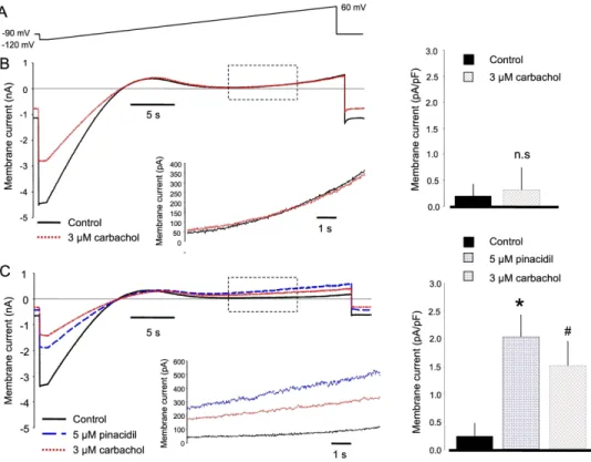
170
171 Results
172 1. Acetylcholine lengthened the APD after pinacidil-mediated action potential shortening
173 Canine Purkinje fibers and ventricular papillary muscles were paced at 500 ms cycle length.
174 In canine Purkinje fibers (PFs; n=15), acetylcholine (5 µM) did not affect the repolarization
175 (233.6±4.7 to 231.7±4.6; Figures 1A and 1E). In contrast, in canine Purkinje fibers (n=8), the
176 IK-ATP activator pinacidil, applied in 5 μM concentration, significantly abbreviated APD90
177 (207.7±7.0 ms vs 113.1±9.1 ms, p<0.05) values. After steady state was reached, acetylcholine
178 was administered. Within 3 minutes, acetylcholine prolonged APD90 to 147.3±7.4 ms,
179 partially reversing the effects of pinacidil (Figures 1B and 1E; p<0.05).
180
181 Similarly, as observed in Purkinje fibers, 5 μM acetylcholine alone failed to influence the
182 APD of the ventricular muscle (APD90: 172.6±5.7 ms vs 172.8±5.3 ms). Pinacidil (n=5;
183 5 μM) pretreatment significantly abbreviated the APD90 value (187.9±4.5 ms vs
184 163.7±6.4 ms, p<0.05), similarly to the effects observed in the case of PFs. After a period of
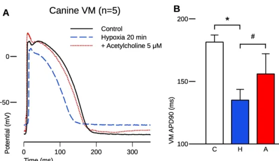
Draft
185 30 minutes, sufficient to reach a steady state, acetylcholine was added to the superfusate.
186 Within 4 minutes, acetylcholine (5 μM) prolonged APD90 to 172.1±7.4 ms (p<0.05), thus
187 partially reversing the effects of pinacidil (Figures 1D and 1E).
188
189 2. Acetylcholine decreased the calculated APD dispersion between PF and VM
190 The changes in the difference between the APD90 values of PF and VM can be used to infer
191 the effects of pinacidil and acetylcholine on the dispersion between these cardiac tissue types
192 (Figure 2). The control APD90 dispersion (9.5%, 20 ms) was significantly increased upon
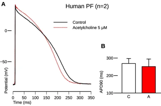
193 5 μM pinacidil application (44.7%, 51 ms). On the other hand, subsequently applied 5 μM
194 acetylcholine markedly decreased the repolarization heterogeneity (16.9%, 28 ms; p<0.05).
195
196 3. Carbachol decreased the pinacidil-induced current activation
197 During ionic current measurements, voltage ramps were used from a holding potential of
198 -90 mV. Membrane potential was hyperpolarized to -120 mV, and then was slowly (over 36 s)
199 depolarized to 60 mV. Ionic currents were analyzed and compared at 0 and +30 mV. We
200 found that carbachol did not change the control current when it was applied without pinacidil
201 (0 mV - control: 0.20±0.2 pA/pF vs 3 μM carbachol: 0.32±0.2 pA/pF, n=6 and +30 mV -
202 control: 0.55±0.4 pA/pF vs 3 μM carbachol: 0.74±0.3 pA/pF, n=6). In contrast, when 5 μM
203 pinacidil was applied first, subsequently employed carbachol significantly reduced the current
204 at both voltages (0 mV – control: 0.24±0.2 pA/pF 5 μM pinacidil: 2.03±0.3 pA/pF 3
205 μM carbachol: 1.51±0.4 pA/pF, n=8, p<0.05. +30 mV - control: 0.78±0.6 pA/pF 5 μM
206 pinacidil: 3.17±0.3 pA/pF 3 μM carbachol: 2.26±0.3 pA/pF, n=8, p<0.05).
207
208 These measurements were carried out with acetylcholine as well. However, we found
209 carbachol to be more stable during the applied long voltage protocol.
210
Draft
211 4. Acetylcholine restored the APD after hypoxia-induced action potential shortening
212 Simulated hypoxia, achieved by gassing the solution with N2 and CO2 instead of O2 and CO2,
213 resulted in a significant abbreviation of APD90 from 181.4±5.7 ms to 135.0±8.6 ms (p<0.05,
214 Figures 4A and 4B), and a decrease in amplitude (103.7±2.8 mV vs 92±3.5 mV). The
215 maximum rate of depolarization was also decreased (185.8±15.8 V/s vs 156.1±20.6 V/s).
216 When applied during hypoxia, 5 μM acetylcholine caused a significant APD90 prolongation to
217 164.4±4.4 ms, partially reversing the effect of hypoxia on the repolarization. AMP returned to
218 a normal range (102.1±1.6 mV), while Vmax remained at 156.0±16.1 V/s.
219
220 5. Acetylcholine caused a slight abbreviation in human Purkinje fibers
221 In human PFs (n=2), acetylcholine in 5 μM concentration caused a slight abbreviation of
222 APD90 from 269.0±28.4 to 251.6±42.85 ms and APD50 from 184.4±20.0 ms to
223 173.3±27.1 ms without affecting other characteristics of the action potential (Figure 5).
224
225 Discussion
226 In this study we investigated the electrophysiological effects of muscarinic agonists on the
227 IK-ATP current. We found that (i) under normal conditions acetylcholine did not influence the
228 action potential duration. (ii) In contrast, when IK-ATP was pharmacologically activated by
229 pinacidil, subsequently applied acetylcholine lengthened the action potential duration as well
230 as (iii) reduced the pinacidil-induced ventricle-Purkinje APD dispersion. (iv) In line with this,
231 carbachol inhibited the IK-ATP that was previously activated by pinacidil. (v) Acetylcholine
232 increased the APD after hypoxia-induced action potential shortening.
233
234 Acetylcholine inhibits the IK-ATP in canine ventricular myocytes
235 It is well known that acetylcholine shortens the atrial APD and has been implicated in atrial
236 fibrillation (Nakayama et al, 1968). Acetylcholine directly affects the GIRK1/4 or
237 Kir3.1/Kir3.4 channels (Nobles et al, 2018; Corey and Clapham, 1998), encoded by KCNJ3
Draft
238 and KCNJ4 genes (Kurachi, 1995). These channels are largely expressed in atrial, SA and AV
239 nodal cells (Galindo et al, 2016; Navarro-Polanco et al, 2013). At the same time, previous
240 studies (Terzic et al, 1994; Ito et al., 1994) claimed that acetylcholine activates the IK-ATP
241 channels, even though the physiological consequences of this effect on the action potential
242 were not clarified.
243
244 The IK-ATP ATP-sensitive potassium channels comprise hetero-octamers consisting of four
245 inward rectifying potassium channel pore-forming subunits (Kir6.1 or Kir6.2, encoded by
246 KCNJ8 and KCNJ11 genes, respectively) and four ATP-binding cassette protein
247 sulphonylurea receptors (SUR1 or SUR2, encoded by ABCC8 and ABCC9 genes,
248 respectively; Inagaki et al, 195). An important feature of the IK-ATP is its closed state under
249 physiological intracellular ATP levels (i. e., under normoxia) and its activation by metabolic
250 stress, when the ratio of ATP/ADP is decreased, e. g., during myocardial ischemia (Deutsch et
251 al., 1991).
252
253 Activation of the sarcolemmal IK-ATP during myocardial ischemia shortens the action potential
254 of various cardiac tissues to different extents, thus it may promote APD dispersion and re-
255 entry type arrhythmias (Janse and Wit, 1989). Accordingly, several investigations found IK-
256 ATP activation to be pro-arrhythmic (Chi et al., 1990), suggesting that sarcolemmal IK-ATP
257 inhibition may prevent arrhythmias induced by myocardial ischemia and ischemia/reperfusion
258 (Billman et al, 1998; Englert et al, 2003; Vajda et al, 2007).
259
260 In our experiments under normal conditions, we found no effect of carbachol on the
261 membrane current (Figure 3) and, similarly, acetylcholine failed to influence the ventricular
262 and Purkinje APDs (Figures 1A and 1C). The observed discrepancy between our and previous
263 results, where an activation of IK-ATP was described upon acetylcholine administration (Terzic
Draft
264 et al, 1994; Ito et al, 1994; Kim et al., 1997), could be the consequence of the species
265 difference and the distinct experimental conditions.
266
267 In contrast, an important, and, to the best of our knowledge, previously not published result of
268 our study is that carbachol is able to suppress the pinacidil-activated IK-ATP. As a consequence,
269 in parallel tissue action potential experiments, acetylcholine lengthened the APD as long as it
270 was previously shortened by the application of IK-ATP-activator pinacidil. Since IK-ATP
271 activation could be arrhythmogenic (Chi et al., 1990) by causing an increase in the APD
272 dispersion, this effect of acetylcholine raises the possibility of a novel antiarrhythmic
273 mechanism of the previously described antiarrhythmic effect of parasympathetic activation
274 during hypoxia (Song et al., 1992; Zuanetti et al., 1987; Collins and Billman, 1989).
275
276 Our experiments conducted under hypoxic conditions provided similar results (i. e.,
277 acetylcholine lengthened the hypoxia-induced shortened ventricular action potential;
278 Figure 4). Even though tissue hypoxia is a complex phenomenon (Carmeliet, 1999), during
279 which several factors change simultaneously (e. g., Ca2+i, Na+i, pH, conductance of gap
280 junctions, membrane potential etc.), it is feasible that IK-ATP activation, as a response to ATP
281 depletion, is an important factor in the observed action potential shortening. Since
282 acetylcholine lengthened the action potential under hypoxic conditions, we suggest IK-ATP
283 inhibition as a possible underlying mechanism.
284
285 Acetylcholine decreased the pinacidil-induced ventricle–Purkinje APD dispersion
286 Free-running Purkinje fibers connect to the ventricular muscle on a small surface area,
287 providing a relatively large-resistance coupling (Tranum-Jensen et al., 1991), and a large sink
288 for current flow that favors conduction blocks more than other parts of the healthy
289 myocardium. Also, due to the weaker electrotonic coupling, the dispersion of repolarization
290 here can be greater than in other areas (Martinez et al., 2018), causing the Purkinje–ventricle
Draft
291 APD ratio to have critical importance in arrhythmia generation. In our experiments, we found
292 significantly greater shortening in Purkinje fibers caused by pinacidil that could be the
293 consequence of the generally weaker repolarization reserve that makes the Purkinje action
294 potential to be more susceptible to any pharmacological interventions (Varró et al, 2000;
295 Baláti et al, 1998). Similarly, acetylcholine exerted larger lengthening in the Purkinje fiber
296 probably by the same reason that ultimately led to reduced ventricle–Purkinje APD
297 dispersion. The reduction of the ventricle–Purkinje fiber APD dispersion could suppress the
298 arrhythmogenic substrate providing a narrower vulnerable period for a critically timed
299 extrasystole to trigger a life-threatening arrhythmia under hypoxic conditions.
300
301 Proposed mechanism
302 Since inhibition of the K-ATP channels is possible by blocking various PKA-mediated
303 pathways (Tinker et al, 2018.), we suggest that the decrease of cAMP levels caused by the
304 activation of cardiac muscarinic receptors using acetylcholine/carbachol was the factor that
305 decreased the density of the IK-ATP current in patch clamp measurements, leading to the
306 subsequent prolongation observed in action potential durations.
307
308 Conclusions
309 We found that muscarinic agonists inhibit the IK-ATP. Therefore, during IK-ATP-mediated action
310 potential shortening, acetylcholine causes asymmetrical action potential lengthening between
311 ventricular muscle and Purkinje fiber that leads to reduced APD dispersion.
312
313 These results suggest that the parasympathetic tone beyond suppressing the catecholaminerg-
314 induced arrhythmogenic triggers (Song et al., 1992) may be also able to reduce the
315 arrhythmogenic substrate under hypoxic conditions.
316 317
Draft
318 Study Limitations
319 (i) In our experiments, the ventricular and Purkinje fiber action potentials were measured
320 from electrically uncoupled tissue samples.
321 (ii) The presented effects were attributed to the M2 muscarinic receptor; nevertheless, the
322 exact level of contribution of other receptor subtypes was not addressed. To achieve this,
323 further studies are needed, utilizing specific agonist and antagonist drugs.
324
325 Acknowledgments
326 We are grateful to Dr. Károly Acsai for his valuable contribution in performing statistical
327 comparisons. This work was supported by grants from the National Research, Development
328 and Innovation Office – NKFIH PD-116011 (for IK), FK-129117 (for NN) and the ÚNKP-
329 18-4, 19-4 and ÚNKP-20-5-SZTE-165 New National Excellence Program of the Ministry for
330 Innovation and Technology (for IK and NN), the János Bolyai Research Scholarship of the
331 Hungarian Academy of Sciences (for NN) and EFOP-3.6.2-16-2017-00006 (LIVE LONGER)
332 and EFOP 3.6.3-VEKOP-16-2017-00009 and Ministry of Human Capacities, Hungary grant
333 20391-3/2018/FEKUSTRAT, and the University of Szeged.
Draft
334 References
335 Baláti, B., Varró, A., & Papp, J. G. (1998). Comparison of the cellular electrophysiological characteristics of 336 canine left ventricular epicardium, M cells, endocardium and Purkinje fibers. Acta Physiologica Scandinavica, 337 164(2), 181–190. https://doi.org/10.1046/j.1365-201X.1998.00416.x
338
339 Billman, G. E., Englert, H. C., & Schölkens, B. A. (1998). HMR 1883, a novel cardioselective inhibitor of the 340 ATP-sensitive potassium channel. Part II: Effects on susceptibility to ventricular fibrillation induced by 341 myocardial ischemia in conscious dogs. The Journal of Pharmacology and Experimental Therapeutics, 286(3), 342 1465–1473.
343
344 Carmeliet, E. (1999). Cardiac ionic currents and acute ischemia: From channels to arrhythmias. Physiological 345 Reviews, 79(3), 917–1017. https://doi.org/10.1152/physrev.1999.79.3.917
346
347 Chi, L., Uprichard, A. C., & Lucchesi, B. R. (1990). Profibrillatory actions of pinacidil in a conscious canine 348 model of sudden coronary death. Journal of Cardiovascular Pharmacology, 15(3), 452–464.
349 https://doi.org/10.1097/00005344-199003000-00016 350
351 Collins, M. N., & Billman, G. E. (1989). Autonomic response to coronary occlusion in animals susceptible to 352 ventricular fibrillation. The American Journal of Physiology, 257(6 Pt 2), H1886-1894.
353 https://doi.org/10.1152/ajpheart.1989.257.6.H1886 354
355 Corey, S., & Clapham, D. E. (1998). Identification of native atrial G-protein-regulated inwardly rectifying K+
356 (GIRK4) channel homomultimers. The Journal of Biological Chemistry, 273(42), 27499–27504.
357 https://doi.org/10.1074/jbc.273.42.27499 358
359 Deutsch, N., Klitzner, T. S., Lamp, S. T., & Weiss, J. N. (1991). Activation of cardiac ATP-sensitive K+ current 360 during hypoxia: Correlation with tissue ATP levels. The American Journal of Physiology, 261(3 Pt 2), H671- 361 676. https://doi.org/10.1152/ajpheart.1991.261.3.H671
362
363 Englert, H. C., Heitsch, H., Gerlach, U., & Knieps, S. (2003). Blockers of the ATP-sensitive potassium channel 364 SUR2A/Kir6.2: A new approach to prevent sudden cardiac death. Current Medicinal Chemistry. Cardiovascular 365 and Hematological Agents, 1(3), 253–271. https://doi.org/10.2174/1568016033477423
Draft
366
367 Harvey, R. D., & Belevych, A. E. (2003). Muscarinic regulation of cardiac ion channels. British Journal of 368 Pharmacology, 139(6), 1074–1084. https://doi.org/10.1038/sj.bjp.0705338
369
370 Higgins, C. B., Vatner, S. F., & Braunwald, E. (1973). Parasympathetic control of the heart. Pharmacological 371 Reviews, 25(1), 119–155.
372
373 Inagaki, N., Gonoi, T., Clement, J. P., Namba, N., Inazawa, J., Gonzalez, G., Aguilar-Bryan, L., Seino, S., &
374 Bryan, J. (1995). Reconstitution of IKATP: An inward rectifier subunit plus the sulfonylurea receptor. Science 375 (New York, N.Y.), 270(5239), 1166–1170. https://doi.org/10.1126/science.270.5239.1166
376
377 Ito, H., Vereecke, J., & Carmeliet, E. (1994). Mode of regulation by G protein of the ATP-sensitive K+ channel 378 in guinea-pig ventricular cell membrane. The Journal of Physiology, 478 ( Pt 1), 101–107.
379 https://doi.org/10.1113/jphysiol.1994.sp020233 380
381 Janse, M. J., & Wit, A. L. (1989). Electrophysiological mechanisms of ventricular arrhythmias resulting from 382 myocardial ischemia and infarction. Physiological Reviews, 69(4), 1049–1169.
383 https://doi.org/10.1152/physrev.1989.69.4.1049 384
385 Kim, D., Watson, M., & Indyk, V. (1997). ATP-dependent regulation of a G protein-coupled K+ channel 386 (GIRK1/GIRK4) expressed in oocytes. The American Journal of Physiology, 272(1 Pt 2), H195-206.
387 https://doi.org/10.1152/ajpheart.1997.272.1.H195 388
389 Koumi, S., Wasserstrom, J. A., & Ten Eick, R. E. (1995). Beta-adrenergic and cholinergic modulation of the 390 inwardly rectifying K+ current in guinea-pig ventricular myocytes. The Journal of Physiology, 486 ( Pt 3), 647–
391 659. https://doi.org/10.1113/jphysiol.1995.sp020841 392
393 Kurachi, Y. (1995). G protein regulation of cardiac muscarinic potassium channel. The American Journal of 394 Physiology, 269(4 Pt 1), C821-830. https://doi.org/10.1152/ajpcell.1995.269.4.C821
395
396 Martinez, M. E., Walton, R. D., Bayer, J. D., Haïssaguerre, M., Vigmond, E. J., Hocini, M., & Bernus, O.
397 (2018). Role of the Purkinje-Muscle Junction on the Ventricular Repolarization Heterogeneity in the Healthy and
Draft
398 Ischemic Ovine Ventricular Myocardium. Frontiers in Physiology, 9, 718.
399 https://doi.org/10.3389/fphys.2018.00718 400
401 Nagy, N., Szél, T., Jost, N., Tóth, A., Gy. Papp, J., & Varró, A. (2015). Novel experimental results in human 402 cardiac electrophysiology: Measurement of the Purkinje fibre action potential from the undiseased human heart.
403 Canadian Journal of Physiology and Pharmacology, 93(9), 803–810. https://doi.org/10.1139/cjpp-2014-0532 404
405 Nakayama, K., Suzuki, Y., & Hashimoto, K. (1968). Sustained atrial fibrillation by acetylcholine infusion into 406 the sinus node artery. The Tohoku Journal of Experimental Medicine, 96(4), 333–339.
407 https://doi.org/10.1620/tjem.96.333 408
409 Navarro-Polanco, R. A., Aréchiga-Figueroa, I. A., Salazar-Fajardo, P. D., Benavides-Haro, D. E., Rodríguez- 410 Elías, J. C., Sachse, F. B., Tristani-Firouzi, M., Sánchez-Chapula, J. A., & Moreno-Galindo, E. G. (2013).
411 Voltage sensitivity of M2 muscarinic receptors underlies the delayed rectifier-like activation of ACh-gated K(+) 412 current by choline in feline atrial myocytes. The Journal of Physiology, 591(17), 4273–4286.
413 https://doi.org/10.1113/jphysiol.2013.255166 414
415 Nobles, M., Montaigne, D., Sebastian, S., Birnbaumer, L., & Tinker, A. (2018). Differential effects of inhibitory 416 G protein isoforms on G protein-gated inwardly rectifying K+ currents in adult murine atria. American Journal 417 of Physiology. Cell Physiology, 314(5), C616–C626. https://doi.org/10.1152/ajpcell.00271.2016
418
419 Pappano, A. J., & Carmeliet, E. E. (1979). Epinephrine and the pacemaking mechanism at plateau potentials in 420 sheep cardiac Purkinje fibers. Pflugers Archiv: European Journal of Physiology, 382(1), 17–26.
421 https://doi.org/10.1007/BF00585899 422
423 Recordati, G., Schwartz, P. J., Pagani, M., Malliani, A., & Brown, A. M. (1971). Activation of cardiac vagal 424 receptors during myocardial ischemia. Experientia, 27(12), 1423–1424. https://doi.org/10.1007/BF02154267 425
426 Song, Y., Thedford, S., Lerman, B. B., & Belardinelli, L. (1992). Adenosine-sensitive afterdepolarizations and 427 triggered activity in guinea pig ventricular myocytes. Circulation Research, 70(4), 743–753.
428 https://doi.org/10.1161/01.res.70.4.743 429
Draft
430 Terzic, A., Tung, R. T., Inanobe, A., Katada, T., & Kurachi, Y. (1994). G proteins activate ATP-sensitive K+
431 channels by antagonizing ATP-dependent gating. Neuron, 12(4), 885–893. https://doi.org/10.1016/0896- 432 6273(94)90340-9
433
434 Tranum-Jensen, J., Wilde, A. A., Vermeulen, J. T., & Janse, M. J. (1991). Morphology of electrophysiologically 435 identified junctions between Purkinje fibers and ventricular muscle in rabbit and pig hearts. Circulation
436 Research, 69(2), 429–437. https://doi.org/10.1161/01.res.69.2.429 437
438 Vajda, S., Baczkó, I., & Leprán, I. (2007). Selective cardiac plasma-membrane K(ATP) channel inhibition is 439 defibrillatory and improves survival during acute myocardial ischemia and reperfusion. European Journal of 440 Pharmacology, 577(1–3), 115–123. https://doi.org/10.1016/j.ejphar.2007.08.016
441
442 Varró, A., & Baczkó, I. (2010). Possible mechanisms of sudden cardiac death in top athletes: A basic cardiac 443 electrophysiological point of view. Pflugers Archiv: European Journal of Physiology, 460(1), 31–40.
444 https://doi.org/10.1007/s00424-010-0798-0 445
446 Varro, A., Baláti, B., Iost, N., Takács, J., Virág, L., Lathrop, D. A., Csaba, L., Tálosi, L., & Papp, J. G. (2000).
447 The role of the delayed rectifier component IKs in dog ventricular muscle and Purkinje fibre repolarization. The 448 Journal of Physiology, 523 Pt 1, 67–81. https://doi.org/10.1111/j.1469-7793.2000.00067.x
449
450 Weiss, J. N., & Venkatesh, N. (1993). Metabolic regulation of cardiac ATP-sensitive K+ channels.
451 Cardiovascular Drugs and Therapy, 7 Suppl 3, 499–505. https://doi.org/10.1007/BF00877614 452
453 Zuanetti, G., De Ferrari, G. M., Priori, S. G., & Schwartz, P. J. (1987). Protective effect of vagal stimulation on 454 reperfusion arrhythmias in cats. Circulation Research, 61(3), 429–435. https://doi.org/10.1161/01.res.61.3.429
Draft
455 Figure Legends
456 Figure 1. Representative traces of Purkinje fiber (A, B) and ventricular muscle preparations
457 (C, D); 5 μM acetylcholine (red dotted lines) alone caused no changes in either preparation
458 type (A, C), while it caused significant prolongation when applied cummulatively after 5 μM
459 pinacidil (B, D, pinacidil effect represented as blue dashed lines). Bars in panel E represent
460 the values of APD90 in each treatment group, from top to bottom corresponding to the traces
461 A to D. Abbreviations under bars: C, control; P, pinacidil, A, acetylcholine. The pacing cycle
462 length was 500 ms. Values are mean ± SEM; *,# p<0.05 RM-ANOVA followed by
463 Bonferroni’s post-hoc test.
464
465 Figure 2. Pinacidil (5 μM) increased the action potential duration dispersion (indicated by
466 ΔAPD90 in percentages, and in ms above the bars) between Purkinje fiber and ventricular
467 muscle preparations, while acetylcholine (5 μM), when applied after pinacidil, decreased
468 dispersion. The pacing cycle length was 500 ms.
469
470 Figure 3. Effect of carbachol on IK-ATP. Ionic currents were measured under a slow voltage
471 ramp protocol (panel A) between -120 mV and 60 mV. The currents were analysed at 0 and
472 30 mV. Panel B demonstrates original representative current traces (left) and bar graphs
473 (right) where 3 μM carbachol (dotted line) failed to influence the control current analysed at
474 0 mV. Inset shows identical current fractions between –3 mV and 45 mV (indicated by dashed
475 rectangle). Current traces in panel C as well as in the inset, illustrate large increase of the
476 membrane current after application of 5 μM pinacidil (blue dashed line) that was inhibited by
477 the subsequently applied 3 μM carbachol (red dotted line). In bar graphs (right), asterisk
478 denotes significant change between control (left column) and pinacidil (middle column),
479 while hash tag indicates significant change between pinacidil (middle column) and carbachol
480 (right column).
481
Draft
482 Figure 4. Representative action potential trace (A) showing that hypoxic conditions caused
483 significant action potential duration abbreviation and decreased mean diastolic potential and
484 amplitude in canine ventricular preparations (blue dashed line), while acetylcholine (5 μM)
485 caused a significant prolongation in action potential duration (red dotted line). Values of
486 APD90 are represented as bars (B). Abbreviations under bars: C, control; H, hypoxia, A,
487 acetylcholine. The pacing cycle length was 500 ms. Values are mean ± SEM; *,#p<0.05,
488 RM-ANOVA followed by Bonferroni’s post-hoc test.
489
490 Figure 5. Representative action potential showing the effect of acetylcholine (5 μM, red
491 dotted line) on a Purkinje fiber taken from a human donor heart (A). Values of APD90 are
492 represented as bars (B). Abbreviations under bars: C, control; A, acetylcholine. The pacing
493 cycle length was 500 ms. Values are mean ± SEM.
Draft
1 Muscarinic agonists inhibit the ATP-dependent potassium current and suppress the
2 ventricle-Purkinje action potential dispersion
3
4 Tibor Magyara,§, Tamás Árpádffy-Lovasa,§, Bence Pásztia, Noémi Tótha, Jozefina Szlováka,
5 Péter Gazdaga, Zsófia Kohajdab, András Gyökeresa, Balázs Györed, Zsolt Gurabia, Norbert
6 Josta,b,c, László Virága,c, Julius Gy. Pappa,b, Norbert Nagya,b,#, István Koncza,*, #
7 8
9 aDepartment of Pharmacology and Pharmacotherapy, Faculty of Medicine, University of
10 Szeged, Szeged, Hungary;
11 bMTA-SZTE Research Group of Cardiovascular Pharmacology, Hungarian Academy of
12 Sciences, Szeged, Hungary
13 cDepartment of Pharmacology and Pharmacotherapy, Interdisciplinary Excellence Centre,
14 University of Szeged, Szeged, Hungary
15 dFaculty of Dentistry, University of Szeged, Hungary
16
17 §Shared first authorship
18 # Shared senior authorship
19
20 *Author for correspondence at:
21 István Koncz MD, PhD
22 Department of Pharmacology & Pharmacotherapy
23 Faculty of Medicine
24 University of Szeged
25 Dóm tér 12,
26 H-6720 Szeged, Hungary
27 E-mail: koncz.istvan@med.u-szeged.hu
Draft
28 Abstract
29 Introduction: Activation of the parasympathetic nervous system has been reported to have an
30 antiarrhythmic role during ischemia-reperfusion injury by decreasing the arrhythmia triggers.
31 Furthermore, it was reported that the parasympathetic neurotransmitter acetylcholine is able to
32 modulate the ATP-dependent K-current (IK-ATP), a crucial current activated during hypoxia.
33 However, the possible significance of this current modulation in the antiarrhythmic
34 mechanism is not fully clarified.
35 Methods: Action potentials were measured using the conventional microelectrode technique
36 from canine left ventricular papillary muscle and free-running Purkinje fibers, under normal
37 and hypoxic conditions. Ionic currents were measured using the whole-cell configuration of
38 the patch clamp method.
39 Results: 5 μM acetylcholine did not influence the action potential duration (APD) either in
40 Purkinje fibers or in papillary muscle preparations. In contrast, it significantly lengthened the
41 APD and suppressed the Purkinje–ventricle APD dispersion when it was administered after
42 5 μM pinacidil application. 3 μM carbachol reduced the pinacidil-activated IK-ATP under
43 voltage-clamp condition. Acetylcholine lengthened the ventricular action potential under
44 simulated ischemia condition.
45 Conclusion: In this study we found that acetylcholine inhibits the IK-ATP and thus suppresses
46 the ventricle-Purkinje APD dispersion. We conclude that parasympathetic tone may reduce
47 the arrhythmogenic substrate exerting a complex antiarrhythmic mechanism during hypoxic
48 conditions.
49
50 Key words: acetylcholine, Purkinje fibers, papillary muscles, hypoxia
Draft
51 Introduction
52 The parasympathetic nervous system has a crucial role in controlling the actual heart rate and
53 impulse propagation via influencing the sinoatrial and atrioventricular nodes (Higgins et al.,
54 1973). The parasympathetic nerve endings operate by releasing acetylcholine that acts on
55 M2-receptors, activating several intracellular signaling routes, and ultimately influencing the
56 cardiac ion channels (Harvey and Belevych, 2003). Even though the parasympathetic nervous
57 system primarily innervates the supraventricular areas of the heart, there are certain important
58 ion channels in the ventricular muscle that are known to be influenced by the release of
59 acetylcholine. It has been previously reported that the inward rectifier potassium current (IK1;
60 Koumi et al., 1995) and the slow component of the delayed rectifier (IKs; Pappano and
61 Carmeliet, 1979) are inhibited, whereas IK-ATP and IK-ACh are activated by acetylcholine via
62 G proteins (Terzic et al, 1994; Ito et al., 1994; Kim et al., 1997).
63
64 The importance of these effects of acetylcholine is underpinned by the fact that the activation
65 ofIK-ATP channels is well known during hypoxia/ischemia, in which situations the duration of
66 the action potential is shortened (Weiss and Venkatesh, 1993). Furthermore, it was reported
67 that vagal activation is also facilitated under ischemia–reperfusion (Recordati et al., 1971).
68 This vagal activation during hypoxia could be antiarrhythmic, since it was reported that
69 increased parasympathetic tone reduces the catecholaminerg-induced early and delayed
70 afterdepolarizations (arrhythmia triggers) (Song et al., 1992), as well as the incidence of
71 ventricular fibrillation (Zuanetti et al., 1987; Collins and Billman, 1989). However, the
72 underlying mechanism of antiarrhythmic effect of M2-receptor activation is not fully clarified.
73 Arrhythmias may develop when an arrhythmogenic substrate (e. g., dispersion of
74 repolarization) and arrhythmia triggers (e.g.: early and delayed afterdepolarizations)
75 simultaneously exist in the heart. The arrhythmogenic substrate could be prominent at
76 Purkinje–ventricle connection because of the relatively weak electrotonic coupling due to low
77 number of gap junctions (Varró and Baczkó, 2010). As a consequence of the different
Draft
78 pharmacological susceptibility of Purkinje fiber and ventricular muscle (Baláti et al, 1998),
79 the activation of IK-ATP may modulate the Purkinje and ventricular action potential duration
80 (APD) to different extents, and the developed APD dispersion may contribute to the onset of
81 arrhythmias.
82
83 The objective of this study was the investigation of the possible effect of acetylcholine on the
84 IK-ATP and on the IK-ATP-mediated action potential dispersion under normal and hypoxic
85 conditions.
86
87 Methods
88 Human tissues
89 Non-diseased human hearts that were unusable for transplantation (based on logistical, not
90 patient-related considerations) were obtained from organ donors. Before cardiac explanation,
91 organ donor patients did not receive medication except dobutamine, furosemide and plasma
92 expanders. The investigations conform to the principles outlined in the Declaration of
93 Helsinki of the World Medical Association. All experimental protocols were approved by the
94 Scientific and Research Ethical Committee of the Medical Scientific Board at the Hungarian
95 Ministry of Health (ETT-TUKEB), under ethical approval No 4991-0/2010-1018EKU
96 (339/PI/010). Human cardiac tissue was stored in cardioplegic solution at 4°C for 4–8 hours.
97
98 Animals
99 All experiments using canine cardiac preparations were carried out in compliance with the
100 Guide for the Care and Use of Laboratory Animals (USA NIH publication NO 85-23, revised
101 1996) and conformed to the Directive 2010/63/EU of the European Parliament. The protocols
102 have been approved by the Ethical Committee for the Protection of Animals in Research of
103 the University of Szeged, Szeged, Hungary (approval number: I-74-24-2017) and by the
Draft
104 Department of Animal Health and Food Control of the Ministry of Agriculture and Rural
105 Development (authority approval number XIII/3331/2017).
106
107 Conventional microelectrode technique
108 Ventricular (papillary or trabecular) muscles were obtained from the right ventricle of canine
109 hearts. Free-running Purkinje fibers were identified as false tendons and isolated from both
110 ventricles of human and canine hearts. Canine hearts were removed through a right lateral
111 thoracotomy from anesthetized (thiopental 30 mg/kg i.v.) mongrel dogs of either sex
112 weighing 10–15 kg. At impalement, Purkinje fibers were observed under a surgical
113 microscope (Zeiss OPMI PRO). The preparations were placed in Locke’s solution and
114 allowed to equilibrate for at least 2 hours while superfused (flow rate 4-5 ml/min) also with
115 Locke’s solution containing (in mM): NaCl 120, KCl 4, CaCl2 2, MgCl2 1, NaHCO3 22, and
116 glucose 11. The pH of this solution was 7.40 to 7.45 when gassed with 95% O2 and 5% CO2
117 at 37 °C. In the experiments where the effects of tissue hypoxia were examined, we changed
118 the gas mixture to 95% N2 and 5% CO2, pH remained at 7.40 to 7.45. All experiments were
119 performed at 37 °C. During the equilibration period, preparations were stimulated at a basic
120 cycle length of 500 ms. Electrical pulses of 0.5–2 ms in duration at twice the diastolic
121 threshold in intensity (S1) were delivered to the preparations through bipolar platinum
122 electrodes. Transmembrane potentials were recorded using glass capillary microelectrodes
123 filled with 3 M KCl (tip resistance: 5 to 15 MΩ). The microelectrodes were coupled through
124 an Ag-AgCl junction to the input of a high-impedance, capacitance-neutralizing amplifier
125 (Experimetria 2011). Intracellular recordings were displayed on a storage oscilloscope
126 (Hitachi V-555) and led to a computer system (APES) designed for on-line determination of
127 the following parameters: resting membrane potential, action potential amplitude, action
128 potential duration at 10% to 90% repolarization and the maximum rate of rise of the action
129 potential upstroke (Vmax). Control recordings were obtained after equilibration period. The
130 compounds used in all experiments were purchased from Sigma/Merck.
Draft
131 2.3. Cell isolation
132 Ventricular myocytes were enzymatically dissociated from the left ventricle of dog hearts.
133 Canine hearts were removed through a right lateral thoracotomy from anesthetized (thiopental
134 30 mg/kg i.v.) mongrel dogs of either sex weighing 10–15 kg. Cardiac myocytes were isolated
135 from the left ventricle, containing an arterial branch through which the segment was perfused
136 on a Langendorff apparatus with solutions in the following sequence: normal Tyrode's
137 solution (containing in mM: 144 mM NaCl, 0.4 mM NaH2PO4, 4 mM KCl, 0.53 mM MgSO4,
138 1.8 mM CaCl2, 5.5 mM Glucose, 5 mM HEPES, pH 7.4 adjusted with NaOH) for 10 min,
139 Ca2+-free Tyrode solution for 10 min and Ca2+-free Tyrode solution containing collagenase
140 (Worthington type II, 0.66 mg/mL). To the final perfusion solution protease (type XIV, 0.12
141 mg/mL) was added at the 15 and the 30 minutes for digestion.
142
143 2.4. Measurement of ionic currents
144 One drop of cell suspension was placed in a transparent recording chamber mounted on the
145 stage of an inverted microscope (Olympus IX51, Tokyo, Japan), and individual myocytes
146 were allowed to settle and adhere to the chamber bottom for at least 5–10 min before
147 superfusion was initiated and maintained by gravity. Only rod-shaped cells with clear
148 striations were used. HEPES-buffered Tyrode’s solution (composition in mM: NaCl 144,
149 NaH2PO4 0.4, KCl 4.0, CaCl2 1.8, MgSO4 0.53, glucose 5.5 and HEPES 5.0, at pH of 7.4)
150 was used as the normal superfusate. During the measurement of IK-ATP, 1 µM nisoldipine was
151 added to the bath solution to block ICaL, IKr was blocked by 0.1 µM dofetilide, and IKs was
152 blocked by 0.5 µM HMR-1556. Micropipettes were fabricated from borosilicate glass
153 capillaries (Science Products GmbH, Hofheim, Germany), using a P-97 Flaming/Brown
154 micropipette puller (Sutter Co, Novato, CA, USA), and had a resistance of 1.5–2.5 MΩ when
155 filled with pipette solution. The membrane currents were recorded with Axopatch-200B
156 amplifiers (Molecular Devices, Sunnyvale, CA, USA) by applying the whole-cell
157 configuration of the patch-clamp technique. The membrane currents were digitized with 250
Draft
158 kHz analogue to digital converters (Digidata 1440A, Molecular Devices, Sunnyvale, CA,
159 USA) under software control (pClamp 8 and pClamp 10, Molecular Devices, Sunnyvale, CA,
160 USA). The composition of the pipette solution (in mM) was the following: KOH 110, KCl
161 40, K2ATP 5, MgCl2 5, EGTA 5, HEPES 10 and GTP 0.1 (pH was adjusted to 7.2 by aspartic
162 acid).
163
164 2.5 Statistical analysis
165 Results are expressed as mean ± S.E.M. Normality of distributions was verified using
166 Shapiro-Wilk test, and homogeneity of variances was verified using Bartlett's test in each
167 treatment group. Statistical comparisons were made using analysis of variance (ANOVA) for
168 repeated measurements, followed by Bonferroni’s post-hoc test. Differences were considered
169 significant when p < 0.05.
170
171 Results
172 1. Acetylcholine lengthened the APD after pinacidil-mediated action potential shortening
173 Canine Purkinje fibers and ventricular papillary muscles were paced at 500 ms cycle length.
174 In canine Purkinje fibers (PFs; n=15), acetylcholine (5 µM) did not affect the repolarization
175 (233.6±4.7 to 231.7±4.6; Figures 1A and 1E). In contrast, in canine Purkinje fibers (n=8), the
176 IK-ATP activator pinacidil, applied in 5 μM concentration, significantly abbreviated APD90
177 (207.7±7.0 ms vs 113.1±9.1 ms, p<0.05) values. After steady state was reached, acetylcholine
178 was administered. Within 3 minutes, acetylcholine prolonged APD90 to 147.3±7.4 ms,
179 partially reversing the effects of pinacidil (Figures 1B and 1E; p<0.05).
180
181 Similarly, as observed in Purkinje fibers, 5 μM acetylcholine alone failed to influence the
182 APD of the ventricular muscle (APD90: 172.6±5.7 ms vs 172.8±5.3 ms). Pinacidil (n=5;
183 5 μM) pretreatment significantly abbreviated the APD90 value (187.9±4.5 ms vs
184 163.7±6.4 ms, p<0.05), similarly to the effects observed in the case of PFs. After a period of