INTRODUCTION
In cellular electrophysiological studies intracellular calcium concentration is often buffered under experimental conditions because of the wide variety of Ca2+-dependent ion currents, including Na+(1, 2), Ca2+(3, 4), K+(5, 6), Cl–(7, 8), Na+-Ca2+
exchanger (9, 10) and nonspecific cation (11, 12) currents in the heart. In voltage clamp experiments, using typically patch pipettes with large pore diameters, buffering of [Ca2+]i is relatively easy, since dialysis of the intracellular compartment with the Ca2+chelator BAPTA is readily performed by the pipette solution. When action potentials are recorded under physiological conditions with conventional sharp microelectrodes this approach is not available due to the small tip diameter of the recording electrode. In this case the cell-permeant acetoxy-methylester form of a Ca2+chelator can be applied to reduce [Ca2+]i. Such experiments have been performed for decades and their results
were interpreted exclusively in terms of buffering [Ca2+]i(13-17) - in spite of the fact that BAPTA-AM (and also EGTA-AM) has been shown to interact with K+channels expressed in HEK cells (18). Since drug-channel interactions may be markedly different in native and expressed ion channels (19), it was reasonable to test the effect of BAPTA-AM on IKr, which current has the greatest influence on ventricular repolarization in larger mammals. For the same reason the effect of BAPTA-AM on action potential morphology was also studied. Canine ventricular cells were chosen in these experiments because the electrophysiological properties of this preparation best resemble those of human myocardium (20, 21).
Our results indicate that in mammalian ventricular cardiomyocytes BAPTA-AM affects APD in two separate ways:
in addition to the consequences of [Ca2+]i buffering, and thus modifying the activity of Ca2+-dependent ionic currents, BAPTA- AM also directly inhibits IKr.
B. HORVATH1,2, N. SZENTANDRASSY1,3, R. VERESS1, D. BARANYAI1, K. KISTAMAS1, J. ALMASSY1, A. TOTH4, J. MAGYAR1,5, T. BANYASZ1, P.P. NANASI1,3
EFFECT OF THE INTRACELLULAR CALCIUM CONCENTRATION CHELATOR BAPTA ACETOXY-METHYLESTER ON ACTION POTENTIAL DURATION IN CANINE
VENTRICULAR MYOCYTES
1Department of Physiology, Faculty of Medicine, University of Debrecen, Debrecen, Hungary;
2Faculty of Pharmacy, University of Debrecen, Debrecen, Hungary;
3Department of Dental Physiology and Pharmacology, Faculty of Dentistry, University of Debrecen, Debrecen, Hungary;
4Department of Pharmacology and Pharmacotherapy, Faculty of Medicine, University of Szeged, Szeged, Hungary;
5Division of Sport Physiology, Department of Physiology, Faculty of Medicine, University of Debrecen, Debrecen, Hungary
Intracellular calcium concentration ([Ca2+]i) is often buffered by using the cell-permeant acetoxy-methylester form of the Ca2+ chelator BAPTA (BAPTA-AM) under experimental conditions. This study was designed to investigate the time- dependent actions of extracellularly applied BAPTA-AM on action potential duration (APD) in cardiac cells. Action potentials were recorded from enzymatically isolated canine ventricular myocytes with conventional sharp microelectrodes.
The effect of BAPTA-AM on the rapid delayed rectifier K+current (IKr) was studied using conventional voltage clamp and action potential voltage clamp techniques. APD was lengthened by 5 µM BAPTA-AM - but not by BAPTA - and shortened by the Ca2+ionophore A23187 in a time-dependent manner. The APD-lengthening effect of BAPTA-AM was strongly suppressed in the presence of nisoldipine, and enhanced in the presence of BAY K8644, suggesting that a shift in the [Ca2+]i- dependent inactivation of L-type Ca2+current may be an important underlying mechanism. However, in the presence of the IKr-blocker dofetilide or E-4031 APD was shortened rather than lengthened by BAPTA-AM. Similarly, the APD-lengthening effect of 100 nM dofetilide was halved by the pretreatment with BAPTA-AM. In line with these results, IKrwas significantly reduced by extracellularly applied BAPTA-AM under both conventional voltage clamp and action potential voltage clamp conditions. This inhibition of IKrwas partially reversible and was not related to the Ca2+chelator effect BAPTA-AM. The possible mechanisms involved in the APD-modifying effects of BAPTA-AM are discussed. It is concluded that BAPTA-AM has to be applied carefully to control [Ca2+]iin whole cell systems because of its direct inhibitory action on IKr.
K e y w o r d s : calcium chelators, intracellular calcium concentration, action potential duration, cardiac ion currents, potassium ion, currents, ventricular myocytes
MATERIALS AND METHODS Isolation of single canine ventricular myocytes
Adult mongrel dogs of either sex were anaesthetized with intramuscular injections of 10 mg/kg ketamine hydrochloride (Calypsol, Richter Gedeon, Hungary) + 1 mg/kg xylazine hydrochloride (Sedaxylan, Eurovet Animal Health BV, The Netherlands) according to protocols approved by the local ethical committee (license No18/2012/DEMAB) in line with the ethical standards laid down in the Declaration of Helsinki in 1964 and its later amendments. The hearts were quickly removed and placed in Tyrode solution containing (in mM) NaCl, 144;
KCl, 5.6; CaCl2, 2.5; MgCl2, 1.2; HEPES, 5; and dextrose, 11; at pH = 7.4. Single myocytes were obtained by enzymatic dispersion using the segment perfusion technique, as described previously (22, 23). Briefly, a wedge-shaped section of the left ventricular wall supplied by the left anterior descending coronary artery was cannulated, dissected and perfused with oxygenized Tyrode solution. After removal of blood the perfusion was switched to a nominally Ca2+-free Joklik solution (Minimum Essential Medium Eagle, Joklik Modification, Sigma-Aldrich Co., St. Louis, MO, USA) for 5 min. This was followed by 35 min perfusion with Joklik solution supplemented with 1 mg/ml collagenase (Type II. Worthington, Chemical Co.) and 0.2% bovine serum albumin (Fraction V., Sigma-Aldrich Co.) containing 50 µM Ca2+. The full transmural section of the middle portion of the left ventricular wall was cut into small pieces and the cell suspension was washed with Joklik solution.
These tissue chunks, however, contained dominantly myocytes of midmyocardial origin. After gradually restoring the normal external Ca2+concentration, the cells were stored in Minimum Essential Medium Eagle (Sigma-Aldrich Co.) until use. Drugs were obtained from Sigma-Aldrich Co.
Recording of action potentials
All electrophysiological measurements were performed at 37°C as previously described (23). Rod-shaped viable cells showing clear striation were sedimented in a plexiglass chamber of 1 ml volume allowing continuous superfusion (at a rate of 2 ml/min) with Tyrode solution. Transmembrane potentials were recorded using 3 M KCl filled sharp glass microelectrodes having tip resistance between 20 and 40 MO.
These electrodes were connected to the input of Multiclamp 700A or 700B amplifiers (Molecular Devices, Sunnyvale, CA, USA). The cells were paced through the recording electrode at a steady frequency of 1 Hz using 1 – 2 ms wide rectangular current pulses having amplitudes of 120% of the diastolic threshold. Since the cytosol was not dialyzed, time-dependent changes in action potential morphology were negligible for the period of our experimental protocol not lasting longer than 40 min. Action potentials were digitized at 200 kHz using Digidata 1332A or 1440A digitizers (Molecular Devices) and stored for later analysis. BAPTA-AM was dissolved in DMSO to yield 5 mM stock solutions, which was diluted into the bathing solution immediately before use.
Conventional voltage clamp
The cells were superfused at 37°C with a modified Tyrode solution supplemented with 1 µM nisoldipine and 1 µM HMR 1556 to prevent the contamination with L-type Ca2+current (ICa) and the slow delayed rectifier K+current (IKs) respectively. The modified Tyrode solution contained (in mM): NaCl, 121; KCl, 4;
CaCl2, 1.3; MgCl2, 1; HEPES, 10; NaHCO3, 25; and glucose, 10;
at pH = 7.35. The osmolarity of this solution was 298 – 303
mOsm. Suction pipettes, made of borosilicate glass, had tip resistances of 2 – 3 MO after filling with pipette solution, containing (in mM) K-aspartate 100, KCl 45, MgCl21, HEPES 5, BAPTA 10, K-ATP 3, at pH = 7.2. Membrane currents were recorded with Multiclamp 700A or 700B amplifier (Molecular Devices) using the whole cell configuration of the patch clamp technique. After establishing high (1 – 10 GO) resistance seal by gentle suction, the cell membrane beneath the tip of the electrode was disrupted by further suction or by applying 1.5 V electrical pulses for 1 ms. Outputs from the amplifier were digitized at 20 kHz under software control (pClamp 10.0, Molecular Devices).
The IKr was activated by 500 ms long depolarizing pulses clamped to +40 mV, arising from the holding potential of –80 mV. This protocol was repeated at a frequency of 0.05 Hz. IKr
was assessed as tail current amplitudes recorded following repolarization to –40 mV for 10 s. Currents were normalized to cell capacitance, determined in each cell using hyperpolarizations from +10 to –10 mV for 15 ms. Series resistance was typically 4 – 8 MO before compensation (usually 50 – 80%). Experiments were discarded when the series resistance was high or substantially increased during the measurement.
Action potential voltage clamp
After establishment of whole-cell configuration, action potentials were recorded in current clamp mode from the myocytes superfused with a modified Tyrode solution described in the ‘conventional voltage clamp’ section. In these experiments no ion channel blocker was applied in the external solution in order to develop normal action potentials. The pipette solution contained (in mM): K-aspartate 130, KCl 30, MgATP 3, HEPES 10, Na2-phosphocreatine 3, EGTA 0.01, cAMP 0.002, KOH 10, at pH = 7.3. As a consequence, the experimental conditions applied in action potential voltage clamp experiments were close to physiological (24, 25). The cells were continuously paced through the recording electrode at steady stimulation frequency of 1 Hz. A previously recorded ‘canonic’ canine midmyocardial action potential was applied to the voltage clamped cells as command voltage. Current traces were recorded continuously under ‘reference’ conditions (in the absence or presence of BAPTA-AM in the bathing medium) and after the application of the specific IKrinhibitor E 4031. IKrwas defined as an E 4031- sensitive current obtained by subtracting the traces obtained in the presence of E 4031 from the ‘reference’ traces. To diminish the consequences of trace-to-trace fluctuations and to reduce noise, 20 consecutive IKrtraces were averaged, and the averaged curve was used for further analysis.
In these experiments, to rule out the changes in current traces caused by the continuous calcium chelating effect of BAPTA- AM, cells were pre-loaded with BAPTA-AM before the measurements. To achieve this, cells were incubated with 5 µM BAPTA-AM for 20 min, then washed twice with Tyrode solution, and stored for at least further 15 minutes in BAPTA-AM-free Tyrode. These BAPTA-AM loaded cells, which did not contract when stimulated through the pipette, were used as control. To test the acute effect of BAPTA-AM on IKrin another group of cells, the superfusate was supplemented with 5 µM BAPTA-AM. In both cases IKrwas identified as E 4031-sensitive current.
Statistical analysis
Results are expressed as mean ± SEM values. Statistical significance of differences from control was evaluated using one-way ANOVA followed by Student’s t-test for paired or unpaired data as pertinent. Differences were considered significant when P was less than 0.05.
RESULTS
Effect of BAPTA-AM on action potential morphology
The most conventional way to study the effect of changes in [Ca2+]ion action potential configuration is loading the cell interior with the Ca2+chelator BAPTA using its cell-permeant acetoxy- methylester form, or alternatively, to load the cells with Ca2+using the Ca2+-ionophore A23187. As presented in Fig. 1, exposure to 5 µM BAPTA-AM lengthened, while to A23187 shortened the duration of action potentials measured at 90% level of repolarization (APD90). These changes developed gradually in a time-dependent manner (Fig. 1C) although in the case of BAPTA- AM there was an initial rapid rise in APD90. Actions of BAPTA- AM and A23187 failed to fully saturate within the recording period of 30 min, probably due to the progressive intracellular accumulation of free BAPTA resulting in a more and more effective buffering of [Ca2+]iin the first case and accumulation of Ca2+in the second one. Furthermore, the changes in APD90were accompanied with characteristic changes in action potential morphology, since reduction of [Ca2+]ishifted the plateau potential to more positive voltages, while its elevation resulted in a marked depression of the plateau (Fig. 1Aand 1B).
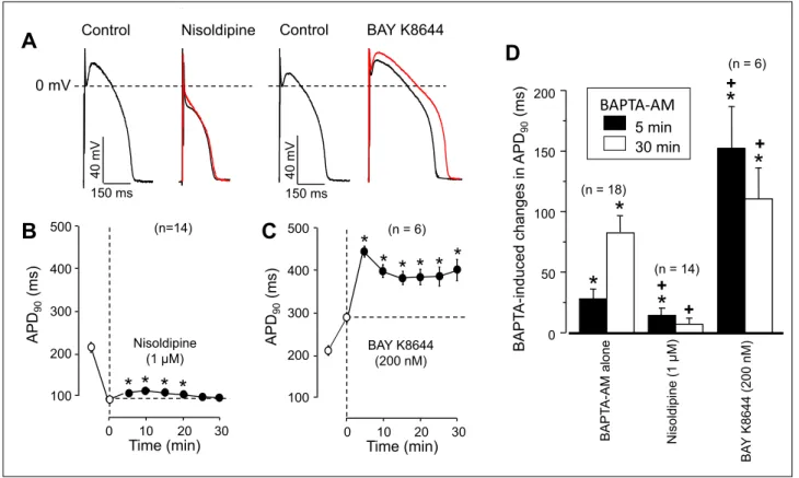
The effect of BAPTA-AM was studied under conditions when the density of ICawas manipulated. In the presence of the Ca2+-channel blocker nisoldipine (26) the BAPTA-AM-induced APD-lengthening was negligible (although statistically significant) and only transient since it disappeared after 20 min exposure to BAPTA-AM (Fig. 2B and 2D). In contrast, increasing ICa-density with BAY K8644 (27) markedly augmented the BAPTA-AM-induced prolongation of APD90
(Fig. 2Cand 2D). It is worthy of note that BAPTA-AM showed
a biphasic effect in the presence of BAY K8644. After 5 min exposure to BAPTA-AM APD90reached its maximum, and after this it shortened gradually reaching a local minimum around 15 min of BAPTA-AM treatment (Fig. 2C).
The effects of nisoldipine and BAY K8644 on action potential configuration (plateau-depression by nisoldipine and elevation by BAY K8644) were qualitatively similar to those seen with A23189 and BAPTA-AM, respectively. Taken these findings, demonstrated in Fig. 1and Fig. 2, together, one might logically conclude that the reduction of [Ca2+]ilengthens while its elevation shortens the action potential, furthermore, these effects are likely mediated viathe Ca2+-dependent inactivation of ICa(22).
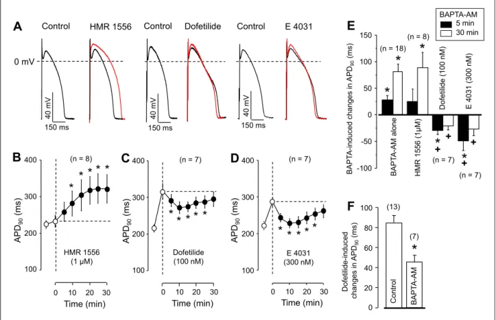
Contrasting to the argumentation above, when the cells were pretreated with IKrblocker (100 nM dofetilide or 300 nM E 4031) BAPTA-AM shortened APD90instead of lengthening it, as it is demonstrated in Fig. 3C-3E. The applied concentrations of IKr
blockers eliminated more than 75% of IKr (28). This shortening effect, however, was temporary - strongly resembling the transient APD90-shortening tendency observed with BAPTA-AM in the presence of BAY K8644 following the immediate APD90
lengthening effect of the former. On the other hand, the APD90- lengthening effect of 100 nM dofetilide was significantly reduced in the presence of 5 µM BAPTA-AM (Fig. 3F). The effect of BAPTA-AM on action potential morphology was sensitive exclusively to the density of IKr, since blocking IKswith 1 µM HMR 1556 (29) failed to alter the effect of BAPTA-AM on action potential duration (Fig. 3B and 3E). The effects of 5 min pretreatment with nisoldipine, BAY K8644, dofetilide and E 4031 (drugs used before the exposure to BAPTA-AM in Fig. 2and Fig.
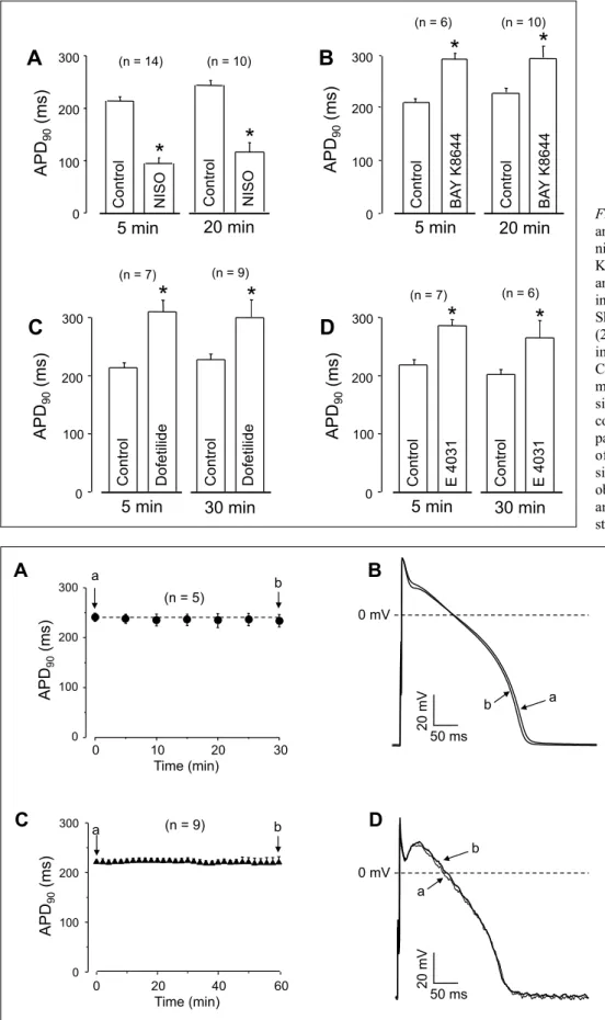
3) on APD90were comparable in magnitude to the those observed after longer exposures, lasting for 20 - 30 min, as shown in Fig. 4.
This is important because any change in APD90recorded in the
B
100 ms40 mV 0 mV
A23187 Control
A
0 mV
100 ms
40 mV
BAPTA-AM Control
100 150 200 250 300
A P D
90(m s)
0 5 10 15 20 25
Time (min)
*
* *
* *
*
* *
* *
BAPTA-AM 5 PM (n = 18) A 23187 1 µM (n = 17)
C
30
*
*
Fig. 1. Effects of changes in [Ca2+]ion action potential duration (APD90). (A), (B): Pairs of representative action potentials recorded from myocytes exposed to 5 µM BAPTA-AM (A) and 1 µM A23187 for 30 min (B). Time-dependent changes in APD90are presented in panel (C). Symbols and bars are means ± SEM, asterisks indicate significant changes from the pre-drug control values. Numbers in parentheses indicate the number of myocytes studied.
Nisoldipine (1 µM)
(n=14)
Time (min)10 20 30
* * *
*
APD90(ms) 100 200 300 400 500
0
B C
BAPTA-AM 5 min 30 min
BAY K8644 (200 nM)
Nisoldipine (1 µM)
BAPTA-AM alone
*
*
*
BAPTA-induced changes in APD90(ms)
*
+(n = 18)
(n = 6)
(n = 14)
*
0 50 100 150
200 +
+
+
D
BAY K8644 (200 nM)
0 10 20 30
* *
Time (min) (n = 6)
*
APD90(ms) 100 200 300 400 500
* * * A
0 mV
150 ms
40 mV
150 ms
40 mV
Control Nisoldipine Control BAY K8644
Fig. 2. Time-dependent effects of BAPTA-AM on action potential duration in the presence of nisoldipine and BAY K8644. (A):
Representative analogue records showing the effects of 5 min exposure to nisoldipine and BAY K 8644 on action potential morphology (black records) Red records indicate superimposed action potentials obtained after 30 min superfusion with BAPTA-AM (in the continuous presence of nisoldipine or BAY K8644). (B), (C): Time-dependent effects of 5 µM BAPTA-AM in the presence of 1 µM nisoldipine (B) and 200 nM BAY K8644 (C) on APD90. Open symbols represent data obtained before the exposure to BAPTA-AM (first symbol: control; second symbol: after 5 min pretreatment with nisoldipine or BAY K8644), filled symbols show data recorded following superfusion with BAPTA-AM in the presence of nisoldipine or BAY K 8644. APD90values measured at 5 and 30 min of BAPTA-AM are presented in panel (D). Symbols, columns and bars are arithmetic means ± SEM, asterisks denote significant differences from pre-BAPTA-AM (control) values, while crosses refer to significant changes comparing to the ‘BAPTA-AM alone’
group (data from Fig. 1C). Numbers in parentheses indicate the number of myocytes studied.
presence of BAPTA-AM cannot be attributed to time-dependent progression of the effect of the drug used for pretreatment.
To demonstrate that not BAPTA - but only BAPTA-AM - is responsible for the observed alterations in action potential morphology, myocytes were superfused with 5 µM BAPTA. No change in action potential duration or in other parameters were observed after 30 min exposure to BAPTA. (Fig. 5Aand 5B) indicating that the presence of the acetoxy-methyl group in the molecule is essential for the development of acute APD- lengthening effect of BAPTA-AM. The stability of our action potentials are documented in Fig. 5Cand 5D. Both the duration as well as the configuration of action potentials failed to alter significantly in untreated canine ventricular cells within the 60 min period of recording.
Effect of BAPTA-AM on IKrcurrent
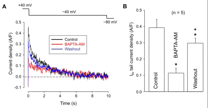
In the first set of experiments, the effect of externally applied BAPTA-AM was studied on IKrin 5 ventricular myocytes using the conventional patch clamp technique. The density of IKrwas evaluated by the amplitude of IKrtail currents recorded upon repolarization to –40 mV. As indicated by Fig. 6, exposure of the cells to 5 µM BAPTA-AM for 5 min decreased the density of IKr
to 32 ± 8 % of the control (from 0.39 ± 0.05 to 0.12 ± 0.03 A/F, P < 0.05, n = 5) in a partially reversible manner, since the current amplitude returned to 76 ± 5% of its control value during a 5 min period of washout with BAPTA-AM-free superfusate. As the
pipette solution contained 10 mM BAPTA in these experiments, suppression of IKr by BAPTA-AM was independent of intracellular Ca2+buffering.
Conventional voltage clamp experiments are not ideal to visualize the consequences of an ion current blockade during the action potential. Therefore action potential voltage clamp experiments have been designed to monitor the changes in IKr
current profile following superfusion with 5 µM BAPTA-AM (Fig. 7). Since this technique is based on pharmacological extraction of IKr(using 1 µM E 4031), measurements had to be performed in two populations of myocytes. The density of IKr
measured in the absence (control) and presence of external BAPTA-AM was 0.50 ± 0.03 and 0.17 ± 0.06 A/F (n = 7 and n
= 5, respectively, P < 0.05), corresponding to a reduction of 66%.
There was a similar difference in the amount of charge carried by IKrduring the action potential: 22 ± 5 versus6 ± 2 mC/F (n = 7 and n = 5, respectively, P < 0.05), a reduction to 27% of the control value. The Ca2+-buffering effect of BAPTA-AM could be excluded in these experiments as well, because the cells had been pre-loaded with BAPTA before their exposure to external BAPTA-AM (see the Methods section for details).
DISCUSSION
In the present study, we have clearly shown that the frequently used Ca2+ chelator BAPTA-AM effectively and
reversibly blocked IKr in canine ventricular myocytes. Our results are in an excellent agreement with the results of Tang et al. (18), who reported an IC50value of 1.3 µM for BAPTA- AM on hERG channels (and similar values for other K+ channels) expressed in HEK 293 cells. These authors also reported an open channel block with BAPTA-AM. Therefore it is somewhat surprising that a similar degree of block was observed under conventional and action potential voltage clamp conditions (reduction to 32 and 34% of the control values, respectively). Probably the consequences of the more positive activation voltage of +40 mV, applied in conventional voltage clamp, compared to the less positive action potential plateau in action potential voltage clamp experiments, was offset by the higher stimulation frequency of 1 Hz used in the latter experimental design. The most important message of both voltage clamp experiments is that external BAPTA-AM must not be used as an experimental Ca2+ chelator when studying the Ca2+dependence of cardiac ion currents in whole cell systems expressing hERG channels, because of the direct inhibitory effect of BAPTA-AM on IKrcurrent.
Based on the present results, the exposure to BAPTA-AM displays a very complex pattern of changes in APD90, so
explanation of the results is necessarily speculative. The rapidly developing acute effect of BAPTA-AM, the significant prolongation of APD90, is very likely due to the BAPTA-AM- induced inhibition of IKr, an effect evident within the initial 5 min of exposure. This explanation apparently contradicts to the finding that the acute lengthening effect of BAPTA-AM was reduced by nisoldipine and augmented by BAY K8644, which might suggest some kind of involvement of ICain the concomitant changes of APD90. However, not only ICa was increased or suppressed by BAY K8644 and nisoldopine, respectively, but also the pre- BAPTA value of APD90 as well as the level of the plateau potential. Consequences of the IKr-blockade strongly increases when APD90is longer (30, 31), as was the case in the presence of BAY K8644, and decreases if APD90is shorter, as was observed in the presence of nisoldipine. In addition, the relative importance of IKrin regulation of APD90strongly increases with positive shifts in the plateau potential due to the faster activation of IKr at more positive voltages (28). This may also contribute to the stronger BAPTA-AM-induced prolongation in the presence of BAY K8644 versus the smaller change observed in nisoldipine.
When the IKr-blocking effect of BAPTA-AM was prevented by pretreatment of the cells with IKr blocking drugs, like
E
E 4031 (300 nM)
Dofetilide (100 nM)
BAPTA-AM alone
* *
BAPTA-induced changes in APD90(ms)
*
(n = 18)
(n = 7) (n = 7) (n = 8)
HMR 1556 (1µM)
* *
-100 -50 0 50 100 150
+ + +
+
BAPTA-AM 5 min 30 min
Dofetilide-induced changes in APD90(ms) 0 20 40 60 80 100
(7) (13)
*
BAPTA-AM
Control
F
Time (min) 0 10 20 30
E 4031 (300 nM)
(n = 7)
* * * * *
100 200 300 400
APD90(ms) Dofetilide
(100 nM) 0 10 20 30
(n = 7)
Time (min)
* * * *
*
100 200 300 400
APD90(ms)
C D
0 10 20 30 Time (min) HMR 1556
(1 µM) APD90(ms)
* *
*
(n = 8)
* *
100 200 300
B
400A
150 ms
40 mV
150 ms
40 mV
150 ms
40 mV
0 mV
Dofetilide Control
Control HMR 1556 Control E 4031
Fig. 3. Time-dependent effects of BAPTA-AM on action potential duration in the presence of different delayed rectifier potassium current blockers. (A): Effects of 5 min exposure to 1 µM HMR 1556, 100 nM dofetilide and 300 nM E 4031 on action potential configuration (black records). Action potentials taken following 30 min BAPTA-AM superfusion (in the continuous presence of HMR 1556, dofetilide or E 4031) are shown in red color. Panels (B) – D) indicate average data obtained with the IKs-blocker HMR 1556 (B), the IKrblocker dofetilide (C) or E-4031 (D). Open symbols represent data measured before the exposure to BAPTA-AM (first symbol:
control; second symbol: 5 min pretreatment with HMR 1556, dofetilide or E 4031), filled symbols show data after superfusion with BAPTA-AM in the presence of HMR 1556, dofetilide or E 4031. (E): Changes in APD90values observed at 5 and 30 min of BAPTA- AM treatment after pretreatment with various K+channel blockers. (F): APD-lengthening effect of 100 nM dofetilide alone and in the presence of 5 µM BAPTA-AM. Symbols, columns and bars are arithmetic means ± SEM, asterisks denote significant differences from pre-BAPTA-AM (control) values, while crosses refer to significant changes comparing to the ‘BAPTA-AM alone’ group. Numbers in parentheses indicate the number of myocytes studied. In panel (F), the asterisk denotes significant difference obtained in the absence and presence of BAPTA-AM.
dofetilide or E 4031, BAPTA-AM initially shortened APD90, an effect likely related to the reduction of [Ca2+]i. The most plausible explanation for this BAPTA-AM-induced shortening is the conversion of the normally inwardly directed Na+/Ca2+
exchange (NCX) current to an outwardly directed one as a consequence of the reduced [Ca2+]i. In line with this, the tendency of APD-shortening observed after the initial BAPTA- AM-induced prolongation in myocytes pretreated with BAY
Control
*
Dofetilide
(n = 7)
300 200 100 0
(n = 6)
Control
*
BAY K8644
5 min
*
Control BAY K8644
(n = 10)
20 min APD90(ms)
0 300 200 100
(n = 10) (n = 14)
Control
Control NISO
5 min 20 min APD(ms)90
*
*
NISO
30 min
Control
*
Dofetilide
APD90(ms)
0 300
200 100
5 min
(n = 9)
30 min
Control
*
E 4031
APD90(ms)
0 300
200 100
5 min
Control
(n = 6)
*
E 4031
(n = 7)
A B
C D
Fig. 4. Comparison of short term and long term effects of 1 µM nisoldipine (A), 200 nM BAY K8644 (B), 100 nM dofetilide (C) and 300 nM E 4031 (D) on APD90
in canine ventricular myocytes.
Short term (5 min) and long term (20 – 30 min) effects were studied in different experiments.
Columns and bars are arithmetic means ± SEM, asterisks denote significant differences from control values, and numbers in parentheses indicate the number of myocytes studied. No significant differences were observed between the long term and short term effects of the studied agents.
A B
Time (min)
0 20 40 60
0 100 200
300 a (n = 9) b
C
0 mV a
b
20 mV
50 ms
D
APD90(ms)
Time (min)
0 10 20 30
0 100 200
300 a b
(n = 5)
APD90(ms)
b a
20 mV
50 ms 0 mV
Fig. 5. Lack of effect of 5 µM externally applied BAPTA on action potential duration (A) and action potential configuration (B).
Stability of action potential duration (C) and action potential morphology (D) in untreated cardiomyocytes.
Superimposed action potentials, recorded from the same cell at the beginning (a) and the end (b) of the recording period are presented in panels (B) and (D). Symbols and bars represent mean ± SEM values, numbers in parentheses indicate the number of myocytes studied.
K8644 was also pronounced probably because the long APD90
and the positive plateau potential favored the reverse mode activity of NCX (32, 33). This effect is in line with the finding that buffering [Ca2+]isignificantly shortened APD90in rat, guinea
pig and ferret cardiomyocytes (34, 35) and with own unpublished results, obtained in guinea pig ventricular cells, indicating that net NCX current was outward when recorded with EGTA-containing patch pipettes.
IKrdensity(A/F) 0.6 0.4 0.2 0 30 20 10
Charge carried (mC/F) 0 Control (7) BAPTA-AM(5)
Control (7) BAPTA-AM(5)
*
*
B A
C
Vm(mV)
Time (ms) Control
BAPTA-AM
IKrdensity (A/F) 50
0
-50
-100 0.6 0.4 0.2 0 -0.2
0 100 200 300
Fig. 7. Effect of BAPTA-AM on IKrunder action potential voltage clamp conditions. (A): The command action potential (above) and representative superimposed IKr current traces recorded in the absence (control) and presence of 5 µM BAPTA- AM applied in the superfusate for 5 min. Both groups of cells were pre-loaded with BAPTA- AM for 20 min in order to load the cells with BAPTA before the measurements. (B) and (C): Peak amplitude of IKr and the total change carried by the current, respectively, measured in absence and presence of extracellular BAPTA-AM.
Columns and bars are arithmetic means ± SEM, asterisks denote significant differences between the groups of cells exposed - or not exposed - to extracellular BAPTA-AM. Numbers in parentheses indicate the number of myocytes studied.
*
(n = 5)
IKrtail current density (A/F) íP9
íP9 +40 mV
Time (s)
0 2 4 6 8 10
-0.1 0.0 0.1 0.2 0.3 0.4 0.5
Current density (A/F)
0.0 0.1 0.2 0.3 0.4 0.5
C on tro l W as ho ut
B A P TA -A M
B A
Control BAPTA-AM Washout
*
+Fig. 6. Effect of BAPTA-AM on IKrunder conventional voltage clamp conditions. (A): Representative set of superimposed IKrtail current traces recorded following repolarization to –40 mV. The current was activated at +40 mV for 500 ms as shown by the applied voltage protocol (above). The cell was exposed to 5 µM BAPTA-AM for 5 min, which was followed by a subsequent 5 min period of washout. (B): Average IKrtail current amplitudes measured before (control) in the presence of, and after the washout of BAPTA-AM.
Columns and bars are arithmetic means ± SEM, asterisks denote significant differences from control, the cross refers to significant changes upon washout of BAPTA-AM, and the number in parentheses indicates the number of myocytes studied.
As documented in Figs. 3C and 3D, the BAPTA-AM- induced APD90-shortening effect was transient. In other words, there was a progressive tendency of prolongation during a long- lasting exposure to BAPTA-AM. This was also evident under control conditions, where no transient shortening was observed (only a small notch after 5 min). The final lengthening tendency of APD90 is likely related to the combination of two further effects of progressive buffering of [Ca2+]i. These are the augmentation of the ICa due to the reduction of its Ca2+- dependent inactivation (22) and offsetting some Ca2+-dependent outward currents, like the Ca2+-dependent Cl- current by buffering [Ca2+]i(7, 8). In summary, buffering of [Ca2+]iresulted in a biphasic response of APD90: a transient initial shortening was followed by progressive prolongation when the IKr-blocking effect of BAPTA-AM was prevented by the pretreatment with dofetilide or E 4031.
Acknowledgements: This work was funded by the National Research Development and Innovation Office (NKFIH- K115397, NKFIH-K109736, NKFIH-PD120794 and OTKA:
ANN-113273). Support was obtained from GINOP-2.3.2.-15- 2016-00040 and EFOP-3.6.2-16-2017-00006 projects, which are co-financed by the European Union and the European Regional Development Fund. Further support was provided by the Janos Bolyai Research Scholarship of the Hungarian Academy of Sciences (to NS and BH) and the University of Debrecen (RH/751/2015) to NS. The work was also supported by the UNKP-17-4-III-DE-201 New National Excellence Program of the Ministry of Human Capacities. The authors thank Miss Eva Sagi for excellent technical assistance.
B. Horvath and N. Szentandrassy have equally contributed to this study.
Conflict of interests: None declared.
REFERENCES
1. Tan HL, Kupershmidt S, Zhang R, et al. A calcium sensor in the sodium channel modulates cardiac excitability Nature 2002; 415: 442-447.
2. Mori M, Konno T, Ozawa T, Murata M, Imoto K, Nagayama K. Novel interaction of the voltage-dependent sodium channel (VDSC) with calmodulin: does VDSC acquire calmodulin-mediated Ca2+-sensitivity? Biochemistry 2000;
39: 1316-1323.
3. Lee A, Wong ST, Gallagher D,et al. Ca2+/calmodulin binds to and modulates P/Q-type calcium channels. Nature1999;
399: 155-159.
4. DeMaria CD, Soong TW, Alseikhan BA, Alvania RS, Yue DT. Calmodulin bifurcates the local Ca2+ signal that modulates P/Q-type Ca2+ channels. Nature 2001; 411:
484-489.
5. Tuteja D, Xu D, Timofeyev V, et al. Differential expression of small-conductance Ca2+-activated K+channels. SK1, SK2, and SK3 in mouse atrial and ventricular myocytes. Am J Physiol Heart Circ Physiol2005; 289: H2714-H2723.
6. Nattel S, Qi XY. Calcium-dependent potassium channels in the heart: clarity and confusion. Cardiovasc Res2014; 101:
185-186.
7. Horvath B, Vaczi K, Hegyi B, et al. Sarcolemmal Ca2+-entry through L-type Ca2+channels controls the profile of Ca2+- activated Cl-current in canine ventricular myocytes. J Mol Cell Cardiol2016; 97: 125-139.
8. Hegyi B, Horvath B, Vaczi K, et al. Ca2+-activated Cl– current is antiarrhythmic by reducing both spatial and
temporal heterogeneity of cardiac repolarization. J Mol Cell Cardiol2017; 109: 27-37.
9. Weber CR, Piacentino V, Ginsburg KS, Houser SR, Bers DM. Na+-Ca2+exchange current and submembrane [Ca2+] during the cardiac action potential. Circ Res 2002; 90:
182-189.
10. Quednau BD, Nicoll DA, Philipson KD. Tissue specificity and alternative splicing of the Na+/Ca2+exchanger isoforms NCX1, NCX2, and NCX3 in rat. Am J Physiol1997; 272:
C1250-C1261.
11. Guinamard R, Chatelier A, Demion M, et al. Functional characterization of a Ca2+-activated non-selective cation channel in human atrial cardiomyocytes. J Physiol (Lond) 2004; 558: 75-83.
12. Guinamard R, Bouvagnet P, Hof T, Liu H, Simard C, Salle L. TRPM4 in cardiac electrical activity. Cardiovasc Res 2015; 108: 21-30.
13. Billman GE, McIlroy B, Johnson JD. Elevated myocardial calcium and its role in sudden cardiac death. FASEB J1991;
5: 2586-2592.
14. Billman GE. Intracellular calcium chelator, BAPTA-AM, prevents cocaine-induced ventricular fibrillation. Am J Physiol Heart Circ Physiol1993; 265: H1529-H1535.
15. Warren M, Huizar JF, Shvedko AG, Zaitsev AV.
Spatiotemporal relationship between intracellular Ca2+
dynamics and wave fragmentation during ventricular fibrillation in isolated blood-perfused pig hearts. Circ Res 2007; 101: e90-e101.
16. Ogawa M, Lin SF, Weiss JN, Chen PS. Calcium dynamics and ventricular fibrillation. Circ Res2008; 102: e52. doi:
10.1161/CIRCRESAHA.108.171538
17. Younes A, Lyashkov AE, Graham D, et al. Ca2+-stimulated basal adenylyl cyclase activity localization in membrane lipid microdomains of cardiac sinoatrial nodal pacemaker cells. J Biol Chem2008; 283: 14461-14468.
18. Tang Q, Jin MW, Xiang JZ, et al. The membrane permeable calcium chelator BAPTA-AM directly blocks human ether a-go-go-related gene potassium channels stably expressed in HEK 293 cells. Biochem Pharmacol2007; 74: 1596-1607.
19. Szentandrassy N, Papp F, Hegyi B, Krasznai Z, Nanasi PP.
Tetrodotoxin blocks native cardiac L-type Ca2+channels but not Cav1.2 channels expressed in HEK cells. J Physiol Pharmacol2013; 64: 807-810.
20. Szabo G, Szentandrassy N, Biro T, et al. Asymmetrical distribution of ion channels in canine and human left ventricular wall: epicardium versus midmyocardium.
Pflugers Arch2005; 450: 307-316.
21. Szentandrassy N, Banyasz T, Biro T, et al. Apico-basal inhomogeneity in distribution of ion channels in canine and human ventricular myocardium. Cardiovasc Res 2005; 65:
851-860.
22. Kistamas K, Szentandrassy N, Hegyi B, et al. Changes in intracellular calcium concentration influence beat-to-beat variability of action potential duration in canine ventricular myocytes. J Physiol Pharmacol2015; 66: 73-81.
23. Szentandrassy N, Horvath B, Vaczi K, et al. Dose-dependent electrophysiological effects of the myosin activator omecamtiv mecarbil in canine ventricular cardiomyocytes. J Physiol Pharmacol2016; 67: 483-489.
24. Chen-Izu Y, Izu LT, Nanasi PP, Banyasz T. From action potential-clamp to ‘onion-peeling’ technique - recording of ionic currents under physiological conditions. In: Patch Clamp Technique, Kaneez FS (ed). InTech 2012, 143-162.
25. Chen-Izu Y, Izu LT, Hegyi B, Banyasz T. Recording of ionic currents under physiological conditions: action potential- clamp and ‘onion-peeling’ techniques. In: Modern Tools of Biophysics, Jue T. (ed). Springer 2017; 31-48.
26. Sanguinetti MC, Kass RS. Voltage-dependent block of calcium channel current in the calf cardiac Purkinje fiber by dihydropyridine calcium channel antagonists. Circ Res 1984; 55: 336-348.
27. Sanguinetti MC, Krafte DS, Kass RS. Voltage-dependent modulation of Ca++ channel current in heart cells by BAY K8644. J Gen Physiol1986; 88: 369-392.
28.Jurkiewicz NK, Sanguinetti MC. Rate-dependent prolongation of cardiac action potentials by a methanesulfonanilide class III antiarrhythmic agent specific block of rapidly activating delayed rectifier K+current by dofetilide. Circ Res1993; 72: 75-83.
29. Thomas GP, Gerlach U, Antzelevitch C. HMR 1556, a potent and selective blocker of slowly activating delayed rectifier potassium current. J Cardiovasc Pharmacol2003;
41: 140-147.
30. Banyasz T, Horvath B, Virag L, et al. Reverse rate dependency is an intrinsic property of canine cardiac preparations. Cardiovasc Res2009; 84: 237-244.
31. Barandi L, Virag L, Jost N, et al. Reverse rate-dependent changes are determined by baseline action potential duration in mammalian and human ventricular preparations. Basic Res Cardiol2010; 105: 315-323.
32. Ginsburg KS, Weber CR, Bers DM. Cardiac Na+Ca2+exchanger: dynamics of Ca2+-dependent activation
and deactivation in intact myocytes. J Physiol (Lond) 2013;
591: 2067-2086.
33. Birinyi P, Toth A, Jona I, et al. The Na+/Ca2+ exchange blocker SEA0400 fails to enhance cytosolic Ca2+transient and contractility in canine ventricular cardiomyocytes.
Cardiovasc Res 2008; 78: 476-484.
34. Janvier NC, Mcmorn SO, Harrison SM, Taggart P, Boyett MR. The role of inward Na+/Ca2+ exchange current in electrical restitution in ferret ventricular cells. J Physiol (Lond) 1997; 504: 301-314.
35. Mitchell M, Powell T, Terrar DA, Twist VW. The effects of ryanodine, EGTA and low-sodium on action potentials in rat and guinea-pig ventricular myocytes: evidence for two inward currents during the plateau. Br J Pharmacol1984;
81: 543-550.
R e c e i v e d : January 2, 2018 A c c e p t e d : February 26, 2018
Author’s address: Prof. Peter P. Nanasi, Department of Physiology and Department of Dental Physiology and Pharmacology, University of Debrecen, H-4012 Debrecen, P.O.Box 22, Hungary.
E-mail: nanasi.peter@med.unideb.hu
![Fig. 1. Effects of changes in [Ca 2+ ] i on action potential duration (APD 90 ). (A), (B): Pairs of representative action potentials recorded from myocytes exposed to 5 µM BAPTA-AM (A) and 1 µM A23187 for 30 min (B)](https://thumb-eu.123doks.com/thumbv2/9dokorg/1399333.117082/3.892.87.811.652.1087/effects-changes-potential-duration-representative-potentials-recorded-myocytes.webp)