International Journal of
Molecular Sciences
Article
Investigation of H 2 S Donor Treatment on Neutrophil Extracellular Traps in Experimental Colitis
Szilvia Török1, Nikoletta Almási1, Zsuzsanna Valkusz2, AnikóPósa1,3, Csaba Varga1and Krisztina Kupai1,2,*
Citation: Török, S.; Almási, N.;
Valkusz, Z.; Pósa, A.; Varga, C.;
Kupai, K. Investigation of H2S Donor Treatment on Neutrophil
Extracellular Traps in Experimental Colitis.Int. J. Mol. Sci.2021,22, 12729.
https://doi.org/10.3390/ijms 222312729
Academic Editors:
Marcin Magierowski and Binghe Wang
Received: 8 October 2021 Accepted: 22 November 2021 Published: 25 November 2021
Publisher’s Note:MDPI stays neutral with regard to jurisdictional claims in published maps and institutional affil- iations.
Copyright: © 2021 by the authors.
Licensee MDPI, Basel, Switzerland.
This article is an open access article distributed under the terms and conditions of the Creative Commons Attribution (CC BY) license (https://
creativecommons.org/licenses/by/
4.0/).
1 Department of Physiology, Anatomy and Neuroscience, Faculty of Science and Informatics, University of Szeged, H-6726 Szeged, Hungary; tszilvia@bio.u-szeged.hu (S.T.);
almasi@expbio.bio.u-szeged.hu (N.A.); paniko@bio.u-szeged.hu (A.P.); vacs@bio.u-szeged.hu (C.V.)
2 Albert Szent-Györgyi Clinical Center, Department of Medicine, Medical Faculty, University of Szeged, H-6720 Szeged, Hungary; valkusz.zsuzsanna@med.u-szeged.hu
3 Interdisciplinary Excellence Center, Department of Physiology, Anatomy and Neuroscience, Faculty of Science and Informatics, University of Szeged, H-6726 Szeged, Hungary
* Correspondence: kupai.krisztina@med.u-szeged.hu; Tel.: +36-62-544-884
Abstract: Inflammatory bowel diseases (IBD) are chronic, immune-mediated disorders, which affect the gastrointestinal tract with intermittent ulceration. It is increasingly clear that neutrophil extracellular traps (NETs) seem to have a role in IBD; however, the associated pathogenesis is still not known. Furthermore, several conventional therapies are available against IBD, although these might have side effects. Our current study aimed to investigate the effects of hydrogen sulfide (H2S) treatment on NETs formation and on the expression of inflammatory mediators in experimental rat colitis. To model IBD, 2,4,6-trinitrobenzenesulfonic acid (TNBS) was administered intracolonically (i.c.) to Wistar–Harlan male rats. Animals were treated (2 times/day) with H2S donor Lawesson’s reagent per os. Our results showed that H2S treatment significantly decreased the extent of colonic lesions. Furthermore, the expression of members of NETs formation: peptidyl arginine deiminase 4 (PAD4), citrullinated histone H3 (citH3), myeloperoxidase (MPO) and inflammatory regulators, such as nuclear transcription factor-kappa B (NF-κB) and high-mobility group box 1 (HMGB1) were reduced in H2S treated group compared to TNBS. Additionally, H2S donor administration elevated the expression of ubiquitin C-terminal hydroxylase L1 (UCHL-1), a potential anti-inflammatory mediator. Taken together, our results showed that H2S may exert anti-inflammatory effect through the inhibition of NETs formation, which suggests a new therapeutic approach against IBD.
Keywords:hydrogen sulfide; inflammation; inflammatory bowel disease; colitis; neutrophil extracel- lular traps
1. Introduction
Inflammatory bowel diseases (IBD) are chronic, immune-mediated disorders, which affect the gastrointestinal (GI) tract with intermittent ulceration. Despite extensive research, the pathogenesis of IBD is not precisely known. IBD occurred worldwide at the turn of the 21st century with stabilized incidence but still high prevalence in industrialized countries and with a continuous increase in developing countries [1]. Crohn’s disease (CD) and ulcerative colitis (UC) are the two major types of IBD with similar symptoms but different epidemiological and clinical features [2]. Both disorders are characterized by phases of relapses and remissions, weight loss, fever, abdominal pain, diarrhea, and rectal bleeding [3]. These symptoms affect the quality of life of the patients, so the primary purposes of IBD therapy are the induction and maintenance of remission along with symptomatic treatments [4]. Currently, several therapeutic options exist [5], although they often cause side effects [6]. Therefore, the development of new therapies and potential therapeutic targets are urgently needed.
Several animal models have been established to investigate IBD, which represent the symptomatic, morphological, and histopathological features of human IBD. Among them,
Int. J. Mol. Sci.2021,22, 12729. https://doi.org/10.3390/ijms222312729 https://www.mdpi.com/journal/ijms
Int. J. Mol. Sci.2021,22, 12729 2 of 16
chemically induced IBD models, such as 2,4,6-trinitrobenzenesulfonic acid (TNBS), dextran sodium sulfate (DSS), oxazolone, or acetic acid colitis models are widely used because of their cost-effectiveness, good reproducibility, and relative simplicity of induction [7]. The TNBS-induced colitis model was originally described by Morris et al. [8]. In rodents, a single intracolonic administration of the hapten reagent (TNBS) dissolved in ethanol results in serious transmural inflammation and ulceration of the colon. As a vehicle, ethanol causes destruction of the mucosal barrier, which enables TNBS to access proteins in the inner layers of the colonic tissue. Interaction of the hapten with colonic proteins initiates Th-1-mediated immune response [9]. Based on the aforementioned features, TNBS-induced colitis model is a suitable tool for investigating the pathogenesis of IBD and developing novel therapeutic options against these ailments.
Hydrogen sulfide (H2S) was known only as a toxic gas for many years, but in the last two decades, it has emerged as an important mediator of normal physiological pathways in various biological systems [10]. H2S is generated endogenously in mammals by many enzymatic and non-enzymatic pathways [11,12]. Several investigations revealed that H2S has a wide range of regulatory functions and plays a role in many physiological and pathological processes [13–16]. In the GI tract, H2S plays a role in normal smooth muscle function [17], mucosal defence [18], and in the stimulation of epithelial secretion [19]. In pathological conditions, H2S seems to reduce inflammation by the enhancement of ulcer healing [20,21].
Neutrophil infiltration is a typical phenomenon in IBD [22]. Neutrophils play a cen- tral role in innate immunity, as they are the first immune cells that migrate to the site of inflammation [23]. In response to infection or inflammatory stimuli, neutrophils can form neutrophil extracellular traps (NETs), a web-like structure, which consists of DNA, histone proteins, granular proteins, such as neutrophil elastase (NE) and myeloperoxidase (MPO), and cytoplasmatic components [24,25]. During NETs formation, ROS-induced peptidyl arginine deiminase 4 (PAD4) activation forms citrullinated histone H3 (citH3), which initiates chromatin decondensation [26]. Then NETs are released into the extracel- lular environment to immobilize and catch pathogens [27,28]. The process of producing and releasing NETs is referred to as NETosis, which was described by Brinkmann et al.
in 2004 [24]. Although NETs play an important role in host defence by protecting from infections, excessive formation of NETs is involved in the pathogenesis of several inflam- matory diseases and tissue damage [27–29]. Accordingly, members of NETs formation can be used as important biomarkers in several ailments [28,30] and for potential therapeutic targets [31–33]. Recently, few studies suggested the relationship between H2S and NETs formation [34,35].
Besides various stimuli, such as pathogens, cytokines and enhanced ROS production, high-mobility group box 1 (HMGB1), a proinflammatory mediator, can also induce NETs formation via Toll-like receptor 4 (TLR4) [36]. Additionally, it is demonstrated that the levels of fecal HMGB1 correlates with the severity of mucosal inflammation; therefore, HMGB1 is suggested as a fecal biomarker of IBD [37]. NF-κB, as a principal regulator of inflammatory processes, is involved in the pathogenesis of several inflammatory diseases, including IBD [38]. Several animal models of inflammatory diseases demonstrated that the obstruction of the NF-κB signalling pathway is an effective treatment by inhibiting inflammation and tissue damage [38–40]. Furthermore, it is increasingly clear that ubiquitin C-terminal hydroxylase L1 (UCHL-1), a deubiquitinating enzyme, may have a role in the reduction of inflammation [41,42]. In addition, in vascular cells, it was shown that UCHL-1 suppressed NF-κB activation via deubiquitinating IκB-α, therefore preventing its proteasomal degradation [43].
Therefore, in this current study, we hypothesized that H2S can inhibit the colonic NETs formation in a rat model of colitis and may contribute to the reduction of inflammation and the healing of ulcers. Our results showed that an exogenously administered H2S donor suppressed the expression of the members of NETs formation and other inflammatory mediators in TNBS-induced colitis. Furthermore, H2S donor treatment increased the levels
Int. J. Mol. Sci.2021,22, 12729 3 of 16
of UCHL-1, a potential candidate for anti-inflammatory processes. Based on our results, we suggest that NETs components may serve as potential therapeutic targets in IBD and H2S administration seems to provide a promising strategy against IBD through the inhibition of NETosis.
2. Materials and Methods 2.1. Experimental Animals
All procedures were performed in accordance with the standards of the European Community guidelines for the Care and Use of Laboratory Animals and were approved by the Institutional Ethics Committee (XX./4799/2015, 15 December 2015) at the University of Szeged.
Male Wistar–Harlan rats (225–250 g) were purchased from Toxi-Coop Ltd. (Budapest, Hungary) and were kept in a conventional animal house under controlled conditions (temperature 22±2◦C; relative humidity 55±10%), with a 12 h light-dark cycle. Standard rat chow and tap water were supplied ad libitum throughout the experiments.
2.2. Drug Preparations
To induce colitis 2,4,6-trinitrobenzenesulfonic acid (TNBS) was prepared in 50%
ethanol (EtOH) and distilled water mixture. Lawesson’s reagent suspended in 0.5% car- boxymethylcellulose (CMC) was used as an H2S donor. As a positive control, sulfasalazine (SASP) was applied, which was dissolved in physiological saline (0.9%). All chemicals were purchased from Sigma-Aldrich (St. Louis, MO, USA) and were prepared freshly before treatments. Animals were anesthetized with thiopental (Tiobarbital Braun, 0.5 g, B. Braun Medical SA, Barcelona, Spain) dissolved in physiological saline (0.9%).
2.3. Experimental Design and Drug Administration
After one week of acclimatization rats were divided randomly into three groups:
absolute control (no treatment,n= 8), EtOH group (0.25 mL 50% ethanol enema,n= 8), and TNBS group (10 mg in 0.25 mL of 50% ethanol,n= 34). The experimental colitis was achieved based on Morris’ method [8]. Following overnight fasting, animals were challenged with a single dose of 0.25 mL TNBS intracolonically (i.c.) with an 8 cm long soft polyethylene tube through the anus under mild anesthesia (thiopental, i.p. 40 mg/kg).
Next, the TNBS group was divided further and treated immediately after TNBS instillation per os for 3 days (twice/day) with the following drugs: Lawesson’s reagent as a H2S donor (TNBS+ H2S donor,n= 16) at dose of 18.75µM/kg/day (dissolved in 0.5% CMC,n= 8);
SASP as a positive control (TNBS+SASP,n= 10) at dose of 50 mg/kg/day (dissolved in physiological saline (0.9%); CMC (0.5%, vehicle of Lawesson’s reagent). The selection of the drug doses was based on our previous findings in the same experimental colitis model [44]. Rats were euthanized (thiopental, i.p. 100 mg/kg) 72 h after TNBS instillation and the distal 8 cm portion of the colon was excised, longitudinally opened, washed with ice-cold physiological saline, and photographed (Panasonic Lumix DMC-TZ6, Panasonic Co., Kadoma, Osaka, Japan) for determination of macroscopic inflammation. Finally, the colon samples were frozen and powdered in liquid nitrogen and kept at−80◦C until used for biochemical analysis.
2.4. Evaluation of Macroscopic Inflammatory Damage
The extents of macroscopically apparent inflammation, ulceration, and tissue necrosis were determined in a randomized manner from the colored images, using proprietary com- puterized planimetry software developed in our laboratory (Stat_2_1_1, Szeged, Hungary), based on planimetrics. The area of macroscopically visible mucosal lesion was calculated and represented as the percentage (%) of the total 8 cm colonic segment area.
Int. J. Mol. Sci.2021,22, 12729 4 of 16
2.5. Measurement of Colonic MPO Enzyme Activity
The colonic inflammatory damage was also determined by the measurement of myeloperoxidase (MPO) activity, employing a modified method described by Bradley et al. [45]. MPO is primarily expressed in neutrophil granulocytes, and the activity of the enzyme is commonly used to quantify neutrophil infiltration. Approximately 30 mg of frozen, powdered colon tissue samples were homogenized (Benchmark Scientific Handheld homogenizer D1000, 13,500 rev/min, 2×10 s, Benchmark Scientific, NJ, MA, USA) in ice-cold phosphate buffer (50 mM, pH 6.0), containing 0.5% hexadecyltrimethylammonium- bromide (HETAB). To further break cell membranes, homogenates were subjected to freeze- thaw cycles four times in liquid nitrogen and a 37◦C water bath. Then, homogenates were centrifuged at 10,000×gfor 15 min at 4◦C. In a 96-well plate, 12µL from each supernatant was mixed with 280µL phosphate buffer (50 mM, pH 6.0) containing 0.167 mg/mLO- dianisidine dihydrochloride (Sigma-Aldrich, St. Louis, MO, USA), and the reaction was initiated with 10µL of 0.03% hydrogen peroxide. After shaking for 90 s, the activity of MPO was detected spectrophotometrically at 490 nm (Benchmark Microplate Reader, Bio-Rad Laboratories, Hercules, CA, USA). Results were expressed asµU/mg protein.
2.6. Expression of the Inflammatory Mediators and NETosis Markers by Western Blot Analyses In order to detect expression changes of inflammatory mediators and NETosis markers Western blot analyses were performed. Approximately 30 mg of frozen, powdered colon samples were homogenized with an ultrasonicator (UP100H Hielscher, 3×10 s, Teltow, Germany) in Radioimmunoprecipitation assay (RIPA) buffer (Merck Millipore, Burlington, MA, USA), supplemented with phenylmethanesulfonyl fluoride (PMSF) (Sigma-Aldrich, St. Louis, MO, USA) with a tissue to homogenization buffer ratio of 1:4. The supernatants were collected by centrifugation at 12,000 rpm for 10 min at 4◦C. After determination of protein concentrations by Bradford assay, 50µg protein of each lysate was mixed with reducing sample buffer (Thermo Scientific, Waltham, MA, USA), heated to 95◦C for 5 min and loaded onto 10% sodium dodecyl sulfate (SDS)-polyacrylamide gels. After separation by electrophoresis (90 V, 2 h), proteins were transferred onto nitrocellulose membranes (Amersham Pharmacia Biotech., Buckinghamshire, UK) for 2.5 h at 35 V. The transfer efficiency was checked by Ponceau staining. Membranes were blocked with 5%
non-fat dry milk, except for citH3 (5% bovine serum albumin (BSA)), in Tris-buffered saline containing 0.05% Tween 20 (TBS-T) overnight at 4◦C. After three times 10 min washing in TBS-T, membranes were incubated with primary antibodies in 1% milk or in case of citH3 in 1% BSA, for 2 h at room temperature (RT) or overnight at 4◦C: anti- PAD4 (1:1000, overnight, Proteintech, Rosemont, IL, USA, 17373-1-AP), anti-citH3 (1:750, 2 h, Abcam, Cambridge, UK, ab5103), anti-MPO (1:1000, overnight, Abcam, Cambridge, UK, ab208670), anti-HMGB1 (1:1000, 2 h, Abcam, Cambridge, UK, ab79823), anti-NF- κB p65 (1:500, 2 h, Abcam, Cambridge, UK, ab16502), anti-UCHL-1 (1:500, 2 h, Abcam, Cambridge, UK, ab108986). Then membranes were rinsed again for 3×10 min in TBS-T and were incubated with anti-rabbit or anti-mouse horseradish peroxidase (HRP)-conjugated secondary antibody (1:5000, DAKO Agilent, Santa Clara, CA, USA) in 1% non-fat dry milk or 1% BSA for 1 h at RT. After washing in TBS-T, membranes were developed with Immobilon Western Chemiluminescent HRP Substrate (Merck Millipore, Burlington, MA, USA) to visualize proteins. Signals were detected by Uvitec gel documentation system (Uvitec Ltd., Cambridge, UK), and densities were analysed by Quantity One Software version 4.5 (Bio-Rad Laboratories, Hercules, CA, USA). Finally, to check the exact amount of the loaded proteins, membranes were stripped, blocked in 5% BSA, and probed with anti-β-actin (1:10,000, Abcam, Cambridge, UK, ab20272) primary antibody in 1% BSA for 2 h at RT, followed by incubation with anti-mouse HRP-conjugated secondary antibody (1:5000; 1 h, RT, DAKO Agilent, Santa Clara, CA, USA), and detected as described above.
Results are presented as relative expressions normalized toβ-actin.
Int. J. Mol. Sci.2021,22, 12729 5 of 16
2.7. Protein Determination
The total protein contents were measured by Bradford assay. Aliquots of 20µL of the diluted samples (25×or 40×with distilled water) were mixed with 980µL distilled water, and 200µL of Bradford reagent was added to each sample. Following 10 min of incubation, samples were assayed spectrophotometrically at 595 nm and compared to bovine serum albumin (BSA) standards. Protein concentration was expressed as mg protein/mL.
2.8. Statistical Analysis
All data are expressed as mean±standard error of the mean (S.E.M.). Statistical analysis was carried out by SigmaPlot 12.0 for Windows (Systat Software Inc., San Jose, CA, USA). Parameters were shown normal distribution, and data were assessed by one-way ANOVA followed by Holm–Sidak post hoc test in all measurements. A probability (p) value of less than 0.05 was accepted as statistically significant.
3. Results
3.1. Protective Effect of Hydrogen Sulfide Donor on the Severity of Inflammation in TNBS-Induced Rat Colitis
To model colitis, 2,4,6-trinitrobenzenesulfonic acid (TNBS) dissolved in 50% ethanol (EtOH) was given intracolonically to rats, and 72 h after challenge, the macroscopic colonic injuries were analysed. Both TNBS and its solvent EtOH provoked serious colonic mucosal damage compared to the untreated healthy controls. EtOH, as the vehicle of TNBS, caused hyperaemia and superficial ulcers of the colonic tissue. However, the administration of TNBS resulted in a more intense injury, as approximately 50% of the examined colonic area were damaged with inflammation and obvious hemorrhagic necrosis. In our previous experiment, we determined the most effective dose of H2S donor Lawesson’s reagent (18.75µM/kg/day), and as a positive control sulfasalazine (SASP) (50 mg/kg/day), which both decreased the severity of the colonic inflammation [44]. Our present study confirmed that treatment with an 18.75µM/kg/day dose of H2S donor significantly reduced the extent of colon lesions compared to the TNBS group (26.89± 2.92 vs. 49.28±3.71%).
Similarly, a statistically significant attenuation was found in the severity of inflammation in the positive control group, treated with SASP (20.49±3.63 vs. 49.28±3.71%). CMC, the vehicle of Lawesson’s reagent, was also tested alone and showed no significant difference in the colonic damage compared to the TNBS group, thus suggesting that CMC has no effect on inflammation. The extent of the macroscopically apparent mucosal injury was expressed as the percentage of the total colonic segmented area (Figure1a), and representative images of the colon are shown in Figure1b–g.
3.2. Oral administration of Hydrogen Sulfide Donor Reduced the Elevated Expressions of PAD4 NETosis Marker by TNBS
In this study, the expression of the NETosis marker PAD4 was determined by western blotting (Figure2). PAD4 showed a statistically significant elevation after TNBS adminis- tration compared to the control group (1.26±0.16 vs. 0.75±0.08 relative expression). The expression of PAD4 was significantly reduced by H2S donor and SASP treatment compared to the TNBS group (0.82±0.07 and 0.74±0.07 vs. 1.26±0.16 relative expression).
Int. J. Mol. Sci.2021,22, 12729 6 of 16
Int. J. Mol. Sci. 2021, 22, x FOR PEER REVIEW 6 of 17
Figure 1. (a) The extent of colonic lesion in the different treatment groups in TNBS-induced rat co- litis. Representative images of colonic inflammation in the experimental groups: (b) absolute control (no treatment); (c) EtOH (50% ethanol, the solvent of TNBS); (d) TNBS (2,4,6-trinitrobenzenesulfonic acid enema); (e) H2S donor (TNBS + 18.75 μM/kg/day Lawesson’s reagent); (f) CMC (TNBS + 0.5%
carboxymethylcellulose, the vehicle of Lawesson’s reagent); (g) SASP (TNBS + 50 mg/kg/day sul- fasalazine, as a positive control). Results are shown as mean + S.E.M.; n = 7–12/group; one-way ANOVA, Holm–Sidak post hoc test, && p < 0.001 absolute control vs. EtOH; ## p < 0.001 absolute control vs. TNBS; ** p < 0.001 TNBS vs. TNBS + treatments.
3.2. Oral administration of Hydrogen Sulfide Donor Reduced the Elevated Expressions of PAD4 NETosis Marker by TNBS
In this study, the expression of the NETosis marker PAD4 was determined by west- ern blotting (Figure 2). PAD4 showed a statistically significant elevation after TNBS ad- ministration compared to the control group (1.26 ± 0.16 vs. 0.75 ± 0.08 relative expression).
The expression of PAD4 was significantly reduced by H2S donor and SASP treatment compared to the TNBS group (0.82 ± 0.07 and 0.74 ± 0.07 vs. 1.26 ± 0.16 relative expression).
Figure 1.(a) The extent of colonic lesion in the different treatment groups in TNBS-induced rat colitis.
Representative images of colonic inflammation in the experimental groups: (b) absolute control (no treatment); (c) EtOH (50% ethanol, the solvent of TNBS); (d) TNBS (2,4,6-trinitrobenzenesulfonic acid enema); (e) H2S donor (TNBS + 18.75µM/kg/day Lawesson’s reagent); (f) CMC (TNBS + 0.5%
carboxymethylcellulose, the vehicle of Lawesson’s reagent); (g) SASP (TNBS + 50 mg/kg/day sulfasalazine, as a positive control). Results are shown as mean + S.E.M.;n= 7–12/group; one-way ANOVA, Holm–Sidak post hoc test, &&p< 0.001 absolute control vs. EtOH; ##p< 0.001 absolute control vs. TNBS; **p< 0.001 TNBS vs. TNBS + treatments.
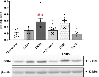
3.3. Alteration in the Expression of Colonic citH3 following Treatments
PAD4-induced citrullination of histone H3 is also used as a marker of NET formation.
Corresponding to the results of PAD4, we observed similar trends in the case of citH3 (Figure3). The expression of the citrullinated protein showed a significant elevation in TNBS-induced inflammation in the colon compared to absolute control and EtOH groups (0.29±0.05 vs. 0.09±0.018 and 0.15±0.02 relative expression); however, both H2S donor and SASP administration significantly decreased the level of citH3 relative to the TNBS group (0.15±0.02 and 0.09±0.01 vs. 0.29±0.05 relative expression).
Int. J. Mol. Sci.Int. J. Mol. Sci. 2021, 22, x FOR PEER REVIEW 2021,22, 12729 7 of 17 7 of 16
Figure 2. Changes of the expression of PAD4 NETosis marker after H2S donor administration. Ab- solute control (no treatment); EtOH (50% ethanol, the solvent of TNBS); TNBS (2,4,6-trinitroben- zenesulfonic acid enema); H2S donor (TNBS + 18.75 μM/kg/day Lawesson’s reagent); CMC (TNBS + 0.5% carboxymethylcellulose, the vehicle of Lawesson’s reagent); SASP (TNBS + 50 mg/kg/day sulfasalazine, as a positive control). Results are presented as mean + S.E.M.; n = 6–13/group; one- way ANOVA, Holm–Sidak post hoc test, # p < 0.05 absolute control vs. TNBS; * p < 0.05 TNBS vs.
TNBS + treatments.
3.3. Alteration in the Expression of Colonic citH3 following Treatments
PAD4-induced citrullination of histone H3 is also used as a marker of NET formation.
Corresponding to the results of PAD4, we observed similar trends in the case of citH3 (Figure 3). The expression of the citrullinated protein showed a significant elevation in TNBS-induced inflammation in the colon compared to absolute control and EtOH groups (0.29 ± 0.05 vs. 0.09 ± 0.018 and 0.15 ± 0.02 relative expression); however, both H2S donor and SASP administration significantly decreased the level of citH3 relative to the TNBS group (0.15 ± 0.02 and 0.09 ± 0.01 vs. 0.29 ± 0.05 relative expression).
Figure 2. Changes of the expression of PAD4 NETosis marker after H2S donor adminis- tration. Absolute control (no treatment); EtOH (50% ethanol, the solvent of TNBS); TNBS (2,4,6-trinitrobenzenesulfonic acid enema); H2S donor (TNBS + 18.75µM/kg/day Lawesson’s reagent); CMC (TNBS + 0.5% carboxymethylcellulose, the vehicle of Lawesson’s reagent); SASP (TNBS + 50 mg/kg/day sulfasalazine, as a positive control). Results are presented as mean + S.E.M.;
n= 6–13/group; one-way ANOVA, Holm–Sidak post hoc test, #p< 0.05 absolute control vs. TNBS;
*p< 0.05 TNBS vs. TNBS + treatments.
Int. J. Mol. Sci. 2021, 22, x FOR PEER REVIEW 8 of 17
Figure 3. The expression of citH3 after TNBS administration and H2S donor treatments in rat colon tissues. Absolute control (no treatment); EtOH (50% ethanol, the solvent of TNBS); TNBS (2,4,6- trinitrobenzenesulfonic acid enema); H2S donor (TNBS + 18.75 μM/kg/day Lawesson’s reagent);
CMC (TNBS + 0.5% carboxymethylcellulose, the vehicle of Lawesson’s reagent); SASP (TNBS + 50 mg/kg/day sulfasalazine, as a positive control). Results are presented as mean + S.E.M.; n = 5–
12/group; one-way ANOVA, Holm–Sidak post hoc test, ## p < 0.001 absolute control vs. TNBS; a p
< 0.05 EtOH vs. TNBS; * p < 0.05 TNBS vs. TNBS + treatments.
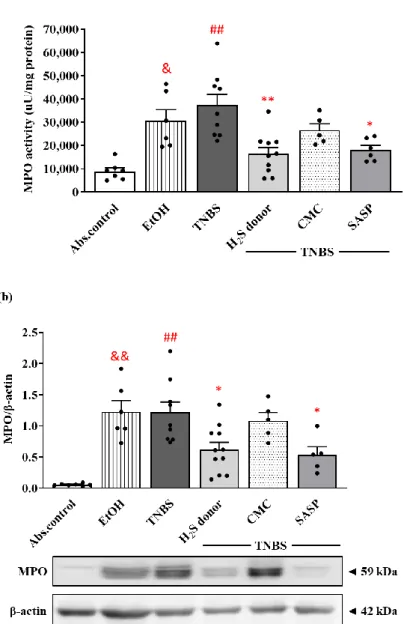
3.4. Hydrogen Sulfide Donor Treatment Suppressed the Activity and Expression of MPO in Rat Colon
As a marker of inflammation, MPO is released by the activation of neutrophils in inflammatory processes, corresponding to the extent of inflammatory damage. EtOH, the vehicle of TNBS, significantly increased the activity of the enzyme compared to the control group (30,568.63. ± 4866.79 vs. 8958.17 ± 1448.31 µ U/mg protein). The induction of colitis by TNBS instillation resulted in an approximately four-fold elevation in MPO activity compared to the control group (35,749.15 ± 4494.80 vs. 8958.17 ± 1448.31 µ U/mg protein).
Treatment with the protective dose of H2S donor significantly diminished the TNBS-in- creased activity of the enzyme, suggesting a lower infiltration of neutrophils (16,372.51 ± 2794.58 vs. 35,749.15 ± 4494.80 µ U/mg protein). Following SASP administration, we also observed a statistically significant reduction in the activity of the colonic MPO compared to the TNBS group (18,059.62 ± 1960.57 vs. 35,749.15 ± 4494.80 µ U/mg protein) (Figure 4a).
As Western blot revealed (Figure 4b), MPO was significantly overexpressed follow- ing EtOH and TNBS challenge similarly to the activity of the enzyme in both groups com- pared to healthy controls (1.23 ± 0.18 and 1.22 ± 0.17 vs. 0.06 ± 0.01 relative expression).
Release of H2S from Lawesson’s reagent caused a significant reduction in the expression of MPO, and equivalent results were seen in the case of the SASP-treated group compared to TNBS (0.62 ± 0.11 and 0.54 ± 0.13 vs. 1.22 ± 0.17 relative expression).
Figure 3. The expression of citH3 after TNBS administration and H2S donor treatments in rat colon tissues. Absolute control (no treatment); EtOH (50% ethanol, the solvent of TNBS); TNBS (2,4,6-trinitrobenzenesulfonic acid enema); H2S donor (TNBS + 18.75µM/kg/day Lawesson’s reagent); CMC (TNBS + 0.5% carboxymethylcellulose, the vehicle of Lawesson’s reagent); SASP (TNBS + 50 mg/kg/day sulfasalazine, as a positive control). Results are presented as mean + S.E.M.;
n= 5–12/group; one-way ANOVA, Holm–Sidak post hoc test, ##p< 0.001 absolute control vs. TNBS;
ap< 0.05 EtOH vs. TNBS; *p< 0.05 TNBS vs. TNBS + treatments.
3.4. Hydrogen Sulfide Donor Treatment Suppressed the Activity and Expression of MPO in Rat Colon
As a marker of inflammation, MPO is released by the activation of neutrophils in inflammatory processes, corresponding to the extent of inflammatory damage. EtOH, the vehicle of TNBS, significantly increased the activity of the enzyme compared to the
Int. J. Mol. Sci.2021,22, 12729 8 of 16
control group (30,568.63.±4866.79 vs. 8958.17±1448.31µU/mg protein). The induction of colitis by TNBS instillation resulted in an approximately four-fold elevation in MPO activity compared to the control group (35,749.15±4494.80 vs. 8958.17±1448.31µU/mg protein). Treatment with the protective dose of H2S donor significantly diminished the TNBS-increased activity of the enzyme, suggesting a lower infiltration of neutrophils (16,372.51±2794.58 vs. 35,749.15±4494.80µU/mg protein). Following SASP adminis- tration, we also observed a statistically significant reduction in the activity of the colonic MPO compared to the TNBS group (18,059.62±1960.57 vs. 35,749.15±4494.80µU/mg protein) (Figure4a).
Int. J. Mol. Sci. 2021, 22, x FOR PEER REVIEW 9 of 17
Figure 4. Alterations of the colonic myeloperoxidase (MPO) enzyme activity (a) and expression (b) following H2S donor administration in TNBS-induced rat colitis. Absolute control (no treatment);
EtOH (50% ethanol, the solvent of TNBS); TNBS (2,4,6-trinitrobenzenesulfonic acid enema); H2S do- nor (TNBS + 18.75 μM/kg/day Lawesson’s reagent); CMC (TNBS + 0.5% carboxymethylcellulose, the vehicle of Lawesson’s reagent); SASP (TNBS + 50 mg/kg/day sulfasalazine, as a positive control).
Results are presented as mean + S.E.M.; n = 5–11/group; one-way ANOVA, Holm–Sidak post hoc test, ## p < 0.001 absolute control vs. TNBS; * p < 0.05, ** p < 0.001 TNBS vs. TNBS + treatments; & p
< 0.05, && p < 0.001 absolute control vs. EtOH.
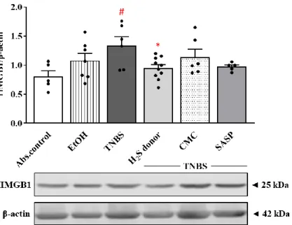
3.5. Hydrogen Sulfide Donor Attenuated the Overexpression of HMGB1 by TNBS
The expression of HMGB1 was also detected using the Western blot technique. The colonic HMGB1 levels showed a statistically significant increase in the TNBS group com- pared to the control group (0.80 ± 0.10 vs. 1.34 ± 0.15 relative expression). Treatment with the protective dose of H2S donor significantly reduced the expression of HMGB1 com- pared to the TNBS group (0.95 ± 0.06 vs. 1.34 ± 0.15 relative expression). Although SASP treatment also caused a decrease in HMGB1 levels, there was no significant difference compared to the TNBS group (Figure 5).
Figure 4.Alterations of the colonic myeloperoxidase (MPO) enzyme activity (a) and expression (b) following H2S donor administration in TNBS-induced rat colitis. Absolute control (no treatment);
EtOH (50% ethanol, the solvent of TNBS); TNBS (2,4,6-trinitrobenzenesulfonic acid enema); H2S donor (TNBS + 18.75µM/kg/day Lawesson’s reagent); CMC (TNBS + 0.5% carboxymethylcellulose, the vehicle of Lawesson’s reagent); SASP (TNBS + 50 mg/kg/day sulfasalazine, as a positive control).
Results are presented as mean + S.E.M.;n= 5–11/group; one-way ANOVA, Holm–Sidak post hoc test, ##p< 0.001 absolute control vs. TNBS; *p< 0.05, **p< 0.001 TNBS vs. TNBS + treatments;
&p< 0.05, &&p< 0.001 absolute control vs. EtOH.
Int. J. Mol. Sci.2021,22, 12729 9 of 16
As Western blot revealed (Figure4b), MPO was significantly overexpressed following EtOH and TNBS challenge similarly to the activity of the enzyme in both groups compared to healthy controls (1.23±0.18 and 1.22±0.17 vs. 0.06±0.01 relative expression). Release of H2S from Lawesson’s reagent caused a significant reduction in the expression of MPO, and equivalent results were seen in the case of the SASP-treated group compared to TNBS (0.62±0.11 and 0.54±0.13 vs. 1.22±0.17 relative expression).
3.5. Hydrogen Sulfide Donor Attenuated the Overexpression of HMGB1 by TNBS
The expression of HMGB1 was also detected using the Western blot technique. The colonic HMGB1 levels showed a statistically significant increase in the TNBS group com- pared to the control group (0.80 ±0.10 vs. 1.34 ±0.15 relative expression). Treatment with the protective dose of H2S donor significantly reduced the expression of HMGB1 compared to the TNBS group (0.95±0.06 vs. 1.34±0.15 relative expression). Although SASP treatment also caused a decrease in HMGB1 levels, there was no significant difference compared to the TNBS group (Figure5).
Int. J. Mol. Sci. 2021, 22, x FOR PEER REVIEW 10 of 17
Figure 5. Alterations of the expression of HMGB1 in the treated groups. Absolute control (no treat- ment); EtOH (50% ethanol, the solvent of TNBS); TNBS (2,4,6-trinitrobenzenesulfonic acid enema);
H2S donor (TNBS + 18.75 μM/kg/day Lawesson’s reagent); CMC (TNBS + 0.5% carboxymethylcellu- lose, the vehicle of Lawesson’s reagent); SASP (TNBS + 50 mg/kg/day sulfasalazine, as a positive control). Results are presented as mean + S.E.M.; n = 5–10/group; one-way ANOVA, Holm–Sidak post hoc test, # p < 0.05 absolute control vs. TNBS; * p < 0.05 TNBS vs. TNBS + H2S donor treatment.
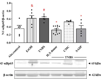
3.6. Effects of Hydrogen Sulfide Donor on the Expression of NF-kB p65 Subunit in TNBS- Induced Rat Colitis
As a pivotal inflammatory transcription factor, NF-κB has been implicated in the pathogenesis of colitis. As Figure 6 shows, EtOH and TNBS significantly enhanced the expression of NF-κB p65 subunit compared to the absolute control group (0.62 ± 0.07 and 0.62 ± 0.05 vs. 0.36 ± 0.08 relative expression). Release of H2S from Lawesson’s reagent resulted in a statistically significant attenuation in the colonic expression of NF-κB p65 subunit compared to TNBS groups (0.33 ± 0.03 vs. 0.62 ± 0.05 relative expression). Orally administered SASP also significantly diminished the level of NF-κB p65 protein compared to the TNBS group (0.35 ± 0.08 vs. 0.62 ± 0.05 relative expression).
Figure 5.Alterations of the expression of HMGB1 in the treated groups. Absolute control (no treat- ment); EtOH (50% ethanol, the solvent of TNBS); TNBS (2,4,6-trinitrobenzenesulfonic acid enema);
H2S donor (TNBS + 18.75µM/kg/day Lawesson’s reagent); CMC (TNBS + 0.5% carboxymethylcel- lulose, the vehicle of Lawesson’s reagent); SASP (TNBS + 50 mg/kg/day sulfasalazine, as a positive control). Results are presented as mean + S.E.M.;n= 5–10/group; one-way ANOVA, Holm–Sidak post hoc test, #p< 0.05 absolute control vs. TNBS; *p< 0.05 TNBS vs. TNBS + H2S donor treatment.
3.6. Effects of Hydrogen Sulfide Donor on the Expression of NF-kB p65 Subunit in TNBS-Induced Rat Colitis
As a pivotal inflammatory transcription factor, NF-κB has been implicated in the pathogenesis of colitis. As Figure6shows, EtOH and TNBS significantly enhanced the expression of NF-κB p65 subunit compared to the absolute control group (0.62±0.07 and 0.62±0.05 vs. 0.36±0.08 relative expression). Release of H2S from Lawesson’s reagent resulted in a statistically significant attenuation in the colonic expression of NF-κB p65 subunit compared to TNBS groups (0.33±0.03 vs. 0.62±0.05 relative expression). Orally administered SASP also significantly diminished the level of NF-κB p65 protein compared to the TNBS group (0.35±0.08 vs. 0.62±0.05 relative expression).
Int. J. Mol. Sci.Int. J. Mol. Sci. 2021, 22, x FOR PEER REVIEW 2021,22, 12729 11 of 17 10 of 16
Figure 6. Changes in the expression of NF-κB p65 subunit following TNBS administration and treat- ment with H2S donor in the colon. Absolute control (no treatment); EtOH (50% ethanol, the solvent of TNBS); TNBS (2,4,6-trinitrobenzenesulfonic acid enema); H2S donor (TNBS + 18.75 µ M/kg/day Lawesson’s reagent); CMC (TNBS + 0.5% carboxymethylcellulose, the vehicle of Lawesson’s rea- gent); SASP (TNBS + 50 mg/kg/day sulfasalazine, as a positive control). Results are presented as mean + S.E.M.; n = 5–12/group; one-way ANOVA, Holm–Sidak post hoc test, # p < 0.05 absolute control vs. TNBS; * p < 0.05 TNBS vs. TNBS + treatments; & p < 0.05 absolute control vs. EtOH.
3.7. Hydrogen Sulfide Donor Administration Elevated the Expression of UCHL-1 Deubiquitinating Enzyme
Investigating the effects of treatments on the expression of UCHL-1 deubiquitinase enzyme by Western blot analysis revealed that both 50% EtOH and TNBS instillation caused a significant decrease in its expression compared to the non-treated control group (0.27 ± 0.04 and 0.18 ± 0.03 vs. 0.69 ± 0.07 relative expression). However, we detected a significantly enhanced expression of UCHL-1 in the H2S donor treated group compared to TNBS (0.42 ± 0.06 vs. 0.18 ± 0.03 relative expression). Similar to Lawesson’s reagent, we observed a significant increase in the positive control (SASP) group compared to TNBS (0.49 ± 0.06 vs. 0.18 ± 0.03 relative expression). Results are presented in Figure 7.
Figure 6.Changes in the expression of NF-κB p65 subunit following TNBS administration and treat- ment with H2S donor in the colon. Absolute control (no treatment); EtOH (50% ethanol, the solvent of TNBS); TNBS (2,4,6-trinitrobenzenesulfonic acid enema); H2S donor (TNBS + 18.75µM/kg/day Lawes- son’s reagent); CMC (TNBS + 0.5% carboxymethylcellulose, the vehicle of Lawesson’s reagent); SASP (TNBS + 50 mg/kg/day sulfasalazine, as a positive control). Results are presented as mean + S.E.M.;
n= 5–12/group; one-way ANOVA, Holm–Sidak post hoc test, #p< 0.05 absolute control vs. TNBS;
*p< 0.05 TNBS vs. TNBS + treatments; &p< 0.05 absolute control vs. EtOH.
3.7. Hydrogen Sulfide Donor Administration Elevated the Expression of UCHL-1 Deubiquitinating Enzyme
Investigating the effects of treatments on the expression of UCHL-1 deubiquitinase enzyme by Western blot analysis revealed that both 50% EtOH and TNBS instillation caused a significant decrease in its expression compared to the non-treated control group (0.27±0.04 and 0.18±0.03 vs. 0.69±0.07 relative expression). However, we detected a significantly enhanced expression of UCHL-1 in the H2S donor treated group compared to TNBS (0.42±0.06 vs. 0.18±0.03 relative expression). Similar to Lawesson’s reagent, we observed a significant increase in the positive control (SASP) group compared to TNBS (0.49±0.06 vs. 0.18±0.03 relative expression). Results are presented in Figure7.
Int. J. Mol. Sci. 2021, 22, x FOR PEER REVIEW 12 of 17
Figure 7. Changes in the colonic expression of UCHL-1 in different treated groups. Absolute control (no treatment); EtOH (50% ethanol, the solvent of TNBS); TNBS (2,4,6-trinitrobenzenesulfonic acid enema); H2S donor (TNBS + 18.75 μM/kg/day Lawesson’s reagent); CMC (TNBS + 0.5% carbox- ymethylcellulose, the vehicle of Lawesson’s reagent); SASP (TNBS + 50 mg/kg/day sulfasalazine, as a positive control). Results are presented as mean + S.E.M.; n = 5–10/group; one-way ANOVA, Holm–Sidak post hoc test, ## p < 0.001 absolute control vs. TNBS; * p < 0.05 TNBS vs. TNBS + treat- ments; && p < 0.001 absolute control vs. EtOH.
4. Discussion
Our current study aimed to investigate the effects of H2S donor treatment on the ex- pression of NETs-related proteins and inflammatory mediators in an experimentally in- duced rat model of IBD. We found that the administration of H2S donor (18.75 µ M/kg/day) attenuated the severity of inflammation in TNBS-induced rat colitis. Our results showed that NETs-associated proteins were significantly increased in colitic colon tissues, while H2S donor treatment significantly decreased the expression of these markers. Addition- ally, the H2S donor attenuated the TNBS-provoked expressions of the inflammatory me- diators HMGB1 and NF-κB (p65 subunit) and elevated the expression of the UCHL-1 en- zyme. These results suggest an inhibitory effect of H2S donor in TNBS-colitis through the inhibition of NETs formation, which offers a new potential therapeutic strategy for pa- tients with IBD. The summary of our results is shown in Figure 8.
Figure 8. Summary of the presumed protective action of H2S donor treatment in experimental rat colitis. 2,4,6-trinitrobenzenesulfonic acid (TNBS); hydrogen sulfide (H2S); peptidyl arginine deimi- nase 4 (PAD4); citrullinated histone H3 (citH3); myeloperoxidase (MPO); high-mobility group box 1 (HMGB1); nuclear transcription factor-kappa B p65 subunit (NF-κB p65); ubiquitin C-terminal hydroxylase L1 (UCHL-1).
Figure 7.Changes in the colonic expression of UCHL-1 in different treated groups. Absolute con- trol (no treatment); EtOH (50% ethanol, the solvent of TNBS); TNBS (2,4,6-trinitrobenzenesulfonic acid enema); H2S donor (TNBS + 18.75µM/kg/day Lawesson’s reagent); CMC (TNBS + 0.5%
carboxymethylcellulose, the vehicle of Lawesson’s reagent); SASP (TNBS + 50 mg/kg/day sul- fasalazine, as a positive control). Results are presented as mean + S.E.M.;n= 5–10/group; one-way ANOVA, Holm–Sidak post hoc test, ##p< 0.001 absolute control vs. TNBS; *p< 0.05 TNBS vs.
TNBS + treatments; &&p< 0.001 absolute control vs. EtOH.
Int. J. Mol. Sci.2021,22, 12729 11 of 16
4. Discussion
Our current study aimed to investigate the effects of H2S donor treatment on the expression of NETs-related proteins and inflammatory mediators in an experimentally in- duced rat model of IBD. We found that the administration of H2S donor (18.75µM/kg/day) attenuated the severity of inflammation in TNBS-induced rat colitis. Our results showed that NETs-associated proteins were significantly increased in colitic colon tissues, while H2S donor treatment significantly decreased the expression of these markers. Additionally, the H2S donor attenuated the TNBS-provoked expressions of the inflammatory mediators HMGB1 and NF-κB (p65 subunit) and elevated the expression of the UCHL-1 enzyme.
These results suggest an inhibitory effect of H2S donor in TNBS-colitis through the inhibi- tion of NETs formation, which offers a new potential therapeutic strategy for patients with IBD. The summary of our results is shown in Figure8.
Int. J. Mol. Sci. 2021, 22, x FOR PEER REVIEW 12 of 17
Figure 7. Changes in the colonic expression of UCHL-1 in different treated groups. Absolute control (no treatment); EtOH (50% ethanol, the solvent of TNBS); TNBS (2,4,6-trinitrobenzenesulfonic acid enema); H2S donor (TNBS + 18.75 μM/kg/day Lawesson’s reagent); CMC (TNBS + 0.5% carbox- ymethylcellulose, the vehicle of Lawesson’s reagent); SASP (TNBS + 50 mg/kg/day sulfasalazine, as a positive control). Results are presented as mean + S.E.M.; n = 5–10/group; one-way ANOVA, Holm–Sidak post hoc test, ## p < 0.001 absolute control vs. TNBS; * p < 0.05 TNBS vs. TNBS + treat- ments; && p < 0.001 absolute control vs. EtOH.
4. Discussion
Our current study aimed to investigate the effects of H2S donor treatment on the ex- pression of NETs-related proteins and inflammatory mediators in an experimentally in- duced rat model of IBD. We found that the administration of H2S donor (18.75 µ M/kg/day) attenuated the severity of inflammation in TNBS-induced rat colitis. Our results showed that NETs-associated proteins were significantly increased in colitic colon tissues, while H2S donor treatment significantly decreased the expression of these markers. Addition- ally, the H2S donor attenuated the TNBS-provoked expressions of the inflammatory me- diators HMGB1 and NF-κB (p65 subunit) and elevated the expression of the UCHL-1 en- zyme. These results suggest an inhibitory effect of H2S donor in TNBS-colitis through the inhibition of NETs formation, which offers a new potential therapeutic strategy for pa- tients with IBD. The summary of our results is shown in Figure 8.
Figure 8. Summary of the presumed protective action of H2S donor treatment in experimental rat colitis. 2,4,6-trinitrobenzenesulfonic acid (TNBS); hydrogen sulfide (H2S); peptidyl arginine deimi- nase 4 (PAD4); citrullinated histone H3 (citH3); myeloperoxidase (MPO); high-mobility group box 1 (HMGB1); nuclear transcription factor-kappa B p65 subunit (NF-κB p65); ubiquitin C-terminal hydroxylase L1 (UCHL-1).
Figure 8. Summary of the presumed protective action of H2S donor treatment in experimental rat colitis. 2,4,6-trinitrobenzenesulfonic acid (TNBS); hydrogen sulfide (H2S); peptidyl arginine deiminase 4 (PAD4); citrullinated histone H3 (citH3); myeloperoxidase (MPO); high-mobility group box 1 (HMGB1); nuclear transcription factor-kappa B p65 subunit (NF-κB p65); ubiquitin C-terminal hydroxylase L1 (UCHL-1).
H2S, as an important gasotransmitter, acts as a vasodilator [46], diminishes leukocyte adherence and migration, and, furthermore, reduces edema development [47]. It was demonstrated that H2S donor molecules and their drug hybrids might be effective for reducing inflammation, resulting in fewer side effects [48–50]. Wallace et al. [51], in their study, revealed that an H2S-releasing NSAID, a derivative of naproxen (known as ATB-346), reduced pain and inflammation with less GI injury than naproxen. Moreover, this H2S-releasing derivative of naproxen has been tested in human phase2 clinical trial effectively [52]. In our investigation, we used the TNBS-induced rat colitis model, which is a widely accepted model to study IBD [8]. TNBS instillation causes serious transmural inflammation with ulcers and hyperaemia in the colon [9]. In line with previous findings from our laboratory [44], we confirmed in the current study that the administration of H2S donor, derived from Lawesson’s reagent, significantly suppressed the severity of inflammation and reduced the extent of ulcers in TNBS-induced rat colitis. The reduction of inflammation was as effective as SASP, a commonly used medical therapy [53]. Chen et al. also reported in dextran sulfate sodium (DSS)-induced colonic inflammation that NaHS treatment decreased inflammation and tissue injury in mice [54]. However, the protective mechanism of H2S is still unclear.
Besides phagocytosis and cytokine and ROS production, activated neutrophils can also form and release NETs [25]. IBD is characterized by intense neutrophil infiltration to the site of inflammation [22], and it is increasingly clear that NETs are involved in the pathogenesis of IBD [55,56]. Therrien et al. [57] showed that the recruitment of activated neutrophils in inflamed mucosa is associated with the severity of disease in IBD. Tao Li et al. [56] investigated the presence of NETs formation markers in plasma from patients with IBD. Their results showed significantly elevated plasma levels of DNA and MPO-DNA complexes in active UC or CD patients compared to those with inactive UC or CD and healthy controls. Additionally, an overexpressed citH3 was observed in the colonic tissues
Int. J. Mol. Sci.2021,22, 12729 12 of 16
of IBD patients. Furthermore, Dinallo et al. [58] provided additional evidence on NETs formation in the inflamed colon of UC patients, demonstrating the co-localization of MPO, NE and citH3 with the DNA by immunofluorescence staining. In our study, we observed the effects of exogenous H2S-releasing agent, Lawesson’s reagent, on the expression of NETs-associated proteins in TNBS-induced colitis. Our western blot analysis showed a high expression of PAD4 in colon samples following TNBS instillation. Similarly to PAD4, citH3 also exhibited an increased expression in the TNBS group. We also measured the activity and expression of another NETs-related protein, MPO, which were elevated after the induction of colitis. However, the administration of H2S donor significantly decreased the expression of all measured NETs-associated proteins. Our results are consistent with the work of Zhang et al. [59], demonstrating an increased expression of citH3, an elevated gene expression of PAD4 and increased activity of MPO enzyme in TNBS-induced colitis in mice.
In their study, the NETs structures were also displayed by immunofluorescence staining and immunohistochemistry. They found that neutrophils formed a web-like structure in the TNBS-treated group, and the treatment with Cl-amidine, a PAD4 inhibitor, led to a reduction in NETs formation. Additionally, Chumanevich et al. measured increased PADI2 and PAD4 levels in DSS-induced colitis in mice [60]. They also demonstrated elevated PAD levels in colon biopsies from UC patients. Furthermore, it was found in animal models that Cl-amidine, a pan-PAD inhibitor, attenuated the severity of several diseases, such as rheumatoid arthritis [61], systemic lupus erythematosus [32], and cancer [62]. In addi- tion, Cl-amidine treatment also reduced NETs formation and suppressed inflammation in DSS- [60] and in TNBS-induced mouse colitis [59]. Furthermore, in a mouse model of lethal lipopolysaccharide (LPS) induced shock, Deng et al. found that treatment with a CitH3 monoclonal antibody attenuated proinflammatory responses and NETs formation [33].
Yang et al. investigated the effects of H2S donor on NETosis in diabetic mice [34]. They found that H2S donor decreased NETs formation, leading to diabetic wound healing, which is consistent with our current findings in TNBS-induced colitis. These results suggest that the molecules promoting NETs formation could be used as biomarkers for IBD, and H2S donor administration may contribute to the attenuation of inflammation and ulcer healing through inhibiting NETosis.
In addition, we examined the expression of the inflammatory mediator HMGB1 in TNBS-induced rat colitis. In line with our previous findings [42], the current results also showed a high expression of HMGB1 in the inflamed colonic tissue. Maeda et al. also de- tected a colonic overexpression of HMGB1 in the DSS-induced mouse model of UC, while its inhibition by anti-HMGB1 antibody resulted in a decreased extent of inflammation [63].
According to Tadie et al., HMGB1 enhances the NETs formation via toll-like receptor 4 (TLR4), contributing to inflammatory processes and tissue damage [36]. However, in our study, the administration of H2S donor markedly attenuated HMGB1 expression. With another H2S donor, NaHS, Zhao et al. also demonstrated the inhibitory effect of H2S on NETs formation through the reduction of HMGB1 levels in a rat model of hyperhomocys- teinemia [35]. These findings, in line with our current results, suggest that the protective effect of H2S may rely on the inhibition of HMGB1 expression.
Furthermore, we tested the effects of H2S donor administration on the expression of NF-κB. NF-κB is one of the key regulators in inflammatory conditions, and its increased activation is typical in patients suffering from IBD [40]. In the current study, we showed an enhanced expression of NF-κB by TNBS, which was then significantly decreased after treatment with Lawesson’s H2S donor. Several studies demonstrated that H2S inhibits the degradation of the inhibitor kappa B-α(IκB-α) subunit, which regulates NF-κB activation and translocation [64,65]. Sun et al. showed that citrullination of NF-κB by PAD4 promotes inflammation; thus, the inhibition of this process seems to be a promising therapeutic strategy in inflammation-associated diseases [66]. In our current study, we demonstrated a decreased expression of PAD4 following H2S donor administration and an H2S-induced NF-κB reduction. Based on these results, we presume that the inhibition of PAD4 ex- pression and the inactivation of NF-κB might contribute to the anti-inflammatory effect
Int. J. Mol. Sci.2021,22, 12729 13 of 16
of H2S. Besides its important role in proteasomal degradation, UCHL-1 as a potential inflammatory mediator is also able to block NF-κB activation by stabilizing IκBα[43]. Here, we demonstrated an attenuated expression of UCHL-1 in rat colitis induced by TNBS, which is consistent with our previous findings [42]. This reduced UCHL-1 expression was significantly restored due to H2S donor treatment.
Limitation
The limitation of our current study is that only one H2S donor, namely Lawesson’s reagent, was tested against TNBS-induced colitis to reduce the extent of the damaged area of the colon. However, it would be interesting to check in the future whether other H2S donors exert this protection.
Our results showed that NETosis played an important part in the pathogenesis of the TNBS colitis model and H2S treatment attenuated the expression of NETs-associated proteins in the colonic tissues. Based on these findings, we concluded that the elevated levels of members of NETs seem to be the consequence of NETosis. The application of immunohistochemical methods may provide morphological evidence of NETosis and may support the protective effects exerted by H2S donor in our colitis model, although the limi- tation of our current study is that we applied only Western blot analysis. NETs formation in the TNBS-induced mouse colitis model was demonstrated by immunohistochemistry;
however, it would be worth clarifying the existence of NETosis in the inflamed colonic tissues of rats with the same method.
5. Conclusions
In conclusion, we found that exogenous H2S decreased inflammation and contributed to ulcer healing by the inhibition of NETs formation and proinflammatory mediators in TNBS-induced rat colitis. Additionally, UCHL-1 seems to contribute to the protective effect of H2S. Our findings suggest that NETs formation is involved in the pathogenesis of IBD, and H2S, through the inhibition of NETs formation, provides a potential therapeutic option against IBD. Our current result is promising; however, it does not provide comprehensive answers. Therefore, further investigations are needed to reveal the exact role of NETosis in TNBS-induced rat colitis and the protective mechanism of H2S in inflammatory conditions.
Author Contributions:Conceptualization, K.K. and S.T.; methodology, S.T., N.A. and K.K.; software, Z.V.; validation, Z.V.; formal analysis, A.P.; investigation, S.T. and N.A.; resources, C.V. and K.K.; data curation, S.T.; writing—original draft preparation, S.T.; writing—review and editing, S.T., N.A. and K.K.; visualization, S.T.; supervision, K.K. All authors have read and agreed to the published version of the manuscript.
Funding:This research received no external funding.
Institutional Review Board Statement:The study was conducted according to the European Union Guidelines for the Care (European Union Directive (2010/63/EU) and the guidelines for the Use of Laboratory Animals and approved by the Institutional Ethics Committee at the University of Szeged (XX./4799/2015, 15 December 2015).
Data Availability Statement:All data analysed in this study are included in this published article.
Acknowledgments: This research was supported by GINOP-2.3.2-15-2016-00030 and Ministry of Human Capacities, Hungary grant 20391-3/2018/FEKUSTRAT, Bolyai János Postdoctoral Research Grant (Krisztina Kupai), BO/00683/21/5, New National Excellence Program of the Ministry for Innovation and Technology from the source of the National Research, Development and Innovation FundÚNKP-21-5-SZTE-597 (Krisztina Kupai).
Conflicts of Interest:The authors declare no conflict of interest.
Int. J. Mol. Sci.2021,22, 12729 14 of 16
References
1. Ng, S.C.; Shi, H.Y.; Hamidi, N.; Underwood, F.E.; Tang, W.; Benchimol, E.I.; Panaccione, R.; Ghosh, S.; Wu, J.C.Y.; Chan, F.K.L.; et al.
Worldwide incidence and prevalence of inflammatory bowel disease in the 21st century: A systematic review of population-based studies.Lancet2017,390, 2769–2778. [CrossRef]
2. Bernstein, C.N.; Fried, M.; Krabshuis, J.H.; Cohen, H.; Eliakim, R.; Fedail, S.; Gearry, R.; Goh, K.L.; Hamid, S.; Khan, A.G.; et al.
World Gastroenterology Organization Practice Guidelines for the diagnosis and management of IBD in 2010.Inflamm. Bowel Dis.
2010,16, 112–124. [CrossRef]
3. Podolsky, D.K. Inflammatory bowel disease.N. Engl. J. Med.2002,347, 417–429. [CrossRef]
4. Walsh, A.; Mabee, J.; Trivedi, K. Inflammatory bowel disease.Prim. Care2011,38, 415–432. [CrossRef]
5. Pithadia, A.B.; Jain, S. Treatment of inflammatory bowel disease (IBD).Pharmacol. Rep.2011,63, 629–642. [CrossRef]
6. Hendrickson, B.A.; Gokhale, R.; Cho, J.H. Clinical aspects and pathophysiology of inflammatory bowel disease.Clin. Microbiol.
Rev.2002,15, 79–94. [CrossRef]
7. Randhawa, P.K.; Singh, K.; Singh, N.; Jaggi, A.S. A review on chemical-induced inflammatory bowel disease models in rodents.
Korean J. Physiol. Pharmacol.2014,18, 279–288. [CrossRef] [PubMed]
8. Morris, G.P.; Beck, P.L.; Herridge, M.S.; Depew, W.T.; Szewczuk, M.R.; Wallace, J.L. Hapten-induced model of chronic inflammation and ulceration in the rat colon.Gastroenterology1989,96, 795–803. [CrossRef]
9. Antoniou, E.; Margonis, G.A.; Angelou, A.; Pikouli, A.; Argiri, P.; Karavokyros, I.; Papalois, A.; Pikoulis, E. The TNBS-induced colitis animal model: An overview.Ann. Med. Surg. (Lond.)2016,11, 9–15. [CrossRef]
10. Abe, K.; Kimura, H. The possible role of hydrogen sulfide as an endogenous neuromodulator.J. Neurosci.1996,16, 1066–1071.
[CrossRef]
11. Stipanuk, M.H.; Beck, P.W. Characterization of the enzymic capacity for cysteine desulphhydration in liver and kidney of the rat.
Biochem. J.1982,206, 267–277. [CrossRef] [PubMed]
12. Wang, R. Two’s company, three’s a crowd: Can H2S be the third endogenous gaseous transmitter?FASEB J.2002,16, 1792–1798.
[CrossRef]
13. Kimura, H. Production and physiological effects of hydrogen sulfide. Antioxid. Redox Signal. 2014,20, 783–793. [CrossRef]
[PubMed]
14. Szabo, C. Hydrogen sulphide and its therapeutic potential.Nat. Rev. Drug Discov.2007,6, 917–935. [CrossRef] [PubMed]
15. Coletta, C.; Szabo, C. Potential role of hydrogen sulfide in the pathogenesis of vascular dysfunction in septic shock.Curr. Vasc.
Pharmacol.2013,11, 208–221.
16. Lowicka, E.; Beltowski, J. Hydrogen sulfide (H2S)—the third gas of interest for pharmacologists.Pharmacol. Rep.2007,59, 4–24.
17. Teague, B.; Asiedu, S.; Moore, P.K. The smooth muscle relaxant effect of hydrogen sulphide in vitro: Evidence for a physiological role to control intestinal contractility.Br. J. Pharmacol.2002,137, 139–145. [CrossRef]
18. Wallace, J.L. Physiological and pathophysiological roles of hydrogen sulfide in the gastrointestinal tract.Antioxid. Redox Signal.
2010,12, 1125–1133. [CrossRef]
19. Ise, F.; Takasuka, H.; Hayashi, S.; Takahashi, K.; Koyama, M.; Aihara, E.; Takeuchi, K. Stimulation of duodenal HCO3−secretion by hydrogen sulphide in rats: Relation to prostaglandins, nitric oxide and sensory neurones. Acta Physiol. (Oxf.) 2011,201, 117–126. [CrossRef]
20. Wallace, J.L.; Dicay, M.; McKnight, W.; Martin, G.R. Hydrogen sulfide enhances ulcer healing in rats.FASEB J.2007,21, 4070–4076.
[CrossRef]
21. Fiorucci, S.; Orlandi, S.; Mencarelli, A.; Caliendo, G.; Santagada, V.; Distrutti, E.; Santucci, L.; Cirino, G.; Wallace, J.L. Enhanced activity of a hydrogen sulphide-releasing derivative of mesalamine (ATB-429) in a mouse model of colitis.Br. J. Pharmacol.2007, 150, 996–1002. [CrossRef]
22. Wallace, K.L.; Zheng, L.B.; Kanazawa, Y.; Shih, D.Q. Immunopathology of inflammatory bowel disease.World J. Gastroenterol.
2014,20, 6–21. [CrossRef]
23. Kolaczkowska, E.; Kubes, P. Neutrophil recruitment and function in health and inflammation. Nat. Rev. Immunol. 2013,13, 159–175. [CrossRef] [PubMed]
24. Brinkmann, V.; Reichard, U.; Goosmann, C.; Fauler, B.; Uhlemann, Y.; Weiss, D.S.; Weinrauch, Y.; Zychlinsky, A. Neutrophil extracellular traps kill bacteria.Science2004,303, 1532–1535. [CrossRef] [PubMed]
25. Mesa, M.A.; Vasquez, G. NETosis.Autoimmune Dis.2013,2013, 651497. [CrossRef]
26. Rohrbach, A.S.; Slade, D.J.; Thompson, P.R.; Mowen, K.A. Activation of PAD4 in NET formation.Front. Immunol.2012,3, 360.
[CrossRef] [PubMed]
27. Yang, H.; Biermann, M.H.; Brauner, J.M.; Liu, Y.; Zhao, Y.; Herrmann, M. New Insights into Neutrophil Extracellular Traps:
Mechanisms of Formation and Role in Inflammation.Front. Immunol.2016,7, 302. [CrossRef]
28. Jorch, S.K.; Kubes, P. An emerging role for neutrophil extracellular traps in noninfectious disease.Nat. Med.2017,23, 279–287.
[CrossRef]
29. Delgado-Rizo, V.; Martinez-Guzman, M.A.; Iniguez-Gutierrez, L.; Garcia-Orozco, A.; Alvarado-Navarro, A.; Fafutis-Morris, M. Neutrophil Extracellular Traps and Its Implications in Inflammation: An Overview.Front. Immunol.2017,8, 81. [CrossRef]
[PubMed]
Int. J. Mol. Sci.2021,22, 12729 15 of 16
30. Pan, B.; Alam, H.B.; Chong, W.; Mobley, J.; Liu, B.; Deng, Q.; Liang, Y.; Wang, Y.; Chen, E.; Wang, T.; et al. CitH3: A reliable blood biomarker for diagnosis and treatment of endotoxic shock.Sci. Rep.2017,7, 8972. [CrossRef]
31. Mutua, V.; Gershwin, L.J. A Review of Neutrophil Extracellular Traps (NETs) in Disease: Potential Anti-NETs Therapeutics.Clin.
Rev. Allergy Immunol.2021,61, 194–211. [CrossRef]
32. Knight, J.S.; Subramanian, V.; O’Dell, A.A.; Yalavarthi, S.; Zhao, W.; Smith, C.K.; Hodgin, J.B.; Thompson, P.R.; Kaplan, M.J. Peptidylarginine deiminase inhibition disrupts NET formation and protects against kidney, skin and vascular disease in lupus-prone MRL/lpr mice.Ann. Rheum. Dis.2015,74, 2199–2206. [CrossRef]
33. Deng, Q.; Pan, B.; Alam, H.B.; Liang, Y.; Wu, Z.; Liu, B.; Mor-Vaknin, N.; Duan, X.; Williams, A.M.; Tian, Y.; et al. Citrullinated Histone H3 as a Therapeutic Target for Endotoxic Shock in Mice.Front. Immunol.2019,10, 2957. [CrossRef] [PubMed]
34. Yang, C.T.; Chen, L.; Chen, W.L.; Li, N.; Chen, M.J.; Li, X.; Zheng, X.; Zhao, Y.Z.; Wu, Y.X.; Xian, M.; et al. Hydrogen sulfide primes diabetic wound to close through inhibition of NETosis.Mol. Cell. Endocrinol.2019,480, 74–82. [CrossRef] [PubMed]
35. Zhao, X.; Zhang, L.; Liu, X.; Zhao, Z.; Zhong, X.; Wang, Y. Exogenous hydrogen sulfide inhibits neutrophils extracellular traps formation via the HMGB1/TLR4/p-38 MAPK/ROS axis in hyperhomocysteinemia rats.Biochem. Biophys. Res. Commun.2021, 537, 7–14. [CrossRef] [PubMed]
36. Tadie, J.M.; Bae, H.B.; Jiang, S.; Park, D.W.; Bell, C.P.; Yang, H.; Pittet, J.F.; Tracey, K.; Thannickal, V.J.; Abraham, E.; et al. HMGB1 promotes neutrophil extracellular trap formation through interactions with Toll-like receptor 4.Am. J. Physiol. Lung Cell. Mol.
Physiol.2013,304, L342–L349. [CrossRef]
37. Palone, F.; Vitali, R.; Cucchiara, S.; Pierdomenico, M.; Negroni, A.; Aloi, M.; Nuti, F.; Felice, C.; Armuzzi, A.; Stronati, L. Role of HMGB1 as a suitable biomarker of subclinical intestinal inflammation and mucosal healing in patients with inflammatory bowel disease.Inflamm. Bowel Dis.2014,20, 1448–1457. [CrossRef]
38. Tak, P.P.; Firestein, G.S. NF-kappaB: A key role in inflammatory diseases.J. Clin. Investig.2001,107, 7–11. [CrossRef]
39. Li, Q.; Verma, I.M. NF-kappaB regulation in the immune system.Nat. Rev. Immunol.2002,2, 725–734. [CrossRef] [PubMed]
40. Atreya, I.; Atreya, R.; Neurath, M.F. NF-kappaB in inflammatory bowel disease.J. Intern. Med.2008,263, 591–596. [CrossRef]
41. Gu, Y.; Ding, X.; Huang, J.; Xue, M.; Zhang, J.; Wang, Q.; Yu, H.; Wang, Y.; Zhao, F.; Wang, H.; et al. The deubiquitinating enzyme UCHL1 negatively regulates the immunosuppressive capacity and survival of multipotent mesenchymal stromal cells.Cell Death Dis.2018,9, 459. [CrossRef]
42. Almasi, N.; Torok, S.; Dvoracsko, S.; Tomboly, C.; Csonka, A.; Barath, Z.; Murlasits, Z.; Valkusz, Z.; Posa, A.; Varga, C.; et al.
Lessons on the Sigma-1 Receptor in TNBS-Induced Rat Colitis: Modulation of the UCHL-1, IL-6 Pathway.Int. J. Mol. Sci.2020,21, 4046. [CrossRef]
43. Takami, Y.; Nakagami, H.; Morishita, R.; Katsuya, T.; Cui, T.X.; Ichikawa, T.; Saito, Y.; Hayashi, H.; Kikuchi, Y.; Nishikawa, T.; et al.
Ubiquitin carboxyl-terminal hydrolase L1, a novel deubiquitinating enzyme in the vasculature, attenuates NF-kappaB activation.
Arterioscler. Thromb. Vasc. Biol.2007,27, 2184–2190. [CrossRef] [PubMed]
44. Kupai, K.; Almasi, N.; Kosa, M.; Nemcsok, J.; Murlasits, Z.; Torok, S.; Al-Awar, A.; Barath, Z.; Posa, A.; Varga, C. H2S confers colonoprotection against TNBS-induced colitis by HO-1 upregulation in rats.Inflammopharmacology2018,26, 479–489. [CrossRef]
[PubMed]
45. Bradley, P.P.; Priebat, D.A.; Christensen, R.D.; Rothstein, G. Measurement of cutaneous inflammation: Estimation of neutrophil content with an enzyme marker.J. Investig. Dermatol.1982,78, 206–209. [CrossRef] [PubMed]
46. Zhao, W.; Zhang, J.; Lu, Y.; Wang, R. The vasorelaxant effect of H2S as a novel endogenous gaseous K(ATP) channel opener.
EMBO J.2001,20, 6008–6016. [CrossRef]
47. Zanardo, R.C.; Brancaleone, V.; Distrutti, E.; Fiorucci, S.; Cirino, G.; Wallace, J.L. Hydrogen sulfide is an endogenous modulator of leukocyte-mediated inflammation.FASEB J.2006,20, 2118–2120. [CrossRef] [PubMed]
48. Sparatore, A.; Santus, G.; Giustarini, D.; Rossi, R.; Del Soldato, P. Therapeutic potential of new hydrogen sulfide-releasing hybrids.
Expert Rev. Clin. Pharmacol.2011,4, 109–121. [CrossRef] [PubMed]
49. Magierowski, M.; Magierowska, K.; Surmiak, M.; Hubalewska-Mazgaj, M.; Kwiecien, S.; Wallace, J.L.; Brzozowski, T. The effect of hydrogen sulfide-releasing naproxen (ATB-346) versus naproxen on formation of stress-induced gastric lesions, the regulation of systemic inflammation, hypoxia and alterations in gastric microcirculation.J. Physiol. Pharmacol.2017,68, 749–756.
50. Marutani, E.; Ichinose, F. Emerging pharmacological tools to control hydrogen sulfide signaling in critical illness.Intensive Care Med. Exp.2020,8, 5. [CrossRef]
51. Wallace, J.L.; Caliendo, G.; Santagada, V.; Cirino, G. Markedly reduced toxicity of a hydrogen sulphide-releasing derivative of naproxen (ATB-346).Br. J. Pharmacol.2010,159, 1236–1246. [CrossRef]
52. Wallace, J.L.; Nagy, P.; Feener, T.D.; Allain, T.; Ditroi, T.; Vaughan, D.J.; Muscara, M.N.; de Nucci, G.; Buret, A.G. A proof-of- concept, Phase 2 clinical trial of the gastrointestinal safety of a hydrogen sulfide-releasing anti-inflammatory drug.Br. J. Pharmacol.
2020,177, 769–777. [CrossRef]
53. Das, K.M.; Farag, S.A. Current medical therapy of inflammatory bowel disease.World J. Gastroenterol.2000,6, 483–489.
54. Chen, X.; Liu, X.S. Hydrogen sulfide from a NaHS source attenuates dextran sulfate sodium (DSS)-induced inflammation via inhibiting nuclear factor-kappaB.J. Zhejiang Univ. Sci. B2016,17, 209–217. [CrossRef]
55. Bennike, T.B.; Carlsen, T.G.; Ellingsen, T.; Bonderup, O.K.; Glerup, H.; Bogsted, M.; Christiansen, G.; Birkelund, S.; Stensballe, A.;
Andersen, V. Neutrophil Extracellular Traps in Ulcerative Colitis: A Proteome Analysis of Intestinal Biopsies.Inflamm. Bowel Dis.
2015,21, 2052–2067. [CrossRef] [PubMed]