DOI: 10.15413/ajsr.2017.0136 ISSN 2315-7712
©2017 Academia Publishing
Research Paper
Differentially expressed microRNAs and their relation to gasotransmitters in TNBS-induced colitis in rat colon
Accepted 18th July, 2017 ABSTRACT
MicroRNAs are well established regulators of Inflammatory Bowel Disease (IBD), such as Crohn’s disease (CD) and ulcerative colitis (UC). Furthermore, gasotransmitters including nitric oxide (NO), carbon monoxide (CO) and hydrogen sulphide (H2S) also seem to play important role in this disease pathogenesis. In the present study, we aimed to investigate the relationship between microRNAs and gasotransmitters in colitis. Male Wistar rats were administered 2, 4, 6-trinitrobenzenesulphonic acid (TNBS, 10 mg/0.25 ml in 50% ethanol) rectally to induce colitis. For the purpose of comparison, we observed miRNA expressions by nanoString nCounter® miRNA Expression Assay and measured the activities and expressions (by Western blot) of the enzymes responsible for gasotransmitter synthesis. In total, 228 miRNA were screened and from these 94 showed upregulation and 63 were downregulated.
We found several miRNAs (miR-26a, miR-200a, miR-200c, miR-21, miR-222, miR-101, miR-28, miR-132, miR-7, miR-141, let-7, miR-196a, miR-30 and miR- 186) that might have an effect on gasotransmitter pathways. This study proved that there are important interactions between gasotransmitters and microRNAs in TNBS-induced rat colitis. Our results are presumably important in IBD diagnostics, pathogenesis modelling and could be a starting-point to further investigations.
Key words: microRNA, IBD, TNBS, gasotransmitters.
INTRODUCTION
Inflammatory bowel disease (IBD) is identified as an immune-mediated disease with autoimmune phenomenon, which may expand to the global gastrointestinal tract (GI) (Wen and Fiocchi, 2004, Chen et al., 2014). Several subtypes of IBD were described with particular regard to the most common forms, Crohn’s disease (CD) and ulcerative colitis (UC) (Archanioti et al., 2011). Despite intensive research on IBD currently, the pathogenesis of this disease is still unknown, although several factors have been revealed to contribute to its development, including genetic, environmental, immunological and most recently, epigenetic factors. Of these, research on microRNAs
(miRNAs) seems to be a new promising area with the potential of being relevant biomarkers for IBD diagnosis or targets for future treatments (Coskun et al., 2012).
MicroRNAs are small (18 to 24 nucleotide), non-coding, evolutionary conserved RNA molecules with the ability to regulate mRNA expression levels through transcriptional repression or degradation of their target mRNA (Kalla et al., 2015). The miRNA mediated gene expression regulation is crucial in several physiological processes (Su et al., 2013; Varga et al., 2013), including cell cycle, differentiation, apoptosis ( Farago et al., 2011 ), cell homeostasis, stress response (Iborra et al., 2013, Varga et Nikoletta Almási1#, Anikó Pósa1#, Amin
Al-awar1, Szilvia Török1, Zoltán Baráth2, János Nemcsók6, Zsolt Murlasits5, Lajos Istvan Nagy4, László G Puskás3,4, Csaba Varga1 and Krisztina Kupai1*
1Deptartment of Physiology Anatomy and Neuroscience, Faculty of Science and Informatics, University of Szeged, Szeged, Hungary.
2Department of Orthodontics and Pediatric Dentistry, Faculty of Dentistry, University of Szeged, Szeged, Hungary.
3Institute of Genetics, Biological Research Center, Hungarian Academy of Sciences, Szeged, Hungary.
4AVIDIN Ltd., Szeged, Hungary.
5College of Arts and Sciences, Sport Science Program, Laboratory Animal Research Center, Qatar University, Doha, Qatar.
6Department of Biology, J. Selye University, Komárno, Slovakia.
#Both authors contributed equally to the research.
*Corresponding author. E-mail:
kupai@bio.u-szeged.hu
al., 2013, 2014), immune development and response (Su et al., 2013) and numerous pathological conditions such as cancer development, stroke and inflammatory processes (Sonkoly and Pivarcsi, 2009).
miRNAs are described recently as important constituents of the innate and adaptive immune system and differential expressions of microRNAs are associated with several immune-mediated diseases such as psoriasis, arthritis and IBD (Dalal and Kwon, 2010). It has been shown that miRNAs are expressed differently in the peripheral blood and intestinal tissues of patients with both CD and UC. Furthermore, discrepancies can be observed in active disease stages as well (Takagi et al., 2010; Wu et al., 2011).
Gasotransmitters, such as nitric oxide (NO), carbon monoxide (CO) and hydrogen sulphide (H2S) are signaling molecules with key functions in cell physiology (Mustafa et al., 2009). It has been shown that gasotransmitter synthesizing enzymes exhibit different activities and expression levels in colitis (Szalai et al., 2014), indicating that these molecules might be important in IBD development. Inducible nitric oxide synthase (iNOS) and constitutive nitric oxide synthase (cNOS) endothelial (eNOS) and neuronal (nNOS), produces NO, the first identified gasotransmitter from L-arginine in response to pro-inflammatory cytokines. Both enzymes’ expressions have been shown to be upregulated in the intestinal epithelium and inflamed colonic mucosa in IBD (Dhillon et al., 2014; Szalai et al., 2014).
Carbon monoxide (CO) is the second described gasotransmitter with physiological vasorelaxant properties (Wang et al., 1997). CO is generated endogenously through the biodegradation of heme, which catalyzed by heme oxygenase (HO) enzymes. In this reaction, besides CO, biliverdin and free Fe2+ also arise, which are responsible for the antioxidant and antiapoptotic effects of HO. There are three isoforms of the HO enzyme, HO-1 (inducible form, also known as hsp32), HO-2 (constitutive isoform) and HO-3 (Ryter and Choi, 2016). These isoforms are encoded by HMOX-1,-2,-3 genes in human and Hmox-1,-2,-3 genes in rat (Kutty et al., 1994). Several studies reported that CO and the HO enzymes play important roles in inflammatory conditions through their anti-inflammatory effects (Hegazi et al., 2005; Sheikh et al., 2011), thus, CO/HO system is becoming one of the main targets in IBD research.
A recently discovered gasotransmitter is H2S, produced from L-cystein through two main pathways in the colon, a pyridoxal-5’-phosphate (P5P) dependent manner, containing two enzymes, cystathionine β-synthase (CBS) and cystathionine γ-lyase (CSE) and another pathway which operates with an intermediate (3- mercaptopyruvate) and requires cysteine aminotransferase (CAT) and a mitochondria localized enzyme, mercaptopyruvate sulfurtransferase (3MST) (Guo et al., 2012; Flannigan et al., 2013).
H2S is essential in various physiological processes
including neuromodulation (Abe and Kimura, 1996), vasorelaxation, colon contractility (Teague et al., 2002), long term potentiation (LTP) (O'Dell et al., 1991), apoptosis and ulcer healing (Wallace et al., 2009). The exact role of H2S in IBD is controversial. It is reported that H2S is elevated in IBD, but its role in this disease is complex and depends on several factors, such as the model used in the investigation, whether the inflammation is acute or chronic and the time of the administration of H2S donors or inhibitors (Whiteman and Winyard, 2011).
In the present study, we aimed to investigate the different expressions of miRNAs, along with the changes of gasotransmitter synthesizing enzyme activities or altered expressions in animal model of colitis. In this context, we objected to identify the link between differentially expressed miRNAs and the changes in enzyme activities and expressions to propose a novel therapeutic target in IBD research.
MATERIALS AND METHODS Animals
All manipulations were performed in accordance with the standards of the European Community guidelines on the care and use of laboratory animals and was approved by the Institutional Ethics Committee at the University of Szeged.
Male Wistar rats (180 to 220 g) were housed in groups.
Food was withdrawn overnight before induction of colitis;
otherwise, the animals had access to food and drinking water ad libitum throughout the experiments. The animal care and research protocols were in accordance with the guidelines of the University of Szeged.
Experimental design
The animals were randomly divided into three groups:
absolute control (no treatment), vehicle (50% ethanol (EtOH)) and TNBS (10 mg/0.25 ml in 50% ethanol).
Animals were fasted for 24 h before treatment. On the first day of experiment colitis was induced by 2, 4, 6- trinitrobenzenesulphonic acid (TNBS; single injection 10 mg in 0.25 ml of 50% ethanol). The intracolonic administration of TNBS was performed with an 8 cm long plastic catheter under transient ether anaesthesia, while the control groups did not receive any treatment. Three days after the induction of colitis the animals were sacrificed and the distal 8 cm portion of the colon dissected, longitudinally opened, gently rinsed with ice- cold physiological saline and photographed for the determination of macroscopic colonic inflammatory damage. The colon was weighed and divided into longitudinal segments to be used for further molecular
and biochemical analyses.
Heme oxygenase activity
Heme oxygenase activity was assessed by measuring bilirubin formation with slight modifications as described by Tenhunen et al. (1968). The colon segment was homogenized (Ultra-turrax T25; 13.500/s; twice for 30 s) in ice-cold 10 mM N-[2-hydroxyethyl] piperazine-N’-[2- ethanesulfonic acid] (HEPES, Sigma-Aldrich), 32 mM sucrose (Sigma-Aldrich), 1 mM dithiothreitol (DTT, Sigma- Aldrich), 0.1 mM EDTA, 10 μg/ml soybean trypsin inhibitor (Sigma-Aldrich), 10 μg/ml leupeptin (Sigma- Aldrich) and 2 μg/ml aprotinin (Sigma-Aldrich); pH 7.4.
The supernatant was collected by centrifugation for 30 min at 20.000 g at 4°C. Incubation was carried out in the dark at 37°C for 60 min with the reaction mixture containing the following ingredients in a final volume of 1.5 ml: 2 mM glucose 6-phosphate (Sigma-Aldrich), 0.14 U/ml glucose 6-phosphate dehydrogenase (Sigma- Aldrich), 15 μM heme, 150 μM β-nicotinamide adenine dinucleotide phosphate (β-NADPH, Sigma-Aldrich), 120 μg/ml rat liver cytosol as a source of biliverdin reductase, 2 mM MgCl2, 100 mM potassium phosphate buffer and 150 μl supernatant. The reaction was stopped by placing the samples on ice. The bilirubin formed was calculated from the difference between optical densities obtained at 460 and 530 nm. One unit of heme oxygenase activity was defined as the amount of bilirubin (nmol) produced/hour/mg protein.
Nitric oxide synthase activity
Nitric oxide synthase activity was determined by quantifying the conversion of [14C]-radiolabelled L- arginine to citrulline by a previously described method with some minor modifications (Boughton-Smith et al., 1993). A segment of colon was homogenized as described for HO activity. Homogenates were centrifuged for 30 min at 20.000 g at 4°C. Samples (40 µl) were incubated for 10 min at 37°C in 100 µl of assay buffer [50 mM KH2PO4, 1.0 mM MgCl2, 50 mM L-valine, 0.2 mM CaCl2, 1.0 mM DTT, 1.0 mM L-citrulline, 15.5 nM L-arginine, 30 µM flavin adenine dinucleotide, 30 µM flavin mononucleotide, 30 M tetrahydro-L-biopterin dihydrochloride, 450 µM β-NADPH and 12 pM of [14C]-L-argininemonohydrochloride (all from Sigma-Aldrich). The reaction was terminated by the addition of 0.5 ml of 1:1 (v/v) suspension of ice-cold DOWEX (Na+-form) in distilled water. The mixture was resuspended with the addition of 850 µl of ice-cold distilled water. The supernatant (970 µl) was removed and radioactivity determined by scintillation counting.
Calcium-dependency of the NOS activity was determined by the addition of 10 µl of ethylene glycol-bis (β-
aminoethyl ether) tetraacetic acid (EGTA; 1 mM, Sigma- Aldrich). NOS activity was confirmed by inhibition with 10 µl of N-nitro-L-arginine-methylester (LNNA; 3.7 mM, Sigma-Aldrich). Inducible NOS was defined as the citrulline formation that was inhibited by LNNA and not EGTA. The constitutive NOS activity was calculated from the difference between citrulline formation that was inhibited by EGTA and the total activity. As the nature of the constitutive isoform (eNOS or nNOS) was not determined, this activity is referred to as cNOS. NOS activity was expressed as pmol/min/mg protein.
CBS and CSE Western blotting
Western blot analysis was used to determine colonic expressions of CBS and CSE in samples from rats with colitis and healthy controls. Colonic tissue was processed and blots prepared as previously described. Proteins were separated on 4 to 20% gradient polyacrylamide gels.
Rabbit polyclonal anti-CSE (1:200), anti-CBS (1:800) were used. Enzyme expression was visualized using a secondary anti-rabbit IgG antibody conjugated to horseradish peroxidase (1:1000) and an enhanced chemiluminescence detection kit on a Chemi-doc gel imaging system (Bio-Rad).
The intensity of the bands was determined and analyzed using ImageLab 2.0 software (Bio-Rad). The expression of each enzyme was normalized to the expression of β-actin.
miRNA expression profile assay by nanoString nCounter®
For the microRNA purification 40 mg tissue was used.
Purification of microRNA was done with the High Pure miRNA Isolation Kit (Roche, Cat. no. 05080576001) as previously described with slight modifications (Coskun et al., 2013). Briefly: tissue was lysed with 400 μl 20%
Binding Buffer, 320 μl Binding Enhancer (Roche) was added, mixed and loaded onto the filter columns (Roche).
Next, the filters were washed in two steps with 500 and 300 μl of Washing Buffer (Roche), then, the RNA was eluted by adding 100 μl Elution Buffer (Roche). All other steps were done according to the manufacturer's recommendations. The quality and quantity was assessed spectrophotometrically (Nanodrop, USA) and with 2100 Bioanalyzer (Agilent).
MicroRNA (150 ng) from the tissue was analyzed using the nanoString nCounter Analysis System. All procedures related to miRNA quantification including sample preparation were carried out as recommended by nanoString Technologies. The detailed protocol for microRNA analysis (sample preparation, hybridization, detection and scanning) was carried out according to the manufacturer’s recommendation and is available at http://www.nanostring.com/products/miRNA (Farago et
Figure 1: (A) Tissue damage parameter (lesion) manifested severe inflammation of 2, 4, 6 trinitrobenzenesulphonic acid (TNBS)- induced colitis in rats. (n=7-10; mean ± SEM; * Compare to the control group, *p<0.05, **p<0.01). Illustrative pictures from (B) absolute control, (C) ethanol (EtOH) and (D) TNBS group animal’s colon.
al., 2011).
Data representation and statistical analysis
Results of the lesion, the activity of HO, cNOS, iNOS and the expression of CBS and CSE are shown as mean ± S.E.M;
statistical comparisons were performed by two tailed Student’s t-test. The significance of the differences between samples was determined by applying the Newman-Keuls test using GraphPad Prism for Windows. In all statistical comparisons, a probability (*) value of less than 0.05 was considered significant.
RESULTS
The severity of lesions in treated rat colon
The severity of lesions in each group was determined. In the control group, colons were healthy, normal with no ulcers found. Figure 1 shows the severity of lesions in the EtOH treated group are significantly increased, but in the TNBS group the lesions were much more pronounced.
miRNA expression changes in TNBS treated rat colon The changes in the expression of 228 miRNAs in TNBS colitis rat colons with digital approach, which captures and
counts individual miRNA transcripts was screened (Mestdagh et al., 2014). 94 upregulated and 63 downregulated miRNAs were found and the significantly changed miRNAs was classified into five clusters, according to their targets and regulated pathways based on the current literature. Tables 1 and 2 present miRNA clusters and their fold change.
Changes in colonic HO activity in TNBS- induced colitis Heme oxygenase enzyme showed increased enzyme activity in the 50% EtOH and the TNBS group as well. Our results were significant in both groups; EtOH (0±0.1 vs 0.2±0.02 compared to control; p<0.05), but we measured much higher HO enzyme activity in the TNBS group (0±0.1 vs 4.7±0.3 compared to control; p<0.001; 0.2±0.02 vs 4.7±0.3 compared to EtOH; p<0.001) (Figure 2).
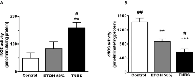
The changes in NOS activities
Both iNOS and cNOS enzyme activities in all three groups were measured. It is clearly shown in our results that iNOS activity was elevated in the 50% EtOH group and it was three-folds higher and significant in TNBS group only (49.3±19.9 vs 158±19.8 compared to control; p<0.01) (Figure 3a). The 50% EtOH and the TNBS treatment also significantly decreased cNOS activity (1428±114 vs.
872±76, control vs EtOH, p<0.01; 1428±114 vs. 571±89,
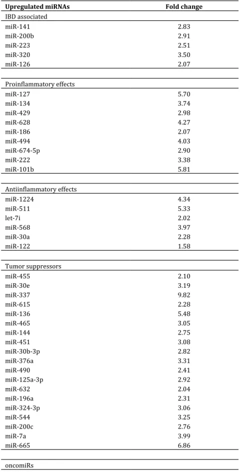
Table 1: Upregulated miRNA expression changes in TNBS colitis rat colon samples (fold change: TNBS compared to control).
Upregulated miRNAs Fold change
IBD associated
miR-141 2.83
miR-200b 2.91
miR-223 2.51
miR-320 3.50
miR-126 2.07
Proinflammatory effects
miR-127 5.70
miR-134 3.74
miR-429 2.98
miR-628 4.27
miR-186 2.07
miR-494 4.03
miR-674-5p 2.90
miR-222 3.38
miR-101b 5.81
Antiinflammatory effects
miR-1224 4.34
miR-511 5.33
let-7i 2.02
miR-568 3.97
miR-30a 2.28
miR-122 1.58
Tumor suppressors
miR-455 2.10
miR-30e 3.19
miR-337 9.82
miR-615 2.28
miR-136 5.48
miR-465 3.05
miR-144 2.75
miR-451 3.08
miR-30b-3p 2.82
miR-376a 3.31
miR-490 2.41
miR-125a-3p 2.92
miR-632 2.04
miR-196a 2.31
miR-324-3p 3.06
miR-544 3.25
miR-200c 2.76
miR-7a 3.99
miR-665 6.86
oncomiRs
miR-652 2.15
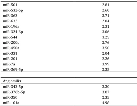
Table 1 contd: Upregulated miRNA expression changes in TNBS colitis rat colon samples (fold change: TNBS compared to control).
miR-501 2.81
miR-532-5p 2.60
miR-362 3.71
miR-632 2.04
miR-196a 2.31
miR-324-3p 3.06
miR-544 3.25
miR-200c 2.76
miR-450a 3.50
miR-331 2.04
miR-201 2.26
miR-7a 3.99
miR-369-5p 2.35
AngiomiRs
miR-342-5p 2.20
miR-376b-5p 3.87
miR-350 2.35
miR-101a 4.98
control compared to TNBS, p<0.001) (Figure 3b).
CBS and CSE enzymes expression changes
By measuring the expression changes of CBS and CSE enzymes using Western blotting, we observed diminished expression levels (CBS: EtOH: 2022,674±137.95, TNBS:
1602.917±337.926 vs control: 2917.169±123.9; CSE:
EtOH: 7320.431±506.35, TNBS: 5835.478±497.34, compared to control: 9370±752.217) in the treated groups as compared to the control and the expression changes were significant only in the TNBS group (p<0.05) (Figure 4a and b).
DISCUSSION
The aim of the present study was to determine the relationship between the differentially expressed miRNAs and gasotransmitter synthesizing enzymes in a TNBS- induced colitis model. Specifically, we screened 228 miRNA’s expression changes and the gasotransmitter synthesizing enzyme activities and expression changes were measured in parallel for comparison.
Inflammatory bowel diseases, such as Crohn’s disease and ulcerative colitis are chronic immune-related gastro- intestinal disorders. Epidemiological studies on IBD showed that this disease is widespread particularly in Northern and Western Europe and North America (Ponder and Long, 2013). However, much lower rates of incidence
are demonstrated in areas of Africa, South America and Asia, suggesting that westernization might be a special risk factor for developing IBD in susceptible individuals (Ye et al., 2015). Owing to its worldwide occurence and the fact that the accurate pathomechanism is still unknown, IBD is one of the most researched areas presently. Furthermore, the diagnosis and the exact characterization of these diseases are cumbersome and unresolved because there is no reliable and existence of non-invasive diagnostic test.
Numerous studies demonstrated that IBD is associated with the differentially expressed genes which contribute to immune responses and healing progression as well (Wu et al., 2010). In recent years, a new potential area of diagnostics was revealed, utilizing the differentially expressed miRNAs, called miRNA profiles, which seems to be a novel diagnostic marker (Fisher and Lin, 2015;
Schaefer et al., 2015). It is becoming increasingly clear that miRNAs, gasotransmitter synthesizing enzymes and the produced gasotransmitters have a crucial role in this disease as well.
The differential expression of microRNAs and their role in IBD have not been extensively studied, but it is a promising new area of research. In our present study, we screened 228 miRNAs expression changes in TNBS induced rats colitis, and found 94 upregulated and 63 downregulated miRNAs. The differentially expressed miRNAs were classified according to their target mRNAs and affected pathways based on current evidence. Five clusters were identified: IBD associated miRNAs, oncomiRs, tumor suppressors, miRNAs which have proinflammatory, or antiinflammatory effects and
Table 2: Downregulated miRNA expression changes in TNBS colitis (fold change: TNBS compared to control).
Downregulated miRNAs Fold change
IBD associated
miR-29a 0.18
miR-132 0.34
miR-19a 0.56
miR-21 0.64
miR-150 0.14
miR-191 0.09
miR-107 0.44
miR-199a-5p 0.23
miR-143 0.09
Proinflammatory effects
miR-23b 0.39
miR-146a 0.35
miR-16 0.27
miR-139-5p 0.31
Antiinflammatory effects
miR-19b 0.64
miR-23a 0.35
miR-93 0.63
Tumor suppressors
miR-26a 0.30
miR-29b 0.06
miR-29c 0.12
miR-30c 0.13
miR-204 0.25
miR-133a 0.23
miR-26b 0.61
miR-290 0.41
miR-203 0.45
miR-28 0.39
miR-215 0.07
miR-192 0.18
miR-148b-3p 0.39
miR-411 0.55
miR-199a-3p 0.10
miR-152 0.21
miR-30d 0.22
miR-145 0.29
miR-324-5p 0.46
miR-154 0.54
miR-205 0.29
miR-15b 0.05
oncomiRs
miR-24 0.25
miR-183 0.18
Table 2 Contd: Downregulated miRNA expression changes in TNBS colitis (fold change: TNBS compared to control).
miR-103 0.22
miR-151 0.30
miR-1249 0.64
miR-96 0.45
miR-106b 0.30
miR-425 0.18
miR-423 0.55
miR-17-5p 0.35
miR-130b 0.53
miR-342-3p 0.07
AngiomiRs
let-7d 0.17
miR-130a 0.14
0 1 2 3 4 5
# # #
* * *
*
C o n t r o l E T O H 5 0 % T N B S
H O a c ti v it y (n m o l b il ir u b in e /h /m g p ro te in )
#
Figure 2: Heme oxygenase (HO) enzyme activity changes in ethanol (EtOH) alone and 2, 4, 6 trinitrobenzenesulphonic acid (TNBS)- induced colitis. HO enzyme activity was determined by measurement of bilirubin formation (n=4-10; mean ± SEM; # Compare to the control group, * compare to the EtOH group; *, # p<0.05, **, ## p<0.01, ***, ### p<0.001).
(Tables 1 and 2).
Several miRNAs were found to be relevant contributors to IBD pathogenesis. For instance, Schaefer et al. (2015) found that in saliva, blood and intestinal samples from IBD patients, that miR-19a, miR-21, miR-31, miR-101, miR- 146a and miR-375 might be diagnostic markers in IBD. In our study, we found the same changes in miR-21, miR-101 and miR-146a expressions, but we did not observe significant changes in miR-19a, miR-31 and miR-375 expressions. Wu et al. (2010) measured miRNAs in IBD patients and found four miRNAs to be significantly over-
expressed; miR-16, miR-21, miR-223 and miR-594. These results do not completely agree with our findings, because we found miR-16 and miR-21 to be downregulated, while we have not seen a significant change in miR-594. This discrepancy might be interpreted by the different species, because Wu et al. (2010) used human samples and we used rat samples for our investigations. Furthermore, Zahm et al. (2014) analyzed the miRNA expression changes in young UC and CD patients and found several miRNAs, miR-192, miR-194, miR-21 and miR-142-3p that were upregulated with the most abundant expression
Figure 3: Colon tissue inducible nitric oxide synthase (iNOS) and constitutive nitric oxide synthase (cNOS) activities measured by enzyme conversion. Bar chart (A) representing iNOS activity, and chart (B) representing cNOS activity.
Groups: control (no treatment); ethanol (EtOH) and 2, 4, 6-trinitrobenzenesulphonic acid (TNBS). Values are represented as means ± S.E.M. for n = 4-10; * Compared to the control group, # compare to the ethanol group; *, # p<0.05, **, ## p<0.01).
Figure 4: The hydrogen sulphide (H2S) producing enzymes (cystathionine β-synthase (CBS), and cystathionine γ-lyase (CSE)) expression changes by Western Blotting in treatment with ethanol (EtOH) or 2,4,6-trinitrobenzenesulphonic acid (TNBS) are demonstrated in (A) and (B) panel (n=4-10; mean ± SEM; * Compared to the control group, # compare to the ethanol group; *,#
p<0.05, **, ## p<0.01,***, ### p<0.001).
changes in miR-21 and 142-3p.
A growing body of research indicates that gasotransmitters have pivotal roles in IBD pathogenesis and disease outcome as well. For example, Aoi et al. (2008) demonstrated that administration of L-NAME, a specific inhibitor of NO production, exacerbated DSS-induced colitis, proposing the protective role of NO in colitis. In turn, application of NO donors such as NOR-3 and NONOates, aggravate DSS-colitis as well, suggesting that both low and excessive levels of NO worsened colitis (Yoshida et al., 2000). It is also suggested that pharmacological induction of HO-1 expression and activation or administration of CO donors can significantly attenuate experimental colitis (Erbil et al., 2007; Uddin et
al., 2013). Fukuda et al. (2014) applied CORM-3 as a CO donor in TNBS- treated mice and described a significant reduction in the inflammatory response compared to TNBS control mice, which received an inactive form of CORM-3.
Furthermore, Takagi et al. (2010) found that inhaled CO in low concentrations (200 ppm) also impacts the antiinflammatory process with a significant inhibition of colonic damage and decreased levels of TNF-α. Moreover, several studies reported the ability of H2S to enhance ulcer healing. Of these, Wallace et al. (2007) measured a high level of H2S in the reepitalization zone of the colonic ulcers which led to enhanced healing of the damaged colonic section. Wallace et al. (2010) also demonstrated in iodocetamide-induced murine colitis, that treatment with
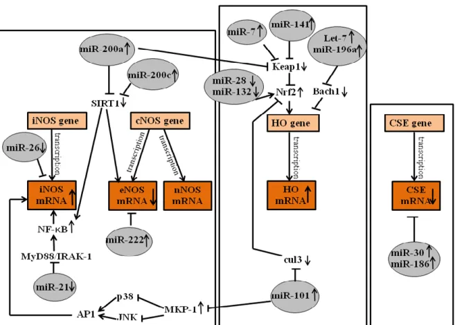
Figure 5: miRNAs which might responsible for the changes in gasotransmitters synthesizing enzymes expressions and activities. iNOS: inducible nitric oxide synthase; NF-κB: nuclear factor kappa-light-chain-enhancer of activated B cells; Myd88:
myeloid differentiation primary response gene 88; IRAK-1: interleukin-1 receptor-associated kinase-1; SIRT1: sirtuin1; MKP- 1: map kinase phosphatase-1; JNK: c-Jun N-terminal kinases; AP1: activator-protein 1; cul3: cullin 3; Nrf2: nuclear factor erythroid 2-related factor; Keap1: Kelch-like ECH-associated protein-1; CSE: cystathionine-γ-lyase,cNOS: constitutive nitric oxide synthase, eNOS: endothelial nitric oxide synthase, nNOS: neuronal nitric oxide synthase, HO: heme oxygenase
H2S donors, such as Lawesson’s Reagent or NaHS, cause amarked decrease in the severity of colitis. To test the effects of H2S in DSS-induced colitis, Hirata et al. (2011) administered a CSE inhibitor, DL-propargylglycine (PAG) and they found that this procedure worsened colitis.
In our work, gasotransmitter synthesizing enzyme activities were increased the case of iNOS and HO enzymes in TNBS-treated rats, while the cNOS enzyme showed decreased activity. There are several studies which are in line with our findings (Yue et al., 2001); they showed that TNBS colitis increased the activity of iNOS enzyme with a concomitant protective effect. Wang et al. (2008) reported that HO-1 activity increased after the rectal induction of colitis by TNBS enema. Furthermore in our previous study, we showed the same changes about HO activity in the same animal model and our results pointed out that the HO-1 activity increased very rapidly and dramatically in the colon after TNBS treatment. The expression changes in CSE and CBS enzymes are controversial. In our study, in accordance with Wallace et al. (2009), the H2S synthesizing CSE and CBS expressions were reduced in the treated groups and unlike others we measured a significant reduction in the TNBS group. Other investigations showed
that H2S producing enzyme expressions were enhanced in inflammation. For instance, Flannigan et al. (2013) and Distrutti et al. (2006) found an elevated expression of this enzyme in inflamed mucosa compared to healthy colon.
Furthermore, Hirata et al. (2011) showed a time- dependent increase in CSE and CBS expression, but they did not measure significant change at day 3 of their experiment as compared to day 0.
In the present study we demonstrated for the first time that there might be relationships between gasotransmitters and microRNAs in IBD. Our results suggest that miRNAs are differentially expressed in TNBS- induced colitis and there are several miRNAs (miR-26a, miR-21, miR-222, miR-200a, miR-200c, miR-101, miR-28, miR-132, miR-7, miR-141, Let-7, miR-196a, miR-186 and miR-30) which might regulate gasotransmitter synthesizing enzyme mRNAs (Figure 5).
We also found out that the downregulated expression of miR-26a might elevate iNOS activity. Zhu et al. (2013) demonstrating that iNOS is a direct target for miR-26a, thus, suppress its expression. In our TNBS- induced colitis colon samples, the miR-26a showed downregulated expression, thus, iNOS might be able to increase. It was
also observed that nuclear factor kappa-light-chain enhancer of activated B cells (NF-κB), a transcription factor, induces the expression of iNOS enzyme. miR-21 alleviates the expression levels of myeloid differentiation primary response gene 88 (MyD88) and interleukin-1 receptor-activated kinase (IRAK-1), which are well known regulators of NF-κB (Xu et al., 2014). In our study, miR-21 showed downregulated expression, which means that MyD88 and IRAK-1 are not repressed, thus, it might contribute to an increase in NF-κB and iNOS expression.
Furthermore, it is also suggested that MAP kinase phosphatase-1 (MKP-1) is a target for miR-101 and the increased expression of miR-101 is associated with an elevated level of MKP-1. The upregulated MKP-1 is able to inhibit p38 and c-Jun N-terminal kinase (JNK), which allows an increase of activator protein 1 (AP1) and thus iNOS expression (Gao et al., 2014).
We suggest that three miRNAs (miR-200a, miR-200c and miR-222) regulate the eNOS and nNOS enzymes (also termed as cNOS). Evangelista et al. (2013) found that upregulation of miR-222 decreases eNOS enzyme.
Furthermore, miR-200a and miR-200c target the sirtuin 1 (SIRT1) 3’ UTR sequence and thus, contribute to a decrease in SIRT1 levels. SIRT1 was found to upregulate eNOS enzyme’s expression, but miR-200a and miR-200c seems to prevent this effect through the initiation of SIRT1 mRNA degradation (Triggle et al., 2012, Carlomosti et al., 2017). In addition, the miR-200a and miR-200c induced repression of SIRT1 might contribute to the upregulation of the iNOS enzyme as well through NF-κB (Yamakuchi, 2012).
HO-1 is induced as a protective mechanism against colonic oxidative damage. Hence, induction of HO-1 might have a potential role in the management of colitis. Several transcription factors are responsible for regulation of HO enzymes. One of these is the nuclear factor erythroid 2- related factor 2 (Nrf2) which is repressed by Kelch ECH associating protein 1 (Keap1) (Reichard et al., 2007) and ubiquitilated by cullin 3 (cul3) (Kim et al., 2014) is able to attach to ARE (antioxidant response element) sequences, thus, inducing HO-1 expression (Reichard et al., 2007). We found several miRNAs, which appears to have an effect on HO-1 activation or repression. We suggest that several miRNAs contribute to an increase in Nrf2 and thus, HO-1 expression according to other investigator’s studies: miR- 200a (Eades et al., 2011), miR-7 (Kabaria et al., 2015), miR-141 (Shi et al., 2015), which target the 3’ UTR sequence of Keap1. Furthermore, cul3, which is also responsible for the inhibition of Nrf2 is inhibited by the increased expression of miR-101 and may contribute to increased HO-1 expression (Schaefer et al., 2015). Another pathway that regulates HO expression is mediated by Bach1, which is responsible for tonic repression of the HMOX1 gene. Hou et al. (2012) found increased expression of Let-7 family members while Go et al. (2016) reported that miR-196a is able to repress Bach1, which allows increased HO-1-expression. Our results also suggest that
the downregulation of miR-28 and miR-132, based on Yang et al. (2011) and Stachurska et al. (2013) findings might permit Nrf2 to increase and thereby activate HO expression.
In our study, the H2S generating CSE enzyme expression was reduced and this effect might be explained by the upregulated miR-30 (Shen et al., 2015) and miR-186 (Yao et al., 2016) expressions, which target CSE’s 3’ UTR sequence and contribute to degradation of CSE mRNA.
Conclusion
Based on our results and on the current literature, we suggest several miRNAs, which might have a regulatory effect on gasotransmitter synthesizing enzymes: miR-26a, miR-21, miR-222, miR-200a, miR-200c, miR-101, miR-28, miR-132, miR-7, miR-141, Let-7, miR-196a, miR-186 and miR-30. Our results may indicate that miRNAs could serve as biomarkers for IBD, aiding early diagnosis and leading to the development of personalized therapies.
ACKNOWLEDGMENTS
This research was realized in the frames of TÁMOP 4.2.4.
A/2-11-1-2012-0001 (to Csaba Varga) National Excellence Program-elaborating and operating an inland student and researcher personal support system. This project was also supported by GINOP-2.3.2-15-2016-00030.
REFERENCES
Abe K, Kimura H (1996). The possible role of hydrogen sulfide as an endogenous neuromodulator. J Neurosci. 16(3):1066-1071.
Aoi Y, Terashima S, Ogura M, Nishio H, Kato S, Takeuchi K (2008). Roles of nitric oxide (NO) and NO synthases in healing of dextran sulfate sodium-induced rat colitis. J. Physiol. Pharmacol. 59(2):315-336.
Archanioti P, Gazouli M, Theodoropoulos G, Vaiopoulou A, Nikiteas N (2011). Micro-RNAs as regulators and possible diagnostic bio-markers in inflammatory bowel disease. J. Crohns Colitis. 5(6):520-524.
Boughton-Smith NK, Evans SM, Laszlo F, Whittle BJ, Moncada S (1993).
The induction of nitric oxide synthase and intestinal vascular permeability by endotoxin in the rat. Br. J. Pharmacol. 110(3):1189- 1195.
Carlomosti F, D'Agostino M, Beji S, Torcinaro A, Rizzi R, Zaccagnini G, Maimone B Di Stefano V, De Santa F, Cordisco S, Antonini A, Ciarapica R, Dellambra E, Martelli F, Avitabile D, Capogrossi MC, Magenta A (2017). Oxidative Stress-Induced miR-200c Disrupts the Regulatory Loop Among SIRT1, FOXO1, and eNOS. Antioxid Redox Signal.
Chen WX, Ren LH, Shi RH (2014). Implication of miRNAs for inflammatory bowel disease treatment: Systematic review. World J.
Gastrointest Pathophysiol. 5(2):63-70.
Coskun M, Bjerrum JT, Seidelin JB, Troelsen JT, Olsen J, Nielsen OH (2013). miR-20b, miR-98, miR-125b-1*, and let-7e* as new potential diagnostic biomarkers in ulcerative colitis. World J. Gastroenterol.
19(27):4289-4299.
Coskun M, Bjerrum JT, Seidelin JB, Nielsen OH (2012). MicroRNAs in inflammatory bowel disease--pathogenesis, diagnostics and therapeutics. World J. Gastroenterol. 18(34):4629-4634.
Dalal SR, Kwon JH (2010). The Role of MicroRNA in Inflammatory Bowel Disease. Gastroenterol. Hepatol. NY. 6(11):714-722.
Dhillon SS, Mastropaolo LA, Murchie R, Griffiths C, Thoni C, Elkadri A, Xu W, Mack A, Walters T, Guo C, Mack D, Huynh H, Baksh S, Silverberg MS, Brumell JH, Snapper SB, Muise AM (2014). Higher activity of the inducible nitric oxide synthase contributes to very early onset inflammatory bowel disease. Clin. Transl. Gastroenterol. 5:e46.
Distrutti E, Sediari L, Mencarelli A, Renga B, Orlandi S, Russo G, Caliendo G, Santagada V, Cirino G, Wallace JL, Fiorucci S (2006). 5-Amino-2- hydroxybenzoic acid 4-(5-thioxo-5H-[1,2]dithiol-3yl)-phenyl ester (ATB-429), a hydrogen sulfide-releasing derivative of mesalamine, exerts antinociceptive effects in a model of postinflammatory hypersensitivity. J. Pharmacol. Exp. Ther. 319(1):447-458.
Eades G, Yao Y, Yang M, Zhang Y, Chumsri S, Zhou Q (2011). miR-200a regulates SIRT1 expression and epithelial to mesenchymal transition (EMT)-like transformation in mammary epithelial cells. J. Biol. Chem.
286(29):25992-26002.
Erbil Y, Giris M, Abbasoglu SD, Barbaros U, Yanik BT, Necefli A, Olgac V, Toker GA (2007). Effect of heme oxygenase-1 induction by octreotide on TNBS-induced colitis. J. Gastroenterol. Hepatol. 22(11):1852-1858.
Evangelista AM, Deschamps AM, Liu D, Raghavachari N, Murphy E (2013). miR-222 contributes to sex-dimorphic cardiac eNOS expression via ets-1. Physiol. Genomics. 45(12):493-498.
Farago N, Feher LZ, Kitajka K, Das UN, Puskas LG (2011). MicroRNA profile of polyunsaturated fatty acid treated glioma cells reveal apoptosis-specific expression changes. Lipids Health Dis. 10:173.
Farago N, Zvara A, Varga Z, Ferdinandy P, Puskas LG (2011).
Purification of high-quality micro RNA from the heart tissue. Acta Biol.
Hung. 62(4):413-425.
Fisher K, Lin J (2015). MicroRNA in inflammatory bowel disease:
Translational research and clinical implication. World J. Gastroenterol.
21(43):12274-12282.
Flannigan KL, Ferraz JG, Wang R, Wallace JL (2013). Enhanced synthesis and diminished degradation of hydrogen sulfide in experimental colitis: a site-specific, pro-resolution mechanism. PLoS One 8(8):e71962.
Fukuda W, Takagi T, Katada K, Mizushima K, Okayama T, Yoshida N, Kamada K, Uchiyama K, Ishikawa T, Handa O, Konishi H, Yagi N, Ichikawa H, Yoshikawa T, Cepinskas G, Naito Y, Itoh Y (2014). Anti- inflammatory effects of carbon monoxide-releasing molecule on trinitrobenzene sulfonic acid-induced colitis in mice. Dig. Dis. Sci.
59(6):1142-1151.
Gao, Y, Liu F, Fang L, Cai R, Zong C, Qi Y (2014). Genkwanin inhibits proinflammatory mediators mainly through the regulation of miR- 101/MKP-1/MAPK pathway in LPS-activated macrophages. PLoS One 9(5):e96741.
Go H, La P, Namba F, Ito M, Yang G, Brydun A, Igarashi K, Dennery PA (2016). MiR-196a regulates heme oxygenase-1 by silencing Bach1 in the neonatal mouse lung. Am. J. Physiol. Lung. Cell Mol. Physiol.
311(2):L400-411.
Guo W, Kan JT, Cheng ZY, Chen JF, Shen YQ, Xu J, Wu D, Zhu YZ (2012).
Hydrogen sulfide as an endogenous modulator in mitochondria and mitochondria dysfunction. Oxid. Med. Cell Longev. 878052.
Hegazi RA, Rao KN, Mayle A, Sepulveda AR, Otterbein LE, Plevy SE (2005). Carbon monoxide ameliorates chronic murine colitis through a heme oxygenase 1-dependent pathway. J. Exp. Med. 202(12):1703- 1713.
Hirata I, Naito Y, Takagi T, Mizushima K, Suzuki T, Omatsu T, Handa O, Ichikawa H, Ueda H, Yoshikawa T (2011). Endogenous hydrogen sulfide is an anti-inflammatory molecule in dextran sodium sulfate- induced colitis in mice. Dig. Dis. Sci. 56(5):1379-1386.
Horvath K, Varga C, Berko A, Posa A, Laszlo F, Whittle BJ (2008). The involvement of heme oxygenase-1 activity in the therapeutic actions of 5-aminosalicylic acid in rat colitis. Eur. J. Pharmacol. 581(3):315-323.
Hou W, Tian Q, Steuerwald NM, Schrum LW, Bonkovsky HL (2012). The let-7 microRNA enhances heme oxygenase-1 by suppressing Bach1 and attenuates oxidant injury in human hepatocytes. Biochem. Biophys.
Acta 1819(11-12):1113-1122.
Iborra M, Bernuzzi F, Correale C, Vetrano S, Fiorino G, Beltran B, Marabita F, Locati M, Spinelli A, Nos P, Invernizzi P, Danese S (2013).
Identification of serum and tissue micro-RNA expression profiles in different stages of inflammatory bowel disease. Clin. Exp. Immunol.
173(2):250-258.
Kabaria S, Choi DC, Chaudhuri AD, Jain MR, Li H, Junn E (2015).
MicroRNA-7 activates Nrf2 pathway by targeting Keap1 expression.
Free Radic. Biol. Med. 89:548-556.
Kalla R, Ventham NT, Kennedy NA, Quintana JF, Nimmo ER, Buck AH, Satsangi J (2015). MicroRNAs: new players in IBD. Gut. 64(3):504-517.
Kim JH, Lee KS, Lee DK, Kim J, Kwak SN, Ha KS, Choe J, Won MH, Cho BR, Jeoung D, Lee H, Kwon YG, Kim YM (2014). Hypoxia-responsive microRNA-101 promotes angiogenesis via heme oxygenase-1/vascular endothelial growth factor axis by targeting cullin 3. Antioxid. Redox.
Signal. 21(18):2469-2482.
Kutty RK, Kutty G, Rodriguez IR, Chader GJ, Wiggert B (1994).
Chromosomal localization of the human heme oxygenase genes: heme oxygenase-1 (HMOX1) maps to chromosome 22q12 and heme oxygenase-2 (HMOX2) maps to chromosome 16p13.3. Genomics.
20(3):513-516.
Mestdagh P, Hartmann N, Baeriswyl L, Andreasen D, Bernard N, Chen C, Cheo D, D'Andrade P, DeMayo M, Dennis L, Derveaux S, Feng Y, Fulmer-Smentek S, Gerstmayer B, Gouffon J, Grimley C, Lader E, Lee KY, Luo S, Mouritzen P, Narayanan A, Patel S, Peiffer S, Ruberg S, Schroth G, Schuster D, Shaffer JM, Shelton, S. Silveria, U. Ulmanella, V.
Veeramachaneni, F. Staedtler, T. Peters, T. Guettouche EJ, Wong L, Vandesompele J (2014). Evaluation of quantitative miRNA expression platforms in the microRNA quality control (miRQC) study. Nat.
Methods. 11(8):809-815.
Mustafa AK, Gadalla MM, Snyder SH (2009). Signaling by gasotransmitters. Sci. Signal. 2(68):re2.
O'Dell TJ, Hawkins RD, Kandel ER, Arancio O (1991). Tests of the roles of two diffusible substances in long-term potentiation: evidence for nitric oxide as a possible early retrograde messenger. Proc. Natl. Acad. Sci.
USA. 88(24):11285-11289.
Ponder A, Long MD (2013). A clinical review of recent findings in the epidemiology of inflammatory bowel disease. Clin. Epidemiol. 5:237- 247.
Reichard JF, Motz GT, Puga A (2007). Heme oxygenase-1 induction by NRF2 requires inactivation of the transcriptional repressor BACH1.
Nucleic. Acids. Res. 35(21):7074-7086.
Ryter SW, Choi AM (2016). Targeting heme oxygenase-1 and carbon monoxide for therapeutic modulation of inflammation. Transl. Res.
167(1):7-34.
Schaefer JS, Attumi T, Opekun AR, Abraham B, Hou J, Shelby H, Graham DY, Streckfus C, Klein JR (2015). MicroRNA signatures differentiate Crohn's disease from ulcerative colitis. BMC Immunol. 16:5.
Sheikh SZ, Hegazi RA, Kobayashi T, Onyiah JC, Russo SM, Matsuoka K, Sepulveda AR, Li F, Otterbein LE, Plevy SE (2011). An anti- inflammatory role for carbon monoxide and heme oxygenase-1 in chronic Th2-mediated murine colitis. J. Immunol. 186(9):5506-5513.
Shen Y, Shen Z, Miao L, Xin X, Lin S, Zhu Y, Guo W, Zhu YZ (2015).
miRNA-30 family inhibition protects against cardiac ischemic injury by regulating cystathionine-gamma-lyase expression. Antioxid Redox Signal. 22(3):224-240.
Shi L, Wu L, Chen Z, Yang J, Chen X, Yu F, Zheng F, Lin X (2015). MiR- 141 Activates Nrf2-Dependent Antioxidant Pathway via Down- Regulating the Expression of Keap1 Conferring the Resistance of Hepatocellular Carcinoma Cells to 5-Fluorouracil. Cell Physiol.
Biochem. 35(6):2333-2348.
Sonkoly E, Pivarcsi A (2009). microRNAs in inflammation. Int. Rev.
Immunol. 28(6): 535-561.
Stachurska A, Ciesla M, Kozakowska M, Wolffram S, Boesch- Saadatmandi C, Rimbach G, Jozkowicz A, Dulak J, Loboda A (2013).
Cross-talk between microRNAs, nuclear factor E2-related factor 2, and heme oxygenase-1 in ochratoxin A-induced toxic effects in renal proximal tubular epithelial cells. Mol. Nutr. Food Res. 57(3):504-515.
Su X, Qian C, Zhang Q, Hou J, Gu Y, Han Y, Chen Y, Jiang M, Cao X (2013). miRNomes of haematopoietic stem cells and dendritic cells identify miR-30b as a regulator of Notch1. Nat. Commun. 4:2903.
Szalai Z, Szasz A, Nagy I, Puskas LG, Kupai K, Kiraly A, Berko AM, Posa A, Strifler G, Barath Z, Nagy LI, Szabo R, Pavo I, Murlasits Z, Gyongyosi M, Varga C (2014). Anti-inflammatory effect of recreational exercise in TNBS-induced colitis in rats: role of NOS/HO/MPO system.
Oxid Med. Cell Longev. 925981.
Takagi T, Naito Y, Mizushima K, Akagiri S, Suzuki T, Hirata I, Omatsu T,
Handa O, Kokura S, Ichikawa H, Yoshikawa T (2010). Inhalation of carbon monoxide ameliorates TNBS-induced colitis in mice through the inhibition of TNF-alpha expression. Dig. Dis. Sci. 55(10):2797-2804.
Takagi T, Naito Y, Mizushima K, Hirata I, Yagi N, Tomatsuri N, Ando T, Oyamada Y, Isozaki Y, Hongo H, Uchiyama K, Handa O, Kokura S, Ichikawa H, Yoshikawa T (2010). Increased expression of microRNA in the inflamed colonic mucosa of patients with active ulcerative colitis. J.
Gastroenterol. Hepatol. 25(1):129-133.
Teague B, Asiedu S, Moore PK (2002). The smooth muscle relaxant effect of hydrogen sulphide in vitro: evidence for a physiological role to control intestinal contractility. Br. J. Pharmacol. 137(2):139-145.
Tenhunen R, Marver HS, Schmid R (1968). The enzymatic conversion of heme to bilirubin by microsomal heme oxygenase. Proc. Natl. Acad. Sci.
USA. 61(2):748-755.
Triggle CR, Samuel SM, Ravishankar S, Marei I, Arunachalam G, Ding H.(2012). "The endothelium: influencing vascular smooth muscle in many ways. Can. J. Physiol. Pharmacol. 90(6):713-738.
Uddin MJ, Jeong SO, Zheng M, Chen Y, Cho GJ, Chung HT, JoeY (2013).
Carbon monoxide attenuates dextran sulfate sodium-induced colitis via inhibition of GSK-3beta signaling. Oxid. Med. Cell Longev. 210563.
Varga ZV, Kupai K, Szucs G, Gaspar R, Paloczi J, Farago N, Zvara A, Puskas LG, Razga Z, Tiszlavicz L, Bencsik P, Gorbe A, Csonka C, Ferdinandy P, Csont T (2013). MicroRNA-25-dependent up-regulation of NADPH oxidase 4 (NOX4) mediates hypercholesterolemia-induced oxidative/nitrative stress and subsequent dysfunction in the heart. J.
Mol. Cell Cardiol. 62:111-121.
Varga ZV, Zvara A, Farago N, Kocsis GF, Pipicz M, Gaspar R, Bencsik P, Gorbe A, Csonka C, Puskas LG, Thum T, Csont T, Ferdinandy P (2014).
MicroRNAs associated with ischemia-reperfusion injury and cardioprotection by ischemic pre- and postconditioning: protectomiRs.
Am. J. Physiol. Heart Circ. Physiol. 307(2):H216-227.
Wallace JL (2010). Physiological and pathophysiological roles of hydrogen sulfide in the gastrointestinal tract. Antioxid Redox Signal.
12(9):1125-1133.
Wallace JL, Vong L, McKnight W, Dicay M, Martin GR (2009).
Endogenous and exogenous hydrogen sulfide promotes resolution of colitis in rats. Gastroenterol. 137(2):569-578. 578 e561.
Wallace JL, Dicay M, McKnight W, Martin GR (2007). Hydrogen sulfide enhances ulcer healing in rats. FASEB J. 21(14):4070-4076.
Wang R, Wang Z, Wu L (1997). Carbon monoxide-induced vasorelaxation and the underlying mechanisms. Br. J. Pharmacol. 121(5):927-934.
Wen Z, Fiocchi C (2004). Inflammatory bowel disease: autoimmune or immune-mediated pathogenesis? Clin. Dev. Immunol. 11(3-4):195-204.
Whiteman M, Winyard PG (2011). Hydrogen sulfide and inflammation:
the good, the bad, the ugly and the promising. Expert Rev. Clin.
Pharmacol. 4(1):13-32.
Wu F, Guo NJ, Tian H, Marohn M, Gearhart S, Bayless TM, Brant SR, Kwon JH (2011). Peripheral blood microRNAs distinguish active ulcerative colitis and Crohn's disease. Inflamm. Bowel. Dis. 17(1):241- 250.
Wu F, Zhang S, Dassopoulos T, Harris ML, Bayless TM, Meltzer SJ, Brant SR, Kwon JH (2010). Identification of microRNAs associated with ileal and colonic Crohn's disease. Inflamm. Bowel Dis. 16(10):1729-1738.
Xu X, Kriegel AJ, Jiao X, Liu H, Bai X, Olson J, Liang M, Ding X (2014).
miR-21 in ischemia/reperfusion injury: a double-edged sword?
Physiol. Genomics. 46(21):789-797.
Yamakuchi M (2012). MicroRNA Regulation of SIRT1. Front. Physiol. 3:68.
Yang M, Yao Y, Eades G, Zhang Y, Zhou Q (2011). MiR-28 regulates Nrf2 expression through a Keap1-independent mechanism. Breast Cancer Res. Treat. 129(3):983-991.
Yao Y, Zhang H, Chen HP, Li L, Xie W, Lan G, Zhao ZW, Zheng XL, Wang ZB, a Tang CK (2016). MicroRNA-186 promotes macrophage lipid accumulation and secretion of pro-inflammatory cytokines by targeting cystathionine gamma-lyase in THP-1 macrophages.
Atherosclerosis. 250:122-132.
Ye Y, Pang Z, Chen W, Ju S, Zhou C (2015). The epidemiology and risk factors of inflammatory bowel disease. Int. J. Clin. Exp. Med.
8(12):22529-22542.
Yoshida Y, Iwai A, Itoh K, Tanaka M, Kato S, Hokari R, Miyahara T, Koyama H, Miura S, Kobayashi M (2000). Role of inducible nitric oxide synthase in dextran sulphate sodium-induced colitis. Aliment Pharmacol. Ther. 14(1):26-32.
Yue G, Lai PS, Yin K, Sun FF, Nagele RG, Liu X, Linask KK, Wang C, Lin KT, WongYP (2001). Colon epithelial cell death in 2,4,6- trinitrobenzenesulfonic acid-induced colitis is associated with increased inducible nitric-oxide synthase expression and peroxynitrite production. J. Pharmacol. Exp. Ther. 297(3):915-925.
Zahm AM, Hand NJ, Tsoucas DM, Le Guen CL, Baldassano RN, Friedman JR (2014). Rectal microRNAs are perturbed in pediatric inflammatory bowel disease of the colon. J. Crohns. Colitis. 8(9):1108-1117.
Zhu H, Vishwamitra D, Curry CV, Manshouri R, Diao L, Khan A, Amin M (2013). NPM-ALK up-regulates iNOS expression through a STAT3/microRNA-26a-dependent mechanism. J. Pathol. 230(1): .82- 94.
Cite this article as:
Almási N, Pósa A, Al-awar A, Török S, Baráth Z, Nemcsók J, Murlasits Z, Nagy LI, Puskás LG, Varga C, Kupai K (2017).
Differentially expressed microRNAs and their relation to gasotransmitters in TNBS-induced colitis in rat colon. Acad. J.
Sci. Res. 5(9): 277-289.
Submit your manuscript at
http://www.academiapublishing.org/ajsr