BMI is an important driver of b-cell loss in type 1 diabetes upon diagnosis in 10 to 18-year-old children
A Lauria1,*, A Barker2,*, N Schloot3,†, N Hosszufalusi4, J Ludvigsson5, C Mathieu6, D Mauricio7, M Nordwall5,8, B Van der Schueren6, T Mandrup-Poulsen9,14, W A Scherbaum10, I Weets11,12, F K Gorus11,12, N Wareham2, R D Leslie13and P Pozzilli1,13
1Department of Endocrinology and Diabetes, University Campus Bio-Medico, Via Alvaro del Portillo, Rome 21 00128, Italy,2MRC Epidemiology Unit, Cambridge, UK,3Institute for Clinical Diabetology, German Diabetes Centre, Leibniz-Institute for Diabetes Research and Clinic for Metabolic Diseases Heinrich Heine University, Dusseldorf, Germany,4Semmelweis University, 3rd Department of Internal Medicine, Linkoping University, Linkoping, Sweden,
5Division of Pediatrics, Department of Clinical and Experimental Medicine, Faculty of Health Sciences, Linko¨ping University, Linko¨ping, Sweden,6Laboratory for Experimental Medicine and Endocrinology, Katholieke Universiteit Leuven, Leuven, Belgium,7Department Endocrinology, Hospital Arnau de Vilanova, Lleida, Spain,8Pediatric Clinic, Vrinnevi Hospital, Norrko¨ping, Sweden,9Department of Biomedical Sciences, University of Copenhagen, Copenhagen, Denmark,10Department of Endocrinology, Diabetes and Rheumatology, Heinrich Heine University, Dusseldorf, Germany,11Diabetes Research Center and Academic Hospital (UZ Brussel), Vrije Universiteit Brussel (VUB), Brussel, Belgium,12Belgian Diabetes Registry (BDR), Brussels, Belgium,13Centre of Diabetes, Blizard Institute, Barts and The London School of Medicine, Queen Mary University of London, London, UK and14Department of Molecular Medicine and Surgery, Karolinska Institutet, Stockholm, Sweden
*(A Lauria and A Barker contributed equally to this work)
†N Schloot is now at Lilly Germany GmbH, BU Diabetes Medical, Dusseldorf, Germany
Correspondence should be addressed to P Pozzilli Email
p.pozzilli@unicampus.it
Abstract
Objective: Body weight-related insulin resistance probably plays a role in progression to type 1 diabetes, but has an uncertain impact following diagnosis. In this study, we investigated whether BMI measured at diagnosis was an independent predictor of C-peptide decline 1-year post-diagnosis.
Design: Multicentre longitudinal study carried out at diagnosis and up to 1-year follow-up.
Methods: Data on C-peptide were collected from seven diabetes centres in Europe. Patients were grouped according to age at diagnosis (!5 years,nZ126;O5 years!10 years,nZ295;O10 years!18 years,nZ421;O18 years,nZ410). Linear regression was used to investigate whether BMI was an independent predictor of change in fasting C-peptide over 1 year.
Models were additionally adjusted for baseline insulin dose and HbA1c.
Results: In individuals diagnosed between 0 and 5 years, 5 and 10 years and those diagnosedO18 years, we found no association between BMI and C-peptide decline. In patients aged 10–18 years, higher BMI at baseline was associated with a greater decline in fasting C-peptide over 1 year with a decrease (b95% CI;Pvalue) of 0.025 (0.010, 0.041) nM/kg per m2 higher baseline BMI (PZ0.001). This association remained significant after adjusting for gender and differences in HbA1c and insulin dose (bZ0.026, 95% CIZ0.0097, 0.042;PZ0.002).
Conclusions: These observations indicate that increased body weight and increased insulin demand are associated with more rapid disease progression after diagnosis of type 1 diabetes in an age group 10–18 years. This should be considered in studies ofb-cell function in type 1 diabetes.
European Journal of Endocrinology (2015)172, 107–113
EuropeanJournalofEndocrinology
Introduction
Type 1 diabetes is considered an autoimmune disorder caused by inflammatoryb-cell destruction, and presenting in childhood is characterised by loss of weight, ketoaci- dosis, acute onset and insulin dependency. In contrast, type 2 diabetes is considered a metabolic disorder usually presenting in adults, associated with obesity and insulin resistance(1).
However, insulin resistance may influence disease progression following diagnosis also in type 1 diabetes.
Yki-Jarvinen & Koivisto(2)showed that newly diagnosed type 1 diabetes patients who went into remission of insulin therapy had greater insulin sensitivity than patients who continued to require insulin therapy. In addition, they found that more than a year post-diagnosis, insulin sensitivity in these patients was associated with body weight and was significantly reduced(2). Ludvigsson et al.(3) described in a national cohort of patients with type 1 diabetes that those with higher BMI had higher C-peptide at diagnosis, but then lost relatively more C-peptide during the first years after diagnosis.
The concept that insulin resistance may play a key role in the aetiology of type 1 diabetes is described by the accelerator hypothesis (4) and by the wider b-cell stress hypothesis(5).
The accelerator hypothesis argues that type 1 and type 2 diabetes are disorders that are aggravated by increased insulin resistance due to body weight gain, set against different genetic backgrounds, with the autoim- munity of type 1 diabetes being an expected response to upregulated b-cell response in individuals with genetic predisposition to heightened immunoreactivity.
Support for this hypothesis comes from a number of sources. In addition to the increase in incidence of type 2 diabetes that has paralleled increase in obesity, there has been an equally striking rise in the incidence of type 1 diabetes (6). Rapid weight gain during the first years of life has been found to increase the incidence of type 1 diabetes(7) and a number of studies have also shown a positive correlation between the year of onset of type 1 diabetes and BMI(8, 9, 10). More recently, we have shown that the decline in fasting C-peptide is more rapid in those patients with an earlier age of onset of type 1 diabetes, and that the factors influencing the decline in fasting C-peptide following diagnosis differ substantially accor- ding to the age of onset(11). Several studies have reported data showing that the age of onset of type 1 diabetes is inversely related with BMI, with children diagnosed at younger ages typically being heavier than those diagnosed
later (9, 12, 13, 14). Genetic studies have demonstrated that the proportion of high-risk HLA haplotypes has declined over time and been replaced by protective HLA haplotypes, arguing that the environmental contri- bution to disease risk (i.e. insulin resistance associated with increased obesity) has become more prominent in type 1 diabetes aetiology alongside the secular trends in obesity(15, 16). Finally, several studies indicate that in individuals at risk for the development of type 1 diabetes, insulin resistance is an independent risk factor for disease development (17, 18, 19, 20), and in studies of identical twins the co-twin who goes on to develop type 1 diabetes is more insulin resistant than the twin who does not (21). However it is important to add that other studies question these findings, and hence the theory is controversial(22).
Although some of the evidence summarised above suggests a potential key role for insulin resistance in type 1 diabetes aetiology, there has been no direct demonstration that insulin resistance in type 1 diabetes is detrimental to b-cell function even though, as mentioned above, it has been shown that C-peptide decline is more rapid in type 1 diabetic patients with higher BMI(3).
In order to investigate whether body weight influ- ences loss of C-peptide in type 1 diabetes, we have investigated the association between BMI measured at age of onset and change in fasting C-peptide levels in the first year post-diagnosis using longitudinal data from a large data set of European type 1 diabetes patients recruited from seven different centres.
Subjects and methods Subjects
From a cohort of 3929 Caucasian patients with type 1 diabetes of seven European registries (Belgian Diabetes Registry, Leuven Database, Hungary Database, Spain Database, Sweden Database, Germany Database, Italy Database IMDIAB), we included 1252 patients for whom data on fasting C-peptide, BMI, HbA1c levels and insulin dose at baseline and additionally for fasting C-peptide at 1 year follow-up were available. Eighty-one patients were placebo-treated subjects from immune intervention trails. In each study centre, type 1 diabetes was diagnosed according to ADA and WHO criteria. A detailed description of the cohort has been published elsewhere(11).
EuropeanJournalofEndocrinology
Measurements
Fasting, stimulated C-peptide and HbA1c were measured by standardised assays at each centre after an overnight pause in long-acting insulin; fasting glucose was required to be 70–200 mg/dl at the time of sampling. Baseline C-peptide was measured within the first 12 weeks since initiation of insulin therapy. Different methods were used for C-peptide determination, but all had similar detection limit (0.01 nM), and each centre covered patients of different age groups. Weight and height were measured using standardised procedures and used to calculate BMI (weight/height2).
Statistical methods
Statistical analysis was performed with Stata 11.2 (StataCorp. 2009, Stata Statistical Software: Release 11.2, TX, USA). Participants were stratified into four groups according to their age at onset of type 1 diabetes: very young (%5 years), pre-pubertal (O5 and %10 years), pubertal (O10 and %18 years) and adult (18–65 years), and all analyses were performed for the whole study population and for each group.
To investigate the influence of BMI on change in fasting C-peptide over 1 year (D C-peptide0–1), linear regression models were used with change in fasting C-peptide (C-peptideYear-1KC-peptidebaseline) as an out- come variable and baseline BMI as the independent variable of interest. The models were adjusted for change in BMI over 1 year (DBMIK1) for subjects with available data about BMI after 1 year from the diagnosis (nZ780).
The models were further adjusted for age of onset, sex, baseline HbA1c and baseline insulin dose to investigate whether differences in these variables explained any observed association between BMI and D C-peptide0–1. D C-peptide0–1 was included in models as an untrans- formed variable since data were normally distributed. All models were also further adjusted for study centre to account for potential differences in the method of measurement of C-peptide and HbA1c.
In addition to analysing data using untransformed BMI variables, for children in the 0–5, 5–10 and 10–18 year age groups, we also performed regression analysis with BMI transformed to z-scores by the least mean squares (LMS) method (23) using the 1990 British Growth reference (zanthro command in Stata), which accounts for the fact that differences in BMI between children of different ages can be a result of differences in growth and development rather than solely adiposity. However, since
the use of untransformed BMI or BMIz-scores did not alter our results, data are reported for untransformed BMI variables given the ease of interpretation and comparison with results in adults. Finally, potential interactions between baseline variables were investigated by fitting continuous interactions terms in regression models.
To maximise the sample size, we report analyses that are not restricted to those with non-missing values for all parameters of interest (BMI, gender, HbA1c and insulin dose), and hence the sample size for each regression model is slightly different. However, the results were not different when we performed sensitivity analysis focusing solely on individuals with non-missing values for all traits.
Results
Baseline characteristics of study participants according to age of onset are given inTable 1.
Association of BMI with change in fasting C-peptide over 1 year (DC-peptide0–1) in the total cohort
At baseline, individuals with a higher BMI had higher fasting C-peptide levels with a 5.2% (4.0, 6.5) increase per 1 kg/m2higher BMI (PZ3.3!10K16).
Individuals with a higher BMI at baseline had a greater decline inDC-peptide0–1with each 1 kg/m2higher BMI associated with a decrease (b95% CI;Pvalue) ofK0.012 (K0.020,K0.0046; 0.0002) nM in fasting C-peptide. This association remained significant after taking into account the differences in baseline HbA1c levels (PZ0.012) andD BMI0-1 (PZ0.026), but not when variation in baseline insulin dose (PZ0.060) or both HbA1c levels and insulin dose (PZ0.11) was controlled.
Association of BMI with change in fasting C-peptide over 1 year (DC-peptide0–1) by age group
At baseline, differences in BMI were not associated with fasting C-peptide in those diagnosed at %5 years (PZ0.45). In those diagnosed between 5 and 10 years, a larger BMI at baseline was significantly associated with greater fasting C-peptide levels, with each 1 kg/m2higher BMI associated with a 4.8% (2.1, 7.5) increase in fasting C-peptide (PZ0.00052).
BMI was not a predictor of D C-peptide0–1in those diagnosed at%5 years (bZ0.003, 95% CIZK0.022, 0.027;
PZNS) or those diagnosed between 5 and 10 years of age (bZ0.011, 95% CIZK0.0030, 0.026;PZNS).
EuropeanJournalofEndocrinology
At baseline, in individuals diagnosed between 10 and 18 years, a higher BMI was associated with higher fasting C-peptide levels with a 3.3% (0.98, 5.6) increase per 1 kg/m2difference in BMI (PZ0.0054).
In individuals diagnosed between 10 and 18 years, a higher BMI at baseline was associated with a greater decline in fasting C-peptide over 1 year with a decrease ofK0.025 (K0.010,K0.041; 0.001) nM for each 1 kg/m2 higher baseline BMI. This association remained significant even after differences in baseline HbA1c, and insulin dose were taken into account (bZK0.026, 95% CIZK0.0097, K0.042;PZ0.002). Data on BMI 1 year after the diagnosis were available for 264 subjects. In these subjects, the association between baseline BMI and the decline in C-peptide remained significant also after adjustment for D BMI0–1 (bZK0.028, 95% CIZK0.0049, K0.006;
PZ0.012). The detrimental effect of increased BMI at baseline was reduced in those individuals with a larger baseline insulin dose (pinteractionZ0.004).
In individuals diagnosed atO18 years, each 1 kg/m2 higher BMI was associated with a 5.4% (3.3, 7.4) increase in fasting C-peptide (PZ5.0!10K7); however, baseline BMI was not a significant predictor ofDC-peptide0–1.
The association between changes in BMI and D C-peptide0–1in different age of onset groups is shown in Fig. 1.
Inclusion of BMI z-scores in regression models of individuals diagnosed at !18 years of age (in place of untransformed BMI) did not alter the results.
Discussion
This study demonstrates that increased BMI is associated with an accelerated rate of fasting C-peptide decline over 1 year in newly diagnosed type 1 diabetes patients aged 10–18 years. It is assumed that insulin resistance may play a role in the pathophysiology or progression to type 1 diabetes, and the results reported here suggest that, in
those diagnosed with an age of onset of between 10 and 18 years, decreased insulin sensitivity may play an important role for residual C-peptide after diagnosis.
Although increasing insulin secretion strives to maintain normal glucose tolerance in response to reductions in insulin sensitivity, the increased secretory demand in insulin resistant states can potentially be detrimental to b-cell function, possibly due to b-cell dysfunction resulting from the excessive stimulation of b-cells necessary for the maintenance of normoglycaemia in insulin-resistant states(24, 25, 26).
It is well known that a greater BMI is associated with reduction in insulin sensitivity (26, 27), and hence the greater rate of decline of C-peptide in patients with a higher BMI could plausibly result fromb-cell exhaustion by the mechanism described previously.
An important question to be addressed is why the detrimental association of BMI with fasting C-peptide decline is observed only in those individuals aged 10–18 years. A likely explanation is the pubertal transition that occurs during this age range. A previous longitudinal study has shown that during puberty there is a transient decrease in insulin sensitivity, and hence the pubertal transition is an example of an insulin-resistant state(28).
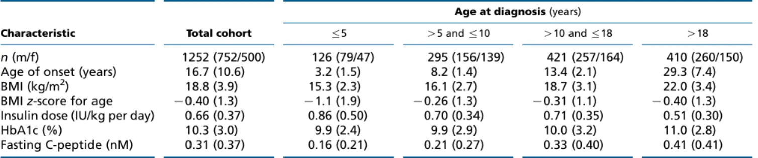
Table 1 Characteristics of population by age of onset. Data are meanGS.D.
Characteristic Total cohort
Age at diagnosis(years)
%5 O5 and%10 O10 and%18 O18
n(m/f) 1252 (752/500) 126 (79/47) 295 (156/139) 421 (257/164) 410 (260/150)
Age of onset (years) 16.7 (10.6) 3.2 (1.5) 8.2 (1.4) 13.4 (2.1) 29.3 (7.4)
BMI (kg/m2) 18.8 (3.9) 15.3 (2.3) 16.1 (2.7) 18.7 (3.1) 22.0 (3.4)
BMIz-score for age K0.40 (1.3) K1.1 (1.9) K0.26 (1.3) K0.31 (1.1) K0.40 (1.3) Insulin dose (IU/kg per day) 0.66 (0.37) 0.86 (0.50) 0.70 (0.34) 0.71 (0.35) 0.51 (0.30)
HbA1c (%) 10.3 (3.0) 9.9 (2.4) 9.9 (2.9) 10.0 (3.2) 11.0 (2.8)
Fasting C-peptide (nM) 0.31 (0.37) 0.16 (0.21) 0.21 (0.27) 0.33 (0.40) 0.41 (0.41)
–0.05 –0.04 –0.03 –0.02 –0.01 0.00 0.01 0.02 0.03
Age of onset group (years)
β (95% CI)
≤5
>5 and ≤10
>10 and ≤18
>18
Figure 1
Association between 1 kg/m2change in BMI andDC-peptide0-1 in age of onset and sex-adjusted models by age of onset group.
EuropeanJournalofEndocrinology
The lack of association between BMI and fasting C-peptide decline over 1 year in younger and older age groups may be due to the fact thatb-cell dysfunction is much more severe in the former and much less severe in the latter(29, 30, 31, 32, 33). Indeed individuals in our study diagnosed at R18 years of age were not obese according to typical BMI cut-offs.
The greater decline in C-peptide observed in patients with a higher BMI could also reflect greater low-grade inflammation associated with obesity(34, 35).
In the SEARCH for Diabetes in Youth study, the decline in b-cell function 12 months after the diagnosis was independent of BMIz-score and insulin resistance among autoantibodies-positive patients with diabetes diagnosed at the age of 10–20 years(36). Different to our data, the SEARCH study used a pathogenesis-based approach for the classification of diabetes; furthermore, the SEARCH study included a wide range of racial and ethnic groups and 23% of the population was not Non-Hispanic White.
Our work may have relevant clinical implications.
Although we are not able to define whether insulin resistance or adiposity is the main determinant of the b-cell decline during the first year after diagnosis, the results presented here may suggest that, especially in individuals diagnosed between 10 and 18 years of age, intervention to reduce insulin resistance (e.g. weight loss) or drugs aimed at improving insulin sensitivity may be a possible component of therapy aimed at preventing fasting C-peptide decline, at least in adolescents. In addition, if the association between BMI and increased C-peptide decline is a result of pro-inflammatory processes associated with obesity, then treatments aimed to reduced inflammation would also be beneficial. Previous studies have shown that with increasing body weight, systemic immune mediators increase not only for type 2 diabetes but also for type 1 diabetes (37, 38). Although anti-IL1 therapy was not effective in a post hoc subanalysis of high BMI subjects(39), properly powered and designed studies also testing other anti-inflammatory interventions are needed to determine this question.
Limitations of our study include the reporting of results using untransformed BMI variables for the ease of interpretation and comparison with the data from adults in our cohorts. To overcome this problem, we repeated all analyses with BMI z-score variables and found no difference in results indicating that increased adiposity is likely to underlie this association.
We have used BMI as a proxy for insulin resistance in this study, and although the two variables are strongly related, it would have been preferable to have performed
the analysis by directly measuring insulin resistance;
however, direct measurement of insulin resistance was not available for analysis. We quantified b-cell function by analysing C-peptide in the fasting state, which may only partially reflectb-cell function in the stimulated state (e.g. after a meal). Although we did have data on stimulated C-peptide, a limited number of patients had sufficient data for analysis; hence we decided to maximise our sample size by using fasting C-peptide alone as our outcome variable. Data are also limited by the use of different C-peptide assays though all assays had similar detection limits.
Moreover, we did not adjust linear regression models for baseline C-peptide. However, the inclusion of fasting C-peptide at baseline in models is actually a matter of debate in statistical analysis. Thus, we decided not to include it as it may introduce a bias(40).
Another limitation is that diabetes-associated auto- immunity was not evaluated in our study and this parameter appears to be a major variable associated with b-cell decline, at least in patients !20 years of age at diagnosis(36).
In conclusion, our work provides supportive evidence for a key role of body mass in type 1 diabetes disease progression after diagnosis. However, additional studies are needed to define whether insulin resistance or adiposity are key factors leading to b-cell decline soon after diagnosis of type 1 diabetes.
Declaration of interest
The authors declare that there is no conflict of interest that could be perceived as prejudicing the impartiality of the research reported.
Funding
This study was supported by Centro Internazionale Studi Diabete.
D Mauricio was supported by a grant from the Instituto Carlos III, Spain (Project FIS 061104).
Author contribution statement
A Barker, A Lauria and P Pozzilli were responsible for the design of the study, acquisition of data, analysis and interpretation of data, writing the manuscript and revising it critically for important intellectual content. N Schloot, N Hosszufalusi, J Ludvigsson, C Mathieu, D Mauricio, M Nordwall, B Van der Schueren, T Mandrup-Poulsen, W A Scherbaum, I Weets and F K Gorus made substantial contributions to the acquisition of data and revised the manuscript critically for important intellectual content. N Wareham made a substantial contribution to the analysis of the data and critical revision of the manuscript for important intellectual content. R D Leslie contributed to discussion and reviewed/edited the manuscript. All authors approved the final version.
EuropeanJournalofEndocrinology
References
1 Alberti KG & Zimmet PZ. Definition, diagnosis and classification of diabetes mellitus and its complications. Part 1: diagnosis and classification of diabetes mellitus provisional report of a WHO consultation.Diabetic Medicine199815539–553. (doi:10.1002/
(SICI)1096-9136(199807)15:7!539::AID-DIA668O3.0.CO;2-S) 2 Yki-Jarvinen H & Koivisto VA. Natural course of insulin resistance in
type I diabetes.New England Journal of Medicine1986315224–230.
(doi:10.1056/NEJM198607243150404)
3 Ludvigsson J, Carlsson A, Deli A, Forsander G, Ivarsson SA, Kockum I, Lindblad B, Marcus C, Lernmark A˚ & Samuelsson U. Decline of C-peptide during the first year after diagnosis of type 1 diabetes in children and adolescents.Diabetes Research and Clinical Practice2013 100203–209. (doi:10.1016/j.diabres.2013.03.003)
4 Wilkin TJ. The accelerator hypothesis: a review of the evidence for insulin resistance as the basis for type I as well as type II diabetes.
International Journal of Obesity200933716–726. (doi:10.1038/
ijo.2009.97)
5 Ludvigsson J. Why diabetes incidence increases – a unifying theory.
Annals of the New York Academy of Sciences20061079374–382.
(doi:10.1196/annals.1375.058)
6 Onkamo P, Vaananen S, Karvonen M & Tuomilehto J. Worldwide increase in incidence of type I diabetes – the analysis of the data on published incidence trends.Diabetologia1999421395–1403.
(doi:10.1007/s001250051309)
7 Johansson C, Samuelsson U & Ludvigsson J. A high weight gain early in life is associated with an increased risk of type 1 (insulin-dependent) diabetes mellitus.Diabetologia19943791–94. (doi:10.1007/
BF00428783)
8 Betts P, Mulligan J, Ward P, Smith B & Wilkin T. Increasing body weight predicts the earlier onset of insulin-dependant diabetes in childhood:
testing the ‘accelerator hypothesis’ (2).Diabetic Medicine200522 144–151. (doi:10.1111/j.1464-5491.2004.01368.x)
9 Kordonouri O & Hartmann R. Higher body weight is associated with earlier onset of type 1 diabetes in children: confirming the ‘Accelerator Hypothesis’.Diabetic Medicine2005221783–1784. (doi:10.1111/
j.1464-5491.2005.01792.x)
10 Patterson CC, Dahlquist GG, Gyurus E, Green A, Soltesz G & Group ES.
Incidence trends for childhood type 1 diabetes in Europe during 1989–2003 and predicted new cases 2005–20: a multicentre prospective registration study.Lancet20093732027–2033. (doi:10.1016/S0140- 6736(09)60568-7)
11 Barker A, Lauria A, Schloot N, Hosszufalusi N, Ludvigsson J, Mathieu C, Mauricio D, Nordwall M, Van der Schueren B, Mandrup-Poulsen Tet al.
Age-dependent decline ofbcell function in type 1 diabetes after diagnosis: a multi-centre longitudinal study.Diabetes, Obesity &
Metabolism201316262–267. (doi:10.1111/dom.12216)
12 Kibirige M, Metcalf B, Renuka R & Wilkin TJ. Testing the accelerator hypothesis: the relationship between body mass and age at diagnosis of type 1 diabetes.Diabetes Care2003262865–2870. (doi:10.2337/
diacare.26.10.2865)
13 Clarke SL, Craig ME, Garnett SP, Chan AK, Cowell CT, Cusumano JM, Kordonouri O, Sambasivan A & Donaghue KC. Increased adiposity at diagnosis in younger children with type 1 diabetes does not persist.
Diabetes Care2006291651–1653. (doi:10.2337/dc06-0277)
14 Dabelea D, D’Agostino RB Jr, Mayer-Davis EJ, Pettitt DJ, Imperatore G, Dolan LM, Pihoker C, Hillier TA, Marcovina SM, Linder Bet al. Testing the accelerator hypothesis: body size,b-cell function, and age at onset of type 1 (autoimmune) diabetes.Diabetes Care200629290–294.
(doi:10.2337/diacare.29.02.06.dc05-1339)
15 Hermann R, Knip M, Veijola R, Simell O, Laine AP, Akerblom HK, Groop PH, Forsblom C, Pettersson-Fernholm K, Ilonen Jet al. Temporal changes in the frequencies of HLA genotypes in patients with type 1
diabetes – indication of an increased environmental pressure?
Diabetologia200346420–425. (doi:10.1007/S00125-003;1045-4) 16 Gillespie KM, Bain SC, Barnett AH, Bingley PJ, Christie MR, Gill GV &
Gale EA. The rising incidence of childhood type 1 diabetes and reduced contribution of high-risk HLA haplotypes.Lancet20043641699–1700.
(doi:10.1016/S0140-6736(04)17357-1)
17 Leslie RD, Taylor R & Pozzilli P. The role of insulin resistance in the natural history of type 1 diabetes.Diabetic Medicine199714327–331.
(doi:10.1002/(SICI)1096-9136(199704)14:4!327::AID-DIA315O3.0.
CO;2-6)
18 Greenbaum CJ. Insulin resistance in type 1 diabetes.Diabetes/Metabo- lism Research and Reviews200218192–200. (doi:10.1002/dmrr.291) 19 Fourlanos S, Narendran P, Byrnes GB, Colman PG & Harrison LC.
Insulin resistance is a risk factor for progression to type 1 diabetes.
Diabetologia2004471661–1667. (doi:10.1007/s00125-004-1507-3) 20 Fourlanos S, Harrison LC & Colman PG. The accelerator hypothesis and
increasing incidence of type 1 diabetes.Current Opinion in Endocrinology, Diabetes, and Obesity200815321–325. (doi:10.1097/MED.
0b013e3283073a5a)
21 Hawa MI, Bonfanti R, Valeri C, Delli Castelli M, Beyan H & Leslie RD.
No evidence for genetically determined alteration in insulin secretion or sensitivity predisposing to type 1 diabetes: a study of identical twins.Diabetes Care2005281415–1418. (doi:10.2337/
diacare.28.6.1415)
22 Winkler C, Marienfeld S, Zwilling M, Bonifacio E & Ziegler AG. Is islet autoimmunity related to insulin sensitivity or body weight in children of parents with type 1 diabetes?Diabetologia2009522072–2078.
(doi:10.1007/s00125-009-1461-1)
23 Cole TJ. The LMS, method for constructing normalized growth standards.European Journal of Clinical Nutrition19904445–60.
24 Poitout V & Robertson RP. Minireview: secondaryb-cell failure in type 2 diabetes – a convergence of glucotoxicity and lipotoxicity.
Endocrinology2002143339–342. (doi:10.1210/endo.143.2.8623) 25 Prentki M & Nolan CJ. Isletbcell failure in type 2 diabetes.Journal of
Clinical Investigation20061161802–1812. (doi:10.1172/JCI29103) 26 Clausen JO, Borch-Johnsen K, Ibsen H, Bergman RN, Hougaard P,
Winther K & Pedersen O. Insulin sensitivity index, acute insulin response, and glucose effectiveness in a population-based sample of 380 young healthy Caucasians. Analysis of the impact of gender, body fat, physical fitness, and life-style factors.Journal of Clinical Investigation 1996981195–1209. (doi:10.1172/JCI118903)
27 Wagenknecht LE, Langefeld CD, Scherzinger AL, Norris JM, Haffner SM, Saad MF & Bergman RN. Insulin sensitivity, insulin secretion, and abdominal fat: the Insulin Resistance Atherosclerosis Study (IRAS) Family Study.Diabetes2003522490–2496. (doi:10.2337/diabetes.52.
10.2490)
28 Goran MI & Gower BA. Longitudinal study on pubertal insulin resistance.Diabetes2001502444–2450. (doi:10.2337/diabetes.50.11.
2444)
29 Vandewalle CL, Decraene T, Schuit FC, De Leeuw IH, Pipeleers DG &
Gorus FK. Insulin autoantibodies and high titre islet cell antibodies are preferentially associated with the HLA DQA1*0301-DQB1*0302 haplotype at clinical type 1 (insulin-dependent) diabetes mellitus before age 10 years, but not at onset between age 10 and 40 years.
The Belgian Diabetes Registry.Diabetologia1993361155–1162.
(doi:10.1007/BF00401060)
30 Awata T, Hagura R, Urakami T & Kanazawa Y. Age-dependent HLA, genetic heterogeneity of IDDM in Japanese patients.Diabetologia1995 38748–749. (doi:10.1007/BF00401850)
31 Graham J, Kockum I, Sanjeevi CB, Landin-Olsson M, Nystrom L, Sundkvist G, Arnqvist H, Blohme G, Lithner F, Littorin Bet al. Negative association between type 1 diabetes and HLA DQB1*0602-DQA1*0102 is attenuated with age at onset, Swedish Childhood Diabetes Study Group.European Journal of Immunogenetics199926117–127.
(doi:10.1046/j.1365-2370.1999.00127.x-i2)
EuropeanJournalofEndocrinology
32 Howson JM, Rosinger S, Smyth DJ, Boehm BO, Group A-ES & Todd JA.
Genetic analysis of adult-onset autoimmune diabetes.Diabetes201160 2645–2653. (doi:10.2337/db11-0364)
33 Lohmann T, Sessler J, Verlohren HJ, Schroder S, Rotger J, Dahn K, Morgenthaler N & Scherbaum WA. Distinct genetic and immunological features in patients with onset of IDDM before and after age 40.Diabetes Care199720524–529. (doi:10.2337/diacare.20.4.524)
34 Ferrante AW Jr. Obesity-induced inflammation: a metabolic dialogue in the language of inflammation.Journal of Internal Medicine2007262 408–414. (doi:10.1111/j.1365-2796.2007.01852.x)
35 Shoelson SE, Herrero L & Naaz A. Obesity, inflammation, and insulin resistance.Gastroenterology20071322169–2180. (doi:10.1053/j.gastro.
2007.03.059)
36 Dabelea D, Mayer-Davis EJ, Andrews JS, Dolan LM, Pihoker C, Hamman RF, Greenbaum C, Marcovina S, Fujimoto W, Linder Bet al.
Clinical evolution ofbcell function in youth with diabetes: the SEARCH for Diabetes in Youth study.Diabetologia2012553359–3368.
(doi:10.1007/s00125-012-2719-6)
37 Pham MN, Hawa MI, Roden M, Schernthaner G, Pozzilli P, Buzzetti R, Scherbaum WA, Seissler J, Hunter S, Leslie RDet al. Increased serum
concentrations of adhesion molecules but not of chemokines in patients with type 2 diabetes compared with patients with type 1 diabetes and latent autoimmune diabetes in adult age: action LADA 5.
Diabetic Medicine201229470–478. (doi:10.1111/j.1464-5491.2011.
03546.x)
38 Pham MN, Hawa MI, Pfleger C, Roden M, Schernthaner G, Pozzilli P, Buzzetti R, Scherbaum W, Seissler J, Kolb Het al. Pro- and anti- inflammatory cytokines in latent autoimmune diabetes in adults, type 1 and type 2 diabetes patients: action LADA 4.Diabetologia2011 541630–1638. (doi:10.1007/s00125-011-2088-6)
39 Moran A, Bundy B, Becker DJ, DiMeglio LA, Gitelman SE, Goland R, Greenbaum CJ, Herold KC, Marks JB, Raskin Pet al.
Interleukin-1 antagonism in type 1 diabetes of recent onset:
two multicentre, randomised, double-blind, placebo-controlled trials.Lancet20133811905–1915. (doi:10.1016/S0140-6736(13) 60023-9)
40 Glymour MM, Weuve J, Berkman LF, Kawachi I & Robins JM. When is baseline adjustment useful in analyses of change? An example with education and cognitive change.American Journal of Epidemiology2005 162267–278. (doi:10.1093/aje/kwi187)
Received 7 July 2014
Revised version received 15 October 2014 Accepted 6 November 2014
EuropeanJournalofEndocrinology